
Fluorescent Sensors for Detecting and Imaging Metal Ions in Biological Systems: Recent Advances and Future Perspectives

Abstract
:1. Introduction
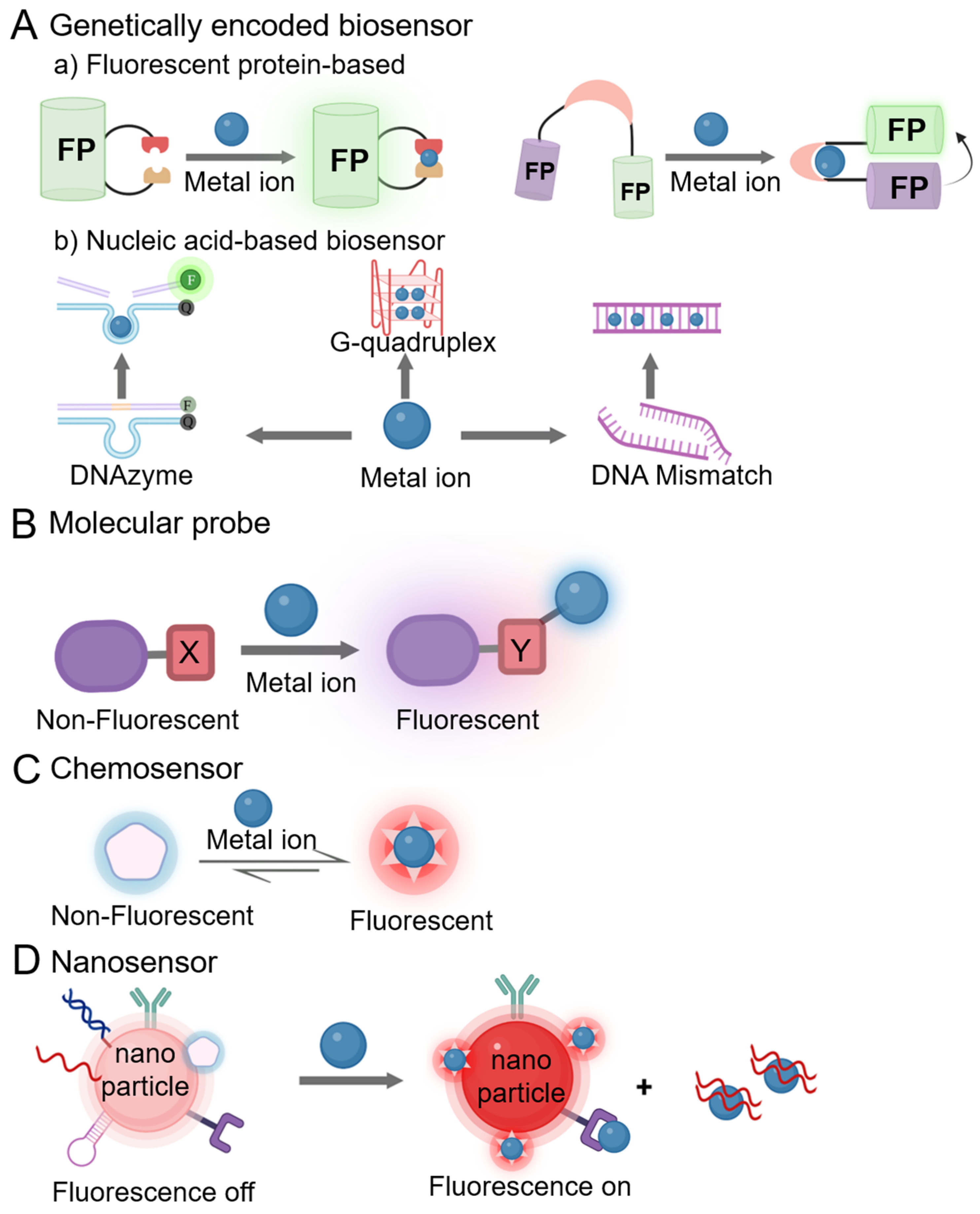
2. Categorization of Fluorescent Sensors for Metal Ions
3. In Vitro Detection of Metal Ions
3.1. Fluorescent Sensors for Essential Metal Ions
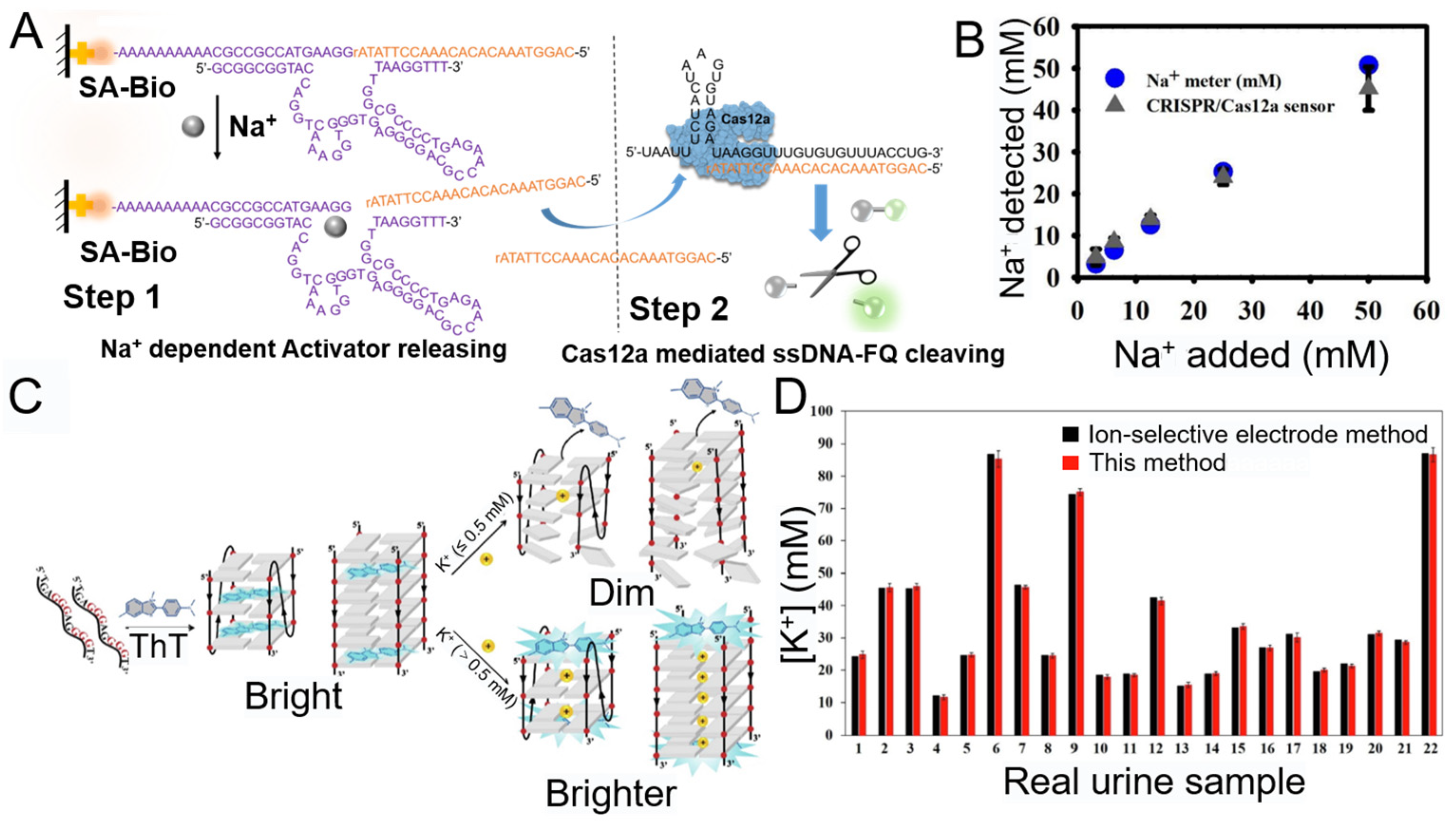
3.1.1. Na+
3.1.2. K+

3.1.3. Zn2+
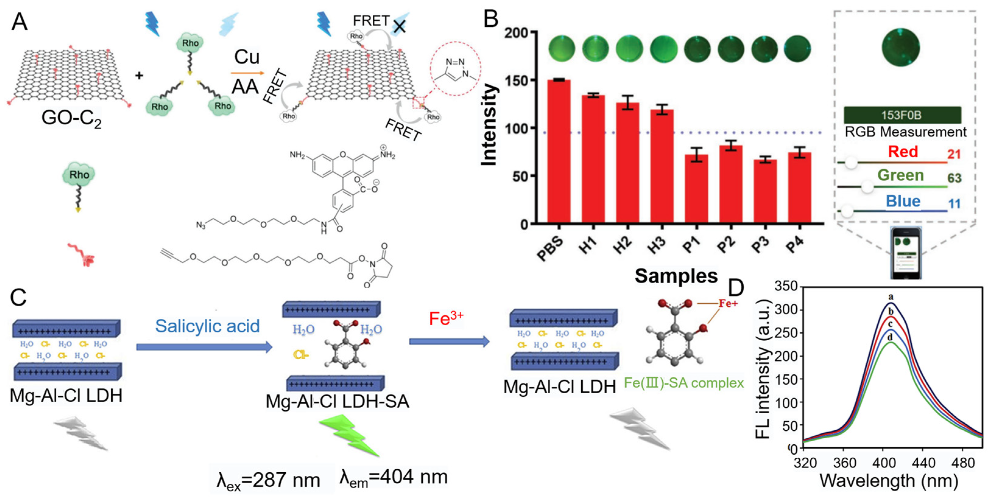
3.1.4. Cu2+
3.1.5. Ca2+
3.1.6. Fe3+
3.2. Fluorescent Sensors for Non-Essential Metal Ions
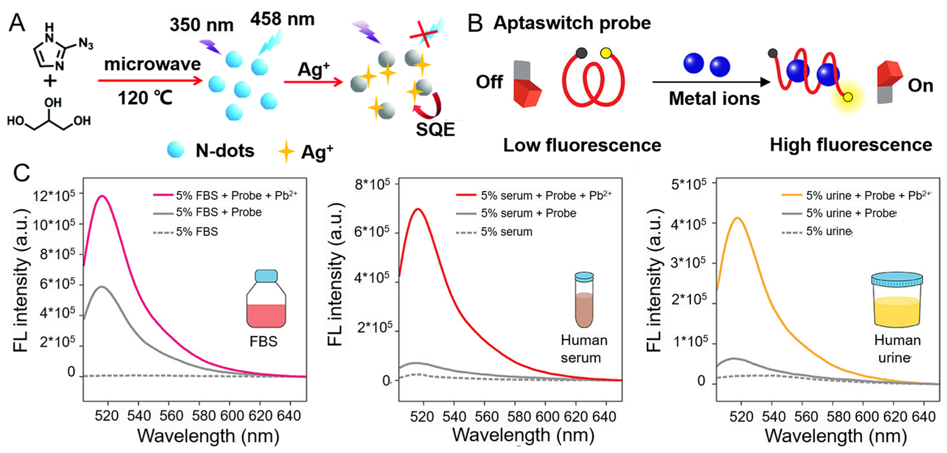
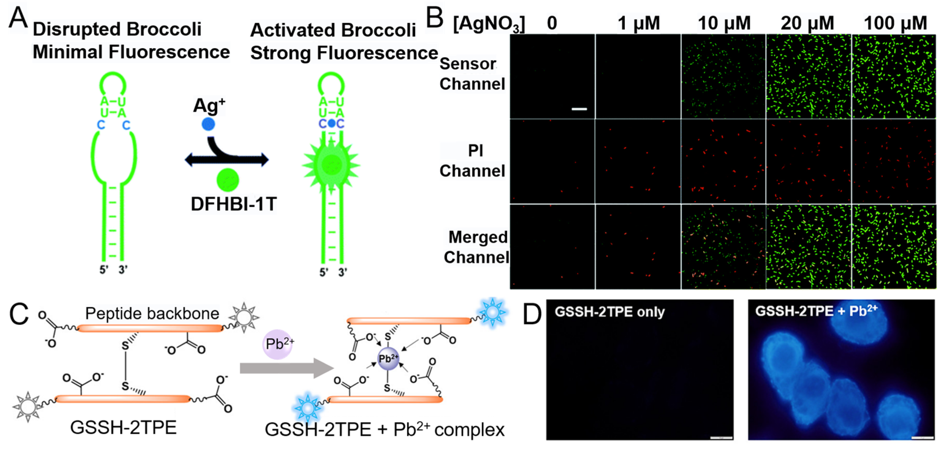
3.2.1. Ag+
3.2.2. Pb2+

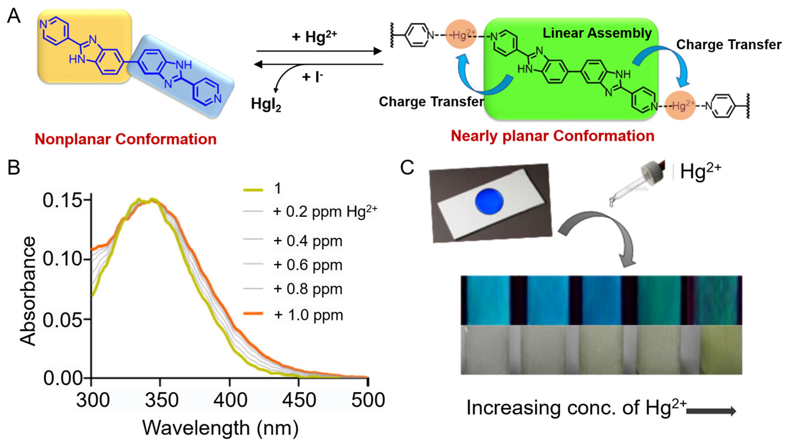
3.2.3. Hg2+
3.2.4. Al3+
3.2.5. Pt4+
4. Intracellular Imaging of Metal Ions
4.1. Fluorescent Sensors for Essential Metal Ions
4.1.1. Na+
4.1.2. K+
4.1.3. Ca2+

4.1.4. Zn2+
4.1.5. Mg2+
4.1.6. Cu2+

4.1.7. Fe2+/Fe3+
4.2. Fluorescent Sensors for Non-Essential Metal Ions
4.2.1. Li+
4.2.2. Ag+
4.2.3. Ni2+

4.2.4. Pb2+
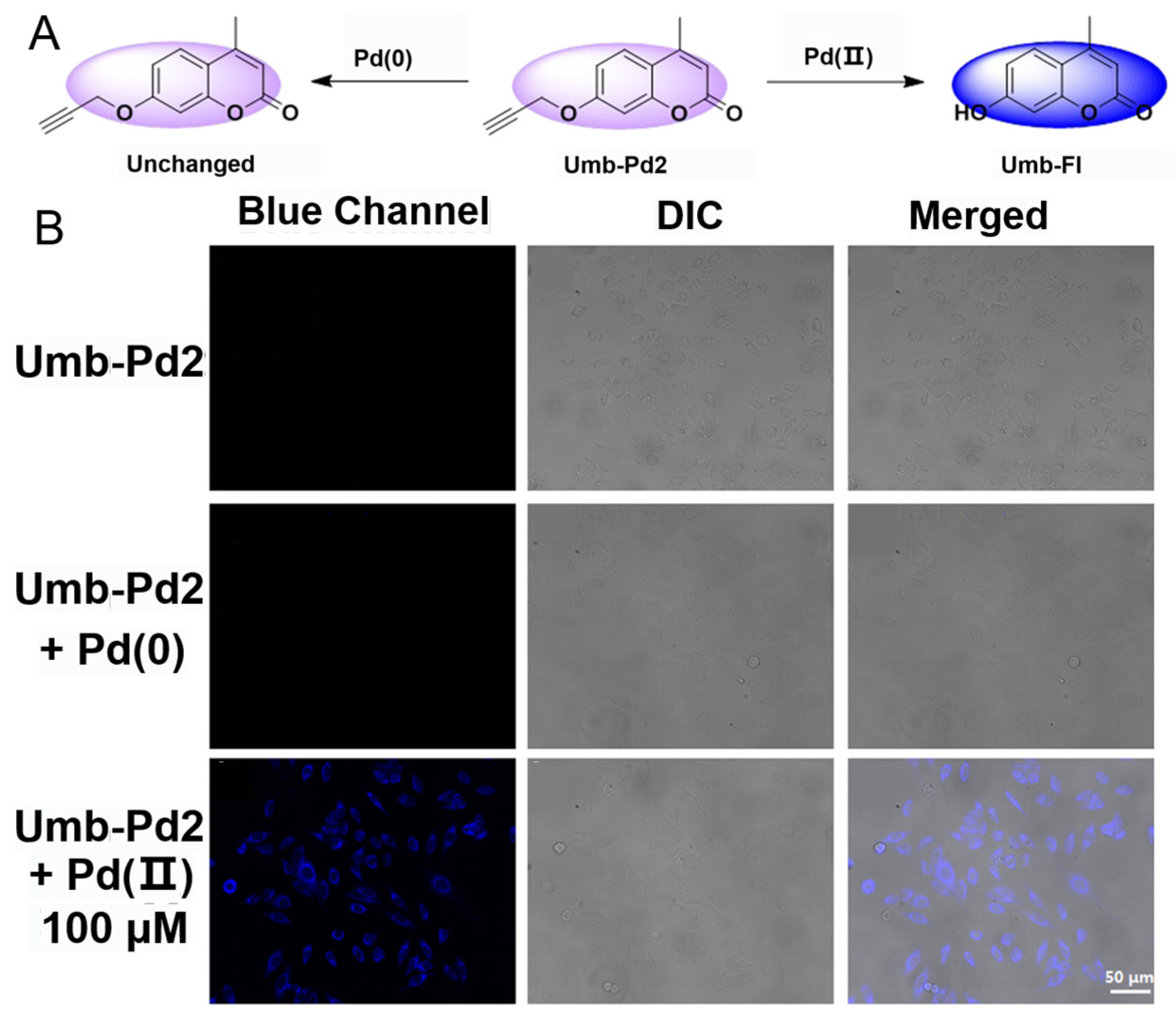
4.2.5. Pd2+
4.2.6. Hg2+
4.2.7. Cd2+
4.2.8. Au3+
4.2.9. Al3+
5. In Vivo Imaging of Metal Ions
5.1. Fluorescent Sensors for Essential Metal Ions
5.1.1. K+
5.1.2. Ca2+

5.1.3. Zn2+
5.1.4. Fe2+/Fe3+
5.1.5. Co2+
5.2. Fluorescent Sensors for Non-Essential Metal Ions
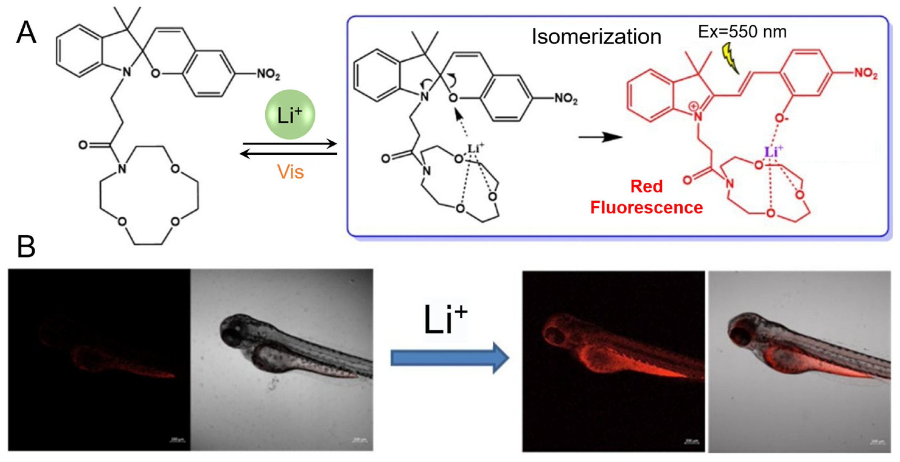
5.2.1. Li+
5.2.2. Pb2+
5.2.3. Sn2+
5.2.4. Cd2+
5.2.5. Hg2+
5.2.6. Ni2+
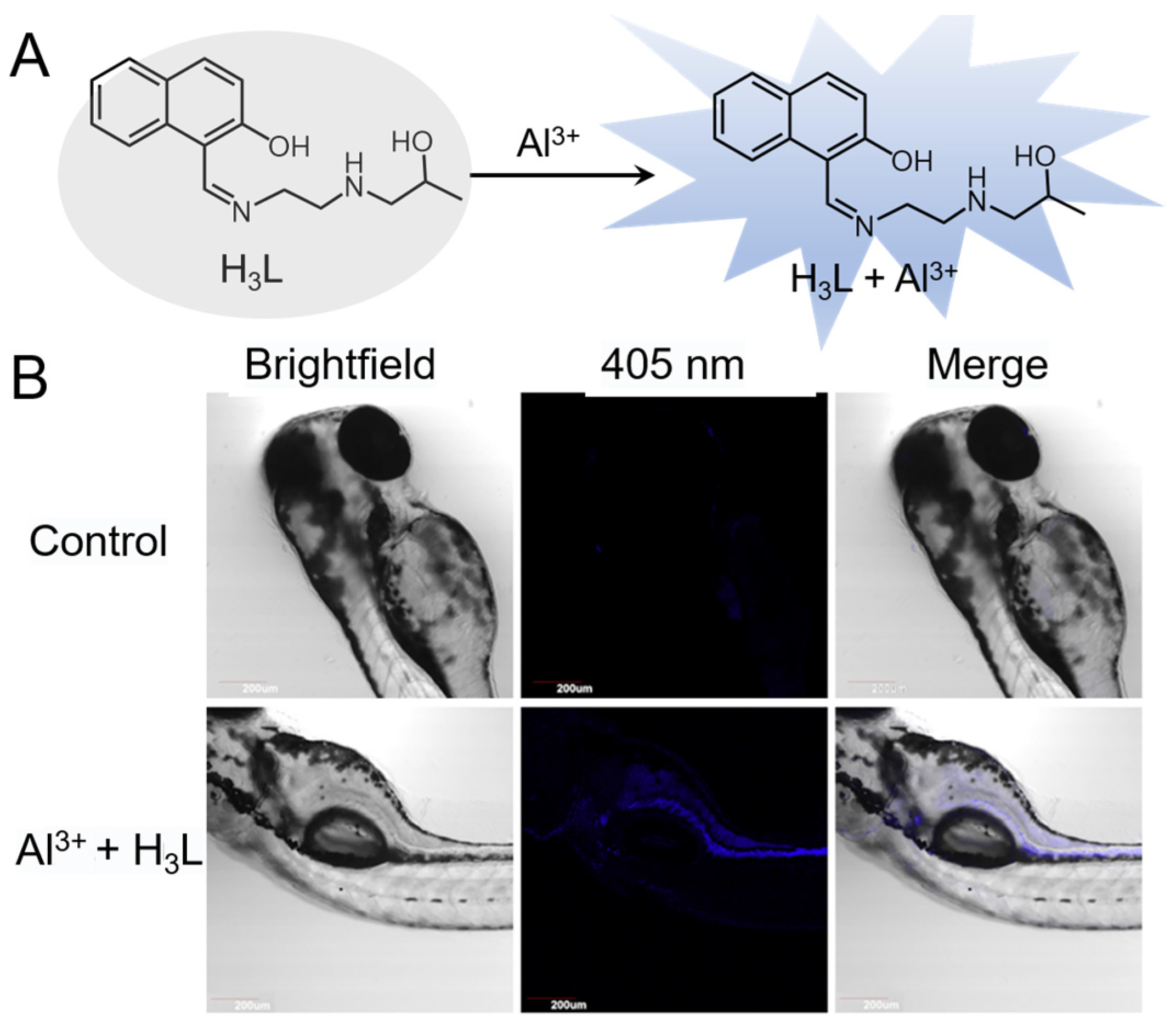
5.2.7. Al3+
6. Conclusions and Future Directions
Funding
Institutional Review Board Statement
Informed Consent Statement
Data Availability Statement
Conflicts of Interest
Abbreviations
| Acronyms | Definition |
| DNA | Deoxyribonucleic acid |
| RNA | Ribonucleic acid |
| ICP-MS | Inductively coupled plasma mass spectrometry |
| AAS | Atomic absorption spectrophotometry |
| FAAS | Flame atomic absorption spectrometry |
| FRET | Fluorescent resonance energy transfer |
| FNA | Functional nucleic acid |
| HCR | Hybrid chain reactions |
| AIE | Aggregation-induced emission |
| BODIPY | Boron-dipyrromethene |
| GQ | G-quadruplex |
| NIR | Near-infrared |
| CD | Carbon dot |
| MOF | Metal-organic framework |
| UV | Ultraviolet |
| HSA | Human serum albumin |
| FBS | Fetal bovine serum |
| PET | Photoinduced electron transfer |
| SERS | Surface-enhanced Raman scattering |
| GFP | Green fluorescent protein |
| TP | Two-photon |
| AuNP | Gold nanoparticle |
| GSH | Glutathione |
| BD | Bipolar disorder |
| ICT | Intramolecular charge transfer |
| UNCP | Upconversion nanoparticle |
| EDTA | Ethylene diamine tetraacetic acid |
| BHQ | Black hole quencher |
References
- Zoroddu, M.A.; Aaseth, J.; Crisponi, G.; Medici, S.; Peana, M.; Nurchi, V.M. The essential metals for humans: A brief overview. J. Inorg. Biochem. 2019, 195, 120–129. [Google Scholar] [CrossRef] [PubMed]
- Jomova, K.; Makova, M.; Alomar, S.Y.; Alwasel, S.H.; Nepovimova, E.; Kuca, K.; Rhodes, C.J.; Valko, M. Essential metals in health and disease. Chem.-Biol. Interact. 2022, 367, 110173. [Google Scholar] [CrossRef] [PubMed]
- Missirlis, F. Regulation and biological function of metal ions in Drosophila. Curr. Opin. Insect Sci. 2021, 47, 18–24. [Google Scholar] [CrossRef] [PubMed]
- Slobodian, M.R.; Petahtegoose, J.D.; Wallis, A.L.; Levesque, D.C.; Merritt, T.J.S. The Effects of Essential and Non-Essential Metal Toxicity in the Drosophila melanogaster Insect Model: A Review. Toxics 2021, 9, 269. [Google Scholar] [CrossRef]
- Lu, Y. Metal ions as matchmakers for proteins. Proc. Natl. Acad. Sci. USA 2010, 107, 1811–1812. [Google Scholar] [CrossRef] [Green Version]
- Carter, K.P.; Young, A.M.; Palmer, A.E. Fluorescent Sensors for Measuring Metal Ions in Living Systems. Chem. Rev. 2014, 114, 4564–4601. [Google Scholar] [CrossRef]
- Zhang, X.-B.; Kong, R.-M.; Lu, Y. Metal Ion Sensors Based on DNAzymes and Related DNA Molecules. Annu. Rev. Anal. Chem. 2011, 4, 105–128. [Google Scholar] [CrossRef] [PubMed] [Green Version]
- Chen, Y.; Bai, Y.; Han, Z.; He, W.; Guo, Z. Photoluminescence imaging of Zn2+ in living systems. Chem. Soc. Rev. 2015, 44, 4517–4546. [Google Scholar] [CrossRef] [PubMed]
- Zhang, J.; Lu, Y. Biocomputing for Portable, Resettable, and Quantitative Point-of-Care Diagnostics: Making the Glucose Meter a Logic-Gate Responsive Device for Measuring Many Clinically Relevant Targets. Angew. Chem. Int. Ed. 2018, 57, 9702–9706. [Google Scholar] [CrossRef]
- Wu, Z.; Fan, H.; Satyavolu, N.S.R.; Wang, W.; Lake, R.; Jiang, J.-H.; Lu, Y. Imaging Endogenous Metal Ions in Living Cells Using a DNAzyme–Catalytic Hairpin Assembly Probe. Angew. Chem. Int. Ed. 2017, 56, 8721–8725. [Google Scholar] [CrossRef]
- Torabi, S.-F.; Wu, P.; McGhee, C.E.; Chen, L.; Hwang, K.; Zheng, N.; Cheng, J.; Lu, Y. In vitro selection of a sodium-specific DNAzyme and its application in intracellular sensing. Proc. Natl. Acad. Sci. USA 2015, 112, 5903–5908. [Google Scholar] [CrossRef] [Green Version]
- Hultin, S. Mosby’s Manual of Diagnostic and Laboratory Tests (4th edn). Ann. Clin. Biochem. 2012, 49, 415. [Google Scholar] [CrossRef] [Green Version]
- Yang, L.; Qing, Z.; Liu, C.; Tang, Q.; Li, J.; Yang, S.; Zheng, J.; Yang, R.; Tan, W. Direct Fluorescent Detection of Blood Potassium by Ion-Selective Formation of Intermolecular G-Quadruplex and Ligand Binding. Anal. Chem. 2016, 88, 9285–9292. [Google Scholar] [CrossRef] [PubMed]
- Chin, D.; Means, A.R. Calmodulin: A prototypical calcium sensor. Trends Cell Biol. 2000, 10, 322–328. [Google Scholar] [CrossRef]
- Jahnen-Dechent, W.; Ketteler, M. Magnesium basics. Clin. Kidney J. 2012, 5, i3–i14. [Google Scholar] [CrossRef] [PubMed] [Green Version]
- Maity, D.; Manna, A.K.; Karthigeyan, D.; Kundu, T.K.; Pati, S.K.; Govindaraju, T. Visible–Near-Infrared and Fluorescent Copper Sensors Based on Julolidine Conjugates: Selective Detection and Fluorescence Imaging in Living Cells. Chem.-Eur. J. 2011, 17, 11152–11161. [Google Scholar] [CrossRef]
- Michielsen, C.M.S.; van Aalen, E.A.; Merkx, M. Ratiometric Bioluminescent Zinc Sensor Proteins to Quantify Serum and Intracellular Free Zn2+. ACS Chem. Biol. 2022, 17, 1567–1576. [Google Scholar] [CrossRef]
- Abdolmohammad-Zadeh, H.; Zamani-Kalajahi, M. A turn-on/off fluorescent sensor based on nano-structured Mg-Al layered double hydroxide intercalated with salicylic acid for monitoring of ferric ion in human serum samples. Anal. Chim. Acta 2019, 1061, 152–160. [Google Scholar] [CrossRef]
- Drennan-Harris, L.R.; Wongwilawan, S.; Tyson, J.F. Trace determination of total mercury in rice by conventional inductively coupled plasma mass spectrometry. J. Anal. At. Spectrom. 2013, 28, 259–265. [Google Scholar] [CrossRef] [Green Version]
- Xiong, C.; Qin, Y.; Hu, B. On-line separation/preconcentration of V(IV)/V(V) in environmental water samples with CTAB-modified alkyl silica microcolumn and their determination by inductively coupled plasma-optical emission spectrometry. J. Hazard. Mater. 2010, 178, 164–170. [Google Scholar] [CrossRef]
- Špirić, Z.; Vučković, I.; Stafilov, T.; Kušan, V.; Frontasyeva, M. Air Pollution Study in Croatia Using Moss Biomonitoring and ICP–AES and AAS Analytical Techniques. Arch. Environ. Contam. Toxicol. 2013, 65, 33–46. [Google Scholar] [CrossRef]
- Wu, P.; He, S.; Luo, B.; Hou, X. Flame Furnace Atomic Absorption Spectrometry: A Review. Appl. Spectrosc. Rev. 2009, 44, 411–437. [Google Scholar] [CrossRef]
- Săcărescu, L.; Chibac-Scutaru, A.-L.; Roman, G.; Săcărescu, G.; Simionescu, M. Selective detection of metal ions, sulfites and glutathione with fluorescent pyrazolines: A review. Environ. Chem. Lett. 2023, 21, 561–596. [Google Scholar] [CrossRef]
- Wu, D.; Chen, L.; Lee, W.; Ko, G.; Yin, J.; Yoon, J. Recent progress in the development of organic dye based near-infrared fluorescence probes for metal ions. Coord. Chem. Rev. 2018, 354, 74–97. [Google Scholar] [CrossRef]
- Denis, M.; Pancholi, J.; Jobe, K.; Watkinson, M.; Goldup, S.M. Chelating Rotaxane Ligands as Fluorescent Sensors for Metal Ions. Angew. Chem. Int. Ed. 2018, 57, 5310–5314. [Google Scholar] [CrossRef] [PubMed] [Green Version]
- Liu, X.; Li, N.; Li, M.; Chen, H.; Zhang, N.; Wang, Y.; Zheng, K. Recent progress in fluorescent probes for detection of carbonyl species: Formaldehyde, carbon monoxide and phosgene. Coord. Chem. Rev. 2020, 404, 213109. [Google Scholar] [CrossRef]
- Wu, L.; Sedgwick, A.C.; Sun, X.; Bull, S.D.; He, X.-P.; James, T.D. Reaction-Based Fluorescent Probes for the Detection and Imaging of Reactive Oxygen, Nitrogen, and Sulfur Species. Acc. Chem. Res. 2019, 52, 2582–2597. [Google Scholar] [CrossRef] [PubMed] [Green Version]
- Liu, X.-L.; Niu, L.-Y.; Chen, Y.-Z.; Yang, Y.; Yang, Q.-Z. A multi-emissive fluorescent probe for the discrimination of glutathione and cysteine. Biosens. Bioelectron. 2017, 90, 403–409. [Google Scholar] [CrossRef]
- Wang, S.-Q.; Wu, Q.-H.; Wang, H.-Y.; Zheng, X.-X.; Shen, S.-L.; Zhang, Y.-R.; Miao, J.-Y.; Zhao, B.-X. Novel pyrazoline-based fluorescent probe for detecting glutathione and its application in cells. Biosens. Bioelectron. 2014, 55, 386–390. [Google Scholar] [CrossRef]
- Niu, P.; Zhu, J.; Wei, L.; Liu, X. Application of Fluorescent Probes in Reactive Oxygen Species Disease Model. Crit. Rev. Anal. Chem. 2022, 31, 1–36. [Google Scholar] [CrossRef]
- Cui, W.-L.; Wang, M.-H.; Yang, Y.-H.; Wang, J.-Y.; Zhu, X.; Zhang, H.; Ji, X. Recent advances and perspectives in reaction-based fluorescent probes for imaging peroxynitrite in biological systems. Coord. Chem. Rev. 2023, 474, 214848. [Google Scholar] [CrossRef]
- Juvekar, V.; Park, S.J.; Yoon, J.; Kim, H.M. Recent progress in the two-photon fluorescent probes for metal ions. Coord. Chem. Rev. 2021, 427, 213574. [Google Scholar] [CrossRef]
- Wan, H.; Xu, Q.; Gu, P.; Li, H.; Chen, D.; Li, N.; He, J.; Lu, J. AIE-based fluorescent sensors for low concentration toxic ion detection in water. J. Hazard. Mater. 2021, 403, 123656. [Google Scholar] [CrossRef]
- Xia, P.-F.; Ling, H.; Foo, J.L.; Chang, M.W. Synthetic genetic circuits for programmable biological functionalities. Biotechnol. Adv. 2019, 37, 107393. [Google Scholar] [CrossRef] [PubMed]
- Wang, M.; Da, Y.; Tian, Y. Fluorescent proteins and genetically encoded biosensors. Chem. Soc. Rev. 2023, 52, 1189–1214. [Google Scholar] [CrossRef] [PubMed]
- Xu, J.; Jiang, R.; Feng, Y.; Liu, Z.; Huang, J.; Ma, C.; Wang, K. Functional nucleic acid-based fluorescent probes for metal ion detection. Coord. Chem. Rev. 2022, 459, 214453. [Google Scholar] [CrossRef]
- Nagai, T.; Sawano, A.; Park, E.S.; Miyawaki, A. Circularly permuted green fluorescent proteins engineered to sense Ca2+. Proc. Natl. Acad. Sci. USA 2001, 98, 3197–3202. [Google Scholar] [CrossRef] [Green Version]
- Tian, L.; Hires, S.A.; Mao, T.; Huber, D.; Chiappe, M.E.; Chalasani, S.H.; Petreanu, L.; Akerboom, J.; McKinney, S.A.; Schreiter, E.R.; et al. Imaging neural activity in worms, flies and mice with improved GCaMP calcium indicators. Nat. Methods 2009, 6, 875–881. [Google Scholar] [CrossRef] [PubMed] [Green Version]
- Torabi, S.-F.; Lu, Y. Functional DNA nanomaterials for sensing and imaging in living cells. Curr. Opin. Biotechnol. 2014, 28, 88–95. [Google Scholar] [CrossRef] [Green Version]
- Zhou, W.; Saran, R.; Liu, J. Metal Sensing by DNA. Chem. Rev. 2017, 117, 8272–8325. [Google Scholar] [CrossRef] [Green Version]
- Zhao, L.; Ahmed, F.; Zeng, Y.; Xu, W.; Xiong, H. Recent Developments in G-Quadruplex Binding Ligands and Specific Beacons on Smart Fluorescent Sensor for Targeting Metal Ions and Biological Analytes. ACS Sens. 2022, 7, 2833–2856. [Google Scholar] [CrossRef]
- Lake, R.J.; Yang, Z.; Zhang, J.; Lu, Y. DNAzymes as Activity-Based Sensors for Metal Ions: Recent Applications, Demonstrated Advantages, Current Challenges, and Future Directions. Acc. Chem. Res. 2019, 52, 3275–3286. [Google Scholar] [CrossRef]
- Li, J.; Mo, L.; Lu, C.-H.; Fu, T.; Yang, H.-H.; Tan, W. Functional nucleic acid-based hydrogels for bioanalytical and biomedical applications. Chem. Soc. Rev. 2016, 45, 1410–1431. [Google Scholar] [CrossRef] [PubMed] [Green Version]
- Krämer, J.; Kang, R.; Grimm, L.M.; De Cola, L.; Picchetti, P.; Biedermann, F. Molecular Probes, Chemosensors, and Nanosensors for Optical Detection of Biorelevant Molecules and Ions in Aqueous Media and Biofluids. Chem. Rev. 2022, 122, 3459–3636. [Google Scholar] [CrossRef] [PubMed]
- Yang, Y.; Gao, F.; Wang, Y.; Li, H.; Zhang, J.; Sun, Z.; Jiang, Y. Fluorescent Organic Small Molecule Probes for Bioimaging and Detection Applications. Molecules 2022, 27, 8421. [Google Scholar] [CrossRef] [PubMed]
- Xu, M.; Xing, J.; Yuan, B.; He, L.; Lu, L.; Chen, N.; Cai, P.; Wu, A.; Li, J. Organic small-molecule fluorescent probe-based detection for alkali and alkaline earth metal ions in biological systems. J. Mat. Chem. B 2023. [Google Scholar] [CrossRef]
- Chen, S.-Y.; Li, Z.; Li, K.; Yu, X.-Q. Small molecular fluorescent probes for the detection of lead, cadmium and mercury ions. Coord. Chem. Rev. 2021, 429, 213691. [Google Scholar] [CrossRef]
- Lee, H.; Hong, K.-I.; Jang, W.-D. Design and applications of molecular probes containing porphyrin derivatives. Coord. Chem. Rev. 2018, 354, 46–73. [Google Scholar] [CrossRef]
- Chowdhury, S.; Rooj, B.; Dutta, A.; Mandal, U. Review on Recent Advances in Metal Ions Sensing Using Different Fluorescent Probes. J. Fluoresc. 2018, 28, 999–1021. [Google Scholar] [CrossRef]
- Raveendran, A.V.; Sankeerthana, P.A.; Jayaraj, A.; Chinna Ayya Swamy, P. Recent developments on BODIPY based chemosensors for the detection of group IIB metal ions. Results Chem. 2022, 4, 100297. [Google Scholar] [CrossRef]
- Ding, Y.; Zhu, W.-H.; Xie, Y. Development of Ion Chemosensors Based on Porphyrin Analogues. Chem. Rev. 2017, 117, 2203–2256. [Google Scholar] [CrossRef]
- Pooja Pandey, H.; Aggarwal, S.; Vats, M.; Rawat, V.; Pathak, S.R. Coumarin-based Chemosensors for Metal Ions Detection. Asian J. Org. Chem. 2022, 11, e202200455. [Google Scholar] [CrossRef]
- Oshchepkov, A.S.; Oshchepkov, M.S.; Oshchepkova, M.V.; Al-Hamry, A.; Kanoun, O.; Kataev, E.A. Naphthalimide-Based Fluorescent Polymers for Molecular Detection. Adv. Opt. Mater. 2021, 9, 2001913. [Google Scholar] [CrossRef]
- Hazra, A.; Roy, P. A rhodamine based dye for sensing of Group 13 metal ions. Anal. Chim. Acta 2022, 1193, 339378. [Google Scholar] [CrossRef] [PubMed]
- Alam, P.; Leung, N.L.C.; Zhang, J.; Kwok, R.T.K.; Lam, J.W.Y.; Tang, B.Z. AIE-based luminescence probes for metal ion detection. Coord. Chem. Rev. 2021, 429, 213693. [Google Scholar] [CrossRef]
- Vikesland, P.J. Nanosensors for water quality monitoring. Nat. Nanotechnol. 2018, 13, 651–660. [Google Scholar] [CrossRef]
- Yaari, Z.; Yang, Y.; Apfelbaum, E.; Cupo, C.; Settle, A.H.; Cullen, Q.; Cai, W.; Roche, K.L.; Levine, D.A.; Fleisher, M.; et al. A perception-based nanosensor platform to detect cancer biomarkers. Sci. Adv. 2021, 7, eabj0852. [Google Scholar] [CrossRef] [PubMed]
- Kumar, V.; Guleria, P.; Mehta, S.K. Nanosensors for food quality and safety assessment. Environ. Chem. Lett. 2017, 15, 165–177. [Google Scholar] [CrossRef]
- Auffan, M.; Rose, J.; Bottero, J.-Y.; Lowry, G.V.; Jolivet, J.-P.; Wiesner, M.R. Towards a definition of inorganic nanoparticles from an environmental, health and safety perspective. Nat. Nanotechnol. 2009, 4, 634–641. [Google Scholar] [CrossRef] [PubMed]
- Sears, M.E.; Kerr, K.J.; Bray, R.I. Arsenic, Cadmium, Lead, and Mercury in Sweat: A Systematic Review. J. Environ. Public Health 2012, 2012, 184745. [Google Scholar] [CrossRef] [Green Version]
- Zeng, Z.; Ma, R.; Liu, C.; Xu, Y.; Li, H.; Liu, F.; Sun, S. A crab-like fluorescent probe for Ga(III) detection in body fluids and biological tissues. Sens. Actuator B-Chem. 2017, 250, 267–273. [Google Scholar] [CrossRef]
- Zheng, P.; Li, M.; Jurevic, R.; Cushing, S.K.; Liu, Y.; Wu, N. A gold nanohole array based surface-enhanced Raman scattering biosensor for detection of silver(i) and mercury(ii) in human saliva. Nanoscale 2015, 7, 11005–11012. [Google Scholar] [CrossRef] [Green Version]
- Ding, Y.; Tang, Y.; Zhu, W.; Xie, Y. Fluorescent and colorimetric ion probes based on conjugated oligopyrroles. Chem. Soc. Rev. 2015, 44, 1101–1112. [Google Scholar] [CrossRef] [Green Version]
- Ma, L.; Liu, J. An in Vitro–Selected DNAzyme Mutant Highly Specific for Na+ under Slightly Acidic Conditions. ChemBioChem 2019, 20, 537–542. [Google Scholar] [CrossRef] [PubMed] [Green Version]
- Xiong, Y.; Zhang, J.; Yang, Z.; Mou, Q.; Ma, Y.; Xiong, Y.; Lu, Y. Functional DNA Regulated CRISPR-Cas12a Sensors for Point-of-Care Diagnostics of Non-Nucleic-Acid Targets. J. Am. Chem. Soc. 2020, 142, 207–213. [Google Scholar] [CrossRef]
- Kaur, G.; Kaur, N. Estimation of sodium ions using easily engineered organic nanoparticles-based turn-on fluorescent sensor: Application in biological and environmental samples. Sens. Actuator B-Chem. 2018, 265, 134–141. [Google Scholar] [CrossRef]
- Qiao, H.; Bai, J.; Zhang, S.; Li, C. A guanosine-based 2-formylphenylborate ester hydrogel with high selectivity to K+ ions. RSC Adv. 2020, 10, 28536–28540. [Google Scholar] [CrossRef]
- Cheng, Y.; Cheng, M.; Hao, J.; Jia, G.; Monchaud, D.; Li, C. The noncovalent dimerization of a G-quadruplex/hemin DNAzyme improves its biocatalytic properties. Chem. Sci. 2020, 11, 8846–8853. [Google Scholar] [CrossRef] [PubMed]
- Cheng, Y.; Cheng, M.; Hao, J.; Miao, W.; Zhou, W.; Jia, G.; Li, C. Highly Selective Detection of K+ Based on a Dimerized G-Quadruplex DNAzyme. Anal. Chem. 2021, 93, 6907–6912. [Google Scholar] [CrossRef] [PubMed]
- Chitbankluai, K.; Thavarungkul, P.; Kanatharana, P.; Kaewpet, M.; Buranachai, C. Newly found K+-Thioflavin T competitive binding to DNA G-quadruplexes and the development of a label-free fluorescent biosensor with extra low detection limit for K+ determination in urine samples. Spectroc. Acta Part A-Molec. Biomolec. Spectr. 2022, 276, 121244. [Google Scholar] [CrossRef]
- Mazumdar, D.; Nagraj, N.; Kim, H.-K.; Meng, X.; Brown, A.K.; Sun, Q.; Li, W.; Lu, Y. Activity, Folding and Z-DNA Formation of the 8-17 DNAzyme in the Presence of Monovalent Ions. J. Am. Chem. Soc. 2009, 131, 5506–5515. [Google Scholar] [CrossRef] [PubMed] [Green Version]
- Xing, S.; Lin, Y.; Cai, L.; Basa, P.N.; Shigemoto, A.K.; Zheng, C.; Zhang, F.; Burdette, S.C.; Lu, Y. Detection and Quantification of Tightly Bound Zn2+ in Blood Serum Using a Photocaged Chelator and a DNAzyme Fluorescent Sensor. Anal. Chem. 2021, 93, 5856–5861. [Google Scholar] [CrossRef]
- Chen, F.; Xiao, F.; Zhang, W.; Lin, C.; Wu, Y. Highly Stable and NIR Luminescent Ru–LPMSN Hybrid Materials for Sensitive Detection of Cu2+ In Vivo. ACS Appl. Mater. Interfaces 2018, 10, 26964–26971. [Google Scholar] [CrossRef] [PubMed]
- He, S.-J.; Xie, Y.-W.; Chen, Q.-Y. A NIR-BODIPY derivative for sensing copper(II) in blood and mitochondrial imaging. Spectroc. Acta Part A-Molec. Biomolec. Spectr. 2018, 195, 210–214. [Google Scholar] [CrossRef]
- Zheng, W.; Li, H.; Chen, W.; Zhang, J.; Wang, N.; Guo, X.; Jiang, X. Rapid Detection of Copper in Biological Systems Using Click Chemistry. Small 2018, 14, 1703857. [Google Scholar] [CrossRef] [PubMed]
- Huang, R.; Xu, Y.; Du, J.; Guan, Q.; Cai, X.; Li, F.; Wang, J.; Chen, W. A fluorescent sensor based on the cascade signal amplification strategy for ultra-sensitive detection of Cu2+. Nanoscale 2023, 15, 1806–1812. [Google Scholar] [CrossRef] [PubMed]
- Falcone, E.; Gonzalez, P.; Lorusso, L.; Sénèque, O.; Faller, P.; Raibaut, L. A terbium(iii) luminescent ATCUN-based peptide sensor for selective and reversible detection of copper(ii) in biological media. Chem. Commun. 2020, 56, 4797–4800. [Google Scholar] [CrossRef]
- Flora, K.; Brennan, J.D. Fluorometric Detection of Ca2+ Based on an Induced Change in the Conformation of Sol−Gel Entrapped Parvalbumin. Anal. Chem. 1998, 70, 4505–4513. [Google Scholar] [CrossRef]
- Lin, Y.; Zheng, Y.; Guo, Y.; Yang, Y.; Li, H.; Fang, Y.; Wang, C. Peptide-functionalized carbon dots for sensitive and selective Ca2+ detection. Sens. Actuator B-Chem. 2018, 273, 1654–1659. [Google Scholar] [CrossRef]
- Zhang, Y.; Yuan, S.; Day, G.; Wang, X.; Yang, X.; Zhou, H.-C. Luminescent sensors based on metal-organic frameworks. Coord. Chem. Rev. 2018, 354, 28–45. [Google Scholar] [CrossRef]
- Li, W.-Y.; Yang, S.; Li, Y.-A.; Li, Q.-Y.; Guan, Q.; Dong, Y.-B. Synthesis of an MOF-based Hg2+-fluorescent probe via stepwise post-synthetic modification in a single-crystal-to-single-crystal fashion and its application in bioimaging. Dalton Trans. 2019, 48, 16502–16508. [Google Scholar] [CrossRef] [PubMed]
- Bagheri, M.; Masoomi, M.Y. Sensitive Ratiometric Fluorescent Metal-Organic Framework Sensor for Calcium Signaling in Human Blood Ionic Concentration Media. ACS Appl. Mater. Interfaces 2020, 12, 4625–4631. [Google Scholar] [CrossRef] [PubMed]
- Hou, L.; Song, Y.; Xiao, Y.; Wu, R.; Wang, L. ZnMOF-74 responsive fluorescence sensing platform for detection of Fe3+. Microchem. J. 2019, 150, 104154. [Google Scholar] [CrossRef]
- Deng, J.; Hu, J.; Zhao, J.; An, N.; Liang, K.; Wang, Q.; Zhang, Z.; Wu, R.; Zhang, F. Eco friendly synthesis of fluorescent carbon dots for the sensitive detection of ferric ions and cell imaging. Arab. J. Chem. 2021, 14, 103195. [Google Scholar] [CrossRef]
- Feng, J.; Zhao, X.; Bian, W.; Tang, X. Microwave-assisted synthesis of nitrogen-rich carbon dots as effective fluorescent probes for sensitive detection of Ag+. Mat. Chem. Front. 2019, 3, 2751–2758. [Google Scholar] [CrossRef]
- Xu, J.; Liu, M.; Zhao, W.; Wang, S.; Gui, M.; Li, H.; Yu, R. DNAzyme-based cascade signal amplification strategy for highly sensitive detection of lead ions in the environment. J. Hazard. Mater. 2022, 429, 128347. [Google Scholar] [CrossRef]
- Mathivanan, J.; Liu, H.; Gan, J.; Chandrasekaran, A.R.; Sheng, J. Fluorescent Aptaswitch for Detection of Lead Ions. ACS Appl. Bio Mater. 2022, 5, 5089–5093. [Google Scholar] [CrossRef]
- Shen, G.; Zhang, H.; Xiang, J.; Yang, F.; Wu, S.; Wang, W.; Du, N.; Zhang, J.; Sun, T.; Tang, Y. Direct detection of potassium and lead (II) ions based on assembly-disassembly of a chiral cyanine dye/TBA complex. Talanta 2019, 201, 490–495. [Google Scholar] [CrossRef]
- Yoon Lee, J.; Kumar Mehta, P.; Subedi, S.; Lee, K.-H. Development of ratiometric fluorescent probes based on peptides for sensing Pb2+ in aquatic environments and human serum. Spectroc. Acta Part A-Molec. Biomolec. Spectr. 2023, 294, 122502. [Google Scholar] [CrossRef]
- Xu, H.; Geng, F.; Jiang, X.; Shao, C.; Wang, Y.; Wang, K.; Qu, P.; Xu, M.; Ye, B.-C. Design of metal-ion-triggered assembly of label-free split G-quadruplex/duplex DNA for turn-on detection of Hg2+ in fetal calf serum. Sens. Actuator B-Chem. 2018, 255, 1024–1030. [Google Scholar] [CrossRef]
- Geng, F.; Jiang, X.; Wang, Y.; Shao, C.; Wang, K.; Qu, P.; Xu, M. DNA-based dual fluorescence signals on and ratiometric mercury sensing in fetal calf serum with simultaneous excitation. Sens. Actuator B-Chem. 2018, 260, 793–799. [Google Scholar] [CrossRef]
- Dey, N. Coordination-driven reversible supramolecular assembly formation at biological pH: Trace-level detection of Hg2+ and I− ions in real life samples. Spectroc. Acta Part A-Molec. Biomolec. Spectr. 2022, 267, 120447. [Google Scholar] [CrossRef] [PubMed]
- Zhang, Y.; Zhao, Y.; Wu, Y.; Zhao, B.; Wang, L.; Song, B.; Huang, C. Benzoindole-based bifunctional ratiometric turn-on sensor with an ICT effect for trapping of H+ and Al3+ in dual-channel cell imaging and samples. Spectroc. Acta Part A-Molec. Biomolec. Spectr. 2021, 247, 119123. [Google Scholar] [CrossRef]
- Chang, M.; Zhang, M.; Hu, H.; Liang, S. Highly selective fluorescence detection of Pt4+ over Pd2+ and Pt2+ using a polyethyleneimine-based nanosensor prepared via facile three-component reaction. Spectroc. Acta Part A-Molec. Biomolec. Spectr. 2022, 279, 121466. [Google Scholar] [CrossRef] [PubMed]
- García-Valle, F.M.; Tabernero, V.; Cuenca, T.; Mosquera, M.E.G.; Cano, J. Intramolecular C–F Activation in Schiff-Base Alkali Metal Complexes. Organometallics 2019, 38, 894–904. [Google Scholar] [CrossRef]
- Ananthan Karthick, K.; Shankar, B.; Kubendran Aravind, M.; Ashokkumar, B.; Tamilselvi, A. Small-Molecule Fluorescent Probe: Ratiometric and Selective Detection of Sodium Ions for Imaging and Solid-State Sensing Applications. ChemistrySelect 2022, 7, e202203235. [Google Scholar] [CrossRef]
- Deng, Z.; Gao, P.; Liu, H.; He, Y.; Zhong, S.; Yang, Y. Cell-Surface-Anchored DNA Sensors for Simultaneously Monitoring Extracellular Sodium and Potassium Levels. Anal. Chem. 2021, 93, 16432–16438. [Google Scholar] [CrossRef]
- Ning, J.; Liu, H.; Sun, X.; Song, G.; Shen, M.; Liao, J.; Su, F.; Tian, Y. Rational Design of a Polymer-Based Ratiometric K+ Indicator for High-Throughput Monitoring Intracellular K+ Fluctuations. ACS Appl. Bio Mater. 2021, 4, 1731–1739. [Google Scholar] [CrossRef] [PubMed]
- Pan, T.; Shen, M.; Shi, J.; Ning, J.; Su, F.; Liao, J.; Tian, Y. Intracellular potassium ion fluorescent nanoprobes for functional analysis of hERG channel via bioimaging. Sens. Actuator B-Chem. 2021, 345, 130450. [Google Scholar] [CrossRef]
- Wang, Z.; Detomasi, T.C.; Chang, C.J. A dual-fluorophore sensor approach for ratiometric fluorescence imaging of potassium in living cells. Chem. Sci. 2021, 12, 1720–1729. [Google Scholar] [CrossRef]
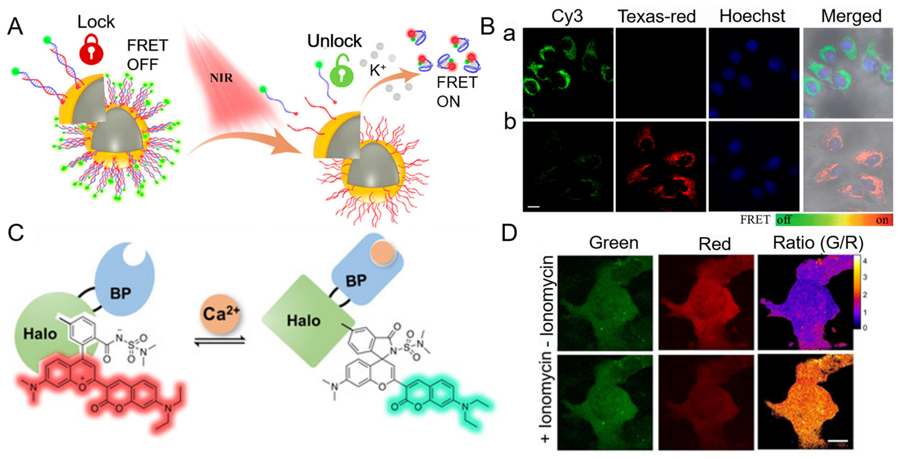
- Cui, M.-R.; Chen, L.-X.; Li, X.-L.; Xu, J.-J.; Chen, H.-Y. NIR Remote-Controlled “Lock–Unlock” Nanosystem for Imaging Potassium Ions in Living Cells. Anal. Chem. 2020, 92, 4558–4565. [Google Scholar] [CrossRef] [PubMed]
- Liu, Z.; Jing, X.; Zhang, S.; Tian, Y. A Copper Nanocluster-Based Fluorescent Probe for Real-Time Imaging and Ratiometric Biosensing of Calcium Ions in Neurons. Anal. Chem. 2019, 91, 2488–2497. [Google Scholar] [CrossRef] [PubMed]
- Chen, D.; Zhao, J.; Zhang, L.; Liu, R.; Huang, Y.; Lan, C.; Zhao, S. Capsicum-Derived Biomass Quantum Dots Coupled with Alizarin Red S as an Inner-Filter-Mediated Illuminant Nanosystem for Imaging of Intracellular Calcium Ions. Anal. Chem. 2018, 90, 13059–13064. [Google Scholar] [CrossRef]
- Pendin, D.; Norante, R.; De Nadai, A.; Gherardi, G.; Vajente, N.; Basso, E.; Kaludercic, N.; Mammucari, C.; Paradisi, C.; Pozzan, T.; et al. A Synthetic Fluorescent Mitochondria-Targeted Sensor for Ratiometric Imaging of Calcium in Live Cells. Angew. Chem. Int. Ed. 2019, 58, 9917–9922. [Google Scholar] [CrossRef]
- Zhou, W.; Saran, R.; Huang, P.-J.J.; Ding, J.; Liu, J. An Exceptionally Selective DNA Cooperatively Binding Two Ca2+ Ions. ChemBioChem 2017, 18, 518–522. [Google Scholar] [CrossRef]
- Li, C.; Chen, P.; Wang, Z.; Ma, X. A DNAzyme-gold nanostar probe for SERS-fluorescence dual-mode detection and imaging of calcium ions in living cells. Sens. Actuator B-Chem. 2021, 347, 130596. [Google Scholar] [CrossRef]
- Wang, L.; Hiblot, J.; Popp, C.; Xue, L.; Johnsson, K. Environmentally Sensitive Color-Shifting Fluorophores for Bioimaging. Angew. Chem. Int. Ed. 2020, 59, 21880–21884. [Google Scholar] [CrossRef]
- Zhu, W.; Takeuchi, S.; Imai, S.; Terada, T.; Ueda, T.; Nasu, Y.; Terai, T.; Campbell, R.E. Chemigenetic indicators based on synthetic chelators and green fluorescent protein. Nat. Chem. Biol. 2023, 19, 38–44. [Google Scholar] [CrossRef]
- van der Linden, F.H.; Mahlandt, E.K.; Arts, J.J.G.; Beumer, J.; Puschhof, J.; de Man, S.M.A.; Chertkova, A.O.; Ponsioen, B.; Clevers, H.; van Buul, J.D.; et al. A turquoise fluorescence lifetime-based biosensor for quantitative imaging of intracellular calcium. Nat. Commun. 2021, 12, 7159. [Google Scholar] [CrossRef] [PubMed]
- Wu, P.; Hwang, K.; Lan, T.; Lu, Y. A DNAzyme-Gold Nanoparticle Probe for Uranyl Ion in Living Cells. J. Am. Chem. Soc. 2013, 135, 5254–5257. [Google Scholar] [CrossRef] [Green Version]
- Li, L.; Feng, J.; Fan, Y.; Tang, B. Simultaneous Imaging of Zn2+ and Cu2+ in Living Cells Based on DNAzyme Modified Gold Nanoparticle. Anal. Chem. 2015, 87, 4829–4835. [Google Scholar] [CrossRef] [PubMed]
- Yang, C.; Yin, X.; Huan, S.-Y.; Chen, L.; Hu, X.-X.; Xiong, M.-Y.; Chen, K.; Zhang, X.-B. Two-Photon DNAzyme–Gold Nanoparticle Probe for Imaging Intracellular Metal Ions. Anal. Chem. 2018, 90, 3118–3123. [Google Scholar] [CrossRef] [PubMed]
- Xiong, M.; Yang, Z.; Lake, R.J.; Li, J.; Hong, S.; Fan, H.; Zhang, X.-B.; Lu, Y. DNAzyme-Mediated Genetically Encoded Sensors for Ratiometric Imaging of Metal Ions in Living Cells. Angew. Chem. Int. Ed. 2020, 59, 1891–1896. [Google Scholar] [CrossRef] [PubMed]
- Zhang, X.; Song, Z.-L.; Chao, Q.; Li, Q.; Kong, R.; Fan, G.-C.; Luo, X. A DNAzyme-based normalized fluorescence strategy for direct quantification of endogenous zinc in living cells. Chem. Commun. 2022, 58, 577–580. [Google Scholar] [CrossRef]
- Yi, D.; Zhao, H.; Zhao, J.; Li, L. Modular Engineering of DNAzyme-Based Sensors for Spatioselective Imaging of Metal Ions in Mitochondria. J. Am. Chem. Soc. 2023, 145, 1678–1685. [Google Scholar] [CrossRef]
- Zhan, J.; Liu, Z.; Liu, R.; Zhu, J.-J.; Zhang, J. Near-Infrared-Light-Mediated DNA-Logic Nanomachine for Bioorthogonal Cascade Imaging of Endogenous Interconnected MicroRNAs and Metal Ions. Anal. Chem. 2022, 94, 16622–16631. [Google Scholar] [CrossRef]
- Jayaraj, A.; Gayathri, M.S.; Sivaraman, G.; Swamy, P.C.A. A highly potential acyclic Schiff base fluorescent turn on sensor for Zn2+ ions and colorimetric chemosensor for Zn2+, Cu2+ and Co2+ ions and its applicability in live cell imaging. J. Photochem. Photobiol. B Biol. 2022, 226, 112371. [Google Scholar] [CrossRef]
- Zhang, G.; Zhao, Y.; Peng, B.; Li, Z.; Xu, C.; Liu, Y.; Zhang, C.; Voelcker, N.H.; Li, L.; Huang, W. A fluorogenic probe based on chelation–hydrolysis-enhancement mechanism for visualizing Zn2+ in Parkinson’s disease models. J. Mat. Chem. B 2019, 7, 2252–2260. [Google Scholar] [CrossRef]
- Wang, P.; Wu, X.; Wu, J.; Liao, Y. Highly selective and sensitive peptide-based fluorescent chemosensor for detection of Zinc(II) ions in aqueous medium and living cells. J. Photochem. Photobiol. A Chem. 2019, 382, 111929. [Google Scholar] [CrossRef]
- Dischler, A.M.; Maslar, D.; Zhang, C.; Qin, Y. Development and Characterization of a Red Fluorescent Protein-Based Sensor RZnP1 for the Detection of Cytosolic Zn2+. ACS Sens. 2022, 7, 3838–3845. [Google Scholar] [CrossRef]
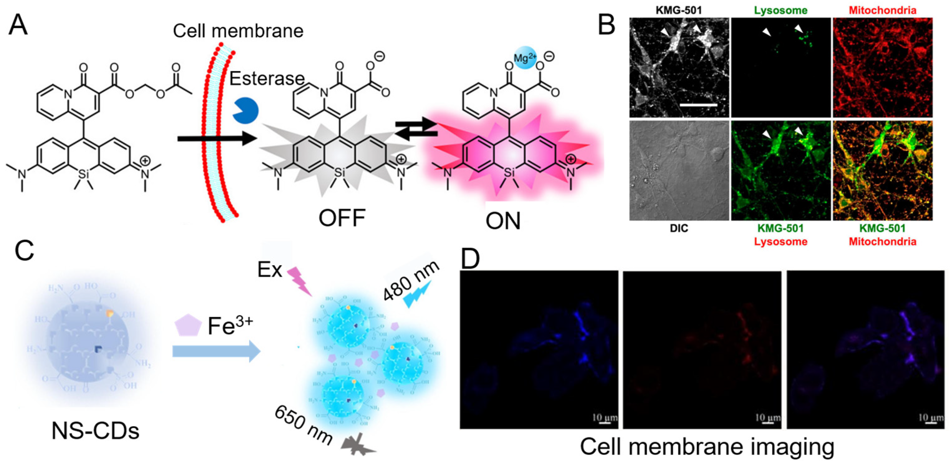
- Murata, O.; Shindo, Y.; Ikeda, Y.; Iwasawa, N.; Citterio, D.; Oka, K.; Hiruta, Y. Near-Infrared Fluorescent Probes for Imaging of Intracellular Mg2+ and Application to Multi-Color Imaging of Mg2+, ATP, and Mitochondrial Membrane Potential. Anal. Chem. 2020, 92, 966–974. [Google Scholar] [CrossRef] [PubMed]
- Sadhanala, H.K.; Aryal, S.; Sharma, K.; Orpaz, Z.; Michaeli, S.; Gedanken, A. Nitrogen-doped carbon dots as a highly selective and sensitive fluorescent probe for sensing Mg2+ ions in aqueous solution, and their application in the detection and imaging of intracellular Mg2+ ions. Sens. Actuator B-Chem. 2022, 366, 131958. [Google Scholar] [CrossRef]
- Yadav, N.; Kumar, R.; Singh, A.K.; Mohiyuddin, S.; Gopinath, P. Systematic approach of chromone skeleton for detecting Mg2+ ion: Applications for sustainable cytotoxicity and cell imaging possibilities. Spectroc. Acta Part A-Molec. Biomolec. Spectr. 2020, 235, 118290. [Google Scholar] [CrossRef]
- Ranee, S.J.; Sivaraman, G.; Pushpalatha, A.M.; Muthusubramanian, S. Quinoline based sensors for bivalent copper ions in living cells. Sens. Actuator B-Chem. 2018, 255, 630–637. [Google Scholar] [CrossRef]
- Chailek, N.; Kaewnok, N.; Petdum, A.; Sirirak, J.; Chaneam, S.; Kamkaew, A.; Girdthep, S.; Wanichacheva, N. Near infrared and colorimetric fluorescence sensor for ultra-selective detection of Cu2+ level with applications in diverse water samples, brain tumor cell and flow injection analysis. J. Photochem. Photobiol. A Chem. 2021, 421, 113533. [Google Scholar] [CrossRef]
- Chang, D.; Shi, L.; Zhang, Y.; Zhang, G.; Zhang, C.; Dong, C.; Shuang, S. Smilax China-derived yellow-fluorescent carbon dots for temperature sensing, Cu2+ detection and cell imaging. Analyst 2020, 145, 2176–2183. [Google Scholar] [CrossRef] [PubMed]
- Wang, Z.; Jia, N.; Zhou, X.; Han, J.; Bu, H. Cu(I)-Catalyzed Click Reaction-Triggered 3D DNA Walker for Constructing an “OFF–ON” Fluorescent Biosensor for Cu2+ Detection. ACS Appl. Bio Mater. 2021, 4, 3571–3578. [Google Scholar] [CrossRef]
- Liang, C.; Xie, X.; Shi, Q.; Feng, J.; Zhang, D.; Huang, X. Nitrogen/sulfur-doped dual-emission carbon dots with tunable fluorescence for ratiometric sensing of ferric ions and cell membrane imaging. Appl. Surf. Sci. 2022, 572, 151447. [Google Scholar] [CrossRef]
- Ge, G.; Li, L.; Chen, M.; Wu, X.; Yang, Y.; Wang, D.; Zuo, S.; Zeng, Z.; Xiong, W.; Guo, C. Green Synthesis of Nitrogen–Doped Carbon Dots from Fresh Tea Leaves for Selective Fe3+ Ions Detection and Cellular Imaging. Nanomaterials 2022, 12, 986. [Google Scholar] [CrossRef]
- Cai, H.; Xu, H.; Chu, H.; Li, J.; Zhang, D. Fabrication of multi-functional carbon dots based on “one stone, three birds” strategy and their applications for the dual-mode Fe3+ detection, effective promotion on cell proliferation and treatment on ferric toxicosis in vitro. J. Mat. Chem. B 2021, 9, 767–782. [Google Scholar] [CrossRef]
- Diao, Q.; Guo, H.; Yang, Z.; Luo, W.; Li, T.; Hou, D. A rhodamine-6G-based “turn-on” fluorescent probe for selective detection of Fe3+ in living cells. Anal. Methods 2019, 11, 794–799. [Google Scholar] [CrossRef]
- Wang, Y.; Liu, F.; Pu, C.; Tong, Z.; Wang, M.; Wang, J. Galactose-imidazole mediated dual-targeting fluorescent probe for detecting Fe3+ in the lysosomes of hepatocytes: Design, synthesis and evaluation. Biosens. Bioelectron. 2022, 204, 114083. [Google Scholar] [CrossRef] [PubMed]
- Rattanopas, S.; Piyanuch, P.; Wisansin, K.; Charoenpanich, A.; Sirirak, J.; Phutdhawong, W.; Wanichacheva, N. Indole-based fluorescent sensors for selective sensing of Fe2+ and Fe3+ in aqueous buffer systems and their applications in living cells. J. Photochem. Photobiol. A Chem. 2019, 377, 138–148. [Google Scholar] [CrossRef]
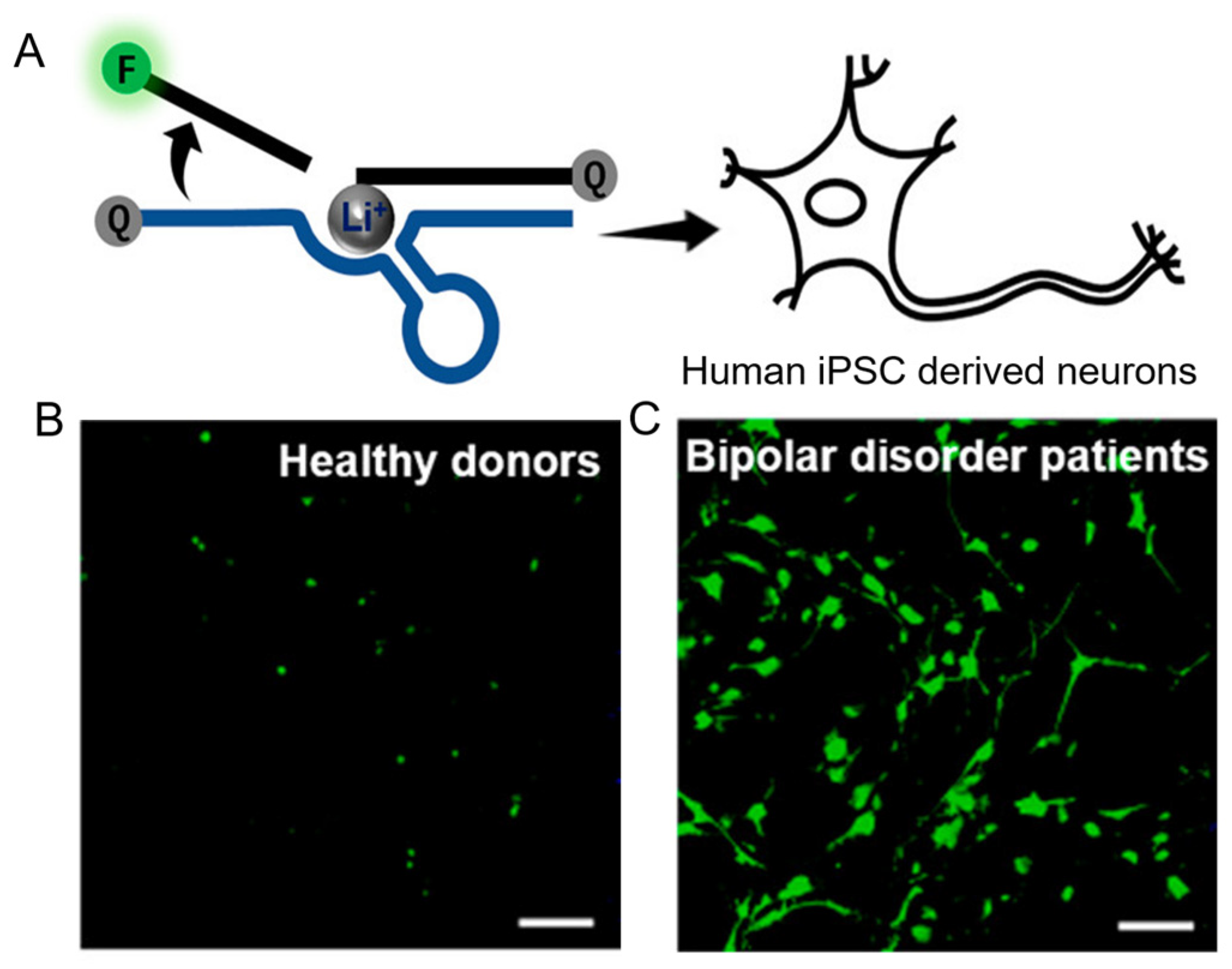
- McGhee, C.E.; Yang, Z.; Guo, W.; Wu, Y.; Lyu, M.; De Long, C.J.; Hong, S.; Ma, Y.; McInnis, M.G.; O’Shea, K.S.; et al. DNAzyme-Based Lithium-Selective Imaging Reveals Higher Lithium Accumulation in Bipolar Disorder Patient-Derived Neurons. ACS Central Sci. 2021, 7, 1809–1820. [Google Scholar] [CrossRef] [PubMed]
- Jiao, Y.; Gao, Y.; Meng, Y.; Lu, W.; Liu, Y.; Han, H.; Shuang, S.; Li, L.; Dong, C. One-Step Synthesis of Label-Free Ratiometric Fluorescence Carbon Dots for the Detection of Silver Ions and Glutathione and Cellular Imaging Applications. ACS Appl. Mater. Interfaces 2019, 11, 16822–16829. [Google Scholar] [CrossRef] [PubMed]
- Yu, Q.; Shi, J.; Mudiyanselage, A.P.K.K.K.; Wu, R.; Zhao, B.; Zhou, M.; You, M. Genetically encoded RNA-based sensors for intracellular imaging of silver ions. Chem. Commun. 2019, 55, 707–710. [Google Scholar] [CrossRef] [PubMed]
- Tang, H.-Y.; Gao, Y.; Li, B.; Li, C.-W.; Guo, Y. Reaction-based colorimetric and ratiometric fluorescent probe for highly selective detection of silver ions. Sens. Actuator B-Chem. 2018, 270, 562–569. [Google Scholar] [CrossRef]
- Bu, F.; Zhao, B.; Kan, W.; Wang, L.; Song, B.; Wang, J.; Zhang, Z.; Deng, Q.; Yin, G. A phenanthro[9,10-d]imidazole-based AIE active fluorescence probe for sequential detection of Ag+/AgNPs and SCN− in water and saliva samples and its application in living cells. Spectroc. Acta Part A-Molec. Biomolec. Spectr. 2019, 223, 117333. [Google Scholar] [CrossRef]
- Yu, M.; Liu, B.; Guo, J.; Wu, F. A sustainable modificatory 1, 2- alternate thiacalix[4]arene for detection of silver ion. Dyes Pigment. 2023, 210, 110983. [Google Scholar] [CrossRef]
- Soleja, N.; Mohsin, M. Real time quantification of intracellular nickel using genetically encoded FRET-based nanosensor. Int. J. Biol. Macromol. 2019, 138, 648–657. [Google Scholar] [CrossRef]
- Wang, X.; Liu, C.; Zhu, H.; Cheng, S.; Zhang, Y.; Su, M.; Rong, X.; Yu, M.; Sheng, W.; Zhu, B. An ultra-sensitive and highly selective fluorescent probe for nickel ions and its environmental and biological applications. Sens. Actuator B-Chem. 2022, 369, 132300. [Google Scholar] [CrossRef]
- Assiri, M.A.; Junaid, H.M.; Waseem, M.T.; Hamad, A.; Shah, S.H.; Iqbal, J.; Rauf, W.; Shahzad, S.A. AIEE active sensors for fluorescence enhancement based detection of Ni2+ in living cells: Mechanofluorochromic and photochromic properties with reversible sensing of acid and base. Anal. Chim. Acta. 2022, 1234, 340516. [Google Scholar] [CrossRef] [PubMed]
- Gu, Y.-Q.; Shen, W.-Y.; Zhou, Y.; Chen, S.-F.; Mi, Y.; Long, B.-F.; Young, D.J.; Hu, F.-L. A pyrazolopyrimidine based fluorescent probe for the detection of Cu2+ and Ni2+ and its application in living cells. Spectroc. Acta Part A-Molec. Biomolec. Spectr. 2019, 209, 141–149. [Google Scholar] [CrossRef] [PubMed]
- Gui, S.; Huang, Y.; Zhu, Y.; Jin, Y.; Zhao, R. Biomimetic Sensing System for Tracing Pb2+ Distribution in Living Cells Based on the Metal–Peptide Supramolecular Assembly. ACS Appl. Mater. Interfaces 2019, 11, 5804–5811. [Google Scholar] [CrossRef] [PubMed]
- Mehta, P.K.; Jeon, J.; Ryu, K.; Park, S.-H.; Lee, K.-H. Ratiometric fluorescent detection of lead ions in aquatic environment and living cells using a fluorescent peptide-based probe. J. Hazard. Mater. 2022, 427, 128161. [Google Scholar] [CrossRef]
- Wen, J.; Lv, Y.; Xia, P.; Liu, F.; Xu, Y.; Li, H.; Chen, S.-S.; Sun, S. A water-soluble near-infrared fluorescent probe for specific Pd2+ detection. Biorg. Med. Chem. 2018, 26, 931–937. [Google Scholar] [CrossRef]
- Wang, D.; Hou, H.; Chen, W.; Wu, Y.; Peng, X.; Song, F. A turn-on fluorescent probe for palladium(II) detection with a large Stokes shift and lysosomes-targeting ability. Tetrahedron Lett. 2022, 102, 153932. [Google Scholar] [CrossRef]
- Ashwin, B.C.M.A.; Sivaraman, G.; Stalin, T.; Yuvakkumar, R.; Muthu Mareeswaran, P. Selective and sensitive fluorescent sensor for Pd2+ using coumarin 460 for real-time and biological applications. J. Photochem. Photobiol. B Biol. 2018, 183, 302–308. [Google Scholar] [CrossRef]
- Zhang, X.-P.; Yuan, Q.; Qi, Y.-L.; Zheng, D.-J.; Liu, Q.-X.; Wang, B.-Z.; Yang, Y.-S.; Zhu, H.-L. An umbelliferone-derivated fluorescent sensor for selective detection of palladium(II) from palladium(0) in living cells. Spectroc. Acta Part A-Molec. Biomolec. Spectr. 2019, 220, 117134. [Google Scholar] [CrossRef]
- Reagen, S.; Wu, Y.; Liu, X.; Shahni, R.; Bogenschuetz, J.; Wu, X.; Chu, Q.R.; Oncel, N.; Zhang, J.; Hou, X.; et al. Synthesis of Highly Near-Infrared Fluorescent Graphene Quantum Dots Using Biomass-Derived Materials for In Vitro Cell Imaging and Metal Ion Detection. ACS Appl. Mater. Interfaces 2021, 13, 43952–43962. [Google Scholar] [CrossRef]
- Sun, Y.; Wang, L.; Zhou, J.; Qin, D.; Duan, H. A new phenothiazine-based fluorescence sensor for imaging Hg2+ in living cells. Appl. Organomet. Chem. 2020, 34, e5945. [Google Scholar] [CrossRef]
- Zhong, W.; Wang, L.; Qin, D.; Zhou, J.; Duan, H. Two Novel Fluorescent Probes as Systematic Sensors for Multiple Metal Ions: Focus on Detection of Hg2+. ACS Omega 2020, 5, 24285–24295. [Google Scholar] [CrossRef] [PubMed]
- Ding, G.; Wu, L.; Feng, H.; Liu, Y.; Li, J.; Si, H.; Yao, X.; He, M.; He, W. The specific binding of a new 1,2,3-triazole to three blood proteins and it’s appended rhodamine complex for selective detection of Hg2+. Spectroc. Acta Part A-Molec. Biomolec. Spectr. 2020, 228, 117728. [Google Scholar] [CrossRef] [PubMed]
- Wang, G.; Xu, W.; Liu, Y.; Fu, N. Ultrasensitive detection and high-contrast bioimaging of Hg2+ using monothiosquaraine-based fluorescent probe via hydrogen bond promoted desulfurization. Microchem. J. 2022, 179, 107481. [Google Scholar] [CrossRef]
- Lin, W.; Xie, X.; Wang, Y.; Chen, J. A New Fluorescent Probe for Selective Cd2+ Detection and Cell Imaging. Z. Anorg. Allg. Chem. 2019, 645, 645–648. [Google Scholar] [CrossRef]
- Wang, P.; An, Y.; Liao, Y. A novel peptide-based fluorescent chemosensor for Cd(II) ions and its applications in bioimaging. Spectroc. Acta Part A-Molec. Biomolec. Spectr. 2019, 216, 61–68. [Google Scholar] [CrossRef]
- Guo, Y.; Li, T.; Xie, L.; Tong, X.; Tang, C.; Shi, S. Red pitaya peels-based carbon dots for real-time fluorometric and colorimetric assay of Au3+, cellular imaging, and antioxidant activity. Anal. Bioanal. Chem. 2021, 413, 935–943. [Google Scholar] [CrossRef]
- Bhattacharyya, A.; Makhal, S.C.; Guchhait, N. CHEF-Affected Fluorogenic Nanomolar Detection of Al3+ by an Anthranilic Acid–Naphthalene Hybrid: Cell Imaging and Crystal Structure. ACS Omega 2018, 3, 11838–11846. [Google Scholar] [CrossRef] [Green Version]
- Berrones-Reyes, J.; Muñoz-Flores, B.M.; Gómez-Treviño, A.; Treto-Suárez, M.A.; Páez-Hernández, D.; Schott, E.; Zarate, X.; Jiménez-Pérez, V.M. Novel fluorescent Schiff bases as Al3+ sensors with high selectivity and sensitivity, and their bioimaging applications. Mater. Chem. Phys. 2019, 233, 89–101. [Google Scholar] [CrossRef]
- Wang, C.-X.; Ai, S.-L.; Wu, B.; Huang, S.-W.; Liu, Z. Biotinylated and fluorophore-incorporated polymeric mixed micelles for tumor cell-specific turn-on fluorescence imaging of Al3+. J. Mat. Chem. B 2020, 8, 3557–3565. [Google Scholar] [CrossRef]
- Fenn, W.O. The Rôle of Potassium in Physiological Processes. Physiol. Rev. 1940, 20, 377–415. [Google Scholar] [CrossRef]
- Ning, J.; Lin, X.; Su, F.; Sun, A.; Liu, H.; Luo, J.; Wang, L.; Tian, Y. Development of a molecular K+ probe for colorimetric/fluorescent/photoacoustic detection of K+. Anal. Bioanal. Chem. 2020, 412, 6947–6957. [Google Scholar] [CrossRef] [PubMed]
- Liu, J.; Pan, L.; Shang, C.; Lu, B.; Wu, R.; Feng, Y.; Chen, W.; Zhang, R.; Bu, J.; Xiong, Z.; et al. A highly sensitive and selective nanosensor for near-infrared potassium imaging. Sci. Adv. 2020, 6, eaax9757. [Google Scholar] [CrossRef] [PubMed] [Green Version]
- Liu, J.; Li, F.; Wang, Y.; Pan, L.; Lin, P.; Zhang, B.; Zheng, Y.; Xu, Y.; Liao, H.; Ko, G.; et al. A sensitive and specific nanosensor for monitoring extracellular potassium levels in the brain. Nat. Nanotechnol. 2020, 15, 321–330. [Google Scholar] [CrossRef]
- Akerboom, J.; Chen, T.-W.; Wardill, T.J.; Tian, L.; Marvin, J.S.; Mutlu, S.; Calderón, N.C.; Esposti, F.; Borghuis, B.G.; Sun, X.R.; et al. Optimization of a GCaMP Calcium Indicator for Neural Activity Imaging. J. Neurosci. 2012, 32, 13819. [Google Scholar] [CrossRef] [PubMed] [Green Version]
- Dana, H.; Sun, Y.; Mohar, B.; Hulse, B.K.; Kerlin, A.M.; Hasseman, J.P.; Tsegaye, G.; Tsang, A.; Wong, A.; Patel, R.; et al. High-performance calcium sensors for imaging activity in neuronal populations and microcompartments. Nat. Methods 2019, 16, 649–657. [Google Scholar] [CrossRef]
- Zarowny, L.; Aggarwal, A.; Rutten, V.M.S.; Kolb, I.; Patel, R.; Huang, H.-Y.; Chang, Y.-F.; Phan, T.; Kanyo, R.; Ahrens, M.B.; et al. Bright and High-Performance Genetically Encoded Ca2+ Indicator Based on mNeonGreen Fluorescent Protein. ACS Sens. 2020, 5, 1959–1968. [Google Scholar] [CrossRef]
- Shemetov, A.A.; Monakhov, M.V.; Zhang, Q.; Canton-Josh, J.E.; Kumar, M.; Chen, M.; Matlashov, M.E.; Li, X.; Yang, W.; Nie, L.; et al. A near-infrared genetically encoded calcium indicator for in vivo imaging. Nat. Biotechnol. 2021, 39, 368–377. [Google Scholar] [CrossRef]
- Li, M.; Xing, Y.; Zou, Y.; Chen, G.; You, J.; Yu, F. Imaging of the mutual regulation between zinc cation and nitrosyl via two-photon fluorescent probes in cells and in vivo. Sens. Actuator B-Chem. 2020, 309, 127772. [Google Scholar] [CrossRef]
- Wu, L.; Liu, Y.; Wu, X.; Li, Y.; Du, J.; Qi, S.; Yang, Q.; Xu, H.; Li, Y. A novel Near-Infrared fluorescent probe for Zn2+ and CN– double detection based on dicyanoisfluorone derivatives with highly sensitive and selective, and its application in Bioimaging. Spectroc. Acta Part A-Molec. Biomolec. Spectr. 2022, 267, 120621. [Google Scholar] [CrossRef]
- Yang, Z.; Loh, K.Y.; Chu, Y.-T.; Feng, R.; Satyavolu, N.S.R.; Xiong, M.; Nakamata Huynh, S.M.; Hwang, K.; Li, L.; Xing, H.; et al. Optical Control of Metal Ion Probes in Cells and Zebrafish Using Highly Selective DNAzymes Conjugated to Upconversion Nanoparticles. J. Am. Chem. Soc. 2018, 140, 17656–17665. [Google Scholar] [CrossRef]
- Wang, X.; Kim, G.; Chu, J.L.; Song, T.; Yang, Z.; Guo, W.; Shao, X.; Oelze, M.L.; Li, K.C.; Lu, Y. Noninvasive and Spatiotemporal Control of DNAzyme-Based Imaging of Metal Ions In Vivo Using High-Intensity Focused Ultrasound. J. Am. Chem. Soc. 2022, 144, 5812–5819. [Google Scholar] [CrossRef] [PubMed]
- Vijay, N.; Wu, S.P.; Velmathi, S. Turn on fluorescent chemosensor containing rhodamine B fluorophore for selective sensing and in vivo fluorescent imaging of Fe3+ ions in HeLa cell line and zebrafish. J. Photochem. Photobiol. A Chem. 2019, 384, 112060. [Google Scholar] [CrossRef]
- Chang, D.; Zhao, Z.; Niu, W.; Shi, L.; Yang, Y. Iron ion sensing and in vitro and in vivo imaging based on bright blue-fluorescent carbon dots. Spectroc. Acta Part A-Molec. Biomolec. Spectr. 2021, 260, 119964. [Google Scholar] [CrossRef] [PubMed]
- Chang, D.; Zhao, Z.; Shi, L.; Liu, W.; Yang, Y. Lysosome-targeted carbon dots for colorimetric and fluorescent dual mode detection of iron ion, in vitro and in vivo imaging. Talanta 2021, 232, 122423. [Google Scholar] [CrossRef] [PubMed]
- Zhu, M.; Zhao, Z.; Liu, X.; Chen, P.; Fan, F.; Wu, X.; Hua, R.; Wang, Y. A novel near-infrared fluorimetric method for point-of-care monitoring of Fe2+ and its application in bioimaging. J. Hazard. Mater. 2021, 406, 124767. [Google Scholar] [CrossRef] [PubMed]
- Zheng, J.; Feng, S.; Gong, S.; Xia, Q.; Feng, G. In vivo imaging of Fe2+ using an easily obtained probe with a large Stokes shift and bright strong lipid droplet-targetable near-infrared fluorescence. Sens. Actuator B-Chem. 2020, 309, 127796. [Google Scholar] [CrossRef]
- Wen, X.; Wen, G.; Li, W.; Zhao, Z.; Duan, X.; Yan, W.; Trant, J.F.; Li, Y. Carbon dots for specific “off-on” sensing of Co2+ and EDTA for in vivo bioimaging. Mater. Sci. Eng. B 2021, 123, 112022. [Google Scholar] [CrossRef]
- Stubing, D.B.; Heng, S.; Abell, A.D. Crowned spiropyran fluoroionophores with a carboxyl moiety for the selective detection of lithium ions. Org. Biomol. Chem. 2016, 14, 3752–3757. [Google Scholar] [CrossRef] [Green Version]
- Kang, J.; Li, E.; Cui, L.; Shao, Q.; Yin, C.; Cheng, F. Lithium ion specific fluorescent reversible extraction-release based on spiropyran isomerization combining crown ether coordination and its bioimaging. Sens. Actuator B-Chem. 2021, 327, 128941. [Google Scholar] [CrossRef]
- Huang, L.; Chen, F.; Zong, X.; Lu, Q.; Wu, C.; Ni, Z.; Liu, M.; Zhang, Y. Near-infrared light excited UCNP-DNAzyme nanosensor for selective detection of Pb2+ and in vivo imaging. Talanta 2021, 227, 122156. [Google Scholar] [CrossRef] [PubMed]
- Ravichandiran, P.; Prabakaran, D.S.; Maroli, N.; Boguszewska-Czubara, A.; Masłyk, M.; Kim, A.R.; Kolandaivel, P.; Ramalingam, P.; Park, B.-H.; Han, M.-K.; et al. Mitochondria-targeted dual-channel colorimetric and fluorescence chemosensor for detection of Sn2+ ions in aqueous solution based on aggregation-induced emission and its bioimaging applications. J. Hazard. Mater. 2021, 415, 125593. [Google Scholar] [CrossRef]
- Wang, J.H.; Liu, Y.M.; Chao, J.B.; Wang, H.; Wang, Y.; Shuang, S. A simple but efficient fluorescent sensor for ratiometric sensing of Cd2+ and bio-imaging studies. Sens. Actuator B-Chem. 2020, 303, 127216. [Google Scholar] [CrossRef]
- Chen, C.-G.; Vijay, N.; Thirumalaivasan, N.; Velmathi, S.; Wu, S.-P. Coumarin-based Hg2+ fluorescent probe: Fluorescence turn-on detection for Hg2+ bioimaging in living cells and zebrafish. Spectroc. Acta Part A-Molec. Biomolec. Spectr. 2019, 219, 135–140. [Google Scholar] [CrossRef]
- Wang, X.; Cheng, S.; Liu, C.; Zhang, Y.; Su, M.; Rong, X.; Zhu, H.; Yu, M.; Sheng, W.; Zhu, B. A novel ratiometric fluorescent probe for the detection of nickel ions in the environment and living organisms. Sci. Total Environ. 2022, 840, 156445. [Google Scholar] [CrossRef] [PubMed]
- Tai, A.W.; Lien, E.J.; Lai, M.M.C.; Khwaja, T.A. Novel N-hydroxyguanidine derivatives as anticancer and antiviral agents. J. Med. Chem. 1984, 27, 236–238. [Google Scholar] [CrossRef]
- Tian, H.; Qiao, X.; Zhang, Z.-L.; Xie, C.-Z.; Li, Q.-Z.; Xu, J.-Y. A high performance 2-hydroxynaphthalene Schiff base fluorescent chemosensor for Al3+ and its applications in imaging of living cells and zebrafish in vivo. Spectroc. Acta Part A-Molec. Biomolec. Spectr. 2019, 207, 31–38. [Google Scholar] [CrossRef]
- Lian, W.-J.; Wang, X.-T.; Xie, C.-Z.; Tian, H.; Song, X.-Q.; Pan, H.-T.; Qiao, X.; Xu, J.-Y. Mixed-ligand copper(ii) Schiff base complexes: The role of the co-ligand in DNA binding, DNA cleavage, protein binding and cytotoxicity. Dalton Trans. 2016, 45, 9073–9087. [Google Scholar] [CrossRef]
- Wang, Q.; Yang, L.; Wang, H.; Song, J.; Ding, H.; Tang, X.-H.; Yao, H. A highly selective and sensitive turn-on fluorescent probe for the detection of Al3+ and its bioimaging. Luminescence 2017, 32, 779–785. [Google Scholar] [CrossRef] [PubMed]


| Analytes | Normal Level Range in Biological System | Reference |
|---|---|---|
| Na+ | 135–145 mM (serum) | [9,10,11] |
| K+ | 3.5–5.4 mM (serum), 19–66 mM (urea) | [12,13] |
| Ca2+ | 10−6 M (intracellular), 10−3 M (extracellular fluid) | [14] |
| Mg2+ | 0.65–1.05 mM (serum) | [15] |
| Cu2+ | 1.4–2.1 mg/kg (adult human body) | [16] |
| Zn2+ | 12–16 μM (serum) | [17] |
| Fe3+ | 14–32 μM (serum) | [18] |
Disclaimer/Publisher’s Note: The statements, opinions and data contained in all publications are solely those of the individual author(s) and contributor(s) and not of MDPI and/or the editor(s). MDPI and/or the editor(s) disclaim responsibility for any injury to people or property resulting from any ideas, methods, instructions or products referred to in the content. |
© 2023 by the authors. Licensee MDPI, Basel, Switzerland. This article is an open access article distributed under the terms and conditions of the Creative Commons Attribution (CC BY) license (https://creativecommons.org/licenses/by/4.0/).
Share and Cite
Shi, Y.; Zhang, W.; Xue, Y.; Zhang, J. Fluorescent Sensors for Detecting and Imaging Metal Ions in Biological Systems: Recent Advances and Future Perspectives. Chemosensors 2023, 11, 226. https://doi.org/10.3390/chemosensors11040226
Shi Y, Zhang W, Xue Y, Zhang J. Fluorescent Sensors for Detecting and Imaging Metal Ions in Biological Systems: Recent Advances and Future Perspectives. Chemosensors. 2023; 11(4):226. https://doi.org/10.3390/chemosensors11040226
Chicago/Turabian StyleShi, Yang, Wenxian Zhang, Yi Xue, and Jingjing Zhang. 2023. "Fluorescent Sensors for Detecting and Imaging Metal Ions in Biological Systems: Recent Advances and Future Perspectives" Chemosensors 11, no. 4: 226. https://doi.org/10.3390/chemosensors11040226