Review

A Critical Overview of Enzyme-Based Electrochemical Biosensors for L-Dopa Detection in Biological Samples

Department of Sciences, University of Basilicata, Via dell’Ateneo Lucano 10, 85100 Potenza, Italy
*
Author to whom correspondence should be addressed.
Chemosensors 2023, 11(10), 523; https://doi.org/10.3390/chemosensors11100523
Submission received: 27 July 2023 / Revised: 26 September 2023 / Accepted: 2 October 2023 / Published: 5 October 2023
(This article belongs to the Collection Electrochemical Biosensors for Medical Diagnosis)

Abstract

:
L-Dopa is an intermediate amino acid in the biosynthesis of endogenous catecholamines, such as dopamine. It is currently considered to be the optimal dopaminergic treatment for Parkinson’s disease, a neurodegenerative disorder affecting around 1% of the population. In an advanced stage of the disease, complications such as dyskinesia and psychosis are caused by fluctuations in plasma drug levels. Real-time monitoring of L-Dopa levels would be advantageous for properly adjusting drug dosing, thus improving therapeutic efficacy. Electrochemical methods have advantages such as easy-to-use instrumentation, fast response time, and high sensitivity, and are suitable for miniaturization, enabling the fabrication of implantable or wearable devices. This review reports on research papers of the past 20 years (2003–2023) dealing with enzyme-based biosensors for the electrochemical detection of L-Dopa in biological samples. Specifically, amperometric and voltammetric biosensors, whose output signal is a measurable current, are discussed. The approach adopted includes an initial study of the steps required to assemble the devices, i.e., electrode modification and enzyme immobilization. Then, all issues related to their analytical performance in terms of sensitivity, selectivity, and capability to analyze real samples are critically discussed. The paper aims to provide an assessment of recent developments while highlighting limitations such as poor selectivity and long-term stability, and the laborious and time-consuming fabrication protocol that needs to be addressed from the perspective of the integrated clinical management of Parkinson’s disease.

1. Introduction

L-Dopa, levodopa, or L-3,4-dihydroxyphenylalanine (LD) belongs to the catecholamine family, a class of compounds synthesized from the amino acid L-tyrosine containing both an amine group and the 1,2-dihydroxy substituted benzene ring, or catechol. LD is an aromatic amino acid analog and represents the precursor of dopamine (DP) and norepinephrine (NP), which are two important neurotransmitters acting on psychomotor and emotional functions (chemical structures are shown in Figure 1) [1].
LD is present in the brain, skin, and adrenal glands, and is involved in different pathological conditions. In the striatum, it performs a modulatory action as a potentiator for presynaptic β-adrenergic receptors, therefore facilitating the release of DP; it is also an enhancer of the post-synaptic D-2 DP receptors. In addition, its possible role as a neurotransmitter at the level of the primary baroreceptor afferents in the nucleus tractus solitarius has been reported. Therefore, it performs an important physiological function in the area involved in the baroreceptive reflex and the regulation of blood pressure [2].
From a historical point of view, LD was considered biologically inactive for a long time, i.e., a mere precursor compound involved in the biosynthesis of catecholamines. Only in 1961–67 was LD revalued as the most effective pharmacological treatment against Parkinson’s disease (PD), proving to be a revolutionarily effective therapeutic agent [2].
Parkinson’s disease (PD) is second only to Alzheimer’s disease (AD) as the most common worldwide neurodegenerative disease of the central nervous system: approximately 1% of the population over the age of 60 suffers from it [3,4,5]. The diagnostic symptoms of PD are mainly motor disabling, e.g., tremors, bradykinesia, postural instability, rigidity, loss of coordinated movements, slow movement (akinesia), and the absence of postural reflexes with bone weakening, causing falls and shortened life expectancy [6,7,8]. The onset of PD also causes psychological symptoms such as depression (40% of PD patients) [3], as well as anxiety and cognitive and decision-making impairment [9,10]. Although the etiology of PD is still being studied and investigated, it is well known that the disease arises from the death of the dopaminergic neuronal cells of the substantia nigra pars compacta, a brain area functional to motor physiological control and coordination [11]. The consequent formation of Lewy bodies (i.e., aggregates made up of α-synuclein and ubiquitin), which interfere with the normal functioning of dopaminergic neurons, cause irreversible degeneration [12]. LD can improve PD symptoms, as it is the physiological precursor of DP. On the other hand, DP cannot be considered for drug treatment since it is unable to cross the blood–brain barrier (BBB) and reach the sites involved in the disease. Currently, in pharmacological formulations, LD is combined with inhibitors of DOPA-decarboxylase (benserazide or carbidopa) and catechol-O-methyltransferase (entacapone or tolcapone). These compounds inhibit LD decarboxylation and ortho-methylation at the synaptic level, respectively, increasing the LD half-life and the amount that can reach the central nervous system [13,14] (Figure 2).
DP replacement therapy with the introduction of LD is still the most effective and best-tolerated medical therapy for motor symptom control [15], but it presents some disadvantages, too. As PD progresses, its long-term intake can induce serious side effects, e.g., motor fluctuations (called on–off phenomenon), orthostatic hypotension, hallucinations, and dyskinesia, but also psychosis and orthostatic hypotension in 80% of patients [16,17]. Because of these reasons, extended-release LD formulations coupled with drugs have been developed to prolong the half-life bioavailability and reduce side effects [18,19,20]. Recently, specific natural sources rich in LD have also been considered to be possible adjuvants in the treatment of PD. The intake of LD-containing food could compensate for drug fluctuations in plasma that cause motor symptoms. Furthermore, the availability of a natural source of LD would be advantageous, considering that the synthesis of the drug is chemically expensive due to the metal catalyst and the advanced technologies [21]. In particular, it has been shown that some species of the Fabaceae family contain large amounts of LD [22]. Mucuna pruriens seeds contain the most significant amount of analyte (up to 10%) [23,24]. Broad bean (Vicia faba L.) has also been identified as a rich source of LD [25].
Considering the drug fluctuations and the high interpatient variability of the response to LD therapy, the real-time monitoring of LD levels in biological samples would allow the drug dosage to be adapted based on patient condition. In this way, it would be possible to reduce side effects and enhance the efficacy of drug treatment.
The methods used for LD detection are mainly based on high-performance liquid chromatography (HPLC) [26,27] coupled with UV detection and mass spectrometry [28,29]. Capillary zone electrophoresis has been employed rarely [30]. Spectrophotometric [31], fluorescence [32], and chemiluminescence methods [33,34] have also been reported. High sensitivity and selectivity have been achieved. Nevertheless, complex sample pretreatment, along with the use of sophisticated instrumentation and long response times, makes such techniques not suitable for the real-time monitoring of LD.
Conversely, cheap and easy-to-use instrumentation, fast response time, and minimal sample pretreatment are among the main advantages of electrochemical methods, which also show high reproducibility and sensitivity, and are suitable for miniaturization [35], allowing for the realization of implantable or wearable devices for the in vivo monitoring of biomolecules.
To date, as recently reviewed, several electrochemical methods for LD detection have been proposed as valid alternatives to conventional ones [35,36,37]. Levodopa can easily undergo oxidation thanks to the presence of the catechol moiety, following an EqCiEq mechanism [38]. To be precise, LD is oxidized to dopaquinone by a two-electron process involving deprotonation. The oxidation potential of LD is influenced by the pH of the solution, which plays an important role in the overall electrode process. In particular, at moderate acidic and neutral pH, the amine group in dopaquinone undergoes deprotonation. The deprotonated amine group is involved in the intramolecular Michael addition reaction with the o-benzoquinone ring, generating cyclodopa as a product. Cyclodopa can then oxidize into dopachrome (Figure 3).
Electrochemical sensors work based on the direct detection of LD through its oxidation on the electrode surface. A drawback typical of such detection is the presence of endogenous compounds in biological samples, such as ascorbic acid or uric acid, which oxidize at the detection potential, generating a superimposed current signal. Alternatively, an electrochemical biosensor can be used, which, according to the IUPAC definition [39], is a “self-contained integrated device, which is capable of providing specific quantitative or semiquantitative analytical information using a biological recognition element (biochemical receptor) which is retained in direct spatial contact with an electrochemical transduction element”.
In this last case, LD oxidation into dopaquinone is catalyzed by a poliphenoloxidase-type enzyme—the biochemical receptor—and the electrochemical signal is typically generated following the dopaquinone reduction at the working electrode. In addition to all the advantages of the electrochemical methods listed above, the operating mode of biosensors should allow the reduction of interference problems from compounds prone to oxidation. On the other hand, since dopaquinone is the detected compound, operational conditions should be properly adjusted to limit the extent of the cyclization process, which is favored at alkaline pH values.
The aim of this review is to provide an assessment of amperometric biosensors for the detection of LD in biological samples, highlighting the progress made in this field in recent years while critically evaluating the drawbacks that still need to be addressed.
It is worth noting that a wider definition of biosensors focused on this type of analyte has often been employed. Modified electrodes, containing non-biologically active elements based on different inorganic or organic materials with good electrical conductivity and catalytic properties, have been reported as biosensors because they have been employed for biomonitoring, i.e., for monitoring biomolecules such as LD. Throughout this review, we will consider only biosensors for LD containing an enzyme as a biorecognition element, according to the IUPAC convention. Among electroanalytical devices for LD detection, chemical sensors have been more extensively investigated with respect to biosensors. Indeed, in the development of biosensors, crucial issues must be considered. In addition to the realization of an efficient conductive surface as an electrochemical transducer, which is also required in the case of chemical sensors, a proper method for enzyme immobilization must be designed. Following a brief description of the enzymes used, both these aspects are then elaborated on in the review, with particular reference to the complex and time-consuming modification protocols often required to assure high sensitivity and adequate stability. A critical discussion is then presented on the strategies adopted to solve the interference problems deriving from the wide substrate specificity of the enzymes used. Finally, the possibility of employing “noninvasive” biological samples to monitor LD levels during drug treatment is described. The realization of “wearable devices” is detailed as a possible perspective for integrated clinical management of PD.

2. Enzymes for L-Dopa Biosensing
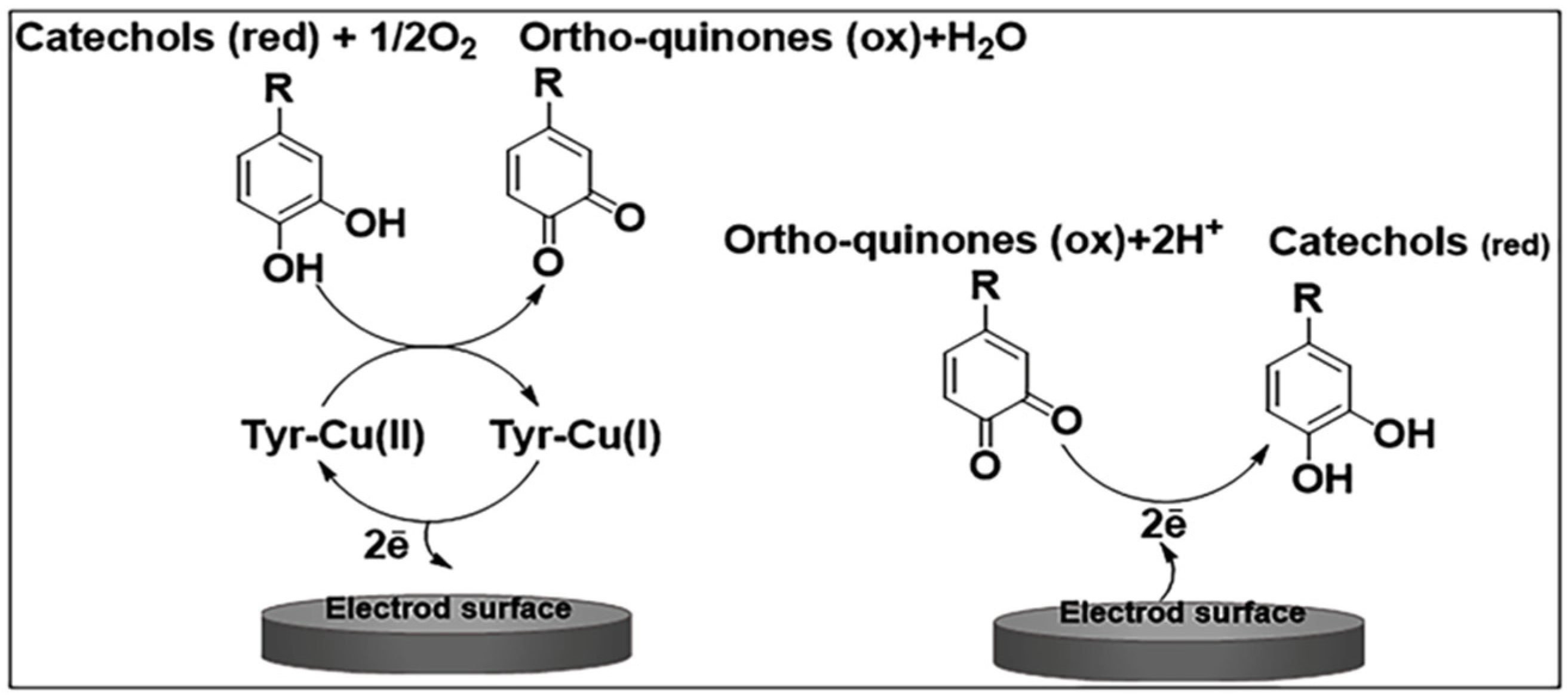
Polyphenol oxidases (PPO, EC 1.10.3.1) are copper-containing enzymes that can mediate reactions based on electron transfer in the absence of cofactors. They catalyze the oxidation of phenolic compounds by employing oxygen as a co-substrate, showing good stability but low specificity towards the substrate [40]. PPOs are grouped into tyrosinase (TYR) and catechol oxidase (CO). Although TYR exerts the double catalytic activity of cresolase, through the hydroxylation of monophenols to diphenols, and catecholase, through the oxidation of o-diphenols, CO can only convert diphenols into o-diquinones [41,42]. To date, the origin of the functional discrimination between TYR and CO is unclear [43]. The quinone resulting from PPO activity is an unstable species, which leads to the formation of brown polymeric compounds, which, for example, in plants, exert a protective function against damage caused by insects or pathogens [44]. Indeed, PPOs are widely distributed in nature among living organisms, occurring ubiquitously in plants but also in other organisms such as fungi, mammals, and prokaryotes [42].
Two highly conserved copper-binding domains are present in plant PPOs, showing high homology with that of bacterial, fungal, or mammalian tyrosinases. Generally, PPOs are part of the type III copper enzyme family comprising hemocyanins and laccases.
Laccase is a multicopper enzyme belonging to the oxidoreductase family. It catalyzes the oxidation of several compounds, such as diphenols and polyphenols, polyamines, aromatic amines, etc. Based on spectroscopic characteristics, the active site of the multicopper enzyme can be classified into three groups: copper type I, copper type II, and two copper type III [45]. This enzyme can mediate the oxidation of aromatic and non-aromatic compounds, particularly phenolic, by an oxygen-dependent reaction with the reduction of oxygen to water, and exhibits p-hydroxylation activity [45,46]. Generally, a laccase enzymatic reaction leads to the formation of phenoxy radical (semiquinone), which is oxidized to o-quinone in the second stage [47].
Despite the low substrate specificity, PPOs are considered to be interesting enzymes to use in the field of biosensors due to their stability [48]. The enzyme generally employed to realize biosensors for LD determination is tyrosinase.

Tyrosinase

Tyrosinase (EC 1.14.18) is a copper metalloenzyme, first isolated in extracts of mushroom by Bourquelot and Bertrand in 1985 [49] and widespread in animals, plants, fungi, microorganisms, and insects [47,50].
Specifically, it is a type III copper protein showing double activity in the presence of molecular oxygen, namely monophenolase or cresolase activity (o-hydroxylation of monophenols, e.g., tyrosine and o-cresol to o-diphenols) and diphenolase or catecholase activity (oxidation of o-diphenols e.g., catechol and LD to the corresponding o-quinones) (Figure 4).
From quinone species, unstable intermediates are produced following enzymatic and nonenzymatic reactions, which then polymerize to form brown-colored pigments. Indeed, in fungi and vertebrates, the enzyme tyrosinase plays a key role in melanin biosynthesis [50,51]. Also, in mammals, tyrosinase is involved in melanin production since it occurs in melanocytes, which are dedicated cells in the skin, hair follicles, and eyes that synthesize this pigment [52]. Moreover, a correlation has been reported between the activity of tyrosinase involved in neuromelanin production and neuronal damage in PD [53].
The structural properties of the enzyme and its distribution in tissues and cells differ depending on the particular source, so no common protein is observed [54]. Indeed, as well as the primary structure, the active site can also show substantial differences. On the other hand, all tyrosinases share the same binuclear type III copper center, which contains two copper atoms. In their active site, these atoms, known as CuA and CuB, are associated with six histidine molecules [55]. The copper atoms present in the active site bind atmospheric oxygen, catalyzing the ortho-hydroxylation of monophenols and oxidation of o-diphenols. During these two reactions, the active center can assume four possible oxidation states. Tyrosinase in its native form is present mainly as met-tyrosinase [Cu(II)2], whose structure contains two copper ions binding a hydroxyl ion. This form of the enzyme can oxidize catechols, while phenols can bind to it without undergoing oxidation. The catechol oxidation causes the reduction of met-tyrosinase to deoxy-tyrosinase. Both coppers are now in the Cu(I) oxidation state [Cu(I)2]. Then, the conversion of deoxy-tyrosinase to oxy-tyrosinase [Cu(II)2·O2] occurs by binding dioxygen. As shown in Figure 5, in the active site of oxy-tyrosinase, the two oxygen atoms are retained between the copper ions. This form of the enzyme can oxidize phenols, following the mechanism of mono-oxygenase, and catechols, following an oxidase mechanism, to o-quinones. However, it may happen that oxy-tyrosinase occasionally oxidizes a catechol through the mono-oxygenase mechanism, considering it a phenol. This minor pathway causes the irreversible formation of an inactive form of tyrosinase [Cu(II)Cu(0)], in which one of the copper atoms has been reduced to the Cu(0) state. This causes its diffusion out of the active center [56].
To explore the behavior of tyrosinase inhibitors, both natural and synthetic, tyrosinase isolated from Agaricus bisporus (a mushroom species) is commonly used due to its high homology with mammalian analog. Various enzyme inhibitors, both from synthetic and natural sources (fungi, plants, bacteria), have been identified. The putative inhibitors of tyrosinases have been investigated in the presence of a monophenol and diphenol as a substrate, tyrosine, and LD, respectively, and their activity has been evaluated on the basis of dopachrome formation [57,58].
The term inhibitor, in the case of the enzyme tyrosinase, can be misleading because it is sometimes referred to, generically, as an inhibitor of melanogenesis in the formation of the pigment melanin rather than as a true inhibitor that provides a direct interaction with the enzyme [57,58].
Putative tyrosinase inhibitors can be classified as follows: (i) reducing agents, such as ascorbic acid used as a melanogenesis inhibitor, which prevent the formation of melanin from dopachrome as they are able to reduce the oxidized o-dopaquinone to LD; (ii) compounds behaving like o-dopaquinone scavengers, such as species containing thiol functionalities, which react with dopaquinone and therefore hinder melanogenesis; (iii) some phenolic compounds, which behave like enzyme substrates, generating quinoid products and preventing the formation of dopachrome; (iv) nonspecific enzyme inactivators such as acids or bases, which inhibit the enzyme, i.e., denature it “non-specifically”; (v) “suicide substrates”, which are tyrosinase inactivators that can be catalyzed by tyrosinase, forming covalent bonds; and (vi) specific tyrosinase inhibitors. Among these types of compounds, only specific tyrosinase inactivators and inhibitors can be considered “true inhibitors” since they effectively bind to the enzyme and inhibit its activity [57].
The peculiarity of possessing both monophenol hydroxylase and diphenoloxidase activity justifies the important biotechnological applications of tyrosinase. It is widely employed in environmental technology for the detoxification of phenol-containing wastewater, in the pharmaceutical field to produce o-diphenols such as LD and dopamine, and in biosensing technologies to detect phenolic compounds [52]. This last application takes advantage of the broad spectrum of substrates that can be catalyzed by tyrosinase, e.g., L-tyrosine, LD, catechol, caffeic acid, tyramine, phenol, dopamine, pyrogallol, and so on [59]. As will be discussed later on, the enzyme’s poor specificity, despite widening the field of application of tyrosinase biosensors to various potentially detectable analytes, compromises the biosensor selectivity since different tyrosinase substrates generally occur as endogenous species in real samples, such as biological ones.

3. Tyrosinase-Based Biosensors

3.1. Tyrosinase Immobilization Method

Almost all biosensors proposed for LD detection make use of tyrosinase as a biological recognition element. The biocomponent (enzyme, DNA probe, antibody, etc.) has a key role in the realization of biosensors with high performance. The analytical features of a biosensor, including good operational and storage stability, high sensitivity, short response time, and high reproducibility, are strongly influenced by the enzymatic immobilization strategy adopted [60]. The immobilization method should preserve the tertiary structure of the enzyme to retain its catalytical activity after immobilization on the electrode surface. Moreover, enzyme adhesion on the electrode support should be sufficiently robust to prevent its possible release in solution during biosensor usage.
To date, various strategies for immobilizing tyrosinase have been employed. These differ in terms of the complexity and preparation time of the biosensor device. As shown in Table 1, laborious and time-consuming fabrication protocols have often been devised to assure biosensor stability. Adsorption, crosslinking, and co-crosslinking methods have been used by dipping the electrode in the enzyme solution or by drop-casting the solution on the electrode surface. Co-crosslinking constitutes an effective immobilization procedure since it allows the obtaining of enzyme layers with high biocomponent stability and good mechanical properties [61]. It is a valuable tool in the preservation of the catalytic activity of several enzymes for the realization of amperometric biosensors of interest in the clinical field [62,63,64]. With respect to mere crosslinking, the presence of an additional inert protein allows the mitigation of the deactivation of the enzyme and improves the mechanical stability of the immobilized membrane. Regarding tyrosinase immobilization, crosslinking (without the inert protein) [65,66] and co-crosslinking (with BSA as an inert protein) [67,68,69,70] have both been employed in the presence of glutaraldehyde (GLU) as a reticulation agent. The methods developed have often been based on consecutive steps to deposit the proteins and GLU on the electrode [65,67,69]. Generally, tyrosinase solution on the electrode surface is air dried at 4 °C for 24 h [65,67]. An additional time ranging from 30 min [67] up to 2 h [69] or even 24 h [65] has been required for GLU deposition. The employment of a deposition solution made of both tyrosinase and GLU allows the shortening of the air-drying time to 12 h [66]. The immobilization time can be further reduced to 2 h [68] and even just to 20 min [70] when tyrosinase, GLU, and BSA are simultaneously present in the co-crosslinking solution. In the latter case, an additional step has been devised to improve the mechanical stability of the immobilized layer, consisting of dipping the electrode in a GLU solution for 15 min and then air drying for 30 min.
The operational stability of the above sensors has been tested on short time scales extended mainly to one or two weeks [65,67] or has not been reported at all [66]. The best performances have been achieved by Cembalo et al. [70], whose device after three weeks showed a sensitivity of around 80% of the initial value, and Pinho et al. [69], who reported a stability of more than a month. In the latter case, however, a very laborious multistep procedure was employed for tyrosinase immobilization, with timescales of more than 24 h. First, the gold surface was modified with amine functionalities following the chemisorption of cysteamine; the electrode was then left immersed in a GLU solution for 2 h at room temperature. On the surface of the gold electrode, an aliquot of tyrosinase solution was cast, and the sensor was placed at 4 °C for 24 h. To avoid the nonspecific absorption of proteins, the electrode was kept in contact with a BSA solution for 15 min at 25 °C.
As well as crosslinking and co-crosslinking, another protocol has been developed to immobilize tyrosinase through intermolecular interaction between the enzyme and nanocomposite previously deposited on the electrode surface [71,72]. The time for immobilization was around 24 h, and appreciable stability over 17 days [72] and 28 days [71] was achieved.
A mixed tyrosinase and ZIF-8/GO solution was dripped onto the electrode surface and allowed to dry overnight by Xiao et al. [73], realizing a device with stability limited to 7 days. Further immobilization methods have been based on the deposition of tyrosinase in a mixture with an immobilizing polymeric hydrogel [74] for 24 h or on the incorporation of tyrosinase into a mixture of graphite powder and mineral oil [75]. A stability test was performed via repetitive measurements at 10-min intervals over 110-min periods.
Table 1. Immobilization methods adopted and stability of the relevant biosensors.
Immobilization MethodTime for Enzyme ImmobilizationStabilityReference
Drop-casting of enzyme solution and immobilization by intermolecular interaction with the electrode nanocomposite24 hAfter 17 days, 83.07% of enzyme activity was retained[72]
Drop-casting of enzyme solution and immobilization by electrostatic interaction with the electrode nanocomposite After 28 days, 64% of enzyme activity was retained[71]
Drop-casting of a mixture of tyrosinase, BSA, and GLU and immobilization by co-crosslinking; then electrode immersion in a GLU solution-20′ for co-crosslinking
-15′ in GLU solution
-drying for 30′
After about three weeks, sensitivity was around 80% of the initial value[70]
Immersion of the electrode in a solution of tyrosinase and BSA for protein absorption and then co-crosslinking by GLU-24 h for protein adsorption
-30′ for GLU crosslinking
Enzyme activity maintained for at least two weeks[67]
Dripping on the electrode of a mixed tyrosinase and ZIF-8/GO solutionDrying overnight-Stable current response for 50 continuous cycles in artificial sweat at the scan rate of 50 mV/s
-Good stability after storage for 7 days
[73]
Drop-casting of an enzyme solution and then crosslinking by GLUKept overnight after GLU additionAfter one week, 97.3% of the initial response was retained[65]
Enzyme-containing paste (graphite powder, tyrosinase, and mineral oil) WE1: stability test carried out over a 110 min period by recording the SWV response for 100 μM L-Dopa in artificial ISF at 10 min time intervals (SD = 2.5, n = 11).
WE2: stability test performed using a 100 μM L-Dopa in artificial ISF over 110 min period via repetitive measurements at 10 min intervals (SD 2%; n = 11)
WE1 and WE2: stability test in the presence of common ISF proteins by repetitive measurements at 10 min intervals over a 2 h period (11 and 14% decrease of the response of the nonenzymatic and enzymatic sensors, respectively)
[75]
Drop-casting of a mixture of tyrosinase and GLU and immobilization by crosslinking12 hStability tested continuously for about 1500 s[66]
Drop-casting of a mixture of tyrosinase, BSA, and GLU and immobilization by co-crosslinking2 hWhen not in use, the biosensors were stored for up to five days from the fabrication at 4 °C[68]
(1) Chemisorption of cysteamine on the gold surface
(2) Dipping of the aminated gold surface in GLU solution
(3) Drop-casting of tyrosinase
(4) Enzyme electrode is blocked
with PBS containing BSA
(1) 18 h
(2) 2 h
(3) 24 h
(4) 15′
The electrodes were stable for more than a month when stored at 4 °C[69]
Deposition of tyrosinase in a mixture with an immobilizing polymeric hydrogel24 h [74]

3.2. Electrode Modification and Detection Mode

Biosensor performance strictly depends on the electrode modification adopted. Many efforts have been made to improve LD detection in terms of both sensitivity and selectivity. With progress in nanotechnology, nanoscale materials such as nanoparticles or nanotubes have been employed to enhance surface area considerably, therefore allowing the achievement of low detection limits. Composites of nanoparticles with conducting polymers (CP) have also been employed in LD biosensing. Table 2 synthesizes the electrode modifications adopted to detect LD and the performances of the relevant biosensors in terms of linearity, sensitivity, and limit of detection. It is worth noting that some articles report the limit of detection concentration values higher than the lower limit of the linear range. The data reported, therefore, require critical evaluation.
CPs are regarded as top candidates for building biosensors. They can offer fast and sensitive measurements because of their special structure, which allows them to participate actively in the transduction mechanism [76]. The precise interactions between the biorecognition elements and the target molecules result in changes in the electrical and optical properties of these materials when a biosensor built of CPs is added to a solution containing target molecules. Electrical read-out methods, including cyclic voltammetry, amperometry, and electrochemical impedance spectroscopy, can be used to detect these changes. The oxidation and reduction potentials of the CPs utilized in the sensor determine the best measuring technique.
PANI is a polymer widely used in the design of biosensors thanks to its advantages of chemical and mechanical stability and its possession of dual-redox couple electrochemical behavior [77]. “Third-generation” biosensors are based on direct electron transfer from the enzyme to the working electrode mediated by the conducting polymer. In particular, PANI exhibits electrical features suitable to promote electron tunneling between the active site of the enzyme and the hydrophilic surface of the electrode.
This detection strategy has been employed by Mollamohadi et al. [71]. In particular, PANI was copolymerized with a modified biopolymer, carboxymethyl starch, to enhance the immobilization of tyrosinase. Carboxylated starch is a biodegradable and non-toxic polysaccharide used for loading more enzymes onto the PANI’s nanocomposites. After copolymerization, multiwalled carbon nanotubes are employed to compensate for the reduction in current transmission in the polyaniline backbone induced by the biopolymer. In addition to providing a biocompatible environment for the enzyme catalytic activity that is similar to biological membranes, carboxymethyl starch can reduce steric hindrance, enabling easier access of the substrate to the enzyme’s active site. As previously described, the immobilized tyrosinase is in oxy-tyrosinase form, with two copper ions (Cu(II)). Upon the conversion of LD to dopaquinone, met-tyrosinase is generated, which retains the oxidation state of the active site’s copper ions [Cu(II)]. As the reaction proceeds, the increase of the substrate concentration promotes the diphenols to bind to both oxy and met-oxy forms. Then, an enhanced amount of ortho-quinone is produced, and the copper ions of the active sites are reduced to Cu(I), causing the formation of deoxy-tyrosinase. During these processes, two electrons are transferred to the electrode surface, causing a gradual increase of the anodic peak current acquired by differential pulse voltammetry (DPV) in the potential range of 0.05–0.4 V. The modified electrode shows quasi-reversible peaks with a formal potential of 0.1 V, attributable to the Cu(II) ion couple present in the active site of the enzyme. The biosensor exhibited linearity towards LD in the concentration range of 10–300 µM, with a sensitivity of 0.035 µA/µM and a limit of detection of 32 µM.
The modification strategy adopted is similar to that previously reported by Aliya et al. [72]. In that case, a sulfonated starch-graft-polyaniline/graphene electrically conductive nanocomposite was realized. Graphene increases the surface-to-volume ratio, allowing for enhanced enzyme loading on the electrode surface. The negatively charged sulfonate groups in the starch increase the solubility of the polymer and, at the same time, provide a biocompatible microenvironment for enzyme stabilization thanks to electrostatic interactions occurring within the positive charges of amine groups on the surface of the tyrosinase. Electron transfer between the tyrosinase enzyme and the electrode surface is therefore improved. L-Dopa is electrocatalytically oxidized to form dopaquinone, which is then reduced at the electrode surface, producing a Faradaic current. DPV is employed for detection in the potential range of 0.1–0.3 V. The biosensor exhibited linearity towards L-Dopa in the concentration range 0.5–109 µM, with a sensitivity of 0.0002 µA/µM and a limit of detection of 15 µM, which is almost the half of that of Mollamohammadi [71].
In Figure 6, the two detection modes are reported based on direct enzyme oxidation and the reduction of enzymatically produced quinone.
PANI films have been employed by Fang et al. [67] as nanocomposites containing Au nanodendrites, which are deposited on the electrode surface electrochemically by applying a constant voltage of 0 V from a 10 mM HAuCl4/0.1 M HCl solution. Recently, dendritic gold nanostructures have been successfully used among the various shaped nanostructures due to their superhydrophobic properties, high surface-to-volume ratio, and enhanced electrocatalytic activity [78]. The introduction of a dendrite-like nanostructure during biosensor assembly allows the promotion of the rate of conversion of LD at the electrode surface and the increase in its active area, therefore improving sensitivity. A Nafion membrane is deposited on the Au nanodendrites, and then PANI is electropolymerized on it. The resulting three-dimensional modification is characterized by a porous structure that allows the increase in the enzyme loading on the sensor. The novelty of the proposed device is the employment of a differential structure (Figure 7). A flexible differential microneedle array is realized on a flexible polyimide substrate. Six electrodes are arranged into two groups, each containing a working electrode, WE1 or WE2, a counter electrode, CE1 or CE2, and a reference electrode, RE1 or RE2. Tyrosinase is only immobilized on the surface of WE1. LD is catalytically oxidized by tyrosinase on the outer layer of WE1, which responds only to electro-active interfering substances susceptible to electrochemical oxidation. On the surface of WE2, there is no tyrosinase, and then both LD and interferents can pass through the outer layers and reach the Au nanodendrite layer, where they are electrochemically oxidized. Chronoamperometry was employed at a detection potential of 0.3 V. The biosensor exhibited linearity towards LD in the concentration range of 0 to 20 µM with a sensitivity of 0.469 nA/µM and limit of detection as low as 0.18 µM.
Au nanodentdrites have been employed by Tai. et al. [66]. They realized a wearable sensor packaged into a sweatband (s-band) on a polyethylene terephthalate (PET) substrate. During amperometric measurement, acquired at a detection potential of 0.34 V, LD is oxidized by tyrosinase to dopaquinone, generating a Faradaic current. To enhance sensitivity, the electrode surface area is largely increased by depositing gold nanodendrites through an overpotential deposition method. Then, a polythionine film is deposited by cyclic voltammetry onto the dendritic gold structure. The biosensor exhibits linearity towards LD in the concentration range of 0–20 µM with a sensitivity of 15 nA/μM. To attest to the applicability of the s-band to LD noninvasive monitoring, the device was covered with sweat solution. The sensitivity in sweat decreased to 1.7 nA/μM due to biofouling activity. The limit of detection was 1.25 μM.
A disposable electrochemical biosensor based on electrosynthesized poly(thionine) was realized by Brunetti et al. [68] for LD determination in undiluted serum samples. An outer crosslinked layer containing tyrosinase is deposited on the top of a screen-printed electrode modified with carbon nanotubes (CNT) and polythionine film. As well as enhancing the sensor’s stability, the polythionine layer exerts the function of a redox mediator. LD is detected from the current response deriving from the electrochemical reduction of dopaquinone, previously generated by the enzyme, and following the reduction of the polymer backbone. Chronoamperograms for different LD concentrations are obtained by applying a constant potential of −0.31 V. The presence of CNT increased the sensor sensitivity to 0.0619 A/M. The biosensor response was linear within the range of 0.8–22.3 μM. A limit of detection of 2.5 µM was achieved.
A carbon screen-printed electrode has been used by Moon et al. [65] to realize an interesting fingertip LD biosensor. In this case, fingertip sweat was instantaneously collected by touching a highly permeable hydrogel. The sweat passes through the hydrogel, reaching the tyrosinase-modified electrode, where sweat LD is enzymatically converted to dopaquinone, which is then reduced by chronoamperometry at an applied potential of −0.3 V. The biosensor exhibits linearity towards LD in the concentration range of 5–30 µM with a limit of detection of 0.3 µM.
Carbon-based electrodes have been employed by Goud et al. [75] for the realization of a wearable electrochemical microneedle sensor for continuous monitoring of LD. A multimodal microneedle sensing platform was designed on the same sensor array patch. Two working electrodes are realized by packing hollow microneedles with different carbon pastes (WE1: unmodified carbon paste; WE2: enzyme-containing carbon paste). At microneedle WE1, direct anodic LD detection is carried out using square-wave voltammetry (SWV) over the potential range −0.4 to 1.0 V. At microneedle WE2, tyrosinase generates dopaquinone, which is detected by chronoamperometry at a detection potential of 0.1 V in the PB solution or 0.3 V in an artificial interstitial fluid (ISF).
At WE1, two linear ranges were observed in PB in the concentration range from 5 to 100 μM and 100 to 300 μM, whereas in ISF, linearity was from 20 to 160 µM. At WE2, the linear range was from 20 to 300 µM in both PB and ISF. Sensitivity in ISF was 0.037 μA/μM at WE1 and 0.048 nA/μM at WE2. Finally, the limit of detection, evaluated in ISF, is 0.5 µM at WE1 and 0.25 µM at WE2.
Recently, an electrochemical wearable, noninvasive biosensor has been proposed for the assessment of LD in sweat [73]. It is based on zeolitic imidazolate nanoparticles grown on the surface of graphene oxide (GO). Tyrosinase is added to the composite. The combination of enzymes with metal–organic frameworks generates a biocomposite able to protect the enzymes. GO is a material suitable for enzyme immobilization since it is characterized by a large surface area, appreciable biocompatibility, and notable thermal stability. The sensor is mounted on a flexible strip made of polyimide that facilitates adhesion to the skin. Screen-printing technology is used for electrode fabrication. The LD present in sweat is oxidized to dopaquinone by tyrosinase, and the current signal produced by chronoamperometry at a detection potential of 0.3 V is monitored. The sensor showed a wide linear response ranging from 1 to 95 μM, a sensitivity of 0.047 µA/µM, and a low limit of detection of 0.45 μM.
Recently, GO was employed by Cembalo et al. [70] for the realization of an LD biosensor with a linear range of 1–210 µM, a sensitivity of 3.21 µA/mM, and a limit of detection of 0.84 µM. The novelty of the proposed sensor is the technique adopted for GO deposition. Because it is quick and easy to perform, GO is mainly deposited on conventional electrode surfaces by drop-casting. However, this technique is characterized by poor reproducibility of spatial distribution and thickness of the deposited layer. Conversely, in this paper, an electrochemical deposition procedure was employed, which is a very reproducible technique suitable for electrodes of any shape and size. Furthermore, it is considered an “eco-friendly” method because aqueous solutions are generally employed.
A notably low limit of detection of 1 nM was achieved with the device proposed by Pinho et al. [69]. Three-dimensional gold nanoelectrode ensembles were prepared and used as the working electrode in an amperometric detector for flow-injection analysis (FIA). The maximum current response was found at a detection potential of −0.200 V. The current response was linear in the range of 10−3 to 10−8 M.
Jubete et al. [74] prepared an electrochemical system built from homemade screen-printed electrodes onto which multiwalled carbon nanotubes were deposited. Concentrations of LD and other catechols were detected in the range of 5 × 10⁻⁷ M to 1 × 10⁻⁵ M. However, sensitivity needs to be improved.

3.3. Selectivity

Selectivity, or anti-interference, is an important requirement in the application of biosensors to the analysis of biological samples. This is particularly true for LD electrochemical monitoring in vivo because its concentration is extremely low compared to other electro-active interferents, such as ascorbic acid and uric acid. Furthermore, tyrosinase substrates naturally occurring in physiological environments, such as tyrosine and dopamine, or deriving from drug administration, such as carbidopa, can directly affect the selectivity of LD biorecognition.
Various strategies have been adopted to improve the selectivity of the sensors, as detailed in Table 3. The most explored one is based on the employment of protective semipermeable membranes. Perfluorosulfonic acid polymer, commonly known as Nafion, is a polymer widely used as a suitable and biocompatible protective layer or as an enzyme-entrapping membrane [79]. Nafion is characterized by a hydrophobic perfluoro backbone and pendant sulfonic acid groups that, at a pH close to neutrality, are negatively charged, therefore preventing anions (e.g., ascorbic acid and uric acid) from crossing the membrane. The selectivity provided by semipermeable membranes such as Nafion is then based on charge-repulsion mechanisms, whereas permselective membranes such as cellulose acetate are based on size exclusion. Insulating polymers are also employed in the realization of electrochemical biosensors. In this case, the compact structure of these thin films, free from defects and pinholes, generates a selective partition or permeation of differently charged compounds [80,81].
Nafion films are usually deposited by solvent casting on an electrode surface. As Table 3 shows, several LD biosensors are based on the employment of Nafion to solve interference problems [65,66,68,70]. Moon et al. [65] investigated the interference from carbidopa at the same concentration ratio as LD (1:4) found in tablets administered to PD patients. A negligible carbidopa interference of around 5% was registered, and, interestingly, even the presence of equal concentrations of carbidopa and LD resulted in a small (20%) interference. Furthermore, no interference was detected in the current response of 10 µM LD from 50 μM caffeine, 100 μM ascorbic acid, 400 μM uric acid, 10 nM dopamine, 100 μM acetaminophen, 10 μM tyrosine and resorcinol. Cembalo et al. [70] investigated the response to potentially interfering substances tested at the concentration values found in plasma samples from PD patients receiving pharmacological treatment. They analyzed 100 μM ascorbic acid, 0.32 µM dopamine, 0.2 μM carbidopa, 0.15 μM serotonin, 20 µM homocysteine, and 2.5 μM tyrosine. Among all compounds tested, only dopamine and tyrosine showed some interference, specifically 20% and 22%, respectively, on the signal relevant to 2.5 μM LD. The authors outlined that, for other tyrosinase biosensors, the dopamine values normally tested are in the range reported for healthy individuals and notably lower than the concentration used in their work. Additionally, LD was often tested at levels above the therapeutic range. Tai et al. [66] investigated 20 μM uric acid, 166 μM glucose, and 16 μM ascorbic acid with respect to 10 µM LD as possible interferents. An error range of 0.35 μM was reported as interference with the LD sensor performance. Brunetti et al., on the other hand, used Nafion for human serum analysis but did not test any potential interferent compound.
Although this commonly used anti-interference method can improve sensor selectivity, it may not be sufficient for in vivo LD monitoring, so other strategies have been developed based on the employment of a dual-sensing platform consisting of parallel, simultaneous, and independent enzymatic and nonenzymatic electrochemical detection. Goud et al. [75] coupled Nafion to two working electrodes that were realized by packing hollow microneedles with different carbon pastes: WE1 contained unmodified carbon paste that allowed for the direct anodic voltammetric detection of L-Dopa; WE2 was filled with an enzyme-containing carbon paste, allowing for biocatalytic detection through the corresponding dopaquinone product. The selectivity of the microneedle sensor array was evaluated by measuring the response of both the electrodes to compounds like ascorbic acid, uric acid, tyrosine, and theophylline taken at three-fold higher concentrations with respect to 50 μM LD. The unmodified electrode and the enzyme-based biosensor showed a highly selective response toward LD detection.
A dual-electrode configuration was employed by Fang et al. [67]: the surface of WE1 was modified with tyrosinase, and the surface of WE2 had no tyrosinase. LD was catalytically oxidized by tyrosinase on the outer layer of WE1, which responds only to electro-active interfering substances susceptible to electrochemical oxidation. On the surface of WE2, there is no tyrosinase, meaning that both LD and interferents can pass through the outer layers and reach the electrode, where they are electrochemically oxidized. The differential current response of the two electrodes (WE2−WE1) is related to the concentration of LD, therefore removing the interference from 50 µM uric acid, 50 µM ascorbic acid, and 200 µM glucose.
In some papers, although no special precautions have been taken, possible interferent substances have been tested. In particular, the behavior of a wearable electrochemical sensor developed by Xiao et al. [73] was investigated in the presence of 20 μM uric acid, 100 μM glucose, 20 μM lactate, and 20 μM ascorbic acid, showing no interference on the LD response at a concentration of 10 μM. Similarly, uric acid and ascorbic acid were investigated by Mollamohammadi et al. [71]. Overlapping signals were detected with 30 µM LD, whereas there was no interference with 40 µM L-Dopa. When analyzing human urine with the device proposed by Pinho et al. [69], glucose, ascorbic acid, and urea taken at a concentration of 10 mM did not show significant interference on the analytical spike of 1 × 10⁻⁶ M L-Dopa. Finally, interference from dopamine and epinephrine has been outlined by Jubete et al. [74].

3.4. Application to Real Sample Analysis

As responses from individuals to LD can vary due to many factors, e.g., dietary intake, age, gender, and drug administration, changes in LD levels can lead to unfavorable fluctuations in the motor and cognitive functions of patients. Therefore, LD monitoring plays an essential role in the treatment of Parkinson’s disease. To address this challenge, a possible solution is the direct monitoring of LD blood or plasma concentration. However, this application requires invasive sampling as well as separate analytical tools, which are poorly compatible with the need for frequent and long-term measurement. The dynamic nature of drug metabolism must be considered. Taking this into account, human sweat constitutes a valid alternative to blood since it contains biomarkers useful to attest to physiological conditions. Sweat sensors can provide useful health information in terms of real-time drug and metabolite concentrations. For this reason, several works proposed in the field of electrochemical biosensors for LD determination are focused on human sweat samples. As well as sweat, tyrosinase-based biosensors have been applied for the analysis of conventional biological samples such as human plasma, serum, and urine, as summarized in Table 3.
These biological matrices are particularly rich in biological metabolites, and, due to their complexity, parameters like the limit of detection, sensitivity, and, above all, selectivity represent the key to efficient electrochemical detection, as previously discussed.
Particularly interesting and innovative are the studies by Xiao et al. [73] and Tai et al. [66], who developed a wearable electrochemical sensor for monitoring LD concentration in human sweat. The sensor proposed by Xiao et al. [73] is constructed on a flexible polyimide (PI) strip that enables conformal fixation to the skin. Moreover, it is integrated with a wireless communication device that enables real-time LD monitoring. The wearable device was used to monitor the time-dependent metabolism of LD in volunteers after the consumption of Vicia faba beans, a natural high-content source of LD [82]. The consumption of 500 g of beans corresponds to an intake of about 250 mg of levodopa. The sensor was placed on several body areas, and sweat was stimulated by stationary cycling. When sweat had accumulated around the sensor, continuous monitoring showed an increase in LD concentration in a lag time of 30 min after bean consumption. The accuracy of the data was ascertained by comparison with results measured by an electrochemical workstation.
Similarly, Tai et al. [66] also developed a wearable sensor packaged in a sweatband (s-band) on a nanodendritic platform. Flexible electrodes were fabricated on PET substrates and placed conformally to the skin. The device was tested on volunteers after they ingested 450 g of fava beans after 12 h of fasting. First, stationary iontophoretic induction was used for sweat extraction. The LD level in the sweat demonstrated an increasing trend for about 47 min. Then, the concentration began to decrease. Interestingly, a slight delay in the pharmacokinetic peak time was observed when a sandwich was consumed before the fava beans, therefore confirming that dietary intake can affect the pharmacokinetic profile of LD in human secretory systems. Although sweating caused by iontophoresis has a limited duration, exercise-induced sweating lasted longer and was therefore used to characterize the drug’s pharmacokinetics more fully. The results from iontophoresis and exercise sweat showed similar values (47 min and 50 min) for the time corresponding to the peak concentration. It is important to note that LD was not detected in sweat when no fava beans were consumed. The outcome shows that the LD sweat trend, which mirrors the measured blood levodopa profile, can be consistently captured by the s-band.
Moon et al. [65] presented the first portable individualized electrochemical drug device for subjects after the administration of an LD/carbidopa (100:25 mg) oral pill, which is the conventional dose of oral medication for PD patients. The device was focused on the dynamic monitoring of drug concentration in naturally secreted fingertip sweat. Unlike the above-discussed vigorous sweat-stimulation methods, natural perspiration at the fingertip constitutes a convenient sweat-sampling method thanks to the high density of eccrine sweat glands and corresponding high sweat rates at the fingertips. The touch-based detection method is “noninvasive” as it is based on the prompt collection of sweat from fingertips onto a permeable hydrogel through which sweat is transported to the tyrosinase-modified electrode. The dynamic profile of LD sweat was (semi)continuously monitored following the administration of a single oral tablet to healthy volunteers. After recording the background current at the electrode for two minutes, the index finger was placed on the gel layer, revesting the electrode for 2 min to allow sweat to diffuse onto the electrode surface. Then, the detection potential was applied, and the current signal was recorded for 2 min. The current started to increase 10 min after taking the pill, reached its peak at 30 min, and then fell back to its background level nearly one hour after pill intake. The slightly different profiles recorded for different subjects can be attributed to the interindividual pharmacokinetic variability of the response to LD, therefore demonstrating the personalized dose–response relationship. Interestingly, the dynamic LD sweat temporal profile was validated by comparison with data obtained by employing capillary blood samples. A similar pharmacokinetic profile was obtained with a short lag time between measurements. These results clearly show that a touch-based sensor can be used successfully to track the variation in LD sweat levels.
Cembalo et al. [70] applied their conventional electrochemical device for LD detection in untreated human plasma from healthy subjects. Satisfactory recoveries from 90.8 to 102.4% were obtained. A disposable electrochemical biosensor was employed by Brunetti et al. [68] for LD determination in undiluted human serum. The sensor was exposed to serum samples spiked with increasing amounts of LD. The calibration curve was derived from relevant chronoamperometric curves. Pinho et al. [69] performed the electroanalysis of urinary LD using tyrosinase immobilized on gold nanoelectrode ensembles. The recovery of LD (1 × 10⁻⁷ M) from spiked urine samples was 96%. Urine samples were analyzed upon deproteinization by adding perchloric acid.
Goud et al. [75] developed an innovative wearable electrochemical sensor based on a three-hollow microneedle array for the continuous minimally invasive orthogonal electrochemical monitoring of LD. The microneedle sensor showed notable analytical performance, and its application as a wearable device was demonstrated in vitro by using an agarose phantom gel that mimics the skin, and ex vivo by penetrating mice skin placed on top of an artificial ISF solution containing LD.
A differential amperometric microneedle biosensor for wearable LD monitoring was also proposed by Fang et al. [67]. The flexible differential microneedle array was fabricated on a flexible polyimide substrate, allowing for the minimally invasive continuous monitoring of LD. The device was first applied for in vitro evaluation in bovine serum albumin with a skin model. The surface of the serum was covered with a layer of rat skin pierced by the tip of the device. The ~3 mm tip was immersed in the serum for the LD test. The device was then implanted subcutaneously into the rat’s abdominal cavity for in vivo continuous monitoring (Figure 8). The current was recorded after LD injection. The abdominal cavity contains abundant blood vessels that favor the absorption of LD soon after injection. A gradual increase in LD concentration was detected in the subcutaneous interstitial fluid. After about 400 s, the current reached the peak value and was constant for the following 900 s. Then, a gradual decrease was observed. The in vivo experiment demonstrated the feasibility of the device for monitoring LD fluctuations in a minimally invasive and continuous fashion.

4. Biosensors Based on Laccase and PPO

Laccases are copper-containing oxidoreductases produced by higher plants and microorganisms, mainly fungi, showing wide substrate specificity. Plant laccase, however, is characterized by rather low activity and stability, therefore encouraging the employment of enzymes from other sources for the realization of laccase-based biosensors. As an alternative to plants, Timur et al. [83] used different sources of laccase to develop thick-film sensors for the determination of phenolic compounds, including LD. The enzymes employed were Laccase Trametes versicolor (TvL) from the white-rot fungus T. versicolor (ATCC 11 235), laccase from genetically modified microorganisms Aspergillus niger (AnL), and crude laccase from Agaricus bisporus tissues (AbT). A three-step procedure was applied for enzyme immobilization: polyaniline (PAn) was electropolymerized by applying an oxidation potential of +0.7 V for 10 min; the sensor was then polarized at a cathodic potential for 15 min; the film was immersed in a solution containing the enzyme, which was immobilized in the polymer layer by electrodeposition at +0.65 V for 5 min. The concentration of the phenolic compound was evaluated at a detection potential of −0.7 V by measuring the oxygen consumption caused by the occurrence of the enzymatic reaction. Linearity was obtained in LD concentration ranges of 2.0–20.0 µM for TvL-based biosensors, 0.4–6.0 µM for AnL-based biosensors, and 1.0–10.0 µM for AbT electrodes. The biosensors were applied for the analysis of wastewater samples.
Leite et al. [84] realized a biosensor for catecholamine determination by employing laccase from Pleurotus ostreatus fungi crude extracts, a suitable source characterized by high enzymatic activities and low total protein quantities, therefore resulting in highly specific activities. Laccase was coupled with peroxidase, and the synergic effect of the bi-enzymatic system was demonstrated. Catecholamine is oxidized by laccase in the presence of molecular oxygen, therefore producing the relevant quinone and hydrogen peroxide via superoxide (O2∙). The catecholamine molecules that have not been oxidized by laccase are, in turn, oxidized by peroxidase into quinones. In this last process, H2O2 produced at the biosensor surface acts as an electron acceptor. The catecholamine quinones produced in both enzymatic processes are then reduced back to the original catecholamines, generating a cathodic current. The synergic catalytic activity of laccase and peroxidase in the same device generated higher cathodic current peaks. No application to the analysis of real samples has been reported.
A bi-enzymatic biosensor employing laccase was also realized by Josypuk et al. [85]. In this case, laccase was used with tyrosinase to realize enzymatic biosensors in flow systems for the amperometric determination of some catecholamines, namely adrenaline, noradrenaline, dopamine, and LD. Unlike the device proposed by Leite [84], the two enzymes exert their activity separately without any synergistic process. Indeed, enzymatic mini-reactors were differently assembled to realize four biosensors: laccase and tyrosinase were each immobilized on two different powders made of mesoporous silica, denoted by the authors as SBA-15 and MCM-41. For the reactor preparation, functionalized silica powder SiO2-NH2 was incubated first with a glutaraldehyde solution and then with an enzyme solution. Catecholamine solution, upon injection in correspondence with the enzymatic reactor, is oxidized into quinone by the corresponding polyphenol oxidase. Quinones are then electrochemically reduced, giving an increase in current proportional to the catecholamine concentration in the injected sample. The best results were achieved with the silica powder of MCM-41 with covalently bonded laccase. This biosensor showed a relative current response of 100% for dopamine, 32.1% for LD, 26.2% for noradrenaline, and 0.71% for adrenaline. The limit of detection for LD was 6.75 µM. A lifetime of several months was achieved. The possibility to apply the Lac-MCM41 biosensor was verified by evaluating the dopamine and noradrenaline content in medical solutions for infusions. No application to the analysis of LD in real samples was reported.
There are a few examples of biosensors for LD based on PPO enzymes. Indeed, many polyphenol biosensors show some drawbacks, such as the lack of operational and storage stability and complexity in the purification steps of the enzyme. Enzymes purified from plants revealed a cheaper and more stable alternative with respect to commercial enzymes for the construction of polyphenol biosensors. Chawla et al. [86] realized a stable and reproducible amperometric polyphenol biosensor based on banana fruit PPOs bonded to a polyvinyl chloride (PVC) membrane. Physisorption was used as a technique to immobilize the polyphenol oxidase onto the PVC membrane after crosslinking through BSA and glutaraldehyde. The detection mechanism starts with LD oxidation into o-quinone by PPO. Then, the electrochemical reduction of o-quinone at the polarized electrode regenerates LD. A bio-electrocatalytic amplification cycle is generated, producing electrons that pass onto the underlying electrode through the pores of the PVC membrane. Linearity was assessed between LD concentrations ranging from 1.25 × 10⁻⁶ M to 1 × 10⁻⁵ M. The detection limit of the biosensor was 7.5 × 10⁻⁷ M. The biosensor stability was monitored over 180 days by repetitive usage 200 times. It was applied for the evaluation of the polyphenol content in commercial tea leaves, alcoholic beverages, and water samples. The recovery of added LD in tea leaf extract (2.5 mM and 5 mM) was 86.48 and 93.50%, respectively.
The same authors [87] realized a similar amperometric polyphenol biosensor employing banana fruit peel PPO bound, this time, to a polyvinyl alcohol (PVA) membrane. PVA shows interesting properties, such as high mechanical strength and resistance to microbial attack. To realize the biosensor, PPO was extracted from ripened banana fruit and covalently linked to the PVA membrane using glutaraldehyde. The detection mode was the same as in the previous work. A linear relationship was obtained in the LD concentration range of 0.5–20 µM, with a limit of detection of 0.5 µM, which was lower than that obtained with PVC. The biosensor was applied to measure polyphenols in tea leaves, alcoholic beverages, and water, showing slightly higher recoveries (92.4 and 95.5% for LD concentrations of 5 µM and 10 µM, respectively).
More recently, crude PPO enzyme extracted from Manilkara Zapota fruit was immobilized on a graphite electrode modified with electrochemically reduced graphene oxide–silver nanocomposite for the electrochemical detection of LD [88]. Reduced graphene oxide (RGO) has become quite popular due to its high surface area and conductivity in a variety of possible applications. The surface of RGO is sprinkled with various metal nanoparticles to increase its sensitivity and conductivity. Due to their biocompatibility, large surface area, and good conductivity, silver nanoparticles are of special interest. A PPO enzyme was deposited by drop-casting the crude enzyme extract onto the modified electrode surface. Chronoamperometric measurements of LD were carried out at the working potential of +0.2 V. The linear range was from 2 to 140 μM. The calculated detection limit of the modified electrode was 1.85 μM. Regarding stability, the sensor did not show any apparent changes in L-Dopa oxidation response for 4 days from construction. Selectivity was also investigated. Interferents like ascorbic acid and uric acid, tested at unspecified concentration levels, caused negligible changes in the current response. To establish its practical applicability, the sensor was employed for the analysis of LD in human urine samples, showing over 99% recovery.
Finally, Sohja et al. [89] developed an enzymatic biosensor for the voltammetric determination of LD in aqueous media by physically immobilizing as catalyst horse-radish peroxidase (HRP) on a glassy carbon electrode modified with p-phenylenediamine (pPDA) as an organic nucleophile chemically bound with functionalized MWCNT. HRP is an enzyme from plant sources that has been widely used for the detection of phenolic compounds thanks to its low cost, high purity, and easy availability. For the immobilization of HRP, silica sol–gel was used as a porous network, which assures advantages such as biocompatibility, large surface areas, and high permeability. The biosensor showed high electrochemical catalytic activity toward the oxidation of LD. The large effective surface area allowed for a low detection limit of 40 nM and a linear range from 0.1 μM to 1.9 μM. The biosensor displayed good stability, retaining 90% of the initial response after 3 weeks and 75% after 60 days. Folic acid and uric acid were tested as possible interferents, giving no contribution to the electrochemical response of LD.

5. Conclusions and Future Perspectives

In this review, the recent achievements in the development of enzyme-based biosensors for LD electrochemical sensing have been discussed. The relevance of LD in the clinical and medical field was underlined as, to date, it is still considered the gold standard for the pharmacological treatment of Parkinson’s disease. Undesirable plasma fluctuations of LD levels as the disease progresses have encouraged the development of wearable electrochemical sensing platforms that can provide new opportunities for the clinical management of PD patients.
Although wearable sensors have already been proposed, substantial improvements are still required for the ultimate realization of an integrated measurement–infusion system, and the prospects are as follows:
  • The dynamic nature of drug metabolism requires continuous and long-term measurements that can be performed if the biosensing device exhibits high operational stability. Unfortunately, in most of the works concerning biosensors for LD, stability has been tested on short time scales. The immobilization technique adopted strongly influences the possibility of preserving the activity and stability of the native enzyme. Crosslinking is an advantageous immobilization technique that has often been employed to immobilize tyrosinase. Multistep protocols have been devised, which are very time-consuming. These aspects should be considered and simplified in future studies. The key could be to properly adjust the concentration values of the protein and crosslinker, which strongly influence the mechanical properties of the resulting enzymatic layer, as recently demonstrated [70].
  • Enzyme activity is very rarely determined after deposition, and its loading is also usually unknown and uncontrolled. Indeed, enzyme solution is usually deposited by drop-casting, which does not allow for precise control of the thickness and spatial distribution of the protein membrane onto the electrode surface. To this aim, as a future outlook, electrochemically assisted procedures could be devised, such as electrophoretic protein deposition, which is applicable to electrodes of any shape and size, resulting in particular suitability for the realization of miniaturized implantable devices.
  • Similar considerations apply to the electrochemical transducer. Many new materials, such as carbon materials (carbon nanotubes, graphene), nanoparticles, nanodendrites, and conducting polymers, have been assembled as nanocomposites for sensor construction to improve their performance in terms of sensitivity and electrocatalytic properties. Although conducting polymers are deposited by electrochemical techniques (CV, chronoamperometry), nanomaterial deposition is mainly performed by drop-casting. Surface distribution and the load of the nanocomposites are, therefore, difficult to control. The development of modification protocols based on electrochemical techniques for both electrode modification and enzyme deposition would be desirable for realizing potentially wearable and/or implantable devices with high reproducibility.
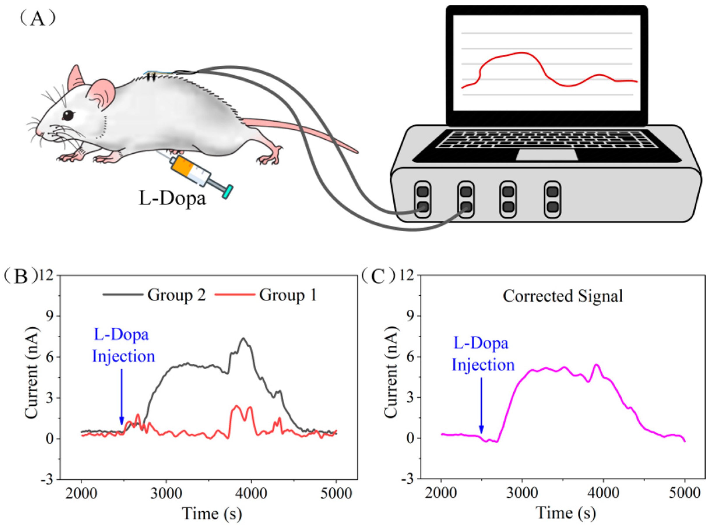
  • Another issue that requires further investigation is the assessment of the biocompatibility of microneedle sensors by on-body testing. For this purpose, it should be noted that the focus of emerging studies is devoted to the possibility of employing biological samples suitable for less invasive analysis. As an alternative to blood, sweat has been used in various studies to evaluate the LD content in humans. Promising results have already been obtained, even if a systematic study must still be carried out to validate the correlation between LD levels in sweat and ISF. Moreover, from a future perspective, clinical testing and validation in PD patients is required. Indeed, all the applications reported have been carried out on biological samples from healthy subjects. Unfortunately, only a few papers have determined the true concentration of LD in biological samples. At the same time, analyte spiking has often been used to validate the clinical utility of the developed biosensor. From a future perspective, the validation of the developed biosensors by a standard reference method would increase the validity of electrochemical devices for clinical application.
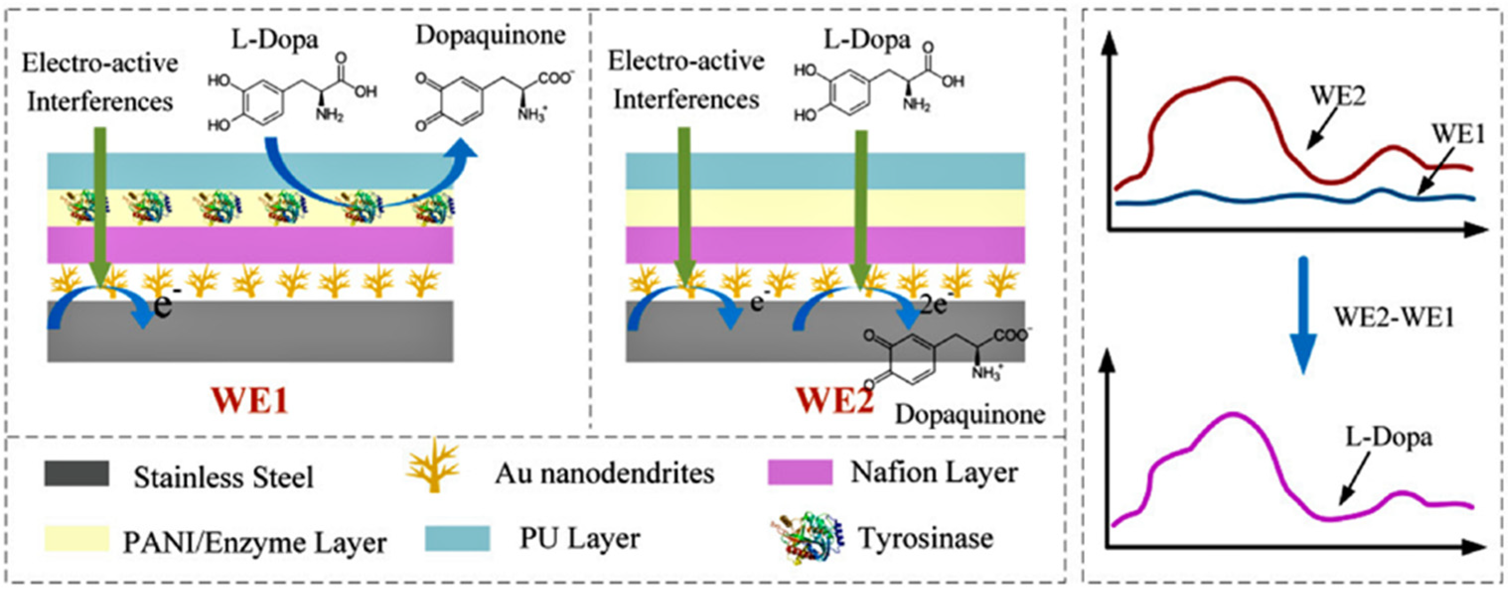
  • Selectivity is a further critical issue arising from the examination of tyrosinase biosensors. As previously discussed, tyrosinase has broad substrate specificity, catalyzing naturally occurring compounds in biological samples such as dopamine, serotonin, adrenaline, and compounds like carbidopa found as a result of drug administration, as well as LD. Among the strategies adopted to solve interference problems, dual-sensing platforms have been proposed based on parallel, simultaneous, and independent enzymatic and nonenzymatic electrochemical detection [67,75]. Nafion has often been used as a protecting membrane towards negatively charged compounds such as ascorbic acid and uric acid. A drawback of this material is its susceptibility to membrane fouling, which limits the operational stability of biosensors. Alternative protective layers should be explored. More or less effective strategies have been developed to solve interference problems. However, this issue has not been properly solved and addressed. Biosensor selectivity has often been evaluated by testing a few potential interferent compounds, as well as at physiological concentration levels typical of healthy individuals, which can differ from those found in plasma from PD patients. Tyrosine and serotonin plasma levels, for instance, are lower than they would be under normal settings [90,91], whereas homocysteine plasma levels rise in levodopa-treated individuals [92]. This aspect should be taken into consideration in future interference studies.
  • The employment of other enzymes as biocatalysts would not help to improve LD biorecognition. PPOs, for example, despite being easily available from fruit sources, show a wide substrate response. In work concerning PPO-based biosensors, these enzymes were employed to detect various catecholamines, among which were LD. Moreover, any application to real sample analysis is not reported, or concerns samples that are different from biological ones.
In conclusion, to date, interesting advances have been achieved in the field of amperometric biosensors for LD detection, but further steps are needed to address the urgent need for effective real-time monitoring.

Author Contributions

Conceptualization, R.C., A.G. and M.A.A.; writing—original draft preparation, C.T., G.C., M.A.A. and R.C.; writing—review and editing, G.B., F.L., A.D.C., C.T. and R.C.; supervision, R.C. All authors have read and agreed to the published version of the manuscript.

Funding

This research received no external funding.

Institutional Review Board Statement

Not applicable.

Informed Consent Statement

Not applicable.

Data Availability Statement

Data are available on request due to privacy restrictions.

Acknowledgments

The authors thank Regione Basilicata for funding a doctoral scholarship concerning the topic of this work.

Conflicts of Interest

The authors declare no conflict of interest.

References

  1. Xing, B.; Li, Y.C.; Gao, W.J. Norepinephrine versus dopamine and their interaction in modulating synaptic function in the prefrontal cortex. Brain Res. 2016, 1641, 217–233. [Google Scholar] [CrossRef]
  2. Hornykiewicz, O. L-DOPA: From a biologically inactive amino acid to a successful therapeutic agent: Historical review article. Amino Acids 2002, 23, 65–70. [Google Scholar] [CrossRef]
  3. Khan, S.T.; Ahmed, S.; Gul, S.; Khan, A.; Al-Harrasi, A. Search for safer and potent natural inhibitors of Parkinson’s disease. Neurochem. Int. 2021, 149, 105135. [Google Scholar] [CrossRef]
  4. Cacabelos, R. Parkinson’s disease: From pathogenesis to pharmacogenomics. Int. J. Mol. Sci. 2017, 18, 551. [Google Scholar] [CrossRef]
  5. Samii, A.; Nutt, J.G.; Ransom, B.R. Parkinson’s disease. Lancet 2004, 29, 1783–1793. [Google Scholar] [CrossRef]
  6. Min, K.; Park, K.; Park, D.H.; Yoo, Y.J. Overview on the biotechnological production of l-DOPA. Appl. Microbiol. Biotechnol. 2015, 99, 575–584. [Google Scholar] [CrossRef]
  7. Fasano, A.; Canning, C.G.; Hausdorff, J.M.; Lord, S.; Rochester, L. Falls in Parkinson’s disease: A complex and evolving picture. Mov. Disord. 2017, 32, 1524–1536. [Google Scholar] [CrossRef]
  8. LeWitt, P.A.; Chaudhuri, K.R. Unmet needs in Parkinson disease: Motor and non-motor. Park. Relat. Disord. 2020, 80, S7–S12. [Google Scholar] [CrossRef]
  9. Dirnberger, G.; Jahanshahi, M. Executive dysfunction in Parkinson’s disease: A review. J. Neuropsychol. 2013, 7, 193–224. [Google Scholar] [CrossRef]
  10. Beitz, J.M. Parkinson’s disease: A review. Front. Biosci. 2014, 6, 65–74. [Google Scholar] [CrossRef]
  11. Balestrino, R.; Schapira, A.H.V. Parkinson disease. Eur. J. Neurol. 2020, 27, 27–42. [Google Scholar] [CrossRef]
  12. Wakabayashi, K.; Tanji, K.; Odagiri, S.; Miki, Y.; Mori, F.; Takahashi, H. The Lewy body in Parkinson’s disease and related neurodegenerative disorders. Mol. Neurobiol. 2013, 47, 495–508. [Google Scholar] [CrossRef]
  13. Salat, D.; Tolosa, E. Levodopa in the treatment of Parkinson’s disease: Current status and new developments. J. Parkinsons. Dis. 2013, 3, 255–269. [Google Scholar] [CrossRef]
  14. Youdim, M.B.H.; Edmondson, D.; Tipton, K.F. The therapeutic potential of monoamine oxidase inhibitors. Nat. Rev. Neurosci. 2006, 7, 295–309. [Google Scholar] [CrossRef]
  15. Poewe, W.; Antonini, A.; Zijlmans, J.C.; Burkhard, P.R.; Vingerhoets, F. Levodopa in the treatment of Parkinson’s disease: An old drug still going strong. Clin. Interv. Aging 2010, 5, 229–238. [Google Scholar] [CrossRef]
  16. Thanvi, B.R.; Lo, T.C.N. Long term motor complications of levodopa: Clinical features, mechanisms, and management strategies. Postgr. Med. J. 2004, 80, 452–458. [Google Scholar] [CrossRef]
  17. Jankovic, J.; Stacy, M. Medical management of levodopa-associated motor complications in patients with Parkinson’s disease. CNS Drugs 2007, 21, 677–692. [Google Scholar] [CrossRef]
  18. Rezak, M. Current Pharmacotherapeutic Treatment Options in Parkinson’s Disease. Dis.-a-Month 2007, 53, 214–222. [Google Scholar] [CrossRef]
  19. Nutt, J.G. Pharmacokinetics and pharmacodynamics of levodopa. Mov. Disord. 2008, 23, 580–584. [Google Scholar] [CrossRef]
  20. Tizabi, Y.; Getachew, B.; Aschner, M. Novel Pharmacotherapies in Parkinson’s Disease. Neurotox. Res. 2021, 39, 1381–1390. [Google Scholar] [CrossRef]
  21. Valdés, R.H.; Puzer, L.; Gomes, M.; Marques, C.E.S.J.; Aranda, D.A.G.; Bastos, M.L.; Gemal, A.L.; Antunes, O.A.C. Production of L-DOPA under heterogeneous asymmetric catalysis. Catal. Commun. 2004, 5, 631–634. [Google Scholar] [CrossRef]
  22. Patil, S.A.; Apine, O.A.; Surwase, S.N.; Jadhav, J.P. Biological sources of L-DOPA: An alternative approach. Adv. Park. Dis. 2013, 2, 81–87. [Google Scholar] [CrossRef]
  23. Lampariello, L.; Cortelazzo, A.; Guerranti, R.; Sticozzi, C.; Valacchi, G. The magic velvet bean of mucuna pruriens. J. Tradit. Complement. Med. 2012, 2, 331–339. [Google Scholar] [CrossRef]
  24. Denne, T. Analysis of Levodopa Content in Commercial Formulations of Mucuna pruriens Seeds Used in Integrative Treatment of Parkinson’s Disease. Mov. Disord. 2019, 34, S37–S38. [Google Scholar]
  25. Tesoro, C.; Ciriello, R.; Lelario, F.; Di Capua, A.; Pascale, R.; Bianco, G.; Dell’Agli, M.; Piazza, S.; Guerrieri, A.; Scrano, L.; et al. Development and Validation of a Reversed-Phase HPLC Method with UV Detection for the Determination of L-Dopa in Vicia faba L. Broad Beans. Molecules 2022, 27, 7468. [Google Scholar] [CrossRef]
  26. Tolokán, A.; Klebovich, I.; Balogh-Nemes, K.; Horvai, G. Automated determination of levodopa and carbidopa in plasma by high-performance liquid chromatography-electrochemical detection using an on-line flow injection analysis sample pretreatment unit. J. Chromatogr. B Biomed. Appl. 1997, 698, 201–207. [Google Scholar] [CrossRef]
  27. Rondelli, I.; Acerbi, D.; Mariotti, F.; Ventura, P. Simultaneous determination of levodopa methyl ester, levodopa, 3-O-methyldopa and dopamine in plasma by high-performance liquid chromatography with electrochemical detection. J. Chromatogr. B Biomed. Sci. Appl. 1994, 653, 17–23. [Google Scholar] [CrossRef]
  28. Tesoro, C.; Lelario, F.; Ciriello, R.; Bianco, G.; Di Capua, A.; Acquavia, M.A. An Overview of Methods for L-Dopa Extraction and Analytical Determination in Plant Matrices. Separations 2022, 9, 224. [Google Scholar] [CrossRef]
  29. César, I.d.C.; Byrro, R.M.D.; de Santana e Silva Cardoso, F.F.; Mundim, I.M.; de Souza Teixeira, L.; Pontes da Silva, E.; Gomes, S.A.; Bonfim, R.R.; Pianetti, G.A. Simultaneous quantitation of levodopa and 3-O-methyldopa in human plasma by HPLC-ESI-MS/MS: Application for a pharmacokinetic study with a levodopa/benserazide formulation. J. Pharm. Biomed. Anal. 2011, 56, 1094–1100. [Google Scholar] [CrossRef]
  30. Zhang, L.; Chen, G.; Hu, Q.; Fang, Y. Separation and determination of levodopa and carbidopa in composite tablets by capillary zone electrophoresis with amperometric detection. Anal. Chim. Acta 2001, 431, 287–292. [Google Scholar] [CrossRef]
  31. Coello, J.; Maspoch, S.; Villegas, N. Simultaneous kinetic-spectrophotometric determination of levodopa and benserazide by bi- and three-way partial least squares calibration. Talanta 2000, 53, 627–637. [Google Scholar] [CrossRef]
  32. Madrakian, T.; Afkhami, A.; Mohammadnejad, M. Simultaneous spectrofluorimetric determination of levodopa and propranolol in urine using feed-forward neural networks assisted by principal component analysis. Talanta 2009, 78, 1051–1055. [Google Scholar] [CrossRef]
  33. He, W.W.; Zhou, X.W.; Lu, J.Q. Capillary electrophoresis-chemiluminecence detection of levodopa and benserazide in Medopar tablet. Chin. Chem. Lett. 2007, 18, 91–93. [Google Scholar] [CrossRef]
  34. Zhao, S.; Bai, W.; Wang, B.; He, M. Determination of levodopa by capillary electrophoresis with chemiluminescence detection. Talanta 2007, 73, 142–146. [Google Scholar] [CrossRef]
  35. State, R.G.; van Staden, J. (KOOS) F. Electrochemical sensors used in the determination of L-Dopa. Electrochem. Sci. Adv. 2022, 2, e2100040. [Google Scholar] [CrossRef]
  36. Beitollahi, H.; Safaei, M.; Tajik, S. Electrochemical deduction of levodopa by utilizing modified electrodes: A review. Microchem. J. 2020, 152, 104287. [Google Scholar] [CrossRef]
  37. Crapnell, R.D.; Banks, C.E. Electroanalytical Overview: The Determination of Levodopa (L-DOPA). ACS Meas. Sci. Au 2023, 3, 84–97. [Google Scholar] [CrossRef]
  38. Eslami, M.; Namazian, M.; Zare, H.R. Electrochemical behavior of 3,4-dihydroxyphenylalanine in aqueous solution. Electrochim. Acta 2013, 88, 543–551. [Google Scholar] [CrossRef]
  39. Thévenot, D.R.; Toth, K.; Durst, R.A.; Wilson, G.S. Electrochemical biosensors: Recommended definitions and classification. Anal. Lett. 2001, 34, 635–659. [Google Scholar] [CrossRef]
  40. Munteanu, F.D.; Lindgren, A.; Emnéus, J.; Gorton, L.; Ruzgas, T.; Csöregi, E.; Ciucu, A.; Van Huystee, R.B.; Gazaryan, I.G.; Lagrimini, L.M. Bioelectrochemical monitoring of phenols and aromatic amines in flow injection using novel plant peroxidases. Anal. Chem. 1998, 70, 2596–2600. [Google Scholar] [CrossRef]
  41. Ziyan, E.; Pekyardimci, Ş. Characterization of polyphenol oxidase from Jerusalem artichoke (Helianthus tuberosus). Turk. J. Chem. 2003, 27, 217–225. [Google Scholar]
  42. Burton, S.G. Biocatalysis with polyphenol oxidase: A review. Catal. Today 1994, 22, 459–487. [Google Scholar] [CrossRef]
  43. Solem, E.; Tuczek, F.; Decker, H. Tyrosinase versus Catechol Oxidase: One Asparagine Makes the Difference. Angew. Chemie-Int. Ed. 2016, 55, 2884–2888. [Google Scholar] [CrossRef]
  44. Gul, I.; Ahmad, M.S.; Naqvi, S.M.S.; Hussain, A.; Wali, R.; Farooqi, A.A.; Ahmed, I. Polyphenol oxidase (PPO) based biosensors for detection of phenolic compounds: A Review. J. Appl. Biol. Biotechnol. 2017, 5, 72–85. [Google Scholar] [CrossRef]
  45. Claus, H. Laccases: Structure, reactions, distribution. Micron 2004, 35, 93–96. [Google Scholar] [CrossRef]
  46. Klabunde, T.; Eicken, C.; Sacchettini, J.C.; Krebs, B.; Henson, M.J.; Solomon, E.I. Crystal structure of a plant catechol oxidase containing a dicopper center. Chemtracts 2000, 13, 97–102. [Google Scholar] [CrossRef]
  47. Raymundo-Pereira, P.A.; Silva, T.A.; Caetano, F.R.; Ribovski, L.; Zapp, E.; Brondani, D.; Bergamini, M.F.; Marcolino, L.H.; Banks, C.E.; Oliveira, O.N.; et al. Polyphenol oxidase-based electrochemical biosensors: A review. Anal. Chim. Acta 2020, 1139, 198–221. [Google Scholar] [CrossRef]
  48. Tarasov, A.; Stozhko, N.; Bukharinova, M.; Khamzina, E. Biosensors Based on Phenol Oxidases (Laccase, Tyrosinase, and Their Mixture) for Estimating the Total Phenolic Index in Food-Related Samples. Life 2023, 13, 291. [Google Scholar] [CrossRef]
  49. Bourquelot, E.; Bertrand, A. Le bleuissement et le noircissement des champignons. Comp. Rend. Soc. Biol. 1985, 47, 582–584. [Google Scholar]
  50. Sánchez-Ferrer, Á.; Neptuno Rodríguez-López, J.; García-Cánovas, F.; García-Carmona, F. Tyrosinase: A comprehensive review of its mechanism. Biochim. Biophys. Acta (BBA)-Protein Struct. Mol. 1995, 1247, 1–11. [Google Scholar] [CrossRef]
  51. Selinheimo, E.; NiEidhin, D.; Steffensen, C.; Nielsen, J.; Lomascolo, A.; Halaouli, S.; Record, E.; O’Beirne, D.; Buchert, J.; Kruus, K. Comparison of the characteristics of fungal and plant tyrosinases. J. Biotechnol. 2007, 130, 471–480. [Google Scholar] [CrossRef]
  52. Zaidi, K.U.; Ali, A.S.; Ali, S.A.; Naaz, I. Microbial tyrosinases: Promising enzymes for pharmaceutical, food bioprocessing, and environmental industry. Biochem. Res. Int. 2014, 2014, 854687. [Google Scholar] [CrossRef]
  53. Greggio, E.; Bergantino, E.; Carter, D.; Ahmad, R.; Costin, G.E.; Hearing, V.J.; Clarimon, J.; Singleton, A.; Eerola, J.; Hellström, O.; et al. Tyrosinase exacerbates dopamine toxicity but is not genetically associated with Parkinson’s disease. J. Neurochem. 2005, 93, 246–256. [Google Scholar] [CrossRef]
  54. Mayer, A.M. Polyphenol oxidases in plants and fungi: Going places? A review. Phytochemistry 2006, 67, 2318–2331. [Google Scholar] [CrossRef]
  55. Nawaz, A.; Shafi, T.; Khaliq, A.; Mukhtar, H.; ul Haq, I.; Thanvi, B.R.; Lo, T.C.N. Tyrosinase: Sources, Structure and Applications. Int. J. Biotechnol. Bioeng. 2017, 3, 135–141. [Google Scholar] [CrossRef]
  56. Ramsden, C.A.; Riley, P.A. Tyrosinase: The four oxidation states of the active site and their relevance to enzymatic activation, oxidation and inactivation. Bioorg. Med. Chem. 2014, 22, 2388–2395. [Google Scholar] [CrossRef]
  57. Chang, T.S. An updated review of tyrosinase inhibitors. Int. J. Mol. Sci. 2009, 10, 2440–2475. [Google Scholar] [CrossRef]
  58. Zolghadri, S.; Bahrami, A.; Hassan Khan, M.T.; Munoz-Munoz, J.; Garcia-Molina, F.; Garcia-Canovas, F.; Saboury, A.A. A comprehensive review on tyrosinase inhibitors. J. Enzyme Inhib. Med. Chem. 2019, 34, 279–309. [Google Scholar] [CrossRef]
  59. Espín, J.C.; Varón, R.; Fenoll, L.G.; Gilabert, M.A.; García-Ruíz, P.A.; Tudela, J.; García-Cánovas, F. Kinetic characterization of the substrate specificity and mechanism of mushroom tyrosinase. Eur. J. Biochem. 2000, 267, 1270–1279. [Google Scholar] [CrossRef]
  60. Sassolas, A.; Blum, L.J.; Leca-Bouvier, B.D. Immobilization strategies to develop enzymatic biosensors. Biotechnol. Adv. 2012, 30, 489–511. [Google Scholar] [CrossRef]
  61. Wiseman, A. Handbook of Enzyme Biotechnology; JohnWiley & Sons, Halsted Press, Inc.: New York, NY, USA, 1985; pp. 147–207. [Google Scholar]
  62. Guerrieri, A.; Ciriello, R.; Crispo, F.; Bianco, G. Detection of choline in biological fluids from patients on haemodialysis by an amperometric biosensor based on a novel anti-interference bilayer. Bioelectrochemistry 2019, 129, 135–143. [Google Scholar] [CrossRef]
  63. Ciriello, R.; De Gennaro, F.; Frascaro, S.; Guerrieri, A. A novel approach for the selective analysis of L-lysine in untreated human serum by a co-crosslinked L-lysine–α-oxidase/overoxidized polypyrrole bilayer based amperometric biosensor. Bioelectrochemistry 2018, 124, 47–56. [Google Scholar] [CrossRef]
  64. Ciriello, R.; Guerrieri, A. A Crosstalk- and Interferent-Free Dual Electrode Amperometric Biosensor for the Simultaneous Determination of Choline and Phosphocholine. Sensors 2021, 21, 3545. [Google Scholar] [CrossRef]
  65. Moon, J.M.; Teymourian, H.; De la Paz, E.; Sempionatto, J.R.; Mahato, K.; Sonsa-ard, T.; Huang, N.; Longardner, K.; Litvan, I.; Wang, J. Non-Invasive Sweat-Based Tracking of L-Dopa Pharmacokinetic Profiles Following an Oral Tablet Administration. Angew. Chemie-Int. Ed. 2021, 60, 19074–19078. [Google Scholar] [CrossRef]
  66. Tai, L.C.; Liaw, T.S.; Lin, Y.; Nyein, H.Y.Y.; Bariya, M.; Ji, W.; Hettick, M.; Zhao, C.; Zhao, J.; Hou, L.; et al. Wearable Sweat Band for Noninvasive Levodopa Monitoring. Nano Lett. 2019, 19, 6346–6351. [Google Scholar] [CrossRef]
  67. Fang, L.; Ren, H.; Mao, X.; Zhang, S.; Cai, Y.; Xu, S.; Zhang, Y.; Li, L.; Ye, X.; Liang, B. Differential Amperometric Microneedle Biosensor for Wearable Levodopa Monitoring of Parkinson’s Disease. Biosensors 2022, 12, 102. [Google Scholar] [CrossRef]
  68. Brunetti, B.; Valdés-Ramírez, G.; Litvan, I.; Wang, J. A disposable electrochemical biosensor for l-DOPA determination in undiluted human serum. Electrochem. Commun. 2014, 48, 28–31. [Google Scholar] [CrossRef]
  69. Pinho, A.; Viswanathan, S.; Ribeiro, S.; Oliveira, M.B.P.P.; Delerue-Matos, C. Electroanalysis of urinary l-dopa using tyrosinase immobilized on gold nanoelectrode ensembles. J. Appl. Electrochem. 2012, 42, 131–137. [Google Scholar] [CrossRef]
  70. Cembalo, G.; Ciriello, R.; Tesoro, C.; Guerrieri, A.; Bianco, G.; Lelario, F.; Acquavia, M.A.; Di Capua, A. An Amperometric Biosensor Based on a Bilayer of Electrodeposited Graphene Oxide and Co-Crosslinked Tyrosinase for L-Dopa Detection in Untreated Human Plasma. Molecules 2023, 28, 5239. [Google Scholar] [CrossRef]
  71. Mollamohammadi, F.; Faridnouri, H.; Zare, E.N. Electrochemical Biosensing of L-DOPA Using Tyrosinase Immobilized on Carboxymethyl Starch-Graft-Polyaniline@MWCNTs Nanocomposite. Biosensors 2023, 13, 562. [Google Scholar] [CrossRef]
  72. Aliya, M.; Zare, E.N.; Faridnouri, H.; Ghomi, M.; Makvandi, P. Sulfonated Starch-Graft-Polyaniline@Graphene Electrically Conductive Nanocomposite: Application for Tyrosinase Immobilization. Biosensors 2022, 12, 939. [Google Scholar] [CrossRef]
  73. Xiao, J.; Fan, C.; Xu, T.; Su, L.; Zhang, X. An electrochemical wearable sensor for levodopa quantification in sweat based on a metal–Organic framework/graphene oxide composite with integrated enzymes. Sens. Actuators B Chem. 2022, 359, 131586. [Google Scholar] [CrossRef]
  74. Jubete, E.; Ochoteco, E.; Loinaz, I.; Pomposo, J.A.; Grande, H.; Linazasoro, G. Electrochemical biosensor development for detection of l-dopa levels in plasma during parkinson illness. In Proceedings of the SENSORS, 2008 IEEE, Lecce, Italy, 26–29 October 2008; pp. 239–241. [Google Scholar] [CrossRef]
  75. Goud, K.Y.; Moonla, C.; Mishra, R.K.; Yu, C.; Narayan, R.; Litvan, I.; Wang, J. Wearable Electrochemical Microneedle Sensor for Continuous Monitoring of Levodopa: Toward Parkinson Management. ACS Sens. 2019, 4, 2196–2204. [Google Scholar] [CrossRef]
  76. Aydemir, N.; Malmström, J.; Travas-Sejdic, J. Conducting polymer based electrochemical biosensors. Phys. Chem. Chem. Phys. 2016, 18, 8264–8277. [Google Scholar] [CrossRef]
  77. Malhotra, B.; Dhand, C.; Lakshminarayanan, R.; Dwivedi, N.; Mishra, S.; Solanki, P.; Venkatesh, M.; Beuerman, R.W.; Ramakrishna, S. Polyaniline-based biosensors. Nanobiosens. Dis. Diagnosis 2015, 4, 25–46. [Google Scholar] [CrossRef]
  78. Zhang, X.; Shi, F.; Yu, X.; Liu, H.; Fu, Y.; Wang, Z.; Jiang, L.; Li, X. Polyelectrolyte Multilayer as Matrix for Electrochemical Deposition of Gold Clusters: Toward Super-Hydrophobic Surface. J. Am. Chem. Soc. 2004, 126, 3064–3065. [Google Scholar] [CrossRef]
  79. Kulkarni, T.; Slaughter, G. Application of semipermeable membranes in glucose biosensing. Membranes 2016, 6, 55. [Google Scholar] [CrossRef]
  80. Carbone, M.E.; Ciriello, R.; Guerrieri, A.; Salvi, A.M. Poly(o-aminophenol) electrosynthesized onto platinum at acidic and neutral ph: Comparative investigation on the polymers characteristics and on their inner and outer interfaces. Int. J. Electrochem. Sci. 2014, 9, 2047–2066. [Google Scholar] [CrossRef]
  81. Carbone, M.E.; Ciriello, R.; Guerrieri, A.; Salvi, A.M. XPS investigation on the chemical structure of a very thin, insulating, film synthesized on platinum by electropolymerization of o-aminophenol (oAP) in aqueous solution at neutral pH. Surf. Interface Anal. 2014, 46, 1081–1085. [Google Scholar] [CrossRef]
  82. Rathod, B.G.; Patel, N.M. Development of validated RP-HPLC method for the estimation of L-Dopa from Mucuna pruriens, its extracts and in Aphrodisiac formulation. Int. J. Pharma Sci. Res. 2014, 5, 508–513. [Google Scholar]
  83. Timur, S.; Pazarloǧlu, N.; Pilloton, R.; Telefoncu, A. Thick film sensors based on laccases from different sources immobilized in polyaniline matrix. Sens. Actuators B Chem. 2004, 97, 132–136. [Google Scholar] [CrossRef]
  84. Leite, O.D.; Lupetti, K.O.; Fatibello-Filho, O.; Vieira, I.C.; de M Barbosa, A. Synergic effect studies of the bi-enzymatic system laccaseperoxidase in a voltammetric biosensor for catecholamines. Talanta 2003, 59, 889–896. [Google Scholar] [CrossRef]
  85. Josypčuk, O.; Barek, J.; Josypčuk, B. Amperometric Determination of Catecholamines by Enzymatic Biosensors in Flow Systems. Electroanalysis 2018, 30, 1163–1171. [Google Scholar] [CrossRef]
  86. Chawla, S.; Narang, J.; Pundir, C.S. An amperometric polyphenol biosensor based on polyvinyl chloride membrane. Anal. Methods 2010, 2, 1106–1111. [Google Scholar] [CrossRef]
  87. Narang, J.; Chawla, S.; Chauhan, N.; Dahiya, M.; Pundir, C.S. Construction of an amperometric polyphenol biosensor based on PVA membrane. J. Food Meas. Charact. 2013, 7, 22–28. [Google Scholar] [CrossRef]
  88. Sandeep, S.; Santhosh, A.S.; Swamy, N.K.; Suresh, G.S.; Melo, J.S.; Nithin, K.S. Electrochemical detection of L-dopa using crude Polyphenol oxidase enzyme immobilized on electrochemically reduced RGO-Ag nanocomposite modified graphite electrode. Mater. Sci. Eng. B Solid-State Mater. Adv. Technol. 2018, 232–235, 15–21. [Google Scholar] [CrossRef]
  89. Shoja, Y.; Rafati, A.A.; Ghodsi, J. Glassy carbon electrode modified with horse radish peroxidase/organic nucleophilic-functionalized carbon nanotube composite for enhanced electrocatalytic oxidation and efficient voltammetric sensing of levodopa. Mater. Sci. Eng. C 2016, 58, 835–845. [Google Scholar] [CrossRef]
  90. Gątarek, P.; Sekulska-Nalewajko, J.; Bobrowska-Korczaka, B.; Pawełczyk, M.; Jastrzębski, K.; Głąbiński, A.; Kałużna-Czaplińska, J. Plasma Metabolic Disturbances in Parkinson’s Disease Patients. Biomedicines 2022, 10, 3005. [Google Scholar] [CrossRef]
  91. Tong, Q.; Zhang, L.; Yuan, Y.; Jiang, S.; Zhang, R.; Xu, Q.; Ding, J.; Li, D.; Zhou, X.; Zhang, K. Reduced plasma serotonin and 5-hydroxyindoleacetic acid levels in Parkinson’s disease are associated with nonmotor symptoms. Park. Relat. Disord. 2015, 21, 882–887. [Google Scholar] [CrossRef]
  92. Paul, R.; Borah, A. L-DOPA-induced hyperhomocysteinemia in Parkinson’s disease: Elephant in the room. Biochim. Biophys. Acta-Gen. Subj. 2016, 1860, 1989–1997. [Google Scholar] [CrossRef]
Figure 1. Chemical structure of L-Dopa, dopamine, and norepinephrine.
Chemosensors 11 00523 g001
Figure 2. Schematic representation of the possible enzymatic conversion pathways of LD before crossing the BBB (red color) and of the enzyme inhibition activity (blue color).
Chemosensors 11 00523 g002
Figure 3. Electrochemical oxidation of L-Dopa in moderate acidic, neutral, and basic pHs.
Chemosensors 11 00523 g003
Figure 4. Phenolic substrate oxidation pathway catalyzed by tyrosinase: ortho-hydroxylation of monophenols (cresolase activity) and oxidation of o-diphenols (catecholase activity).
Chemosensors 11 00523 g004
Figure 5. Schematic representation of the oxidation states of the active center of tyrosinase and its catalytic pathways.
Chemosensors 11 00523 g005
Figure 6. Electron transfer between tyrosinase and the electrode surface during diphenol activity. Left electrode: direct anodic reaction. Right electrode: cathodic reaction of the quinone produced by tyrosinase [71].
Chemosensors 11 00523 g006
Figure 7. Schematic diagram of the surface modification structure of the working electrode WE1 and WE2, and the reaction process of L-Dopa and other electro-active interferences on the electrode surface (left). The current response signal of WE1 and WE2 and the L-Dopa differential detection principle (right) [67].
Chemosensors 11 00523 g007
Figure 8. (A) The in vivo test schematic diagram of the minimally invasive flexible microneedle array. (B) The current response of the two electrode groups on a flexible microneedle array after L-Dopa injection. (C) The flexible microneedle array current response signal to L-Dopa after differential correction [67].
Chemosensors 11 00523 g008
Table 2. Electrode modification, detection mode, and analytical features of the proposed biosensors.
Modified ElectrodeDetection ModeLinear RangeSensitivityLimit of DetectionReference
GC/carboxymethyl starch-graft-PANI /MWCNT nanocompositeDPV (oxidation)10–300 µM0.035 µA/µM32 µM[71]
GC/sulfonated starch-graft-PANI@graphene nanocompositeDPV (reduction)0.5–109 µM0.0002 µA/µM15.0 µM[72]
Differential stainless-steel microneedles
WE1: Aunanodentrites/Nafion/PANI/Tyrosinase/PU
WE2: Aunanodentrites/Nafion/PANI/BSA/PU
Chronoamperometry
(Differential current response, oxidation at 0.3 V)
0–20 µM0.469 nA/µM0.18 µM[67]
Flexible printed gold electrode/Zeolitic imidazolate framework/graphene oxideChronoamperometry
(Oxidation at 0.3 V)
1–95 µM0.047 µA/µM0.45 µM[73]
Screen-printed carbon electrode/porous hydrogelChronoamperometry (reduction at −0.3 V)5–30 µMnot reported0.3 µM[65]
GC/electrochemically deposited GOChronoamperometry (reduction at −0.1 V)1–210 µM3.21 µA/mM0.84 µM[70]
Carbon paste microneedle electrodes
WE1: unmodified carbon past, 65 wt. % graphite powder and 35 wt. % mineral oil
WE2: modified carbon past, 55 wt. % graphite powder, 10 wt. % tyrosinase mushroom enzyme, and
35 wt. % mineral oil
WE1:SWV (oxidation)
WE2: chronoamperometry
(applied potential of 0.1 V in PB solution or 0.3 V in artificial ISF)
WE1:
In PB 5–100 μM and
100–300 μM
In ISF 20–160 µM
WE2:
In PB and ISF 20–300 µM
WE1:
0.037 μA/μM
in ISF
WE2:
0.048 nA/μM
in ISF
WE1:
in ISF 0.5 µM
WE2:
in ISF 0.25 µM
[75]
Au/Cr conductive layer/gold nanodendrites/polythioninAmperometric
(Oxidation at 0.34 V)
0–20 µM15 nA/μM
in PBs
1.7 nA/μM
in sweat
1.25 μM in sweat[66]
Carbon electrode/CNT/polythionine Chronoamperometry
(reduction at −0.31 V)
0.8–22.3 μM0.0619 A/M2.5 µM[68]
Gold nanoelectrode ensembleFIA, amperometric
(reduction at −0.2 V)
10−3–10−8 Mnot reported1 × 10−9 M[69]
MWCNTChronoamperometry
(Reduction at −0.3 V)
not reportednot reportednot reported[74]
Table 3. Anti-interference strategies adopted, compounds investigated, and real samples analyzed.
Anti-Interference StrategyInterferents and LD Tested Real SamplesWearable/System IntegrationReference
uric acid and ascorbic acid 30 µM, LD 30 µM [71]
Differential structure:
-WE1 detects the electrochemical signal of the interferents
-WE2 detects the mixed signal of the interferents and L-Dopa
uric acid and ascorbic acid 50 µM, glucose 200 µM, LD 30 µMIn vitro: bovine serum with a skin model (rat skin)
In vivo: rat’s abdominal cavity
Minimally invasive subcutaneous
biosensor
[67]
uric acid 20 μM, glucose 100 μM, lactate 20 μM, ascorbic acid 20 μM,
LD 10 µM
Sweat levodopa concentrations after
consuming broad beans
(Recovery from 98.85 to 99.79%)
Flexible, wearable electrochemical sensor for the noninvasive in situ detection of LD in sweat.
Integration with a wireless electronic circuit
[73]
Nafion membrane as a protective layerascorbic acid 100 μM, dopamine 0.32 µM, carbidopa 0.2 μM, serotonin 0.15 μM, homocysteine 20 µM, tyrosine 2.5 μM, LD 2.5 μMHuman plasma
(Recovery from 90.8 to 102.4%)
[70]
Nafion membrane as an anti-interference barrierL-Dopa/C-Dopa in 4:1 and 1:1 concentration ratio
Caffeine 50 μM, ascorbic acid 100 μM, uric acid 400 μM, dopamine 10 nM, acetaminophen 100 μM, tyrosine and resorcinol 10 μM, LD 10 µM
Fingertip sweatNoninvasive semi-continuous tracking of sweat LD levels upon administration of standard pill formulations based on a single fingertip touch[65]
Orthogonally measured electrochemical signals, redox, and biocatalytic
Nafion coating as permsaelective
protective layer
ascorbic acid, uric acid, tyrosine, and theophylline 150 μM, LD 50 μM using artificial ISF medium In vitro artificial ISF (pH 7.4)Wearable, can penetrate through skin mimicking phantom gel and mice skin[75]
Nafion coating as an antifouling layerUric acid 20 μM, glucose 166 μM, ascorbic acid 16μM, LD 10 µMSweat generated via iontophoresis and physical activities after Vicia faba consumptionWearable sweatband for prolonged,
continuous, and noninvasive drug monitoring in human subjects after fava bean intake
[66]
Nafion coating as a protective layer Human serum [68]
Glucose, ascorbic acid and urea 10 mM, LD 10−6 MHuman urine
(Recovery 96%)
[69]
dopamine and epinephrine [74]
Disclaimer/Publisher’s Note: The statements, opinions and data contained in all publications are solely those of the individual author(s) and contributor(s) and not of MDPI and/or the editor(s). MDPI and/or the editor(s) disclaim responsibility for any injury to people or property resulting from any ideas, methods, instructions or products referred to in the content.

