Article

Fabric Phase Sorptive Extraction Combined with HPLC-UV for the Quantitation of Amphotericin B in Human Urine

by
Evmorfia Asimakopoulou
1,
Natalia Manousi
2,
Nikoleta Anaxagorou
2,
Abuzar Kabir
3,
Kenneth G. Furton
3 and
Constantinos K. Zacharis
2,*
1
Laboratory of Analytical Chemistry, Department of Chemistry, Aristotle University of Thessaloniki, GR-54124 Thessaloniki, Greece
2
Laboratory of Pharmaceutical Analysis, Department of Pharmaceutical Technology, School of Pharmacy, Aristotle University of Thessaloniki, GR-54124 Thessaloniki, Greece
3
Department of Chemistry and Biochemistry, Florida International University, Miami, FL 33199, USA
*
Author to whom correspondence should be addressed.
Chemosensors 2022, 10(12), 537; https://doi.org/10.3390/chemosensors10120537
Submission received: 19 November 2022 / Revised: 6 December 2022 / Accepted: 13 December 2022 / Published: 15 December 2022
(This article belongs to the Collection Recent Trend in Chromatography for Pharmaceutical Analysis)

Abstract

:
Herein, a fabric phase sorptive extraction-based scheme was reported for the determination of amphotericin B in human urine. The developed method allowed the direct extraction of the analyte from the biological matrix with improved selectivity, repeatability and recovery. Due to the membrane’s engineered affinity towards the analyte, extraction equilibrium was achieved in 30 min. Moreover, no additional sample pretreatment was required due to the high permeability of the FPSE membrane and the small volume of eluting solvent required for quantitative back-extraction of the analytes. The hydrophobic sol–gel polydimethylphenylsiloxane (sol–gel PDMDPheS) coated membrane provided the optimum extraction performance. Important parameters that affect the extraction efficiency (such as sample volume, extraction time, membrane size, stirring rate, ion strength, elution solvent and time) were thoroughly investigated. The analyte was separated from the internal standard (nimesulide) and endogenous compounds of the human urine using a gradient elution program. The proposed assay was linear within the range of 0.10–10.0 μg mL−1 while the relative standard deviation of the repeatability (sr) and within-laboratory reproducibility (sR) were less than 12.7% in all cases. The method exhibited good accuracy which varied between 88.1 to 110.3%. The developed method was successfully applied for the monitoring of amphotericin B concentration in human urine.

Graphical Abstract

1. Introduction

Amphotericin B (AMTB) is a polyene macrolide semi-synthetic antibiotic that was introduced to the market in 1959. It is composed of both a hydrophilic polyhydroxyl and a lipophilic hydrocarbon chain (Figure S1). It was characterized as the gold standard therapeutic drug against several fungal infections and leishmaniasis as well. However, its application is limited due to severe nephrotoxicity, which may lead to kidney failure [1]. AMTB is preferentially administrated parenterally and the monitoring of the dosages and its distribution in the human body during the therapy is an essential parameter for the effectiveness of the treatment.
Many analytical approaches have been proposed for the determination of AMTB in biological fluids. These approaches were recently reviewed by Marena et al. [2]. Among these, separation techniques (HPLC, LC–MS, CE) are predominant ones offering selective and sensitive determinations. Published approaches for the analysis of urinary amphotericin B include HPLC coupled to MS/MS [3,4] and UV detection [5]. Since biological samples are complex matrices that contain phospholipids, proteins, organic compounds and inorganic salts, a sample pretreatment step is usually necessary before the utilization of an instrumental analytical technique [6].
Traditional bioanalytical sample preparation techniques involve liquid–liquid extraction, protein precipitation and solid-phase extraction [6]. However, these protocols usually require laborious and multiple steps, relatively high volumes of organic solvents and in several cases, the extraction efficiency is relatively poor. In the last years, significant effort has been made to overcome the above limitations leading to the development of microextraction techniques. Typical examples include liquid-phase microextraction (LPME), solid-phase microextraction (SPME), microdialysis (MD), electromembrane extraction (EME), capsule phase microextraction (CPME), pipette tip-based microextraction, 3D printed and microfluidic devices to enhance the sensitivity, selectivity and the handling simplicity of the required steps [7,8,9,10,11,12].
Fabric phase sorptive extraction (FPSE) is an environmentally friendly sample preparation approach that takes advantage of the sol–gel technology [13]. The FPSE membrane is made from a flexible substrate (e.g., from polyester, cellulose or cotton) after chemical functionalization with an appropriate sorbent. All FPSE membranes are characterized by permeability resulting in rapid extraction, as well as high resistance in a wide pH range (i.e., 1–12) that makes them compatible with a plethora of samples. Other benefits of FPSE include the relatively high surface area of the membranes and the capability to use them for the direct analysis of biological samples without other requirements for matrix cleanup [14]. Until now, FPSE has been proved to be a useful tool in bioanalysis for the monitoring of a wide variety of compounds (e.g., tetracyclines [15], penicillins [16], anticancer drugs [17] and non-steroidal anti-inflammatory drugs [18]) in biological samples.
Herein, we demonstrated the application of a sol–gel polydimethylphenylsiloxane-coated (sol–gel PDMDPheS) cellulose FPSE membrane for the determination of amphotericin B in human urine. Various experimental parameters that can have an impact on the extraction performance were investigated including the FPSE sorbent chemistry, extraction time, stirring rate, ionic strength, membrane size, sample pH and volume, elution solvent type and amount, and elution time. The analyte and the internal standard (ISTD) were separated under reversed-phase HPLC conditions. Method validation was conducted based on the guidelines for bioanalytical method validation [19] and the proposed protocol was successfully employed for the analysis of the target analyte in human urine.

2. Materials and Methods

2.1. Reagents, Solutions and FPSE Membranes

All analytical reagents were of analytical grade of higher. AMTB (>98%) and nimesulide (≥98.0%)—used as ISTD—were supplied by Sigma-Aldrich (St. Louis, MO, USA). HPLC grade solvents, such as acetonitrile (ACN) and methanol (MeOH) were purchased from Honeywell (Charlotte, NC, USA). Milli-Q water was provided after purification by a B30 system (Adrona SIA, Riga, Latvia).
Stock standards (500 μg mL−1) of AMTB and ISTD were prepared in DMSO and MeOH, respectively, and they were stored at 4 °C. From these solutions, appropriate working standards were prepared in water after appropriate dilution.
For method development, artificial urine was prepared based on the procedure described by Brooks and Keevil [20]. For this purpose, NH4Cl (0.65 g), urea (5 g) citric acid (0.2 g), NaHCO3 (1.05 g), Na2SO4·10H2O (1.6 g), lactic acid (0.05 g), K2HPO4 (0.6 g), CaCl2·2H2O (0.19 g), KH2PO4 (0.48 g), MgSO4·7H2O (0.25 g) and NaCl (2.6 g) were dissolved in 500 mL water and the sample pH was adjusted to 7.0 using diluted hydrochloric acid.
For the preparation of the FPSE membranes, unbleached Muslin cotton (100% cellulose) by Jo-Ann Fabric (Miami, FL, USA) was used. Trifluoroacetic acid and methyltrimethoxysilane were obtained from Sigma-Aldrich (St. Louis, MO, USA), while NaOH and HCl were from Thermo Fisher Scientific (Milwaukee, WI, USA).
Poly(dimethyldiphenylsiloxane) (PDMDPheS), octadecyl trimethoxysilane (C18-TMS)) and poly(dimethyl siloxane) (PDMS) were obtained from Gelest Inc. (Morrisville, PA, USA). The general characteristics of the three different sol–gel FPSE sorbents that were studied (i.e., sol–gel PDMS, sol–gel C18, and sol–gel PDMDPheS) are reported in Table S1. The preparation of the FPSE membrane functionalized with the above-mentioned sorbents is comprehensively described elsewhere [21,22,23].
Urine samples were provided from healthy volunteers who were fully informed regarding the conducted research and their consent was provided in written form. All samples were initially centrifuged at 6000 rpm for 20 min and the supernatant of each sample was individually stored at −18 °C in an appropriate sterilized container. Prior to each extraction, an aliquot of 900 μL οf each sample was mixed with 50 μL of the ISTD solution and 50 μL of the analyte’s standard solution (or H2O for blank) in an extraction vial followed by vortex mixing to ensure sample homogeneity. Using this procedure, the sample matrix was diluted at only 10%.

2.2. HPLC Instrumentation and Conditions
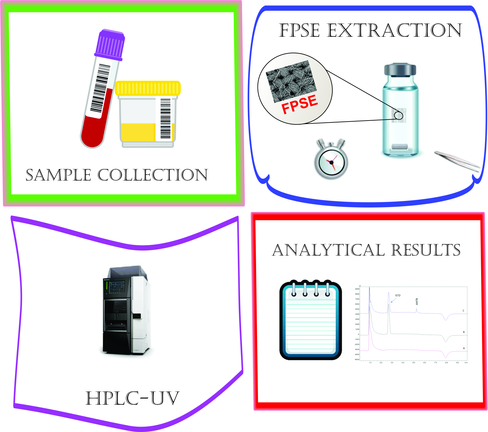
For the analyses, a Shimadzu 2010A HPLC-UV system (Kyoto, Japan) was used. The instrument consisted of a high pressure quaternary gradient pump, a UV detector, an autosampler and a column compartment, while system operation and data processing were carried out using LC Solutions software (vs. 1.25 SP4). The separation of AMTB and the ISTD was performed on a Poroshell 120 EC-C18 column (50 × 4.6 mm, 2.7 μm) from Agilent Technologies (Santa Clara, CA, USA). The target analyte was separated from the ISTD and the matrix interferences using a gradient elution program using citric acid 22 mM (pH 4.2 adjusted with 1 M NaOH): methanol 60:40 v/v (A) and citric acid 22 mM (pH 4.2 adjusted with 1 M NaOH): methanol 20:80 v/v (B). The initial composition of the mobile phase was 50:50, A/B v/v. The composition was changed to 0:100 A/B v/v at 3 min and it was kept constant until 5 min. Finally, the composition was changed back to the initial parameters (i.e., 50:50, A/B v/v) at 5.5 min and the system was equilibrated until 10 min. The mobile phase flow rate was 0.6 mL min−1 and the column was maintained at 30 °C during sample analysis. AMTB and the ISTD were monitored at 380 nm and an injection volume of 10 μL was used. The total analysis time was 10 min.

2.3. FPSE Protocol

Activation: The FPSE membranes were initially immersed sequentially in MeOH for 5 min and in H2O for 5 min in order to remove any impurities that occurred during their preparation and to activate the sol–gel sorbent for the extraction of the target analytes.
Extraction: The membrane was added in a glass vial that contained 1000 μL of sample and a PTFE-coated magnetic micro stir bar. Adsorption was performed under stirring at 350 rpm for 30 min.
Elution: For the desorption of AMTB, the FPSE membranes were inserted into Eppendorf tubes and an aliquot of 250 μL of MeOH was added. Elution was carried out for 2 min and then the extract was injected into the HPLC system.
Wash/regeneration: Accordingly, the membrane was immersed in the initial aliquot of MeOH that was used during the activation step for washing. Finally, the membrane was dried and stored or reused in a further extraction.
The handling of the FPSE membranes during all the reported steps was performed with tweezers to avoid contamination.

2.4. Method Validation

Method validation was performed based on the FDA guidelines for bioanalytical methodologies [19]. Thus, the linearity, accuracy, matrix effect, precision, selectivity, limit of detection (LOD) and limit of quantitation (LOQ) of the FPSE-HPLC-UV method were evaluated.
The evaluation of the sensitivity was conducted through the analysis of drug-free urine samples and their spiked analogues. The investigation of potential carry-over effect was examined by the injection of blank samples following the analysis of real samples spiked sample at the highest level of concentration used in the calibration curve.
To study method accuracy and precision, pooled drug-free human urine was spiked at three different concentrations of AMTB (i.e., 0.10 (LLOQ), 1.00 (MQC) and 10.0 (HQC) μg mL−1. The within-day (intra-day) accuracy and precision were investigated by three analyses performed in the same day for the three different concentrations. The inter-day precision and accuracy were assessed on different consecutive days (n = 3). A threshold of 15% was set as acceptance criterion for the precision and the accuracy % expressed as relative standard deviation (%RSD) and relative error (er%), respectively. For the 0.10 μg mL−1 (LLOQ level), an acceptance value of 20% was set [19].
Calibration curve was constructed by plotting the peak area ratio of AMTB against to ISTD. Six concentration levels were prepared in water and in authentic pooled urine matrix in the working range 0.10–10.0 μg mL−1. For each concentration level, triplicate analyses were performed.
In order to ensure good performance characteristics, the FPSE method was optimized in terms of FPSE material and dimensions, sample pH and volume, adsorption and desorption time, type and quantity of eluent, ionic strength of sample and stirring rate. For the optimization study, artificial urine containing AMTB at a concentration of 2.5 μg mL−1 was used.

3. Results and Discussion

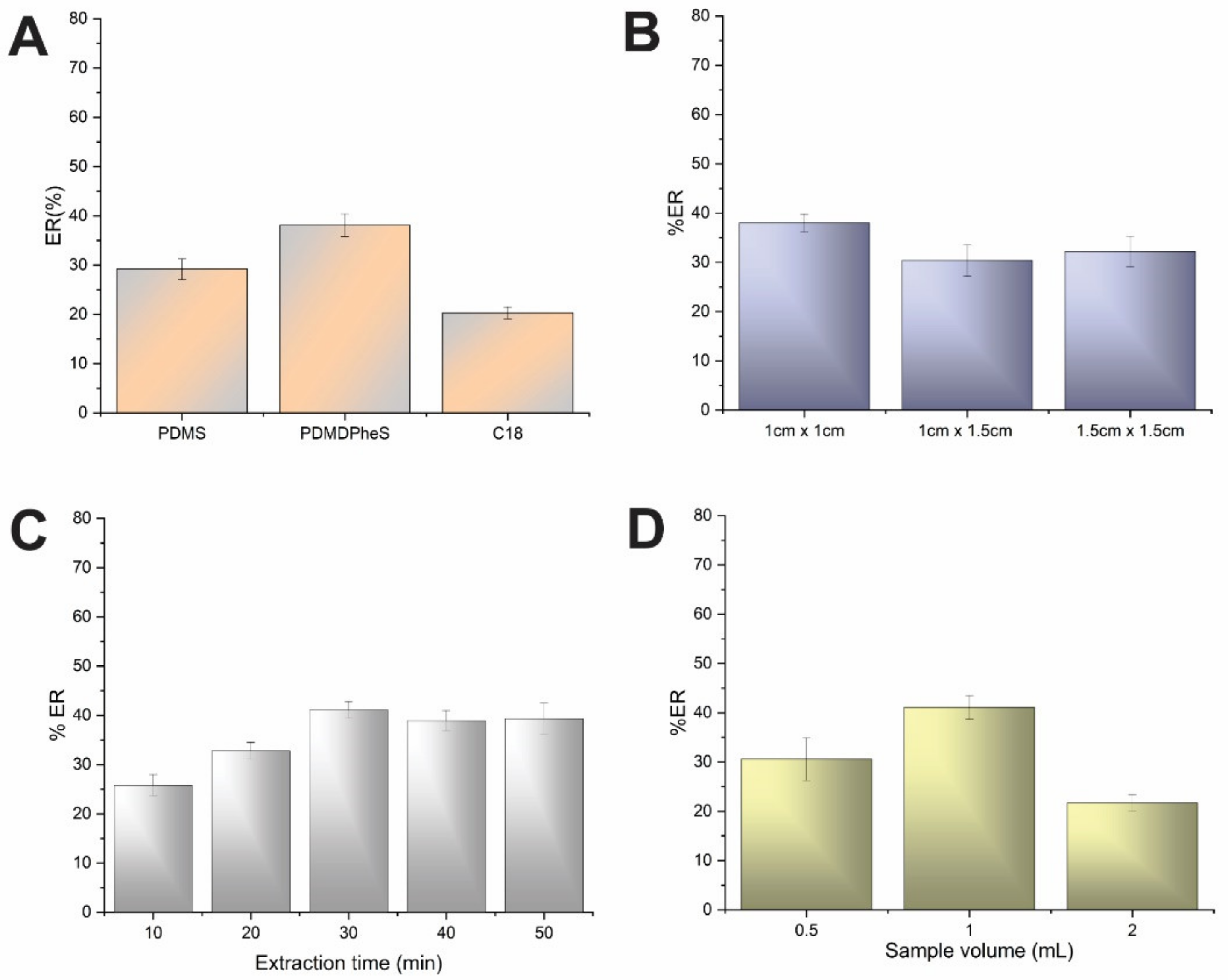
3.1. Effect of FPSE Sorbent Chemistry and Dimensions

The mechanism of FPSE membrane coated with sol–gel sorbent was based on the sorbent’s sponge-like architecture that enables the interactions of its functional groups with the analyte through many interactions such as dipole–dipole interaction, London dispersion, π–π interactions and hydrogen bonding [24]. The host substrate consists of cotton cellulose (100% hydrophilic), has great biocompatibility with the biological samples and therefore enhances the rate of mass transfer resulting in faster equilibrium.
AMTB is a macrocyclic lactose that exhibits amphoteric properties because of the presence of hydrophobic polyene and hydrophilic polyol region. These regions are attached to both a basic mycosamine sugar (pKa 10) and a carboxylic acid group (pKa 5.7) [25]. Based on these properties, three different hydrophobic FPSE membranes were investigated. These experiments were performed using a sample and elution volume of 1 mL each, stirring rate of 600 rpm and extraction time of 30 min. All experiments were performed in triplicate. As it can be seen in Figure 1A, moderate extraction performance was obtained for AMTB for all FPSE sorbents. Slightly higher performance was attained using the sol–gel PDMDPheS material and therefore this membrane was selected for further experiments. This sorbent has methyl and phenyl pendant groups in silanol terminated dimethylsiloxane–diphenylsiloxane copolymer resulting in a nonpolar medium. Apart from the dipole–dipole interactions, various intermolecular forces between the analyte and the extraction membrane including London dispersion, hydrogen bonding and hydrophilic interactions may be present. More specifically, the hydroxyl and amine groups of the AMTB molecule serve as electron donors and can form hydrogen bonds with the functional groups of the sorbent and the cellulose substrate.
Three different FPSE sizes were studied to find out the optimum membrane dimensions. The membranes were cut into rectangular-shaped pieces with surface areas of 1.0, 1.5 and 2.25 cm2. As expected, a higher membrane size required a large amount of sample which is not readily available in the case of biological fluids. As shown in Figure 1B, the extraction recovery (%ER) remained almost stable with the increase in the surface area of the membrane. The size of 1 × 1 cm was finally selected as it the most appropriate for the extraction vial (45 × 15 cm) used in this study.

3.2. Effect of Sample pH and Volume and Extraction Time

As mentioned above the AMTB is a weak base with a primary amino group in the molecule (Figure S1). Based on this, the pH of the aqueous sample affects its polarity and consequently its retention on the PDMDPheS-coated FPSE membrane. Two pH values (3 and 7) were evaluated to find the optimum conditions. The obtained %ER was almost double at neutral pH compared to acidic conditions. This may be attributed to the fact that in neutral conditions AMTB becomes less protonated and therefore interacts to a higher extent with the hydrophobic FPSE membrane.
The extraction time plays an important role on the extraction efficiency since it influences the contact time between the sol–gel-coated FPSE membrane and the sample. During the optimization of this parameter, time intervals of 10–50 min were examined under stirring. The experiments indicated (Figure 1C) that the %ER was linearly increased up to 30 min and leveled off thereafter. Thus, the extraction time of 30 min was chosen for further experiments resulting in %ER ca 40% for the AMTB. This value is acceptable considering that FPSE is an equilibrium technique [24].
The volume of sample is a significant factor affecting the FPSE efficiency as it is directly proportional to the absolute mass of the compounds. In our case, different sample volumes (i.e., 0.5, 1 and 2 mL) were investigated at a fixed absolute amount of AMTB. As illustrated in Figure 1D, the %ER was elevated up to when 1 mL of sample was used and significantly reduced at higher volumes. Finally, the value of 1 mL of sample volume was chosen.

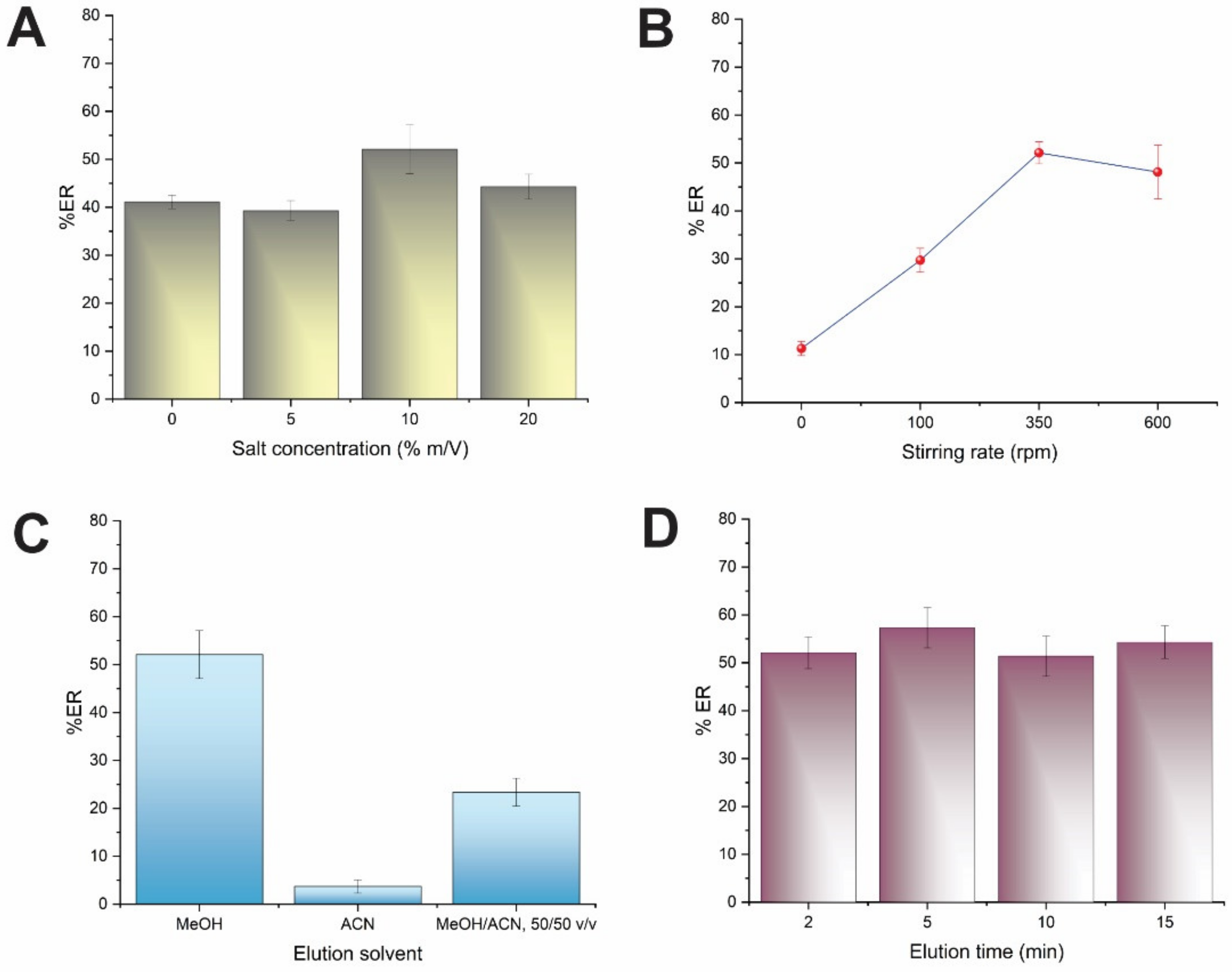
3.3. Effect of Ionic Strength and Stirring Rate

The impact of the sample’s ionic strength on the %ER was investigated by varying the concentration of NaCl between 0 and 20% m/v. Theoretically, the solubility of AMTB in a sample solution of aqueous nature is decreased at higher NaCl concentrations and therefore its presence could benefit the analyte’s interaction with the FPSE sorbent. However, antagonistic phenomena might take place because of an enhancement of the solution’s viscosity leading to reduced mass transfer. It was found that the %ER of the analyte was slightly enhanced at salt concentration up to 10% m/v (Figure 2A). Higher concentrations resulted in a diminished extraction performance possibly because of the above-explained phenomena. A NaCl content of 10% m/v was finally adopted for the subsequent experiments.
Stirring rate also plays a key role on extraction efficiency of the FPSE technique since it affects the mass transfer of the analyte from the sample to the sorbent. In our case, the stirring rate was examined from 0 to 600 rpm (Figure 2B). As it can be observed, the %ER of AMTB was enhanced by increasing the stirring rate up to 350 rpm, while no further increase was observed up to 600 rpm. Thus, further experiments were performed at a stirring rate of 350 rpm.

3.4. Effect of the Elution Solvent, Volume and Time

The investigation of the elution conditions is vital to make sure that appropriate elution of the drug takes place. During the optimization of this step, three different solvents were examined including MeOH, ACN and the mixture of MeOH: ACN, (50:50, v/v). The results are reported in Figure 2C. From these solvents, MeOH provided an almost 10-fold higher %ER compared to ACN. Thus, MeOH was used in further experiments.
The effect of the elution time was investigated in the range of 2–15 min. According to Figure 2D, the %ER was not statistically different at the studied intervals while satisfactory desorption of AMTB was achieved even at 2 min. Thus, this time interval was selected to have an enhanced sample throughput and reduced sample preparation time.
At a final step, the effect of the volume of the eluent on the %ER of the AMTB was examined between 250–1000 μL (Figure S2). It was found that the volume of MeOH used for the elution of AMTB had a non-significant effect on its %ER. According to the principles of green analytical chemistry (GAC) [26], an aliquot of 250 μL was selected to reduce the consumption of organic solvents.

3.5. Method Validation

HPLC-UV chromatograms of the analysis of pooled human urine samples are shown in Figure 3. As is evident, no interfering peaks corresponding to endogenous compounds were recorded at the retention time of AMTB and the ISTD.
Method linearity was assessed using matrix-matched calibration curves in the range of 0.10 to 10.0 μg mL−1. Matrix-matched calibration was used for the quantitation of the drug in the samples due to the non-specific binding of the certain analyte to proteins [27]. The ratio of the peak area of AMTB and the peak area of the ISTD was used as the response to examine the linearity. The regression equation for AMTB was Y = (0.08344 ± 0.00151) X + (0.00837 ± 0.00692). The r2 value was greater than 0.9987 showing acceptable linearity within the examined range. The LLOQ was set at 0.10 μg mL−1 and the LOD (based on the S/N = 3 criterion) was estimated be 0.025 μg mL−1.
The results for the evaluation of method precision and accuracy are presented in Table 1. The intra-day and the inter-day precision were found to be lower than 12.7% and 9.8%, respectively, which is in compliance with the acceptance criteria as described in Section 2.4. The accuracy of the method was expressed as relative recovery (RR %) and was varied between 88.1–110.3% (intra-day) and 96.4–108.3% (inter-day) that also complies with the acceptance criteria that were set.
The reusability of the PDMDPheS-coated FPSE material was investigated. As can be observed in Figure 4 the FPSE membrane was found to be reusable for at least 20 continuous extraction cycles of AMTB in human urine samples with a loss of efficiency of less than 10%.

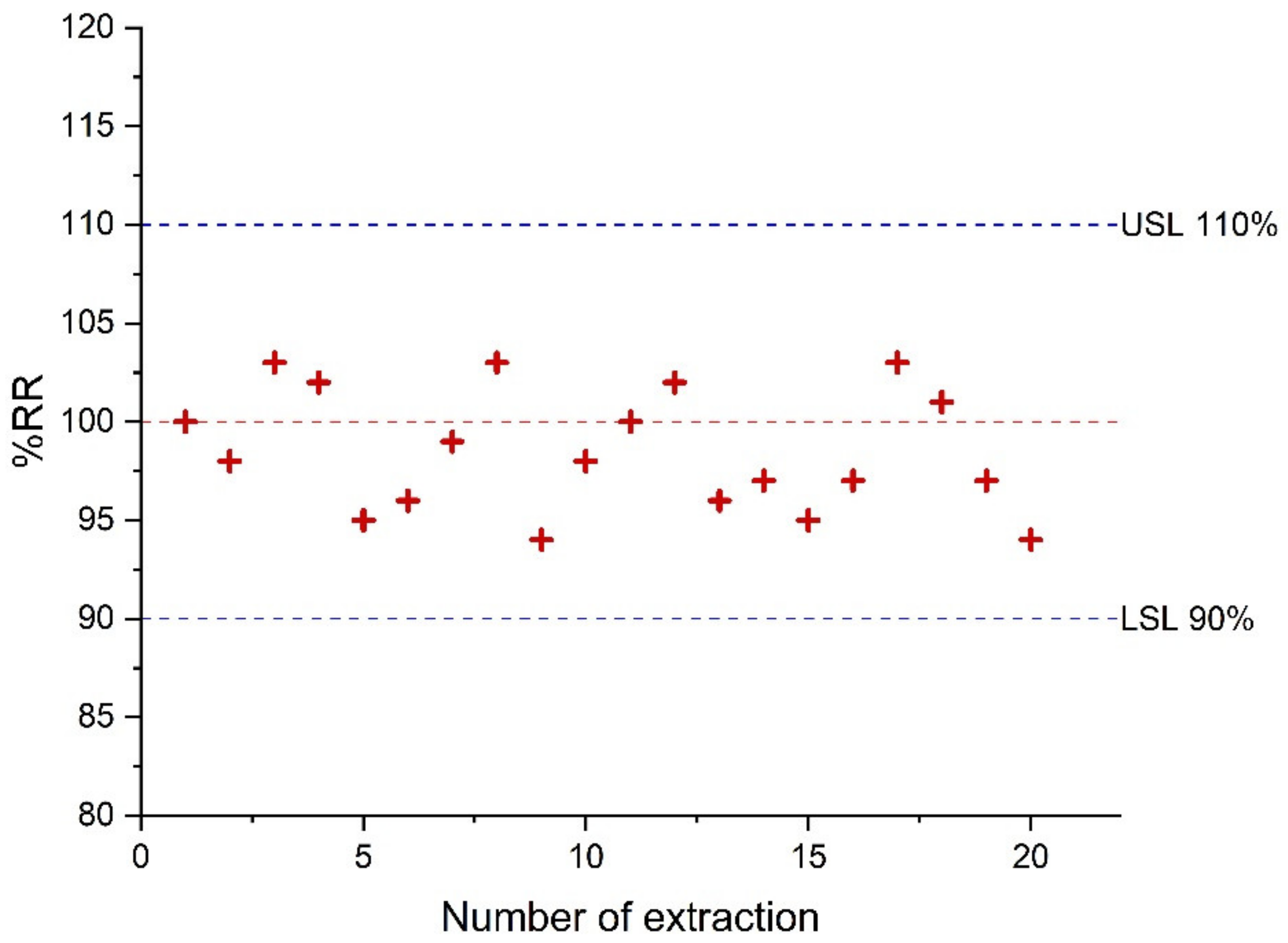
3.6. Sample Stability

The stability of AMTB in unprocessed urine was examined at 0, 4, 8 and 24 h stored at 25 °C and +4 °C. The stability of the analyte in biological matrices is accepted when the deviation from the nominal value is equal to or less than ±15%. No analyte degradation was observed in agreement with other published data [28].

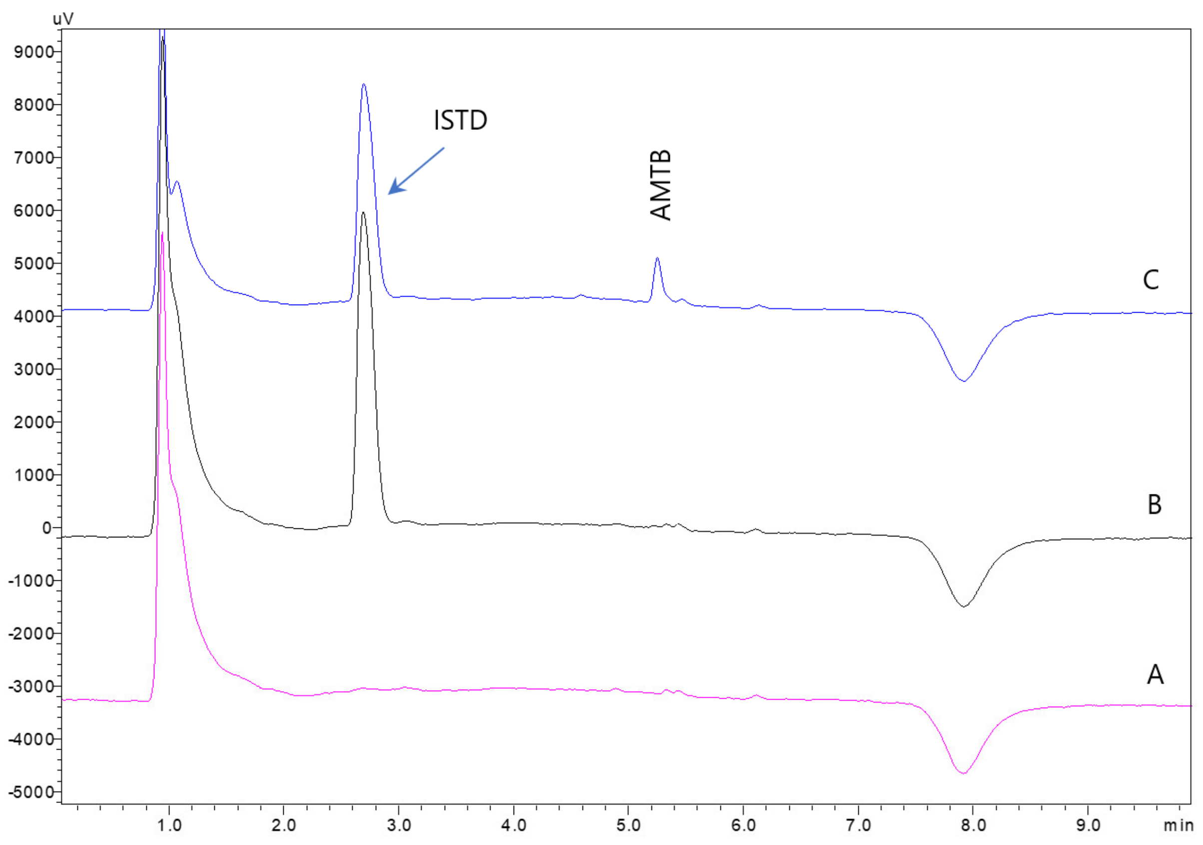
3.7. Application to Human Urine Samples

The applicability and the accuracy of the FPSE-HPLC-UV method was demonstrated by analyzing individual human urine samples. For this purpose, authentic samples spiked at concentration levels of 0.1, 0.25, 1.0 and 10.0 μg mL−1 were analyzed (Table 2). The relative recovery values for all AMTB was 82.6–110.9% and the %RSD was better than 12.0%, demonstrating the successful application of the method for the analysis of real samples. Representative chromatograms are depicted in Figure S3 (Supplementary Material).

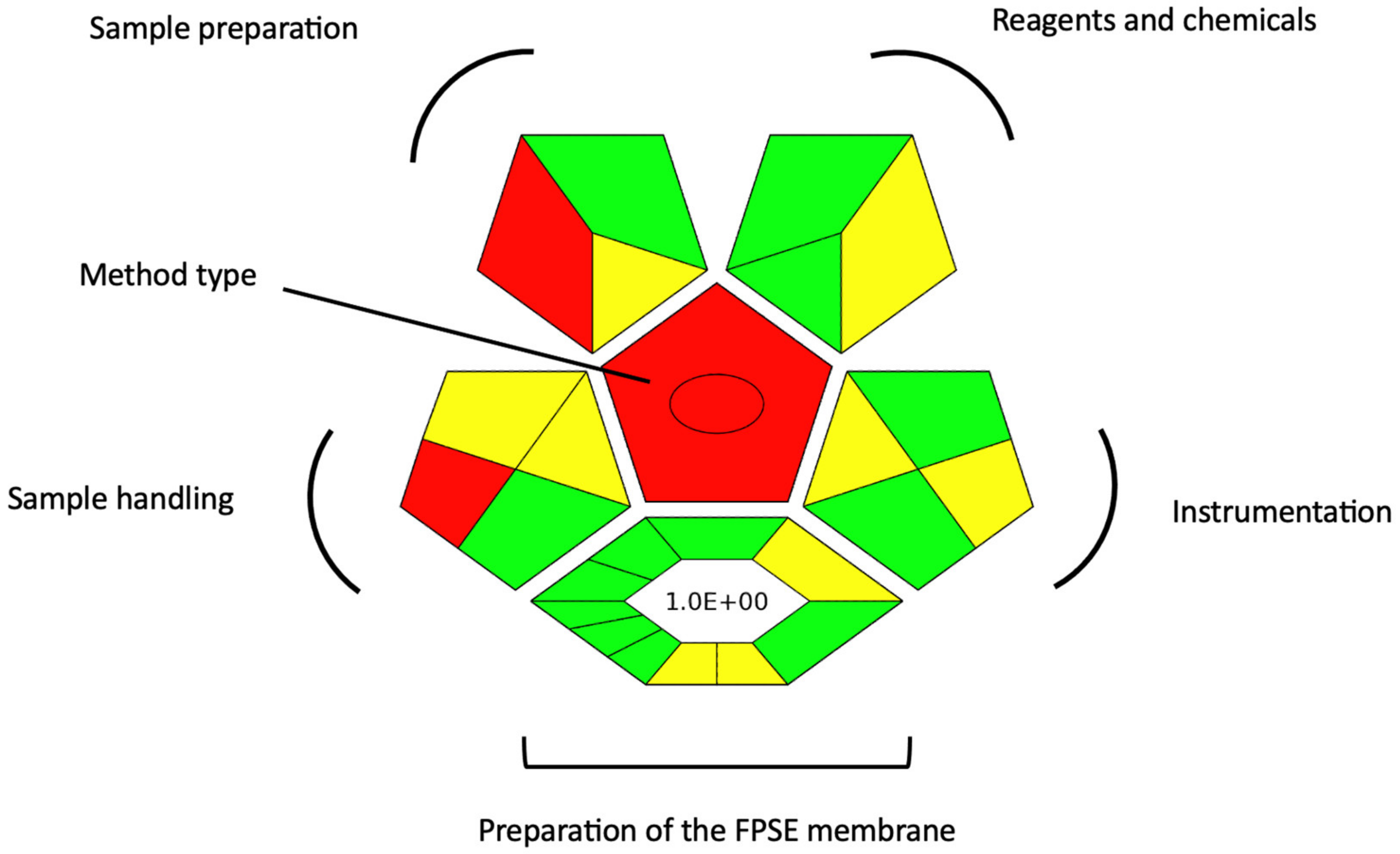
3.8. Evaluation of the Green Character of the FPSE-HPLC-UV Method

In order to investigate the green character of the FPSE-HPLC-UV method and to identify aspects for further improvement, complexGAPI index was used [29]. This index examines the environmental friendliness of an analytical method from the collection of the sample to the final step of the analytical determination, as it was originally introduced in the GAPI index [30]. At the same time, it also takes into account the synthesis used for new extraction platforms, which in our case refers to the sol–gel PDMDPheS-coated FPSE membranes. The complexGAPI pictogram of our method is depicted in Figure 5. Regarding the synthesis of the sol–gel PDMDPheS-coated FPSE membranes, most of the criteria are met as can be discerned from the green color at most of the parts of the hexagon. Among the criteria that are met are the low E-factor of the synthetical route, the reduced waste generation and the high synthesis yield. As for the analytical method, it requires relatively low chemical consumption and it leads to relatively low waste generation, which serves as a benefit compared to the widely used conventional sample preparation techniques. Towards the further improvement of the proposed method is the utilization of green chemicals (e.g., deep eutectic solvents) for the replacement of methanol and the combination of the FPSE protocol with ultra-high performance liquid chromatography for the further reduction of chemicals consumption.

4. Conclusions

In this work, we propose a simple and rapid sol–gel PDMDPheS-coated FPSE protocol combined with HPLC-UV for the monitoring of AMTB in human urine. The analytical method was rapid, economic and showed a green character. Moreover, it eliminated the required sample preparation steps and presented many benefits such as the utilization of low amounts of organic solvents and handling simplicity. The FPSE-HPLC-UV method was validated based on the guidelines of the FDA for bioanalytical methods, exhibiting good performance characteristics. Thus, the proposed procedure can serve as a useful analytical tool for the determination of AMTB for clinical applications.

Supplementary Materials

The following supporting information can be downloaded at: https://www.mdpi.com/article/10.3390/chemosensors10120537/s1, Figure S1: Chemical structure of AMTB; Figure S2: Effect of elution solvent volume on %ER of AMTB; Figure S3: Representative HPLC-UV chromatograms of the analysis blank urine and spiked at concentration levels of 0.25, 1.0 and 5.0 μg mL−1 after FPSE; Table S1: Characteristics of FPSE membranes and the molar ratio of the reagents used for their preparation.

Author Contributions

Conceptualization, C.K.Z.; methodology, C.K.Z., A.K. and N.M.; software, C.K.Z.; validation, E.A., N.A. and N.M.; formal analysis, E.A. and N.A.; investigation, E.A., N.M. and A.K.; data curation, E.A., N.A. and N.M.; writing—original draft preparation, N.M.; writing—review and editing, C.K.Z., A.K. and K.G.F.; supervision, C.K.Z.; project administration, C.K.Z. All authors have read and agreed to the published version of the manuscript.

Funding

This research received no external funding.

Institutional Review Board Statement

Not applicable.

Informed Consent Statement

Written informed consent has been obtained from the volunteers to publish this paper.

Data Availability Statement

Data are contained within the article or in the Supplementary Materials.

Conflicts of Interest

The authors declare no conflict of interest.

References

  1. Patel, P.A.; Patravale, V.B. AmbiOnp: Solid Lipid Nanoparticles of Amphotericin B for Oral Administration. J. Biomed. Nanotechnol. 2011, 7, 632–639. [Google Scholar] [CrossRef] [PubMed]
  2. Davi Marena, G.; Aparecido dos Santos Ramos, M.; Maria Bauab, T.; Chorilli, M.; Davi Marena, Ã.G.; Aparecido dos Santos Ramos, Ã.M.; ıs Maria Bauab, T. A Critical Review of Analytical Methods for Quantification of Amphotericin B in Biological Samples and Pharmaceutical Formulations. Crit. Rev. Anal. Chem. 2020, 52, 555–576. [Google Scholar] [CrossRef] [PubMed]
  3. Lee, J.W.; Petersen, M.E.; Lin, P.; Dressler, D.; Bekersky, I. Quantitation of Free and Total Amphotericin B in Human Biologic Matrices by a Liquid Chromatography Tandem Mass Spectrometric Method. Ther. Drug Monit. 2001, 23, 268–276. [Google Scholar] [CrossRef]
  4. Bekersky, I.; Fielding, R.M.; Dressler, D.E.; Lee, J.W.; Buell, D.N.; Walsh, T.J. Pharmacokinetics, Excretion, and Mass Balance of Liposomal Amphotericin B (AmBisome) and Amphotericin B Deoxycholate in Humans. Antimicrob. Agents Chemother. 2002, 46, 828–833. [Google Scholar] [CrossRef] [PubMed] [Green Version]
  5. Wang, L.H.; Smith, P.C.; Anderson, K.L.; Fielding, R.M. High-Performance Liquid Chromatographic Analysis of Amphotericin B in Plasma, Blood, Urine and Tissues for Pharmacokinetic and Tissue Distribution Studies. J. Chromatogr. B Biomed. Sci. Appl. 1992, 579, 259–268. [Google Scholar] [CrossRef]
  6. Ingle, R.G.; Zeng, S.; Jiang, H.; Fang, W.J. Current Developments of Bioanalytical Sample Preparation Techniques in Pharmaceuticals. J. Pharm. Anal. 2022, 12, 517–529. [Google Scholar] [CrossRef]
  7. Queiroz, M.E.C.; de Souza, I.D.; de Oliveira, I.G.; Grecco, C.F. In Vivo Solid Phase Microextraction for Bioanalysis. TrAC–Trends Anal. Chem. 2022, 153, 116656. [Google Scholar] [CrossRef]
  8. Li, F.; Ceballos, M.R.; Balavandy, S.K.; Fan, J.; Khataei, M.M.; Yamini, Y.; Maya, F. 3D Printing in Analytical Sample Preparation. J. Sep. Sci. 2020, 43, 1854–1866. [Google Scholar] [CrossRef]
  9. Hansen, F.; Øiestad, E.L.; Pedersen-Bjergaard, S. Bioanalysis of Pharmaceuticals Using Liquid-Phase Microextraction Combined with Liquid Chromatography–Mass Spectrometry. J. Pharm. Biomed Anal. 2020, 189, 113446. [Google Scholar] [CrossRef]
  10. Turoňová, D.; Kujovská Krčmová, L.; Švec, F. Application of Microextraction in Pipette Tips in Clinical and Forensic Toxicology. TrAC–Trends Anal. Chem. 2021, 143, 116404. [Google Scholar] [CrossRef]
  11. Paiva, A.C.; Crucello, J.; de Aguiar Porto, N.; Hantao, L.W. Fundamentals of and Recent Advances in Sorbent-Based Headspace Extractions. TrAC–Trends Anal. Chem. 2021, 139, 116252. [Google Scholar] [CrossRef]
  12. Mamounas, G.; Manousi, N.; Kabir, A.; Furton, K.G.; Mystridis, G.A.; Vizirianakis, I.S.; Zacharis, C.K. Designing an “All-in-One” Microextraction Capsule Device for the Liquid Chromatographic-Fluorescence Determination of Doxorubicin and Its Metabolites in Rat Plasma. J. Chromatogr. A 2022, 1680, 463432. [Google Scholar] [CrossRef]
  13. US20140274660A1–Fabric Phase Sorptive Extractors (Fpse)–Google Patents. Available online: https://patents.google.com/patent/US20140274660A1/en (accessed on 16 October 2022).
  14. Parla, A.; Zormpa, E.; Paloumpis, N.; Kabir, A.; Furton, K.G.; Roje, Ž.; Samanidou, V.; Vrček, I.V.; Panderi, I. Determination of Intact Parabens in the Human Plasma of Cancer and Non-Cancer Patients Using a Validated Fabric Phase Sorptive Extraction Reversed-Phase Liquid Chromatography Method with Uv Detection. Molecules 2021, 26, 1526. [Google Scholar] [CrossRef] [PubMed]
  15. Agadellis, E.; Tartaglia, A.; Locatelli, M.; Kabir, A.; Furton, K.G.; Samanidou, V. Mixed-Mode Fabric Phase Sorptive Extraction of Multiple Tetracycline Residues from Milk Samples Prior to High Performance Liquid Chromatography-Ultraviolet Analysis. Microchem. J. 2020, 159, 105437. [Google Scholar] [CrossRef]
  16. Alampanos, V.; Kabir, A.; Furton, K.G.; Samanidou, V.; Papadoyannis, I. Fabric Phase Sorptive Extraction for Simultaneous Observation of Four Penicillin Antibiotics from Human Blood Serum Prior to High Performance Liquid Chromatography and Photo-Diode Array Detection. Microchem. J. 2019, 149, 103964. [Google Scholar] [CrossRef]
  17. Locatelli, M.; Tinari, N.; Grassadonia, A.; Tartaglia, A.; Macerola, D.; Piccolantonio, S.; Sperandio, E.; D’Ovidio, C.; Carradori, S.; Ulusoy, H.I.; et al. FPSE-HPLC-DAD Method for the Quantification of Anticancer Drugs in Human Whole Blood, Plasma, and Urine. J. Chromatogr. B 2018, 1095, 204–213. [Google Scholar] [CrossRef] [PubMed]
  18. Tartaglia, A.; Kabir, A.; D’Ambrosio, F.; Ramundo, P.; Ulusoy, S.; Ulusoy, H.I.; Merone, G.M.; Savini, F.; D’Ovidio, C.; de Grazia, U.; et al. Fast Off-Line FPSE-HPLC-PDA Determination of Six NSAIDs in Saliva Samples. J. Chromatogr. B Analyt. Technol. Biomed. Life Sci. 2020, 1144, 122082. [Google Scholar] [CrossRef]
  19. FDA. Bioanalytical Method Validation Guidance. 2018. Available online: https://www.fda.gov/files/drugs/published/Bioanalytical-Method-Validation-Guidance-for-Industry.pdf (accessed on 10 December 2022).
  20. Brooks, T.; Keevil, C.W. A Simple Artificial Urine for the Growth of Urinary Pathogens. Lett. Appl. Microbiol. 1997, 24, 203–206. [Google Scholar] [CrossRef]
  21. Kumar, R.; Gaurav; Heena; Malik, A.K.; Kabir, A.; Furton, K.G. Efficient Analysis of Selected Estrogens Using Fabric Phase Sorptive Extraction and High Performance Liquid Chromatography-Fluorescence Detection. J. Chromatogr. A 2014, 1359, 16–25. [Google Scholar] [CrossRef]
  22. Aznar, M.; Úbeda, S.; Nerin, C.; Kabir, A.; Furton, K.G. Fabric Phase Sorptive Extraction as a Reliable Tool for Rapid Screening and Detection of Freshness Markers in Oranges. J. Chromatogr. A 2017, 1500, 32–42. [Google Scholar] [CrossRef]
  23. Alcudia-León, M.C.; Lucena, R.; Cárdenas, S.; Valcárcel, M.; Kabir, A.; Furton, K.G. Integrated Sampling and Analysis Unit for the Determination of Sexual Pheromones in Environmental Air Using Fabric Phase Sorptive Extraction and Headspace-Gas Chromatography–Mass Spectrometry. J. Chromatogr. A 2017, 1488, 17–25. [Google Scholar] [CrossRef] [PubMed]
  24. Mazaraki, K.; Kabir, A.; Furton, K.G.; Fytianos, K.; Samanidou, V.F.; Zacharis, C.K. Fast Fabric Phase Sorptive Extraction of Selected β-Blockers from Human Serum and Urine Followed by UHPLC-ESI-MS/MS Analysis. J. Pharm. Biomed Anal. 2021, 199, 114053. [Google Scholar] [CrossRef] [PubMed]
  25. Faustino, C.; Pinheiro, L. Lipid Systems for the Delivery of Amphotericin B in Antifungal Therapy. Pharmaceutics 2020, 12, 29. [Google Scholar] [CrossRef] [PubMed] [Green Version]
  26. Gałuszka, A.; Migaszewski, Z.; Namieśnik, J. The 12 Principles of Green Analytical Chemistry and the SIGNIFICANCE Mnemonic of Green Analytical Practices. TrAC–Trends Anal. Chem. 2013, 50, 78–84. [Google Scholar] [CrossRef]
  27. Bekersky, I.; Fielding, R.M.; Dressler, D.E.; Lee, J.W.; Buell, D.N.; Walsh, T.J. Plasma Protein Binding of Amphotericin B and Pharmacokinetics of Bound versus Unbound Amphotericin B after Administration of Intravenous Liposomal Amphotericin B (AmBisome) and Amphotericin B Deoxycholate. Antimicrob. Agents Chemother. 2002, 46, 834–840. [Google Scholar] [CrossRef] [Green Version]
  28. Pippa, L.F.; Marques, M.P.; da Silva, A.C.T.; Vilar, F.C.; de Haes, T.M.; da Fonseca, B.A.L.; Martinez, R.; Coelho, E.B.; Wichert-Ana, L.; Lanchote, V.L. Sensitive LC-MS/MS Methods for Amphotericin B Analysis in Cerebrospinal Fluid, Plasma, Plasma Ultrafiltrate, and Urine: Application to Clinical Pharmacokinetics. Front. Chem. 2021, 9, 998. [Google Scholar] [CrossRef]
  29. Płotka-Wasylka, J.; Wojnowski, W. Complementary Green Analytical Procedure Index (ComplexGAPI) and Software. Green Chem. 2021, 23, 8657–8665. [Google Scholar] [CrossRef]
  30. Płotka-Wasylka, J. A New Tool for the Evaluation of the Analytical Procedure: Green Analytical Procedure Index. Talanta 2018, 181, 204–209. [Google Scholar] [CrossRef]
Figure 1. Effect of (A) FPSE sorbent type, (B) FPSE membrane size, (C) extraction time and (D) sample volume on %ER of AMTB (n = 3).
Chemosensors 10 00537 g001
Figure 2. Effect of (A) salt concentration, (B) stirring rate, (C) elution solvent and (D) elution time on %ER of AMTB (n = 3).
Chemosensors 10 00537 g002
Figure 3. Representative chromatogram of the FPSE-HPLC-UV analysis of (A) blank human urine sample, (B) spiked with ISTD solution and (C) spiked with ISTD and AMTB at 1.0 μg mL−1.
Chemosensors 10 00537 g003
Figure 4. Reusability of PDMDPheS-coated FPSE membrane. (red crosses indicate the results of each measurement).
Chemosensors 10 00537 g004
Figure 5. ComplexGAPI pictogram for the FPSE-HPLC-UV method. Green colour indicates compliance with the requirements.
Chemosensors 10 00537 g005
Table 1. Intra-day and inter-day precision and accuracy data of the FPSE-HPLC-UV method for the quantitation of AMTB in human urine.
Intra-Day (n = 3)Inter-Day (n = 3)
Added Concentration
(μg mL−1)
Precision
(%RSD)
Accuracy
RR 1 (%)
Precision
(%RSD)
Accuracy
RR (%)
0.10 (LLOQ)12.788.19.896.4
0.25 (LQC)7.091.69.397.7
1.0 (MQC)5.7110.38.4108.3
10.0 (HQC)3.0107.67.4103.5
1 RR: relative recovery.
Table 2. Relative recoveries of the studied drug from human urine by the proposed FPSE-HPLC-UV method.
Added Concentration
(μg mL−1)
Relative Recoveries (%) (RSD%, n = 3)
Sample#1Sample#2Sample#3
0.1089.2 (3.0)106.9 (7.5)82.6 (12.0)
0.25110.7 (4.0)95.4 (9.5)98.3 (9.7)
1.0105.8 (6.3)106.9 (6.7)110.9 (5.0)
10.098.6 (7.0)89.1 (4.8)89.7 (4.6)
Publisher’s Note: MDPI stays neutral with regard to jurisdictional claims in published maps and institutional affiliations.

