Article

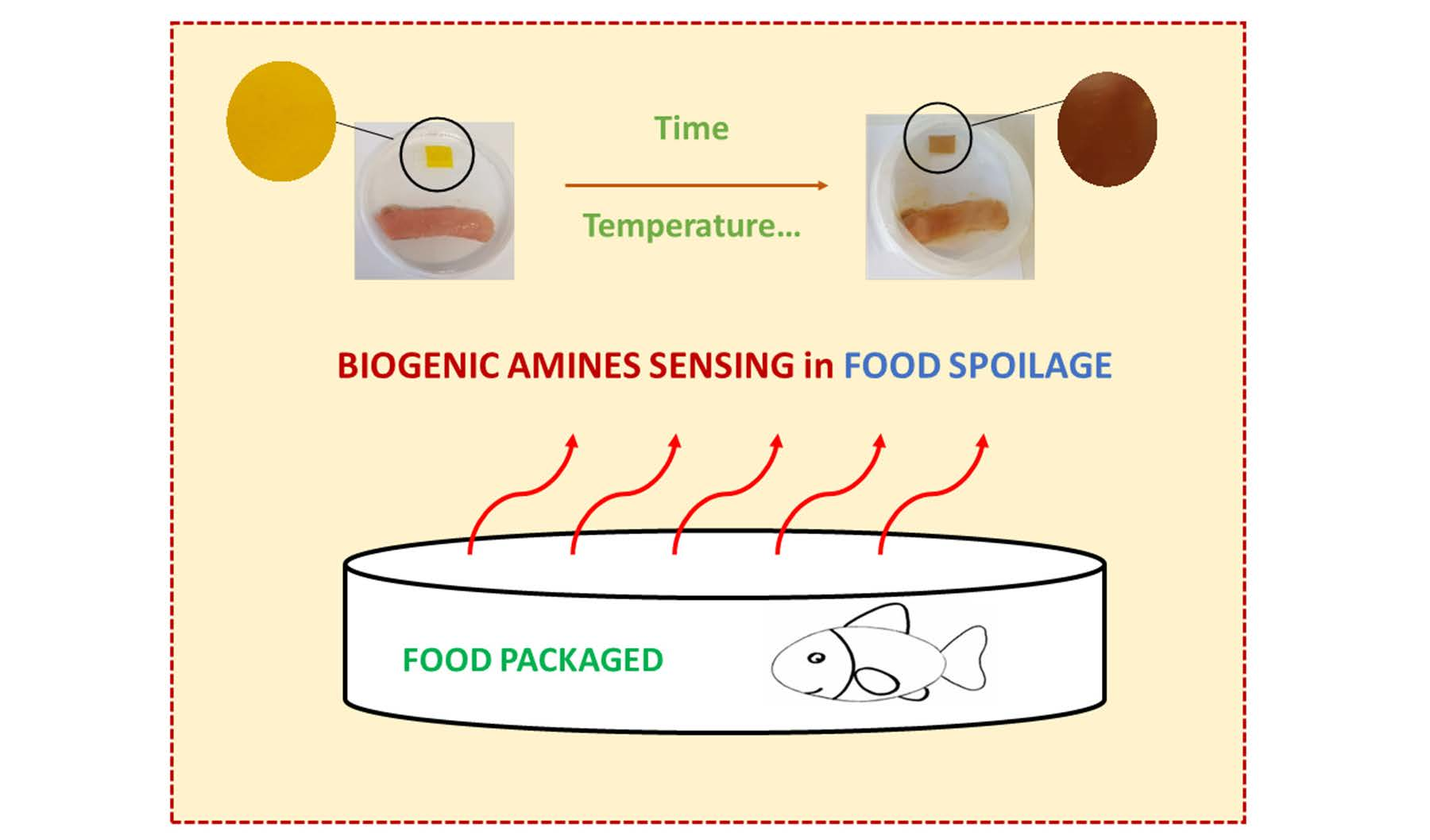
Reversible Colorimetric and Fluorescence Solid Sensors Based on Aryl Hydrazone Derivatives of 1,8-Naphthalimides for Caustic Media and Biogenic Amine Vapors

by
Jesús L. Pablos
1,2,*,
Sabela Fernández-Alonso
1,
Fernando Catalina
1 and
Teresa Corrales
1,*
1
Grupo de Fotoquímica de Polímeros, Instituto de Ciencia y Tecnología de Polímeros (ICTP-CSIC), Juan de la Cierva 3, 28006 Madrid, Spain
2
Departamento Química en Ciencias Farmacéuticas, Facultad de Farmacia, Universidad Complutense de Madrid, Pl. de Ramón y Cajal, s/n, 28040 Madrid, Spain
*
Authors to whom correspondence should be addressed.
Chemosensors 2022, 10(10), 417; https://doi.org/10.3390/chemosensors10100417
Submission received: 16 September 2022 / Revised: 6 October 2022 / Accepted: 9 October 2022 / Published: 13 October 2022
(This article belongs to the Collection Women Special Issue in Chemosensors and Analytical Chemistry)

Abstract

:
Fluorescence and colorimetric solid sensors for caustic media and biogenic amine vapors have been prepared. For this purpose, several hydrazone derivatives of naphthalimides were synthesized and anchored to a photo-crosslinked membrane functionalized with acid chloride groups. The membranes were characterized using different techniques, and their thermal properties and swelling degree were determined. The new naphthalimides and the membranes were evaluated as sensors by determining the change in their spectroscopic properties of absorption and fluorescence with pH. The polymeric sensors exhibit improved stability and can be reused, as a consequence of their solid character and the reversibility of the process. Furthermore, membranes were evaluated as a sensor of trimethylamine vapors through their absorption and fluorescence bands, and the color change in the membrane showed that it could be used to detect basic media with the naked eye. Finally, membranes were packaged in Petri dishes at a controlled temperature with fresh fish bought in the local market. Then, the real chance of using the sensory materials was determined by analyzing the color change in samples.

Graphical Abstract

1. Introduction

In recent years, the development of chemosensors has attracted great attention because of both their potential application in different fields, such as environmental analytics, medical diagnostics and process control, and their advantages as a tool for sensing analytes, including their high sensitivity and selectivity, easy operation, rapid analysis, nondestructive detection, real-time monitoring and cost-effectiveness. A chemosensor is defined as a chemical probe containing photoactive units that teams up with analytes and the interaction is detected by fluorescence changes, or induces a color change which allows the human eye to act as a detector. Among the different type of sensors, naphthalimide derivatives are distinguishable since their thermal, photophysical and electroluminescence properties can be modulated by including different substituents at the 4 position of the naphthalimide structure [1]. Despite a wide variety of naphthalimides that have been reported in the literature, in which different moieties are attached to the structure [2], the design of high-selectivity and -sensitivity fluorescence sensors remains a challenge [3]. Recently, a review of the development of 1,8-naphthalimide-based sensors over the last 5 years was published, including applications for the sensing of biomolecules, metal ions and anions [4]. Additionally, hydrazone-based molecular structures are widely used in organic synthesis, medicinal chemistry and other applications, as well as fluorescent and colorimetric chemosensors [5,6,7,8,9,10,11]. The hydrazone backbone includes an imine carbon with an electrophile character, two nucleophilic nitrogen in both amine and imine groups, and the possible isomerization of the C=N double bond from the conjugation of imine and acid N-H. In certain cases, the long response time or low sensitivity in the presence of analytes is responsible for the weak fluorescence because of quenching effects, such as double-bond isomerization in the excited state, the photoinduced electron transfer (PET) process or the excited-state intramolecular proton-transfer (ESIPT) process [12,13]. Usually, hydrazones are anchored to other fluorophores or aromatic structures such as naphthalimides or coumarins [14,15,16,17,18]. Concerning the target analytes, biogenic amines are of special interest if they can be detected in the vapor phase and also with the naked eye, since they are related to the food freshness of packaged food [19]. In addition, the development of sensors that work under extreme alkaline conditions is considered necessary, since concentrated strong bases, such as caustic ones, are widely used in many industrial processes that cause pollution and equipment corrosion, and the conventional sensors are generally useful for pH ranges of 1–11, but not in high-basicity media.
Furthermore, due to the awareness of the importance of using sensors with a higher stability and lower toxicity, different strategies have been proposed to develop polymeric sensors [20,21,22,23]. Those materials offer the advantages of polymers, which can be processed as manageable films or textiles with better mechanical properties and thermal stability. In previous works, polymerizable naphthalimides have been synthesized and copolymerized with different comonomers, and the functionalization of the surface of polymers with 1,8-naphthalimide derivatives have been reported for pH and Hg2+ detection [24,25,26,27].
In this work, solid sensors for caustic media and biogenic amine vapors have been developed. For this purpose, several hydrazone derivatives of naphthalimides have been synthesized and anchored to a membrane functionalized with acid chloride groups. The membranes were obtained by a photopolymerization reaction, which allows for the design of polymeric sensors with tailor-made properties as a function of the nature and content of monomers selected [28]. The new naphthalimides and the membranes are evaluated as a sensor by determining the change in their spectroscopic properties of absorption and fluorescence with pH. Additionally, the functionalized materials are analyzed as a sensor of trimethylamine vapors through their absorption and fluorescence bands, and the color change in the membrane shows that it could be used to detect basic media with the naked eye. Finally, membranes were packaged in Petri dishes at a controlled temperature with fresh fish bought in the local market, and changes in fluorescence and color could be observed from the beginning and up to 10 days, which is related to the increase in the amine concentration when the fish is decomposing.
The innovation point of our proposal is the real chance to use the sensory materials to detect biogenic amines in fish, and the advantages offered in the materials developed, such as costless novel sensory materials, produce an easy procedure of detection by analyzing the color change in samples. Additionally, as a consequence of their solid character and the reversibility of the process, the polymeric sensors exhibit improved stability and can be reused.

2. Materials and Methods

2.1. Materials and Reagents

In this work, commercially available reagents and solvents were used as received: sodium hydroxide (Panreac, Darmstadt, Germany, 98%), hydrochloric acid (VWR Chemicals, Radnor, PA, USA, 37%), ultrapure Milli-Q water (Millipore, Burlington, MA, USA), trimethylamine (Aldrich, St. Louis, MO, USA, ≥99%), 4-bromo-1,8-naphthalic anhydride (Aldrich, St. Louis, MO, USA, 95%), potassium carbonate (Panreac, Darmstadt, Germany, 99%), 2-aminoethanol (Aldrich, St. Louis, MO, USA, 99%), 4-bromo-1,8-naphthalic anhydride (Aldrich, St. Louis, MO, USA, 95%), hydrazine monohydrate (Aldrich, St. Louis, MO, USA, 99%), benzaldehyde (Aldrich, St. Louis, MO, USA, 98%), salicylaldehyde (Aldrich, St. Louis, MO, USA, 99%), 3-hydroxybenzaldehyde (Aldrich, St. Louis, MO, USA, 99%), 4-hydroxybenzaldehyde (Aldrich, St. Louis, MO, USA, 99%), ethanol (VWR Chemicals, Radnor, PA, USA, 99.95%), dimethylformamide (Scharlau, Cham, Germany, 99.8%), an Irgacure 2959 photoinitiator (BASF, Ludwigshafen, Germany), ethyl acetate (Aldrich, St. Louis, MO, USA, ≥99.5%), hexane (Carlo Erba Reagents, Val-de-Reuil, France, 99%), 2-propanol (Scharlau, Cham, Germany, 99.5%), dichloromethane (Aldrich, St. Louis, MO, USA, ≥99.9%), toluene (Merck, Kenilworth, NJ, USA, ≥99.9%), diethyl ether (Carlo Erba, Val-de-Reuil, France, >99.8%), 1,4-dioxan (Panreac, Darmstadt, Germany, 99%), tetrahydrofuran (Aldrich, St. Louis, MO, USA, 99.9%), chloroform (Scharlau Cham, Germany, 99%), acetone (Scharlau, Cham, Germany, 99.5%), acetonitrile (Aldrich, St. Louis, MO, USA, ≥99.5%), 1-butanol (Panreac, Darmstadt, Germany, 99.5%), methanol (Aldrich, St. Louis, MO, USA, ≥99.8%), coumarin 6 (Aldrich, St. Louis, MO, USA, 98%), deuterium oxide (Euriso-top, Saarbrücken, Germany, 99.9% D), dimethylsulfoxide D6 (Euriso-top, Saarbrücken, Germany, 99.8% D), diiodomethane (Aldrich, St. Louis, MO, USA, 99%).
Before use, the monomers were distilled under a vacuum to remove the inhibitor, 1-vinyl-2-pyrrolidone (Aldrich, St. Louis, MO, USA, ≥97%), butyl acrylate (Aldrich, St. Louis, MO, USA, ≥99%), ethylene glycol dimethacrylate (Aldrich, St. Louis, MO, USA, 98%) and methacryloyl chloride (Aldrich, St. Louis, MO, USA, ≥97%).

2.2. Synthesis of the Aryl Hydrazone Derivatives of 1,8-Naphthalimide

All the naphthalimide derivatives were synthetized using the microwave (MW) method with the same experimental conditions and 20 min of reaction time in all cases. The synthesis pathways, naphthalimide structures and reaction data are shown in Figure 1.

2.2.1. Synthesis of N-(2-Hydroxyethyl)-4-(Hydrazine-1-Yl)-1,8-Naphthalimide (Compound 3)

Compound 2 was synthesized from 4-bromo-1,8-naphthalic anhydride (compound 1) following the procedures described previously in the bibliography [24,29]. A procedure described in the literature was modified and used to synthesize compound 3 [30]. In a pressure-resistant microwave reactor, compound 2 (0.30 g, 0.937 mmol) and hydrazine monohydrate (210 µL, 2.811 mmol) were added in 2-methoxyethanol (10 mL). The reaction mixture was heated at 130 °C under an argon atmosphere and constant stirring (600 rpm) for 20 min. Then, the resulting mixture was cooled at 5 °C and filtered. The solid obtained was washed with water (3 × 30 mL), dried and identified as N-(2-hydroxyethyl)-4-(hydrazine-1-yl)-1,8-naphthalimide (compound 3). Yield: 92%. M.p.: 188.7 ± 2 °C. 1H NMR (400 MHz, DMSO-d6) δ 9.11 (s, 1H), 8.60 (d, J = 8.3 Hz, 1H), 8.40 (d, J = 7.2 Hz, 1H), 8.28 (d, J = 8.6 Hz, 1H), 7.62 (t, J = 7.8 Hz, 1H), 7.24 (d, J = 8.6 Hz, 1H), 4.79 (t, J = 5.8 Hz, 1H), 4.67 (s, 2H), 4.11 (t, J = 6.7 Hz, 2H), 3.57 (d, J = 6.2 Hz, 2H).13C NMR (75 MHz, DMSO) δ 163.92, 163.04, 153.15, 134.17, 130.54, 129.35, 128.19, 124.08, 121.82, 118.42, 107.45, 103.97, 57.99, 41.37. FT-IR (wavenumbers, cm−1): νOH 3461 cm−1; νN-H 3364, 3318 cm−1; νC-H 3070 cm−1 aromatic stretch vibration νC=O 1661, 1632 cm−1; νN-C=O 1602 cm−1; νC-C 1568 cm−1 aromatic ring chain vibrations. EA: theoretical value % C 61.99; % H 4.83; % N 15.49; calculate % C 61.91; % H 4.76; % N 15.41. EI-MS m/z calculated for C14H13N3O3 (M + H+): 272.1; found 272.0.

2.2.2. Synthesis of N-(2-Hydroxyethyl)-4-(Benzaldehydehydrazone-1-Yl) -1,8-Naphthalimide (NBH)

Compound 3 (0.30 g, 1.107 mmol) and benzaldehyde (120 µL, 1.107 mmol) were placed in a pressure-resistant microwave reactor, mixed in ethanol (10 mL) and heated at 90 °C under an argon atmosphere and constant stirring (600 rpm) for 20 min (Figure 1). The reaction mixture was cooled at 5 °C, and the solid separated by filtration, washing with cold ethanol (3 × 30mL) and drying. Then, the solid was identified as compound NBH. Yield: 85%. M.p.: 160.3 ± 2 °C. 1H NMR (400 MHz, DMSO-d6) δ 11.47 (s, 1H), 8.79 (d, J = 8.4 Hz, 1H), 8.48 (d, J = 7.2 Hz, 1H), 8.46 (s, 1H), 8.38 (d, J = 8.5 Hz, 1H), 7.85–7.77 (m, 3H), 7.75 (d, J = 8.5 Hz, 1H), 7.48 (t, J = 7.2 Hz, 2H), 7.43 (d, J = 7.1 Hz, 1H), 4.80 (t, J = 5.9 Hz, 1H), 4.14 (t, J = 6.6 Hz, 2H), 3.60 (q, J = 6.4 Hz, 2H). 13C NMR (101 MHz, DMSO) δ 163.80, 163.10, 146.41, 143.94, 134.64, 133.42, 130.79, 129.55, 129.18, 128.88, 128.17, 126.72, 124.96, 122.12, 118.63, 111.13, 106.88, 57.95, 41.53. FT-IR (wavenumbers, cm−1): νO-H 3363 cm−1; νN-H 3276 cm−1; νC=O 1672 cm−1; νC=N 1633 cm−1; νC-C 1575, 1563 cm−1 aromatic ring chain vibrations. Elemental analysis, theoretical value % C 70.18; % H 4.77; % N 11.69; calculate % C 69.81; % H 4.81; % N 12.09. EI-MS m/z calculated for C21H17N3O3: (M + H+) 360.0; found 360.0 (M+).

2.2.3. Synthesis of N-(2-Hydroxyethyl)-4-(Salicylidene Hydrazone-1-Yl)-1,8-Naphthalimide (NSH)

Compound 3 (0.30 g, 1.107 mmol) and salicylaldehyde (118 µL, 1.107 mmol) were placed in a pressure-resistant microwave reactor, mixed in ethanol (10 mL) and heated at 90 °C under an argon atmosphere and constant stirring (600 rpm) for 20 min (Figure 1). The reaction mixture was cooled at 5 °C, and the solid separated by filtration, washing with cold ethanol (3 × 30 mL) and drying. Then, the solid was identified as compound NSH. Yield: 87%. M.p.: 159.8 ± 2 °C. 1H NMR (400 MHz, DMSO-d6) δ 11.45 (s, 1H), 10.23 (s, 1H), 8.81 (d, J = 9.6 Hz, 2H), 8.48 (d, J = 7.1 Hz, 1H), 8.38 (d, J = 8.5 Hz, 1H), 7.84 (d, J = 6.8 Hz, 1H), 7.78 (t, J = 7.9 Hz, 1H), 7.64 (d, J = 8.5 Hz, 1H), 7.26 (t, J = 7.7 Hz, 1H), 6.96–6.89 (m, 2H), 4.79 (t, J = 5.9 Hz, 1H), 4.14 (d, J = 13.3 Hz, 2H), 3.60 (q, J = 6.4 Hz, 2H). 13C NMR (75 MHz, DMSO) δ 163.82, 163.10, 156.27, 146.30, 141.96, 133.56, 130.86, 130.81, 129.24, 128.25, 126.40, 124.96, 122.10, 120.56, 119.57, 118.59, 116.17, 110.88, 106.43, 57.94, 41.53. FT-IR (wavenumbers, cm−1): νO-H 3354 cm−1; νN-H 3284 cm−1; νC=O 1677 cm−1; νC=N 1636 cm−1; νC-C 1567 cm−1 aromatic ring chain vibrations. Elemental analysis, theoretical value % C 67.19; % H 4.56; % N 11.19; calculate % C 66.50; % H 4.73; % N 11.15. EI-MS m/z calculated for C21H17N3O4: (M + H+) 376.0; found 376.0 (M+).

2.2.4. Synthesis of N-(2-Hydroxyethyl)-4-((Meta-Hydroxy)Benzylidene Hydrazone-1-Yl)-1,8-Naphthalimide N(mHBH)

The synthesis of compound N(mHBH) was similar to the preparation procedure of the compound NSH, but 3-hydroxybenzaldehyde (135 mg, 1.107 mmol) was used as a reactive aldehyde. Yield: 88%. M.p.: 159.5 ± 2 °C. 1H NMR (400 MHz, DMSO-d6) δ 11.43 (s, 1H), 9.63 (s, 1H), 8.79 (d, J = 8.4 Hz, 1H), 8.48 (d, J = 7.2 Hz, 1H), 8.39 (d, J = 7.8 Hz, 2H), 7.79 (t, J = 7.8 Hz, 1H), 7.70 (d, J = 8.5 Hz, 1H), 7.31–7.13 (m, 3H), 6.83 (d, J = 9.1 Hz, 1H), 4.81 (t, J = 5.9 Hz, 1H), 4.13 (t, J = 6.4 Hz, 2H), 3.60 (q, J = 5.9, 5.5 Hz, 2H). 13C NMR (75 MHz, DMSO) δ 163.80, 163.11, 157.74, 146.43, 144.20, 135.90, 133.47, 130.82, 129.93, 129.22, 128.19, 124.97, 122.13, 118.62, 118.33, 116.94, 112.49, 111.06, 106.74, 57.93, 41.53. FT-IR (wavenumbers, cm−1): νO-H 3361 cm−1; νN-H 3319 cm−1; νC=O 1685 cm−1; νC=N 1632 cm−1; νC-C 1572, 1554 cm−1 aromatic ring chain vibrations. Elemental analysis, theoretical value % C 67.19; % H 4.56; % N 11.19; calculate % C 67.09; % H 4.51; % N 11.19. EI-MS m/z calculated for C21H17N3O4: (M + H+) 376.0; found 376.0 (M+).

2.2.5. Synthesis of N-(2-Hydroxyethyl)-4-((Para-Hydroxy)Benzylidene Hydrazone-1-Yl)-1,8-Naphthalimide N(pHBH)

The synthesis of compound N(pHBH) was similar to the preparation procedure of the compound NSH, but 4-hydroxybenzaldehyde (135 mg, 1.107 mmol) was used as a reactive aldehyde. Yield: 86%. M.p.: 162.3 ± 2 °C. 1H NMR (400 MHz, DMSO-d6) δ 11.32 (s, 1H), 9.92 (s, 1H), 8.78 (d, J = 8.5 Hz, 1H), 8.47 (d, J = 7.2 Hz, 1H), 8.40–8.31 (m, 2H), 7.80–7.72 (m, 1H), 7.70–7.60 (m, 3H), 6.87 (d, J = 8.5 Hz, 2H), 4.81 (t, J = 5.9 Hz, 1H), 4.23 (t, J = 6.1 Hz, 2H), 3.57 (t, J = 6.1 Hz, 2H). 13C NMR (75 MHz, DMSO) δ 163.73, 162.94, 159.15, 146.77, 144.69, 133.70, 130.86, 129.27, 128.54, 128.36, 125.62, 124.77, 121.84, 118.48, 115.80, 110.08, 106.46, 57.95, 38.25. FT-IR (wavenumbers, cm−1): νO-H, νN-H 3300 cm−1; νC=O 1676 cm−1; νC=N 1626 cm−1; νC-C 1566, 1557 cm−1 aromatic ring chain vibrations. Elemental analysis, theoretical value % C 67.19; % H 4.56; % N 11.19; calculate % C 67.10; % H 4.52; % N 11.17. EI-MS m/z calculated for C21H17N3O4: (M + H+) 376.0; found 376.0 (M+).

2.3. Membrane Preparation by Photopolymerization (M-Cl) and Functionalization with NBH and NSH

The membrane containing acid chloride (M-Cl) was prepared by a photopolymerization reaction. Subsequently, the membrane was functionalized with two naphthalimide derivatives synthetized in this work: NBH with benzaldehyde hydrazone and NSH with salicylaldehyde hydrazone as substituents in the 4 position of N-(2-hydroxyethyl)-1,8-naphthalimide. The scheme is shown in Figure 2.
In the first step, the crosslinked membrane (M-Cl) was prepared via photopolymerization of a mixture of several monomers: N-vinylpyrrolidone (VP), butyl acrylate (BA), methacryloyl chloride (MACl) as the reactive monomer, and ethylene glycol dimethacrylate (EGDMMA) as the crosslinking agent [31]. The photoinitiator (Irgacure 2959) was added to the mixture at 1% (w/w) and the monomer molar ratio of the feed mixture was VP:57.5/BA:37.5/MACl:5.0/EGDMMA:5.0. After irradiation at 365 nm (dose 20 J cm−2) for 40 min, the transparent crosslinked membrane (100 µm of thickness) was demolded and conditioned under an argon atmosphere at room temperature for 12 h. Then, the crosslinked membranes were washed several times with ethanol and dried under a vacuum at 40 °C to remove the unreacted monomers. The presence of acid chloride was confirmed by identification of characteristic bands in the ATR-FTIR spectra, νC=O acyl chloride 1795 cm−1; νC-Cl 898 cm−1, together with the characteristic comonomer bands of VP νC=O lactam, amide-I 1675 cm−1 and BA, νC=O ester 1725 cm−1; and νC-O (O-CH2-) ester 1161 cm−1. TGA-MS was used for the quantitative determination of chlorine in the M-Cl membrane. The content of 1.4% was determined by an MS analysis of evolved gas at 175 °C as chlorine (EI-MS m/ z: 36 (Cl).
In the second step, strips (1 × 4 cm) of M-Cl membranes were functionalized in the solid state with the benzaldehyde hydrazone derivative (NBH) that contains only one hydroxyl functionality, and also with the salicylaldehyde hydrazone derivative (NSH) since the hydroxyl group of the ortho-position of the salicylic substituent does not react with the M-Cl membrane (Figure 1). For that purpose, the M-Cl membrane was placed in an Ace round-bottom pressure flask, where 20 mL of THF, 10 mg of NBH or NSH naphthalimide derivatives and 500 µL of trimethylamine were added under an argon atmosphere, and heated at 55 °C under a very low stirring speed. After 15h of reaction time, the unreacted acid chloride groups and the unreacted naphthalimide were removed by washing the functionalized membranes M-NBH and M-NSH with cold ethanol and with water thrice. ATR-FTIR and TGA-MS were used to confirm the hydrolysis reaction of the unreacted acid chloride groups through the disappearance of the characteristic peaks of acid chloride. After drying, the transparent, functionalized membranes exhibited an intense yellow-orange color. In the ATR-FTIR spectra, the characteristic peaks of the naphthalimide structures in the membranes were not clearly observed. This may be due to their low intensity and their overlap with the broad absorptions of the membrane functional groups. The content of naphthalimide was quantitatively determined by UV–Vis spectroscopy using the absorption coefficient of each derivative, as seen in Table 1, and previously calculated in water:ethanol (4:1, v/v). All naphthalimide derivatives and membranes were characterized and the spectroscopic properties were studied through the function of pH using UV-Vis and fluorescence spectroscopies.

2.4. Characterization and Procedures

Attenuated Total Reflectance/FT-Infrared Spectroscopy (ATR-FTIR) spectra were obtained using a PerkinElmer BX-FTIR Spectrometer coupled with a MIRacleTMATR accessory from PIKE Technologies. UV spectra were obtained by using a PerkinElmer Lambda 35 Spectrometer. The absorbance at the peak’s maximum of the absorption band was used to determine the aryl hydrazone-naphthalimide content anchored to acrylic membranes. An assessment was made in quintuplicate for each material. Fluorescence spectra were obtained with a PerkinElmer LS 55, and corrected using the response curve of the photomultiplier. Coumarin 6 (ϕF = 0.78 in ethanol) was used to determine the fluorescence quantum yields (ϕF) [33]. 1H-NMR and 13C-NMR spectra were recorded on a Varian-Mercury 400 MHz using DMSO-d6 as a solvent. Mass Spectra (MS) were recorded on a HP 5973-MSD spectrometer.
Differential Scanning Calorimetry (DSC) was undertaken in a METTLER DSC-823e instrument, which was previously calibrated with an indium standard (Tm = 429 K, ΔHm = 25.75 Jg−1). A total of 10 mg of membranes was sealed onto the DSC pans and were heated or cooled under nitrogen, between 30–180 °C, at a rate of 10 °C min−1. The glass transition temperature of the membranes (Tg) was determined in a second scan, and after erasing the previous thermal history of the material in a first scan.
For the Thermogravimetric analysis (TGA), a TGA Q-500 TA Instrument coupled to a Pfeiffer Vacuum ThermoStarTM mass spectrometer was used. The heating rate for the dynamic analysis was 10 °C·min−1, and the nitrogen flow was maintained at a constant of 60 mL·min−1. The weight loss was measured as a function of temperature, and the evolved gas masses were directly monitored.
Elemental analysis (EA) was undertaken in a Carlo Erba EA1108 elemental analyzer (% of C, H, N).
The swelling degree (SD) in the water of the membranes was determined by immersing dried and weighed sample films (Wd) into deionized water at 30 °C. After 24 h, the superficial solvent was removed, and the samples were immediately weighed (Ws). The swelling degree was calculated as follows: SD (%) = ((WsWd)/Wd) × 100, where Ws and Wd are the weight of the swollen and dry samples, respectively.
All reactions were performed in a Anton Paar MonowaveTM 300 microwave synthesis reactor provided with an infrared sensor (IR pyrometer). The reactions were carried out in pressure-resistant 30 mL test tubes sealed with a silicon septum and a magnetic stirring bar was used.
Photopolymerization reactions were undertaken in a Bio-link™ BLX-365 type Bio-link apparatus (Vilbert Lourmat™).
Measurements of pH were undertaken with a Mettler Toledo SevenGo Duo Pro meter with an InLab Expert Pro ISM-ID67 electrode at room temperature. The equipment was previously calibrated with standard buffers. In the measurements from the solutions of the naphthalimide derivatives and membranes, HCl and NaOH solutions in the mixture of water:ethanol (4:1) as a solvent were used. The acid titration by UV/Vis and fluorescence in an aqueous solution was carried out with sodium hydroxide as the basic source and hydrochloric acid as the acid source. The acidity from pH 13 to pH 3 was increased by adding aliquots of the diluted hydrochloric acid. After each addition, the solutions were allowed to equilibrate for 10 min. Then, the pH was measured and the UV/Vis and fluorescence spectra were recorded.
In the case of functionalized membranes, strips of 1 × 4 cm were cut and dipped into 100 mL of water using a homemade support that also slotted into the cell holder of the spectrophotometers. Vials containing 50 mL of aqueous solutions ranging from pH 13 to pH 7 were prepared to study the effect of increasing the medium’s acidity. The film strips were immersed in these vials, starting from the vial with the lowest pH value, and the UV/Vis and fluorescence spectra for each pH were taken after 35 min of conditioning time. The fluorescence detection of amine vapors was performed by adding 100 μL of a 0.3 M water solution of trimethylamine over a cotton piece placed at the bottom of a spectrophotometric cuvette, where the sensory membranes were disposed of to avoid direct contact.
For the visual analysis of the performance of smart labels in packaged food, a set of individual pieces of tuna (20 g) from the local market were packed in Petri dishes and sealed together with strips of 1 × 3 cm of every sensory material. The Petri dishes with packaged foods were studied as both refrigerated (4 °C) and at r.t. (25 °C). Digital pictures of all materials were obtained with a mobile phone every 24 h to assess the process of volatile biogenic amine formation. The sensing performance of the materials was analyzed both qualitatively and quantitatively. The qualitative evaluation was carried out visually by studying the chromogenic responses of sensory strips of every sensory material obtained due to biogenic amine vapors produced by fish spoilage, whereas the quantitative studies were carried out using a digital photograph taken of the sensory polymer strips with a smartphone. Later, the digital images obtained were processed via the treatment of the data to obtain the color definition of each strip, using the three RGB parameters (R = red, G = green, B = blue). After taking the photograph, those parameters were obtained directly for each strip via analysis with a conventional Android smartphone using the app called ColorMeter, which was set to automatically average 11 × 11 (121) pixels. The three RGB parameters of each material were processed using the principal component analysis (PCA), and they were reduced to one, which is called the principal component (PC1). In this way, using the PCA with mathematical software, it is possible to provide an account of >98% of the information on the three RGB parameters, thus permitting the simple elaboration of 2D titration curves ([amine] vs. PC1) with concomitant noise reduction without a significant loss of information.

3. Results

3.1. Synthesis of Hydrazone Derivatives of Naphthalimides (NBH, NSH, N(Mhbh) and N(pHBH))

In this work, four aryl hydrazone derivatives of N-(2-hydroxyethyl) -1,8-naphthalimide were synthetized. The two synthetic steps to obtain N-(2-hydroxyethyl)-4-(hydrazine-1-yl)-1,8-naphthalimide (compound 3) are presented in Figure 1. Firstly, N-(2-hydroxyethyl)-4-bromo-1,8-naphthalimide (compound 2) was synthesized, and secondly, compound 2 reacted with hydrazine, as detailed in the experimental description, to obtain the hydrazine naphthalimide. Then, the synthesis of the hydrazone derivatives was carried out by having compound 3 react with benzaldehyde or hydroxybenzaldehyde, (including OH in the ortho-, meta-, or para-position). All reactions were undertaken by MW irradiation, since it allowed higher yields at shorter reaction times than conventional heating. The new derivatives synthesized are named as follows: NBH (N-(2-hydroxyethyl)-4-(benzaldehyde hydrazone-1-yl)-1,8-naphthalimide), NSH (N-(2-hydroxyethyl)-4-(salicylidene hydrazone-1-yl)-1,8-naphthalimide), N(mHBH) (N-(2-hydroxyethyl)-4-((meta-hydroxy)benzylidene hydrazone-1-yl)-1,8-naphthalimide) and N(pHBH) (N-(2-hydroxyethyl)-4-((para-hydroxy)benzylidene hydrazone-1-yl)-1,8-naphthalimide). The absorption and fluorescence properties and their dependence on pH have been studied for all derivatives, as it is described in Section 3.3.

3.2. Membrane Preparation, Functionalization with Aryl Hydrazone Derivatives of Naphthalimide Sensors and Characterization

In order to study the fluorescence properties of sensors that are free and attached to the polymeric chain, two of the derivatives, NBH and NSH, were selected to be anchored by a Schotten–Baumann reaction to a photo-crosslinked membrane (M-Cl) based on N-vinylpyrrolidone, butyl acrylate and that containing methacryloyl chloride as a reactive monomer. The membrane was prepared by photopolymerization of a mixture of monomers combining the hydrophobic character of BA (37.5%) with the hydrophilic nature of VP (57.5%), and adding MACl (5%) as the reactive monomer for functionalization. In order to control the crosslinking degree of the final membrane, EGDMMA (5% of the mixture) was added as a crosslinking agent. This procedure allowed us to obtain a reversible water-swollen membrane (Figure 2). In all cases, the conversion of the bulk photopolymerization reactions reached values higher than 98% (determined by weight differences), and as it is described in the Materials and Methods, the resulting crosslinked acrylic membrane contained 1.4 % of chlorine in the structure.
The photo-crosslinked membrane was functionalized with the two derivatives, NBH and NSH, that were synthetized in this work, and the membranes were named M-NBH and M-NSH, respectively. This functionalization was accomplished successfully by the heterogeneous reaction under conventional heating in THF at 55 °C for 15 h. After hydrolysis, the unreacted acid chloride group disappeared when the membranes were washed. The amount of naphthalimide grafted to the membranes was determined from the UV–Vis absorbance at the peak maximum and by using the absorption coefficient of M-NBH and M-NSH in water:ethanol (4:1 w/w) (ε460(NBH) = 3.8 × 10⁴ mol−1 cm−1 and ε465(NSH) = 3.3 × 10⁴ mol−1 cm−1) as previously calculated (Table 1). Then, the obtained content of the fluorescence sensor in the membrane was 3.1 × 10⁻³ M and 5.28 × 10⁻⁴ M for M-NSH and M-NBH, respectively. It can be noticed that the content of naphthalimide NSH anchored to the membrane is higher than that of NBH under the same conditions and reaction stoichiometry. This may be due to the better diffusion of the hydrazine NSH inside the photo-crosslinked membrane M-Cl facilitating the reactivity with the acid chloride groups.
The DSC thermograms of the functionalized membranes, M-NBH and M-NSH, exhibited a single glass transition temperature (45 °C), which confirmed copolymer miscibility. Additionally, a similar value of Tg (46 °C) was determined for the photo-crosslinked membrane M-Cl. The thermal stability of the membranes M-Cl, M-NBH and M-NSH was studied by thermogravimetric analysis. The M-Cl membrane showed a weight loss of 1.4%, which was identified via the MS analysis of evolved gas at 175 °C as chlorine (EI-MS m/z: 36 (Cl)). For functionalized membranes, the chlorine weight was not observed, confirming the total hydrolysis of the unreacted acid chloride. The TGA analysis of functionalized membranes showed two peaks: the first was observed at 411 °C, corresponding to the highest weight-loss rate due to the degradation of acrylic components of the membrane, and the second one was detected at 438 °C, which was attributed to the lactam subgroup degradation [34].The degree of swelling of membranes was determined after immersion in water, and the obtained value (approx. SD = 33%) confirmed the hydrophilic character of the membranes, M-NBH and M-NSH, and the adequate swelling for solid sensors in water. Additionally, a reproducible and controlled volume of liquid was retained in the swollen state, and reversible swelling or deswelling in water was observed for the hydrophilic membranes. This behavior is important in order to control the responses to external environmental changes of pH. The functionalized membranes with hydrazone-naphthalimide groups exhibited adequate properties for solid sensor applications, such as easily handled materials, thermal stability, flexibility, optical transparency and dimensional stability to be reused.

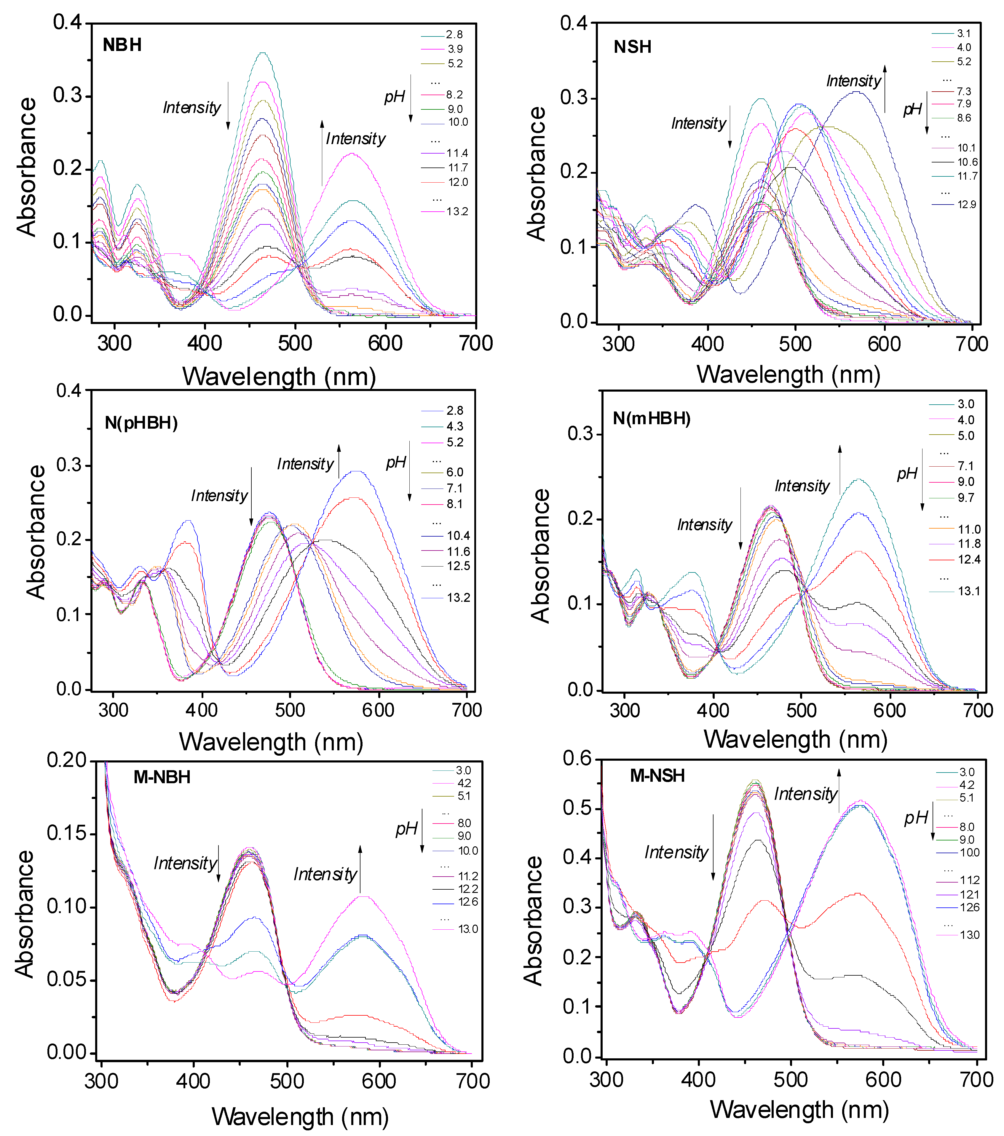
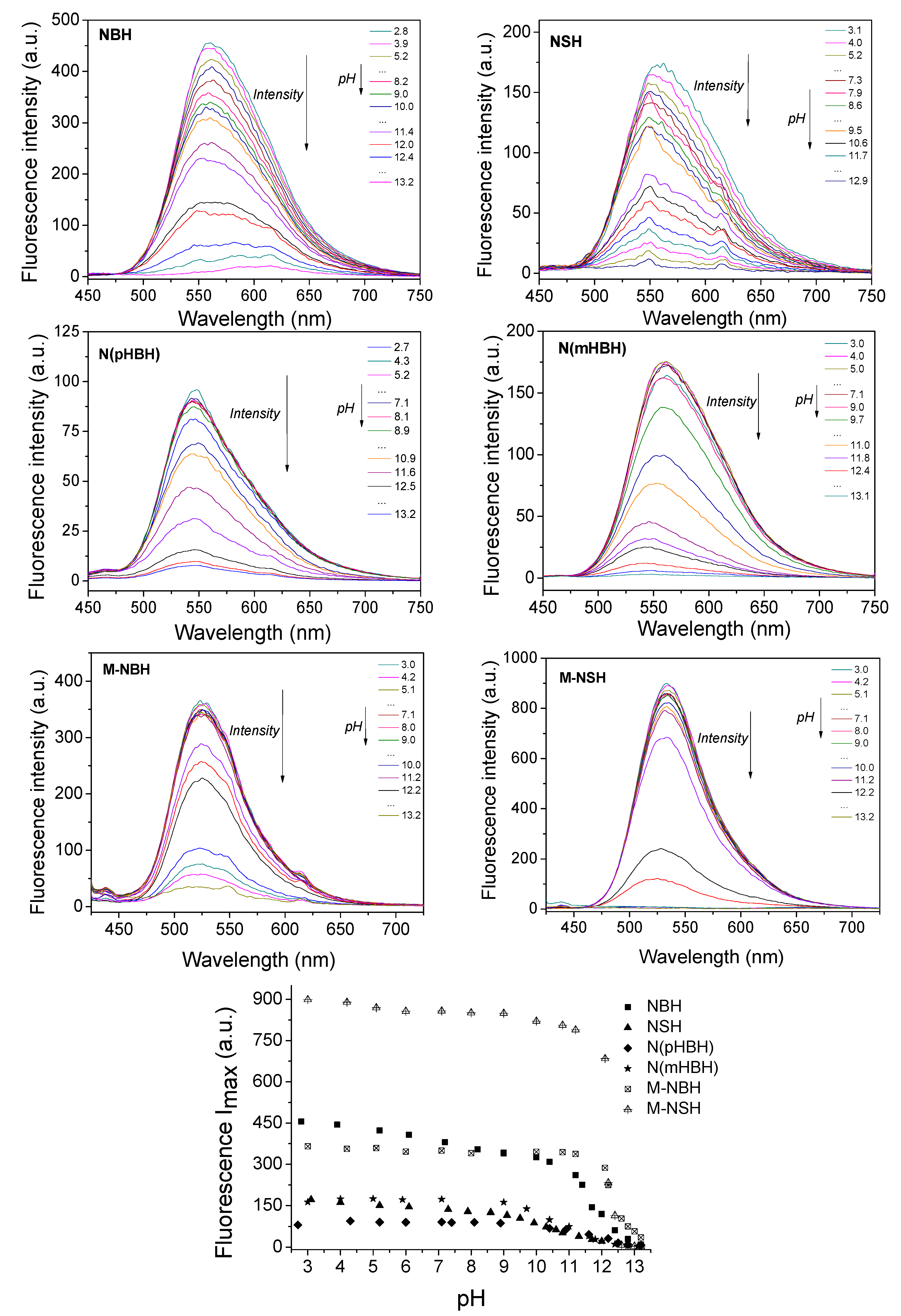
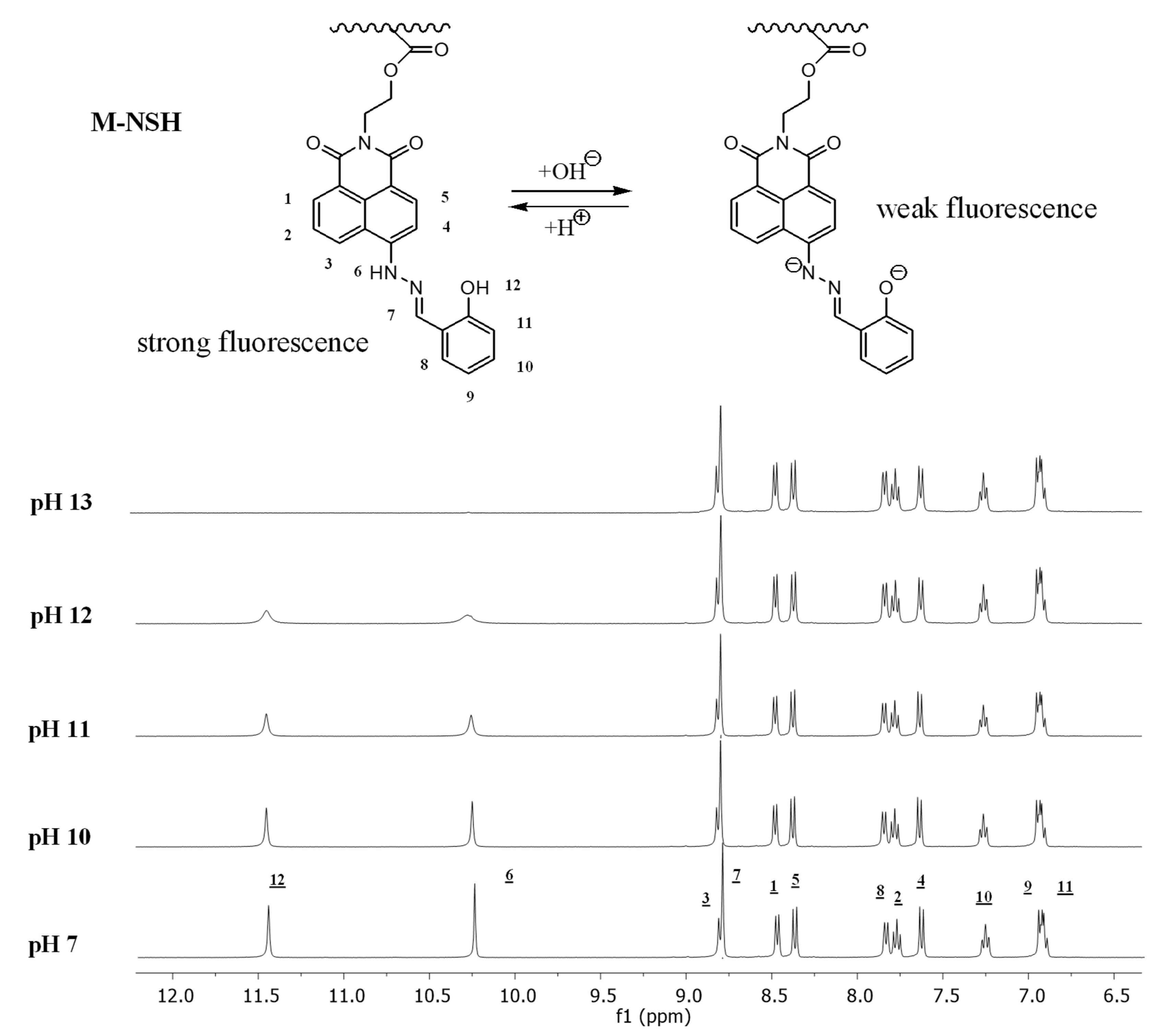
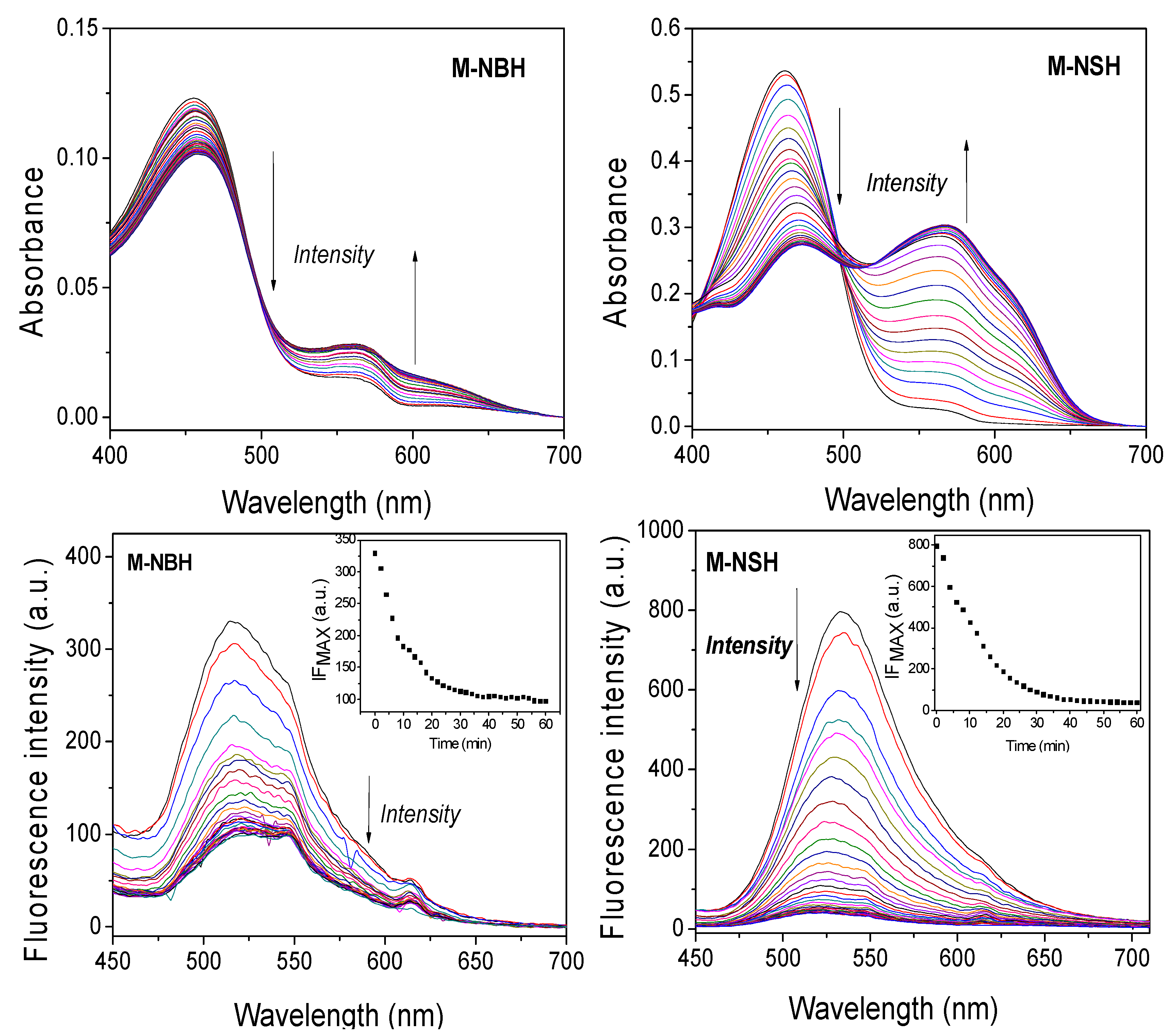
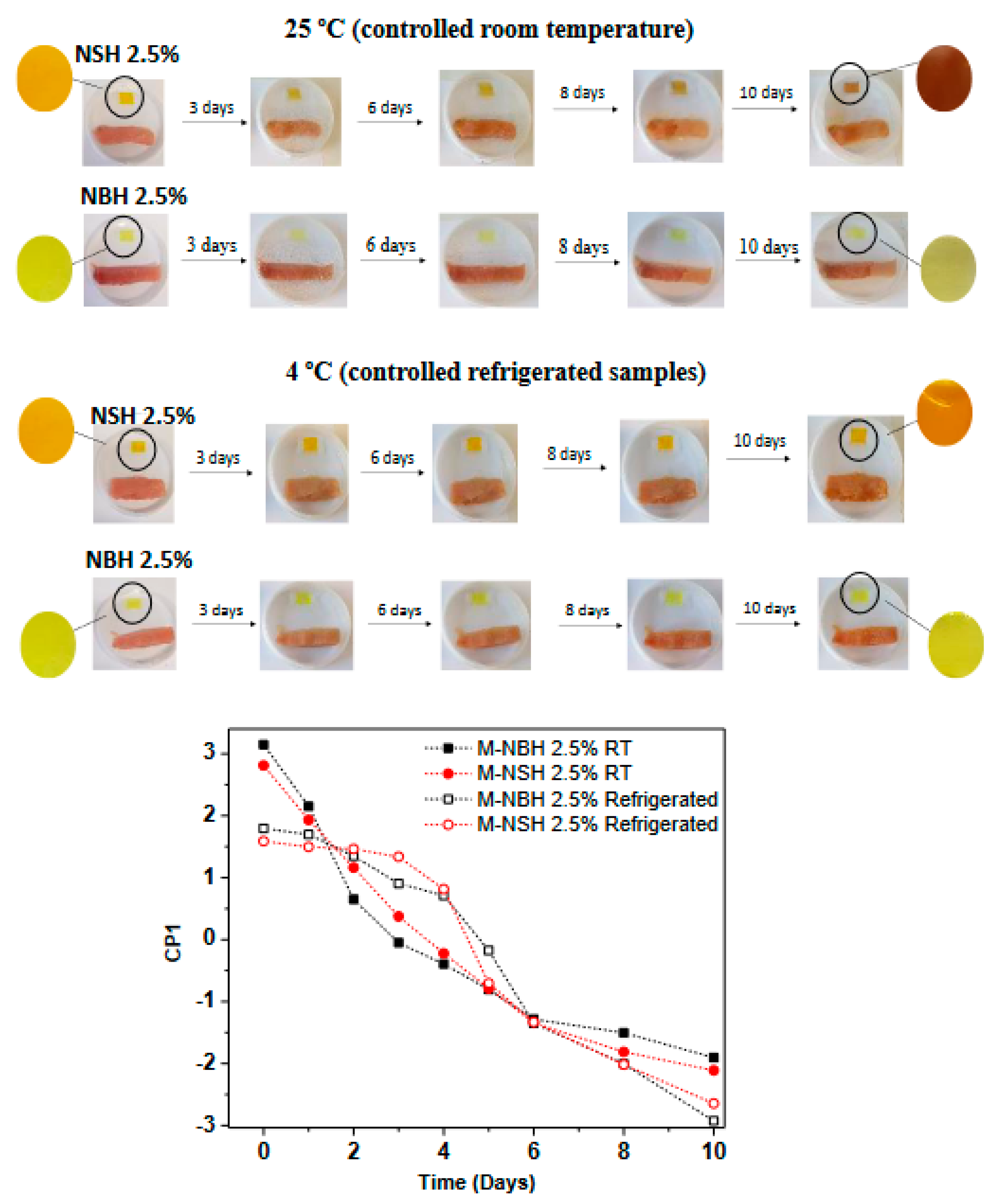
3.3. Effect of pH on the Absorption and Fluorescence Properties of Aryl Hydrazone Derivatives of Naphthalimides and the M-NBH and M-NSH Membranes

Firstly, the absorbance and fluorescence of the hydrazone derivatives and the membranes were studied in water:ethanol (4:1, v/v), as was their pH dependence. Table 1 presents the spectral characteristics, such as the absorption (λABS) and fluorescence (λFLU) maxima in acid (pH = 3) and basic media (pH = 13), the corresponding absorption coefficient (logε), the Stokes shift (Δν), and the quantum yields of fluorescence (ϕFLU). The effect of pH variation on the absorbance spectra of aryl hydrazone derivatives of naphthalimides in solution and the behavior of M-NBH and M-NSH membranes in water:ethanol (4:1, v/v) is illustrated in Figure 3. The fluorescence characteristics as a function of pH for the hydrazine derivatives of naphthalimides and functionalized membranes are shown in Figure 4.
The absorption spectra of the hydrazone derivatives exhibit band shapes in the range of 300–700 nm (Figure 3). Their main absorption peaks were observed around 460–475 and shifted to 565–575 nm when pH values were changed from 3 to 13 in water:ethanol (4:1) by adjusting the pH values of the NaOH and HCl aqueous solutions. The functionalized membranes M-NSH and M-NBH displayed a similar behavior, and their maxima were red-shifted from 460 to 576 and 583, respectively, with a clear isosbestic point at 500 nm when the pH values were changed (Figure 3).
The pKa values of hydrazone derivatives were calculated [32] from the curves of the absorption changes in the presence of increasing the pH from 3 to 13, with the value next to 11 in all cases. In the case of membranes, the pKa values obtained are close to those values measured for the corresponding derivatives in solution, and for M-NSH with the OH substituent (pKa = 10.4), the protonation of the functionalized membrane takes place at a slightly lower pH than the M-NBH (pKa = 11.5).
The fluorescence response of the naphthalimide derivatives and the functionalized membranes at different pH levels was compared, and the results are plotted in Figure 4. The emission of naphthalimide derivatives in solution varied due to different pH values, but had different intervals and slopes of changes. In the basic media, the emission maximum of NBH was slightly shifted to a lower wavelength (4 nm) and it was observed that there was a strong decrease in the fluorescence intensity, which would be due to the deprotonation of the aryl hydrazine and its subsequent photoinduced electron transfer (PET). NSH, N(mHBH) and N(pHBH) showed similar behavior, although it is worth noting the greater shift in NSH (10 nm) and N(pHBH) (15 nm). That effect may be related to the deprotonation of phenol and the stronger electron-donating capability of amine and the phenolate anion, namely, a more effective internal charge transfer structure. For the naphthalimide fluorophores bonded to the membranes though the hydroxyl groups, the fluorescence response to pH is dependent upon the permeability of the membranes, and the restricted mobility of the sensor due to the polymer matrix. As a result, the fluorescence emission of M-NBH and M-NSH decreased at slightly higher pH values than for naphthalimides in solution. Additionally, the emission maxima were observed at a lower wavelength with respect to the free naphthalimides, 562 nm (NBH) to 525 (M-NBH) and 560 nm (NSH) to 533 nm (M-NSH), which is associated with an excited state of higher energy as a consequence of the more restricted relaxation for the probe anchored to the polymer chain. Additionally, no shift in the fluorescence maximum was detected for M-NSH, as it was described for NSH. The factor of fluorescence enhancement for the membranes in acid media was calculated (Table 1) by comparing the increase in the intensities from the basic to acid media. The PET path can be switched off by the protonation of the amine and the fluorescence of the naphthalimide fluorophore is then recovered, as is shown for the derivatives in solution or anchored to the membrane in Figure 4. The enhancement was around 8 times for M-NBH and 40 times for M-NSH. This enhancement in the acid media is higher than the highest recently reported for piperazine naphthalimide derivatives (FE 8–12) [35], and it would confirm their interesting proton “off-on” switch behavior as an optical pH solid sensor.
The quantum yields of fluorescence in acid media are similar for all the naphthalimide derivatives, and as expected, the values are higher than those determined in basic media (Table 1). In polar solvents (water, ethanol), the fluorescence quantum-yield values are relatively low in comparison to those obtained in non-polar solvents, such as hexane. This result could be related to the photo-induced electron transfer from the aryl hydrazone to the fluorophore and the fact that the orbit energy of HOMO becomes lower in polar solvents, which facilitated the PET process. For membranes, the quantum yield of fluorescence could not be calculated due to the difference between the fluorescence standard in solution and the membranes. The pKa for the excited state (pKa*) was determined for naphthalimide derivatives and functionalized membranes. The position of the inflection points in the fluorometric titration is similar to those obtained from the absorbance changes with pH, and similar values to pKa for the ground state were obtained (Table 1).
The reversibility of the pH-induced fluorescence signal of the M-NSH membrane was evaluated by alternating its immersion in HCl solutions (pH = 3.0) and NaOH solutions (pH = 13) until the fluorescence intensity of the membrane reached, again, the original value prior to the next measurement. Ten successive cycles were carried out, as it is shown in Figure 5 for the membrane sensor.
1H NMR studies provided direct evidence of the pH effect on M-NSH. As shown in Figure 6, 1H NMR spectra show the downfield shift of signals corresponding to the proton of the NH group at 10.25 ppm (1H-6) and the proton of the OH group at 11.43 ppm (1H-12) ppm, and their disappearance in basic media.
The behavior for the detection of trimethylamine vapor of functionalized membranes M-NBH and M-NSH was studied since amines are generated through food spoilage and naked-eye detection is a very useful tool for the consumer, as well as for other fields related to food safety and to health and safety at work. The response time of absorbance and fluorescence properties of functionalized films to the basic environment was studied in the vapor phase, and the results are shown in Figure 7. It was observed that the spectroscopic characteristics of naphthalimide NBH and NSH bonded to the polymer were very sensitive to the basic vapor medium. It was necessary to maintain the film for approximately 35 min to determine the effective change in absorption. Both functionalized materials are sensitive to trimethylamine vapors through their absorption and fluorescence bands, although the M-NSH membrane exhibits better behavior in fluorescence decay under vapor treatment, and the fluorescence quenching was observed at 60 min.
The colorimetric sensory behavior toward the basic media of M-NSH was analyzed visually. The membrane was cut and dipped in water:ethanol (4:1) solutions ranging from pH 7 to pH 13 for 35 min. The titration curve was obtained by treatment of the digital picture data, as previously reported [36]. After photographing the samples swelled in different pH values, the three RGB parameters of each material were obtained with an Android app (ColorMeter) and processed using the principal component analysis (PCA), which allows us to reduce the parameters to one principal component (PC1). This gave us an account for >98% of the information on the three RGB parameters, thus permitting the simple elaboration of the titration curves (pH vs. CP1). It was observed that the color of the membrane changed from orange to purple, showing that M-NSH could be used to detect basic media with the naked eye (Figure 8).
To assess the possibility of a real chance to use the sensory materials obtained, a proof of concept related to the performance of the labels was studied. This study was carried out with fresh fish (tuna) bought in the local market and packaged in Petri dishes. Changes were observed for 10 days at a controlled temperature (25 °C and refrigerated at 4 °C) (Figure 9).
A visible color change was observed in room temperature samples in the first days of the experiment and until the end (10 days). On the other hand, the refrigerated samples did not show a significant color change until the fourth day. To analyze the results obtained in the real fish experiment, the color change of the NSH material was determined in terms of CP1 variation, and was calculated after photographing the square samples every day, thus permitting the simple elaboration of 2D titration curves (time (days) vs. CP1) (Figure 9). This variation in PC1 can be related to the variation observed in the 2D titration curve obtained with pH variation (pH vs. CP1) for the samples swelled, which indicates the presence of basic biogenic amine vapors. The fluorescence spectra of every material were obtained (Figure 10), from where a progressive quenching of the fluorescence emission can be observed, which is more pronounced in room temperature samples, and that agrees with the color change observed in digital pictures and principal component data. Additionally, M-NSH showed a greater response to sensitivity than M-NBH, and a higher decrease in fluorescence emission and color change was determined in comparison to M-NBH. That result would be in agreement with those presented above, where the protonation of the functionalized membrane M-NSH with the OH substituent (pKa = 10.4) took place at a slightly lower pH than the M-NBH (pKa = 11.5), and the fluorescence emission for M-NSH decreased at a slightly lower pH than for M-NBH (Figure 4). It could be concluded that the excellent colorimetric and fluorescence detection of strong basic pH (>11) media by the new derivatives allowed for their use as a solid sensor for amine vapors such as trimethylamine (TMA).

4. Conclusions

In this work, several hydrazone derivatives of naphthalimides are synthesized and anchored to a photo-crosslinked membrane functionalized with acid chloride groups. Following that strategy, thermally stable, solid and easily handled membranes are prepared to be applied as fluorescence and colorimetric sensors. The new naphthalimides and the membranes are evaluated as a sensor by determining the change in their spectroscopic properties of absorption and fluorescence with pH. In basic media, a strong decrease is observed in the fluorescence intensity, which would be due to the deprotonation of the aryl hydrazine and its subsequent photoinduced electron transfer (PET). Furthermore, materials are sensitive to trimethylamine vapors through both their absorption and fluorescence bands, and there is a color change in the membrane from orange to purple.
Then, a real chance to use the novel sensory materials has been evaluated by analyzing the response of samples packaged in Petri dishes at a controlled temperature with fresh fish bought in the local market. Additionally, changes in the fluorescence and color of membranes have been observed from the beginning, and up to 10 days. That result is very relevant, since amines are generated through food spoilage and naked-eye detection is a very useful tool for the consumer, as well as for other fields related to food safety and to health and safety at work. Likewise, the polymeric sensors are costless, and as a consequence of their solid character and the reversibility of the process, exhibit improved stability and can be reused.

Author Contributions

Conceptualization, J.L.P., F.C. and T.C.; formal analysis, J.L.P., S.F.-A., F.C. and T.C.; investigation, J.L.P. and S.F.-A.; writing—review and editing, J.L.P., F.C. and T.C. All authors have read and agreed to the published version of the manuscript.

Funding

This study was financed by MINECO (Project Ref. MAT2017- 88923 and Project Ref. PID2021-124926NB-I00)).

Institutional Review Board Statement

Not applicable.

Informed Consent Statement

Not applicable.

Data Availability Statement

Not applicable.

Conflicts of Interest

The authors declare no conflict of interest.

References

  1. Yan, Y.; Zhang, Y.; Xu, H. A Selective “Turn-On” Fluorescent Probe for Recognition of Mercury(II) Ions in Aqueous Solution Based on a Desulfurization Reaction. ChemPlusChem 2013, 78, 628–631. [Google Scholar] [CrossRef] [PubMed]
  2. Poddar, M.; Sivakumar, G.; Misra, R. Donor–acceptor substituted 1,8-naphthalimides: Design, synthesis, and structure–property relationship. J. Mater. Chem. C 2019, 7, 14798–14815. [Google Scholar] [CrossRef]
  3. Gudeika, D. A review of investigation on 4-substituted 1,8-naphthalimide derivatives. Synth. Met. 2020, 262, 116328. [Google Scholar] [CrossRef]
  4. Jain, N.; Kaur, N. A comprehensive compendium of literature of 1,8-Naphthalimide based chemosensors from 2017 to 2021. Coord. Chem. Rev. 2022, 459, 214454. [Google Scholar] [CrossRef]
  5. Diana, R.; Panunzi, B.; Tuzi, A.; Piotto, S.; Concilio, S.; Caruso, U. An Amphiphilic Pyridinoyl-hydrazone Probe for Colorimetric and Fluorescence pH Sensing. Molecules 2019, 24, 3833–3855. [Google Scholar] [CrossRef] [PubMed] [Green Version]
  6. Gemma, S.; Colombo, L.; Forloni, G.; Savini, L.; Fracasso, C.; Caccia, S.; Salmona, M.; Brindisi, M.; Joshi, B.P.; Tripaldi, P.; et al. Pyrroloquinoxaline hydrazones as fluorescent probes for amyloid fibrils. Org. Biomol. Chem. 2011, 9, 5137–5148. [Google Scholar] [CrossRef]
  7. Shao, B.; Baroncini, M.; Qian, H.; Bussotti, L.; Di Donato, M.; Credi, A.; Aprahamian, I. Solution and Solid-State Emission Toggling of a Photochromic Hydrazone. J. Am. Chem. Soc. 2018, 140, 12323–12327. [Google Scholar] [CrossRef] [Green Version]
  8. Sonawane, S.J.; Kalhapure, R.S.; Govender, T. Hydrazone linkages in pH responsive drug delivery systems. Eur. J. Pharm. Sci. 2017, 99, 45–65. [Google Scholar] [CrossRef]
  9. Pereira, T.M.; Kümmerle, A.E. Computational Biology and Chemistry. Chapter Hydrazone-Based Small-Molecule Chemosensors; IntechOpen: London, UK, 2020. [Google Scholar]
  10. Zhou, Y.; Piergentili, I.; Hong, J.; Van der Helm, M.P.; Macchione, M.; Li, Y.; Eelkema, R.; Luo, S. Indoline Catalyzed Acylhydrazone/Oxime Condensation under Neutral Aqueous Conditions. Org. Lett. 2020, 22, 6035–6040. [Google Scholar] [CrossRef]
  11. Shao, B.; Aprahamian, I. pH-Induced Fluorescence and Thermal Relaxation Rate Modulation in a Hydrazone Photoswitch. ChemPhotoChem 2019, 3, 361–364. [Google Scholar] [CrossRef]
  12. Fan, L.; Qin, J.; Li, T.; Wang, B.; Yangn, Z.A. Chromone Schiff-base as Al(III) selective fluorescent and colorimetric chemosensor. J. Lumin. 2014, 155, 84–88. [Google Scholar] [CrossRef]
  13. Qin, J.; Yang, Z.; Fan, L.; Cheng, X.; Li, T.; Wang, B. Design and synthesis of a chemosensor for the detection of Al3+ based on ESIPT. Anal. Methods 2014, 6, 7343–7348. [Google Scholar] [CrossRef]
  14. Kima, S.; Kob, C.; Lima, T.; Yooa, S.; Hamc, H.J.; Kangd, S.-Y.; Kanga, S.; Chob, S.K.; Hana, M. A hydrazone-based turn-on fluorescent probe for peroxynitrite detection and live-cell imaging. Dye. Pigment. 2019, 171, 107762–107768. [Google Scholar] [CrossRef]
  15. Cheshmedzhieva, D.; Ivanova, P.; Stoyanov, S.; Tasheva, D.; Dimitrova, M.; Ivanovc, I.; Ilieva, S. Experimental and theoretical study on the absorption and fluorescence properties of substituted aryl hydrazones of 1,8-naphthalimide. Phys. Chem. Chem. Phys. 2011, 13, 18530–18538. [Google Scholar] [CrossRef] [PubMed]
  16. Dilek, O.; Bane, S.L. Synthesis and Spectroscopic Characterization of Fluorescent Boron Dipyrromethene-Derived Hydrazones. J. Fluoresc. 2011, 21, 347–354. [Google Scholar] [CrossRef] [PubMed] [Green Version]
  17. Aysha, T.; Zain, M.; Arief, M.; Youssef, Y. Synthesis and spectral properties of new fluorescent hydrazone disperse dyes and their dyeing application on polyester fabrics. Heliyon 2019, 5, e02358. [Google Scholar] [CrossRef] [PubMed] [Green Version]
  18. Xiang, Y.; Tong, A.; Jin, P.; Ju, Y. New Fluorescent Rhodamine Hydrazone Chemosensor for Cu(II) with High Selectivity and Sensitivity. Org. Lett. 2006, 8, 2863–2866. [Google Scholar] [CrossRef]
  19. Pablos, J.L.; Vallejos, S.; Muñoz, A.; Rojo, M.J.; Serna, F.; García, F.C.; García, J.M. Solid Polymer Substrates and Coated Fibers Containing 2,4,6-Trinitrobenzene Motifs as Smart Labels for the Visual Detection of Biogenic Amine Vapors. Chem. Eur. J. 2015, 21, 8733–8736. [Google Scholar] [CrossRef]
  20. Qi, P.; Wu, X.; Liu, L.; Yu, H.; Song, S. Hydrazone-Containing Triblock Copolymeric Micelles for pH-Controlled Drug Delivery. Front. Pharmacol. 2018, 9, 12. [Google Scholar] [CrossRef] [Green Version]
  21. Li, P.; Zhang, D.; Zhang, Y.; Lu, W.; Zhang, J.; Wang, W.; He, Q.; Théato, P.; Chen, T. Aggregation-Caused Quenching-Type Naphthalimide Fluorophores Grafted and Ionized in a 3D Polymeric Hydrogel Network for Highly Fluorescent and Locally Tunable Emission. ACS Macro Lett. 2019, 8, 937–942. [Google Scholar] [CrossRef]
  22. Grabchev, I.; Qian, X.; Xiao, Y.; Zhang, R. Novel heterogeneous PET fluorescent sensors selective for transition metal ions or protons: Polymers regularly labelled with naphthalimide. New J. Chem. 2002, 26, 920–925. [Google Scholar] [CrossRef]
  23. Jiang, J.; Xiao, X.; Zhao, P.; Tian, H. Colorimetric naked-eye recognizable anion sensors synthesized via RAFT polymerization. J. Polym. Sci. Part A Polym. Chem. 2010, 48, 1551–1556. [Google Scholar] [CrossRef]
  24. Fernández-Alonso, S.; Corrales, T.; Pablos, J.L.; Catalina, F. Solid fluorescence sensors obtained by functionalization of photocrosslinked water-swollen acrylic membranes with 4-piperazine naphthalimide derivatives. Polymer 2017, 124, 139–150. [Google Scholar] [CrossRef]
  25. Pablos, J.L.; Hernández, E.; Catalina, F.; Corrales, T. Solid Fluorescence pH Sensors Based on 1,8-Naphthalimide Copolymers Synthesized by UV Curing. Chemosensors 2022, 10, 73. [Google Scholar] [CrossRef]
  26. Fernández-Alonso, S.; Corrales, T.; Pablos, J.L.; Catalina, F. A Switchable fluorescence solid sensor for Hg2+ detection in aqueous media based on a photocrosslinked membrane functionalized with (benzimidazolyl)methyl-piperazine derivative of 1,8-naphthalimide. Sens. Actuators B Chem. 2018, 270, 256–2621. [Google Scholar] [CrossRef]
  27. Fernández-Alonso, S.; Corrales, T.; Pablos, J.L.; Catalina, F. Surface modification of poly(ethylene-butyl acrylate) copolymers by microwave methodology and functionalization with 4-dimethylamino-N-(2-hydroxyethyl)-1,8-naphthalimide for acidity sensing. React. Funct. Polym. 2016, 107, 78–861. [Google Scholar] [CrossRef]
  28. Allen, N.S.; Corrales, T.; Edge, M.; Catalina, F.; Blanco-Pina, M.; Green, A. Photochemistry and photopolymerization activities of novel phenylthiobenzophenone and diphenylthiophene photoinitiators. Polymer 1998, 39, 903–910. [Google Scholar] [CrossRef]
  29. Tian, Y.Q.; Shumway, B.R.; Youngbull, C.; Jen, A.K.Y.; Johnson, R.H.; Meldrum, D.R. Dually fluorescent sensing of pH and dissolved oxygen using a membrane made from polymerizable sensing monomers. Sens. Actuators B Chem. 2010, 147, 714–722. [Google Scholar] [CrossRef] [Green Version]
  30. Lee, Y.H.; Tang, Y.; Verwilst, P.; Lin, W.; Kim, J.S. A biotin-guided formaldehyde sensor selectively detecting endogenous concentrations in cancerous cells and tissues. Chem. Commun. 2016, 52, 11247–11250. [Google Scholar] [CrossRef]
  31. Pablos, J.L.; Catalina, F.; Ibeas, S.; Corrales, T. Fluorescent imidazolium-based poly(ionic liquid)s for Fe3+ detection in aqueous medium. J. Photochem. Photobiol. A Chem. 2021, 406, 113015–113024. [Google Scholar] [CrossRef]
  32. Reijenga, J.; Van Hoof, A.; Van Loon, A.; Teunissen, B. Development of Methods for the Determination of pKa Values. Anal. Chem. Insights 2013, 8, 53–71. [Google Scholar] [CrossRef] [Green Version]
  33. Reynolds, G.; Drexhage, K. New coumarin dyes with rigidized structure for flash lamp-pumped dye lasers. Opt. Commun. 1975, 13, 222–225. [Google Scholar] [CrossRef]
  34. Redondo-Foj, B.; Carsí, M.; Ortiz-Serna, P.; Sanchis, M.J.; Vallejos, S.; García, F.; García, J.M. Effect of the Dipole-Dipole interactions in the molecular dynamics of poly(vinylpyrrolidone)-based copolymers. Macromolecules 2014, 47, 5334–5346. [Google Scholar] [CrossRef]
  35. Niu, C.-G.; Zeng, G.-M.; Chen, L.-X.; Shen, G.-L.; Yu, R.-Q. Proton “off-on” behaviour of methylpiperazinyl derivative of naphthalimide: A pH sensor based on fluorescence enhancement. Analyst 2004, 129, 20–24. [Google Scholar] [CrossRef] [PubMed]
  36. Vallejos, S.; Muñoz, A.; Ibeas, S.; Serna, F.; Garcia, F.C.; Garcia, J.M. Solid sensory polymer substrates for the quantification of iron in blood, wine and water by a scalable RGB technique. J. Mater. Chem. A 2013, 1, 15435–15441. [Google Scholar] [CrossRef]
Figure 1. Synthesis of the naphthalimide derivatives and their corresponding microwave (MW) reaction times.
Chemosensors 10 00417 g001
Figure 2. Synthesis of the photo-crosslinked acrylic membrane containing acid chloride groups (M-Cl) and functionalization with aryl-hydrazone derivatives of 1,8-naphthalimide (M-NBH and M-NSH).
Chemosensors 10 00417 g002
Figure 3. Spectroscopic characteristics as a function of pH, of absorption of aryl hydrazone derivatives of naphthalimides, NBH, NSH, N(mHBH), and N(pHBH), and the functionalized membranes M-NBH and M-NSH, in water:ethanol (4:1).
Chemosensors 10 00417 g003
Figure 4. Spectroscopic characteristics as a function of pH (3–13) of fluorescence of aryl hydrazone derivatives of naphthalimides, NBH, NSH, N(mHBH), and the functionalized membranes M-NBH and M-NSH, in water:ethanol (4:1).
Chemosensors 10 00417 g004
Figure 5. Reversibility of the M-NSH membrane sensor.
Chemosensors 10 00417 g005
Figure 6. Reversible pH effect on M-NSH structure together with partial 1H-NMR in DMSO at 25 °C at different pH values.
Chemosensors 10 00417 g006
Figure 7. Spectroscopic characteristics as a function of time of absorption and fluorescence of functionalized membranes, M-NBH and M-NSH, in the presence of trimethylamine vapor.
Chemosensors 10 00417 g007
Figure 8. Response of M-NSH to the pH. Pictures of membranes cut upon dipping for 35 min in water:ethanol solution (4:1) ranging from pH 7 to pH 13. Titration curve obtained by treatment of the digital picture data (CP1 is the principal component of PCA analysis using the red and green values of the RGB parameters).
Chemosensors 10 00417 g008
Figure 9. Upper: picture at different times of membranes (M-NSH and M-NBH) packed and sealed in Petri dishes together with individual pieces of tuna (20 g) from the local market, both refrigerated (4 °C) and at r.t. (25 °C). Lower: response of membranes by analysis of digital picture data (CP1) vs. time of exposure.
Chemosensors 10 00417 g009
Figure 10. Fluorescence spectra and pictures of initial membranes (M-NBH and M-NSH) after exposed to fish in sealed Petri dishes for 10 days at room temperature (25 °C) and refrigerated (4 °C).
Chemosensors 10 00417 g010
Table 1. Parameters for aryl hydrazone derivatives of 1,8-naphthalimide in solution (a) and bonded to the polymer membrane (M-NBH and M-NSH), determined by UV–Vis absorption and fluorescence spectroscopies and pH dependence.
ParameterNBHNSHN(mHBH)N(pHBH)M-NBHM-NSH
λABS-aci326, 465 nm330, 460 nm330, 465 nm330, 475 nm460 nm460 nm
Log εacid(b)4.28 (326 nm)/
4.58 (465 nm)
4.20 (330 nm)/
4.52 (460 nm)
3.95 (330 nm)/
4.38 (475 nm)
3.84 (330 nm)/
4.34 (465 nm)
--
λIsobestic348, 400,
503 nm
365, 400,
530 nm
323, 414,
524 nm
336, 405,
504 nm
417, 500 nm410, 500 nm
λABS-base367, 565 nm386, 568 nm384, 566 nm377, 575 nm583 nm576 nm
Log εbase(b)4.00 (367 nm)/
4.45 (565 nm)
4.17 (386 nm)/
4.5 (568 nm)
4.14 (384 nm)/
4.46 (573 nm)
3.84 (377 nm)/
4.38 (565 nm)
--
pKa(c)11.711.51111.311.510.4
λFLU-acid562 nm560 nm548 nm561 nm525 nm533 nm
ϕFLU-acid(d)0.100.110.0920.095-
ϕFLU pH=7(d)0.0900.0980.0920.095-
ϕFLU ethanol(d)0.0990.110.130.11-
ϕFLU hexane(d)0.170.20.210.19
λFLU-base558 nm550 nm544 nm544 nm525 nm525 nm
ϕFLU-base(d)0.0030.0030.0020.002--
pKa *(c)10.810.210.310.210.910
Δν (e)365035602700368427002980
FE(f)28.531.211.4398.2>40
(a) Measured at 10−5 M in water:ethanol (4:1, v/v). Subscripts “acid” and “base” refer to the limiting value of a given parameter when the acid or base condition is increased until pH = 3 or pH = 13, respectively. Fluorescence emission by excitation at λIsobestic. (b) ε, molar absorptivity in M−1cm−1. (c) Obtained by analysis [32]. (d) Relative to coumarin 6 (ϕF = 0.78 in ethanol). (e) Stokes shift (νFLU–νABS) in frequency (cm−1). (f) Fluorescence enhancement factor FE = AFLU-acid/AFLU-base.
Publisher’s Note: MDPI stays neutral with regard to jurisdictional claims in published maps and institutional affiliations.

