
Recent Developments in Rhodamine-Based Chemosensors: A Review of the Years 2018–2022

Abstract
:1. Introduction
2. Rhodamine-Based Sensors for Metal Ions
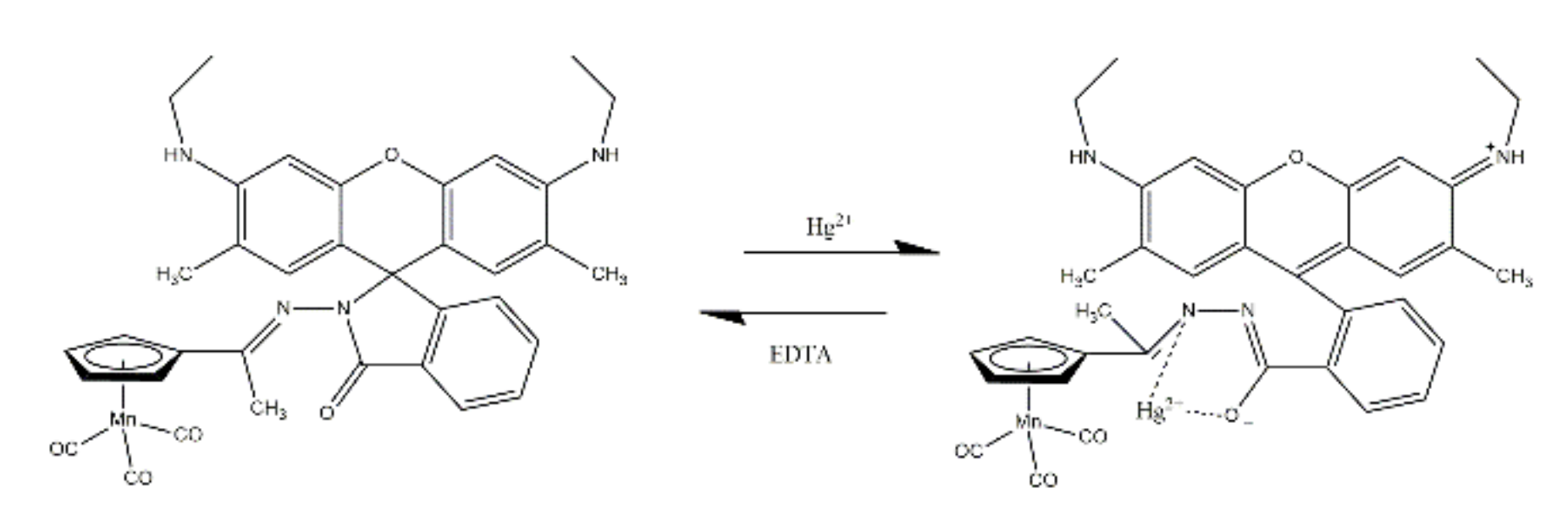
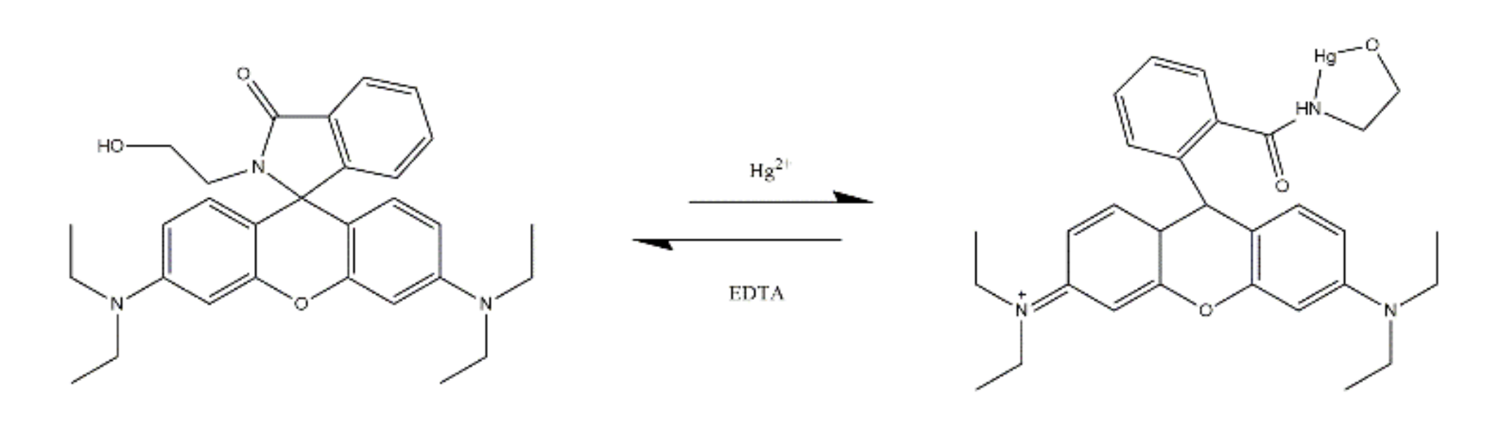
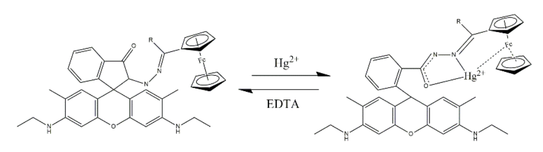
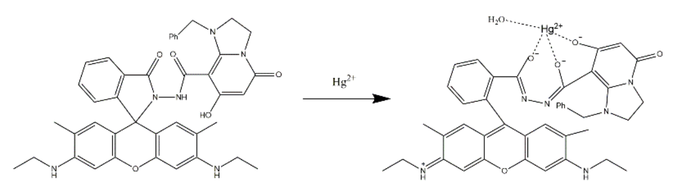
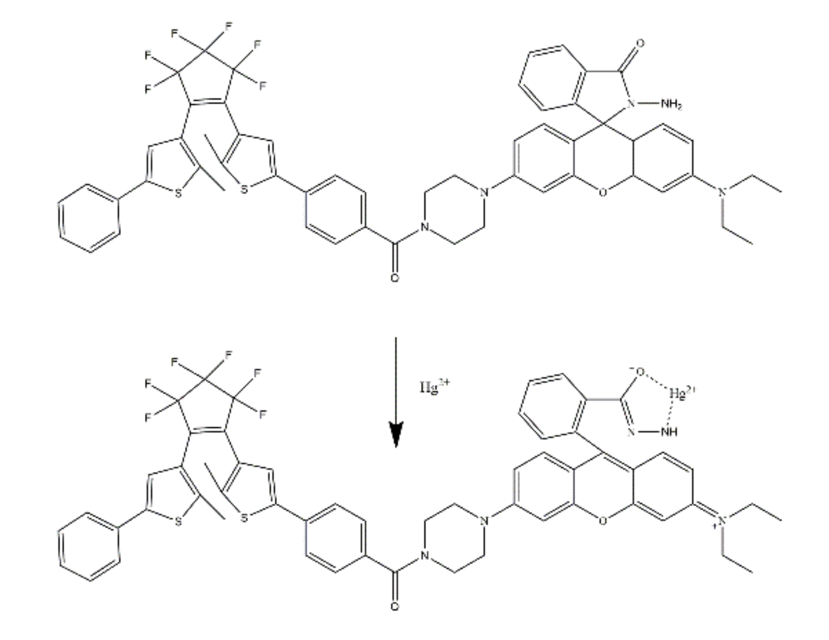
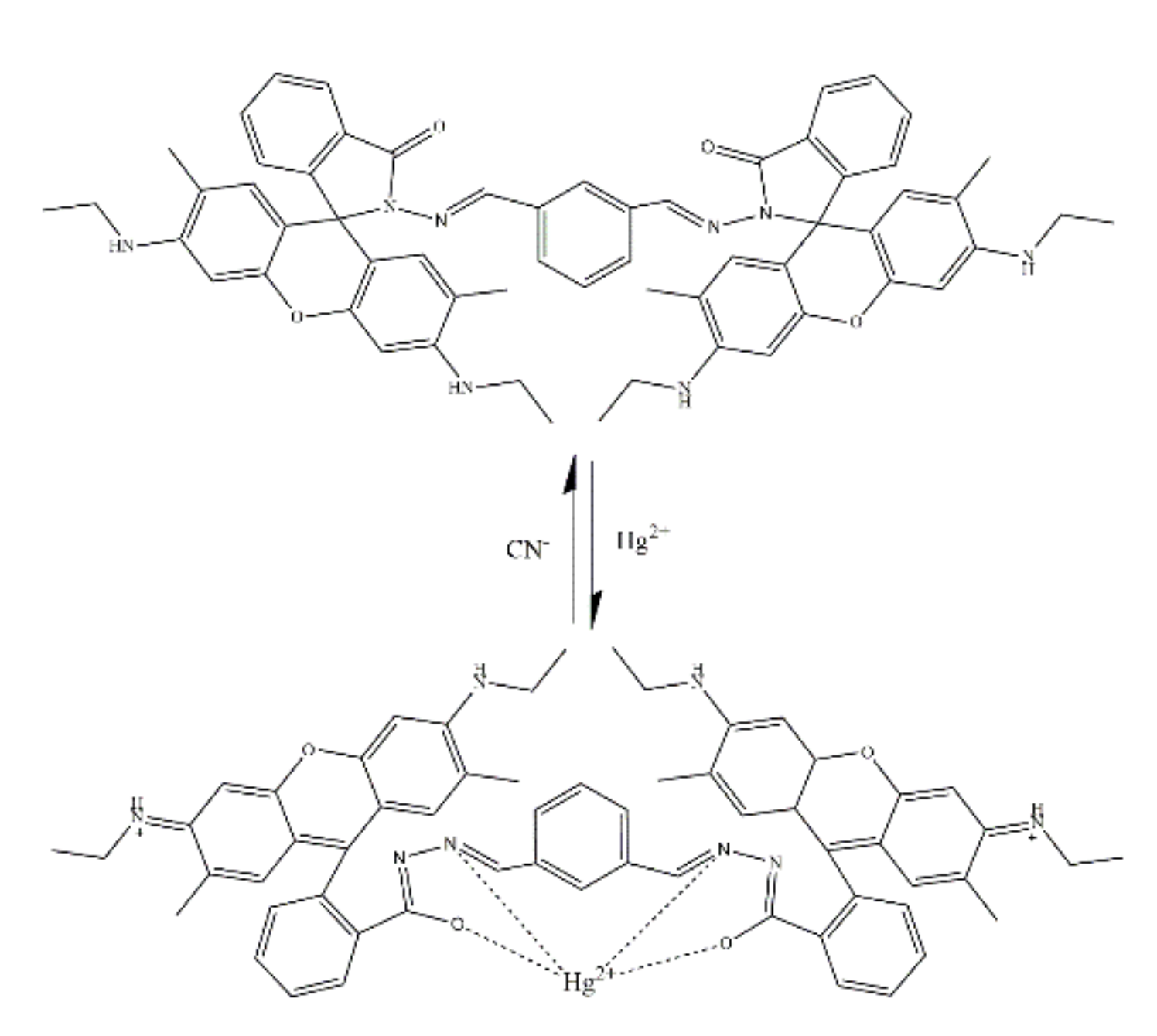
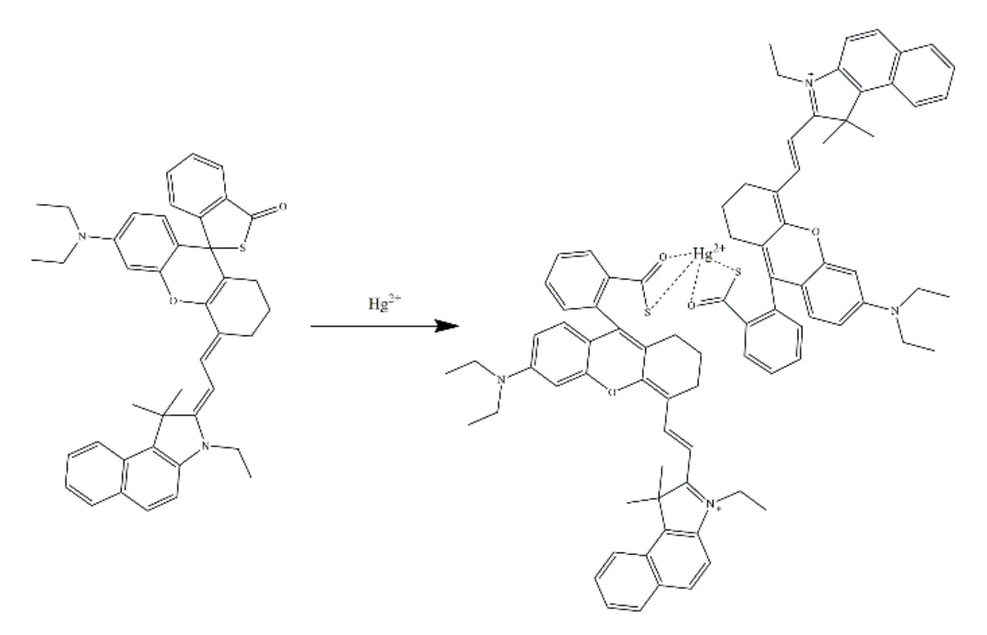
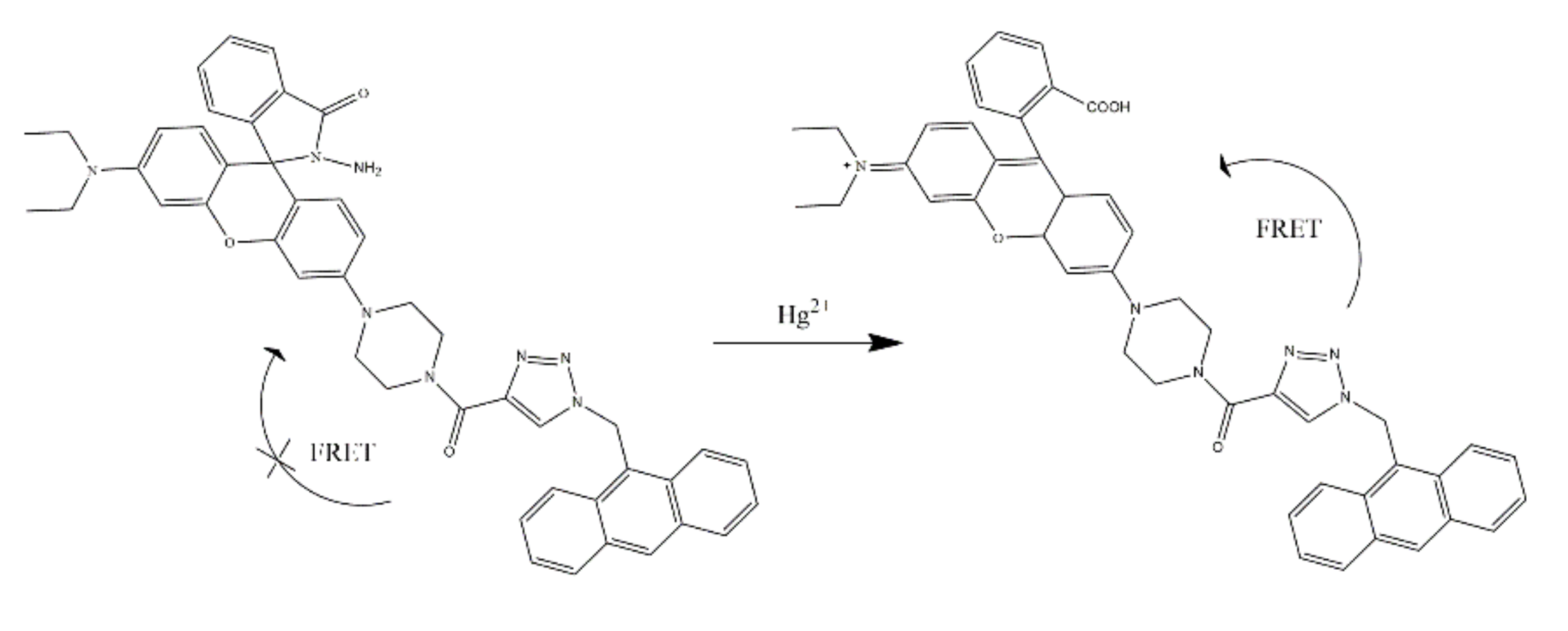
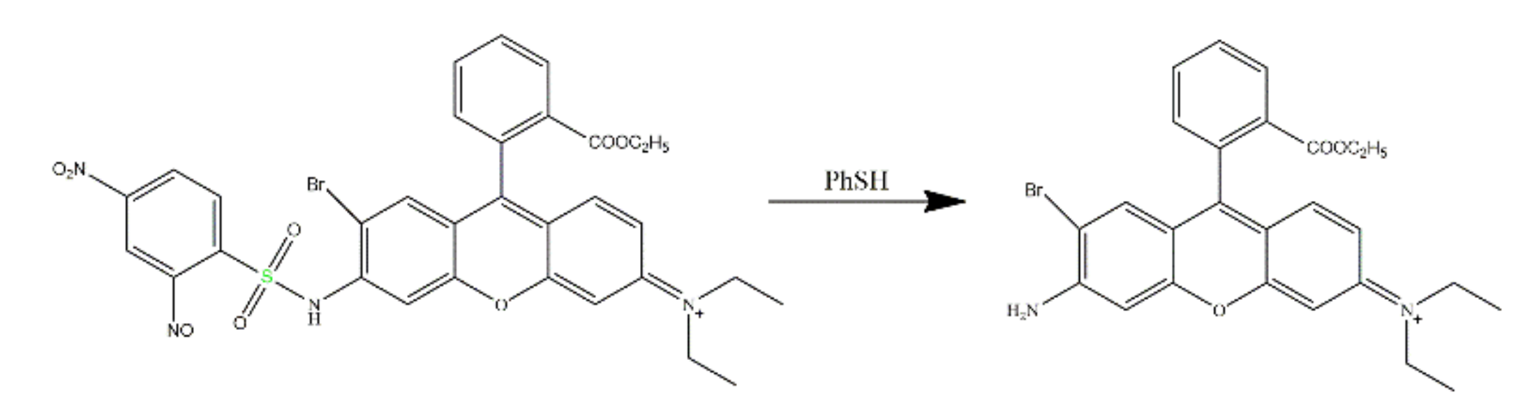
2.1. Rhodamine-Based Sensors for Hg2+
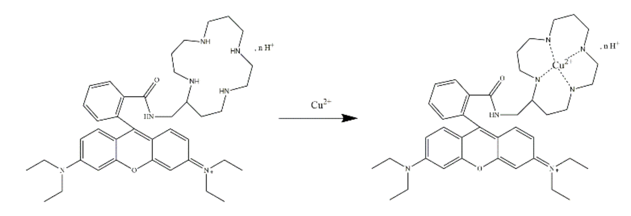
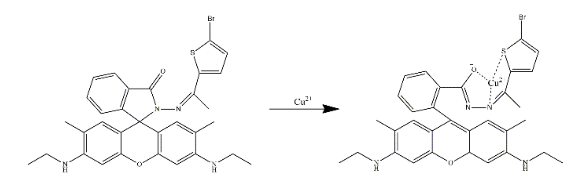
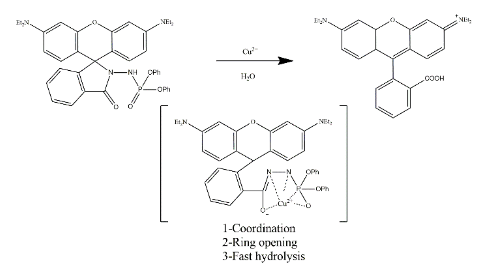
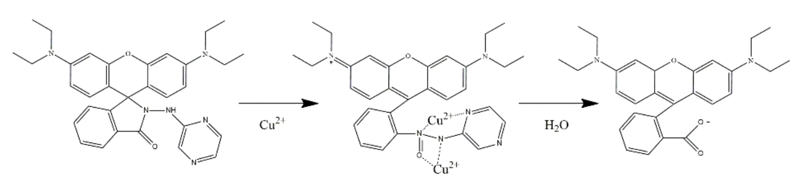
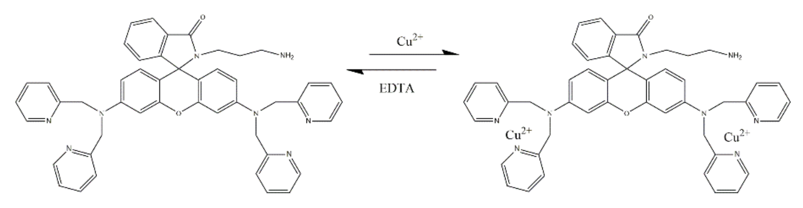
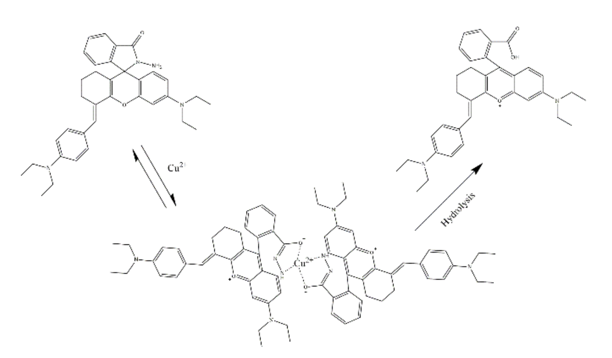
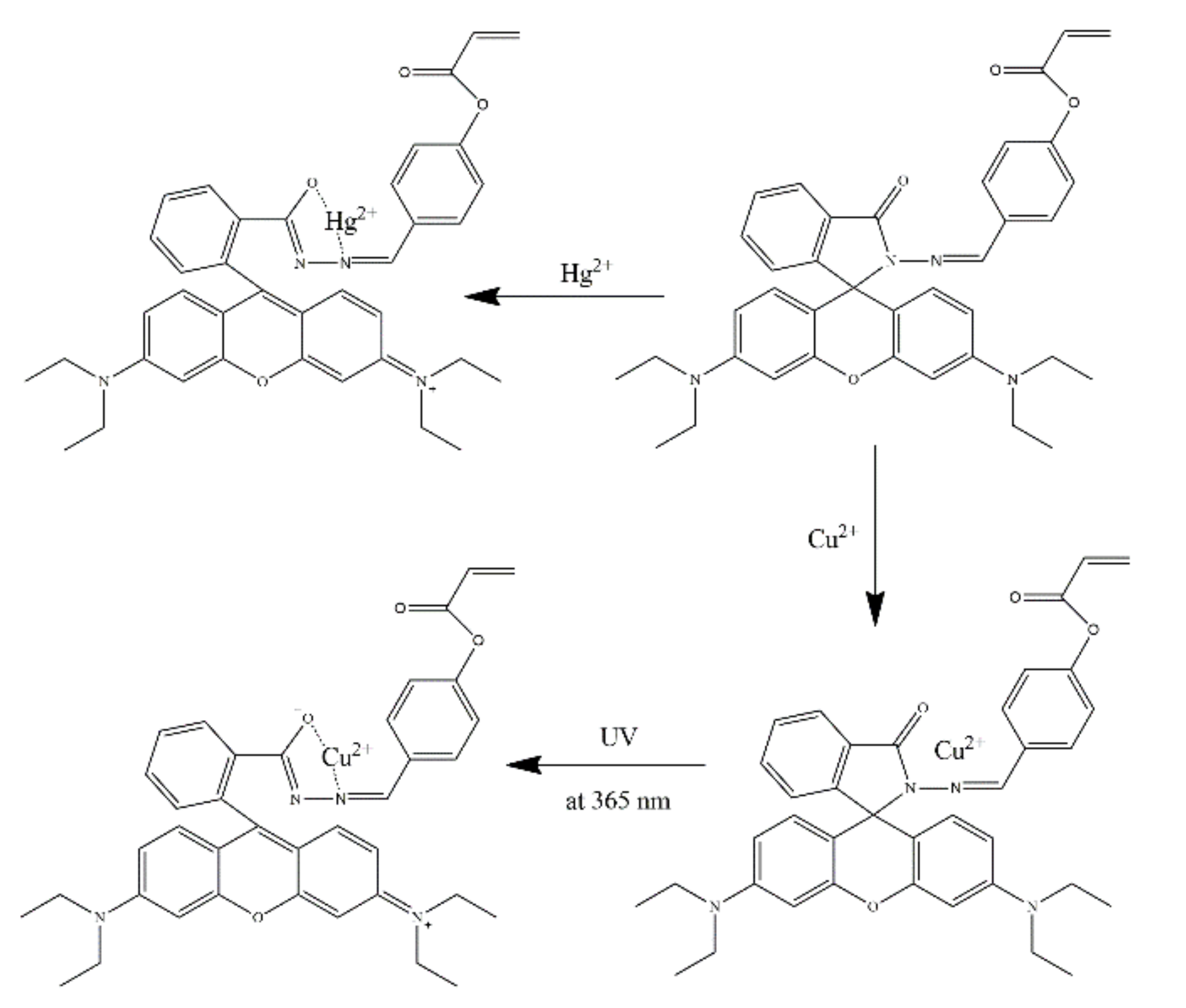
2.2. Rhodamine-Based Sensors for Cu2+
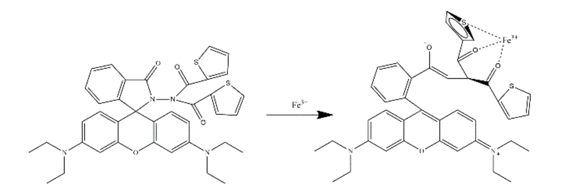
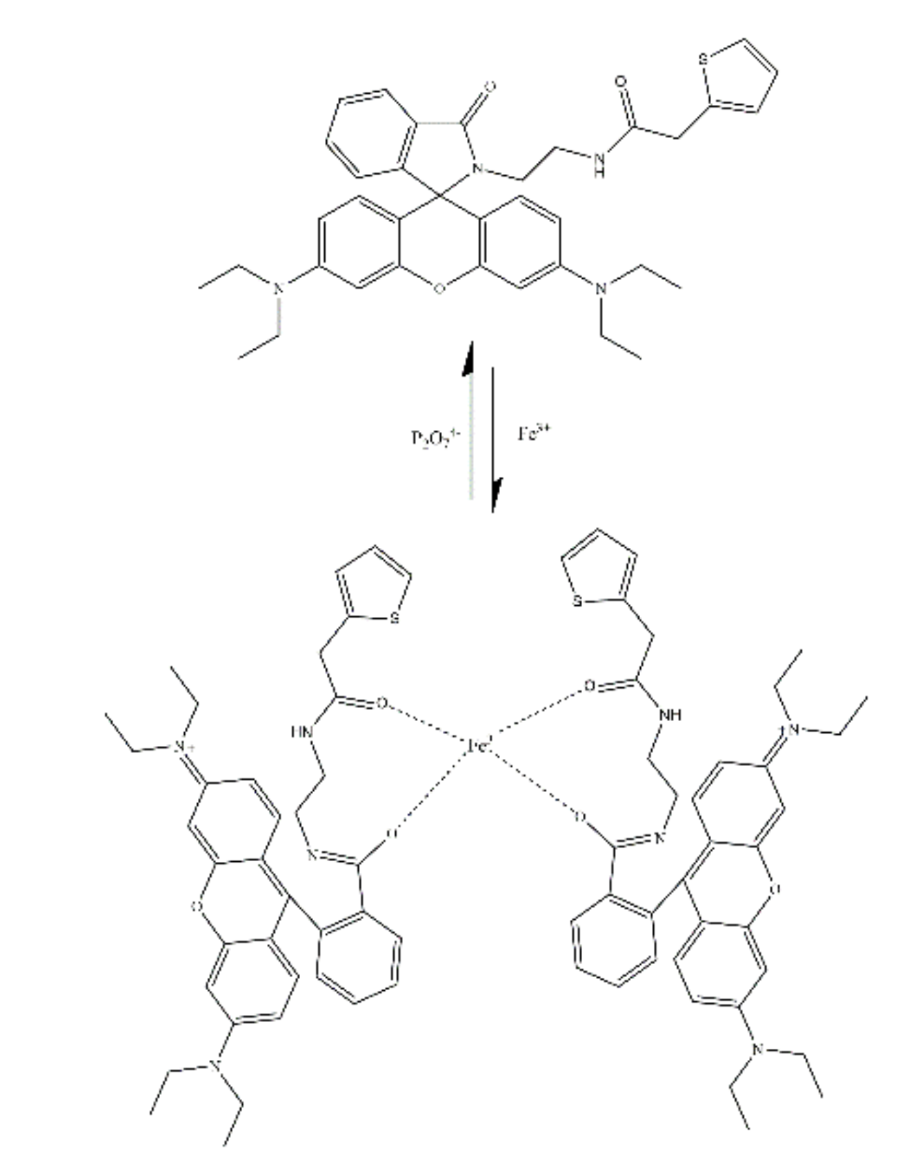
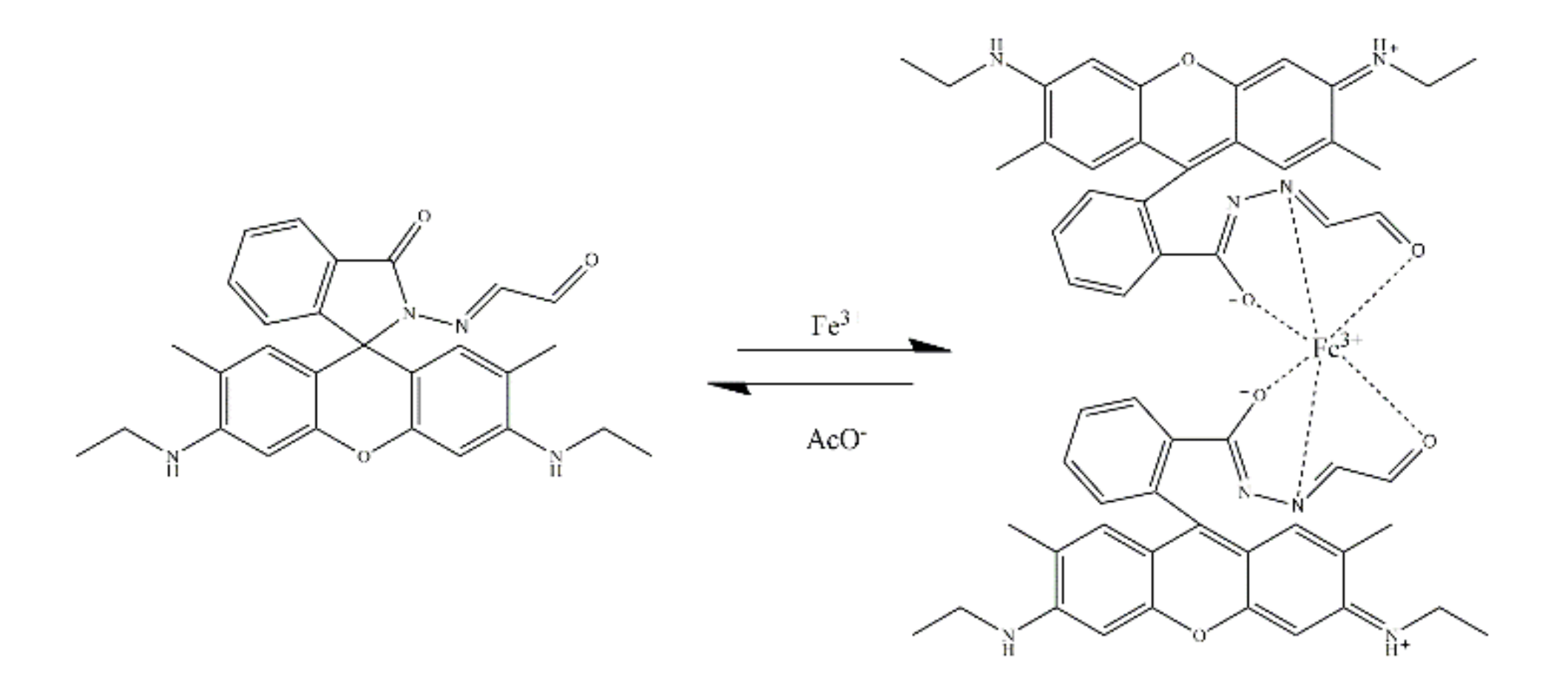
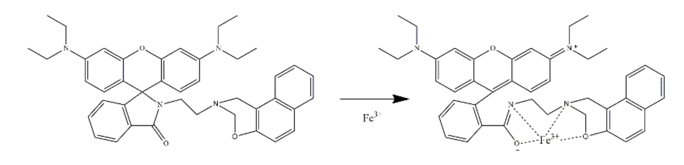
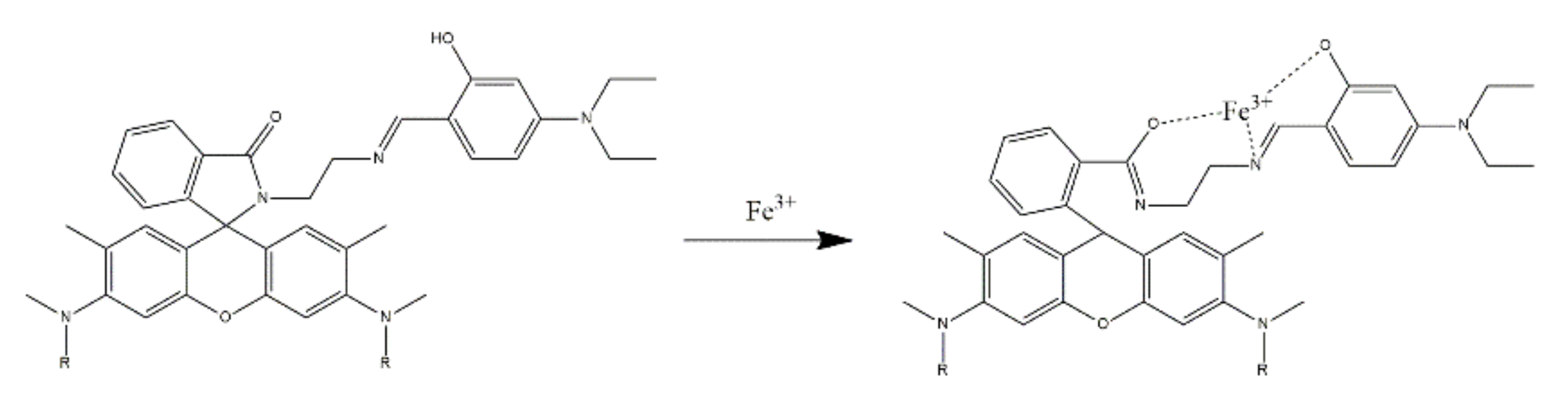
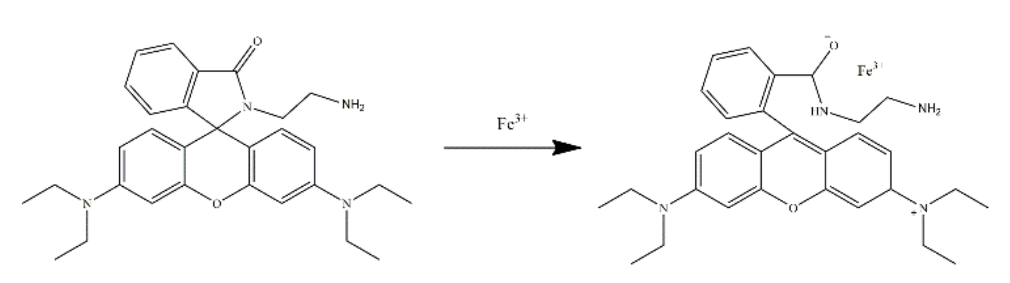
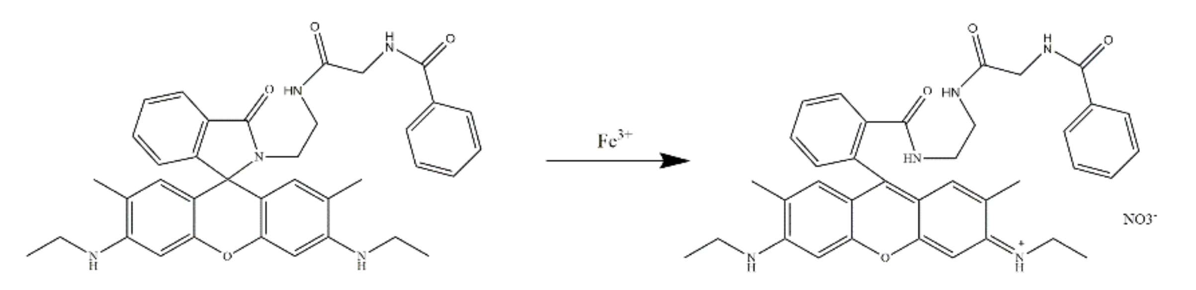
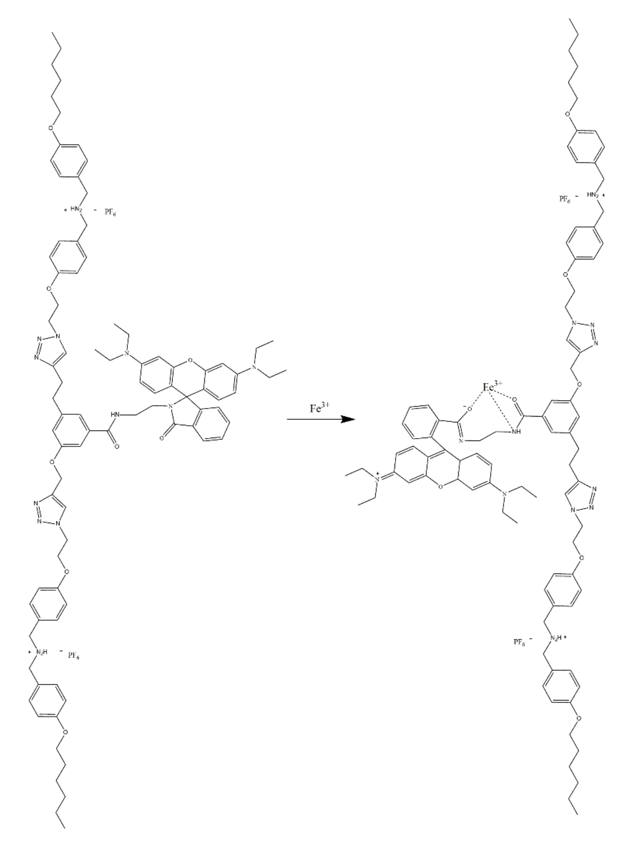
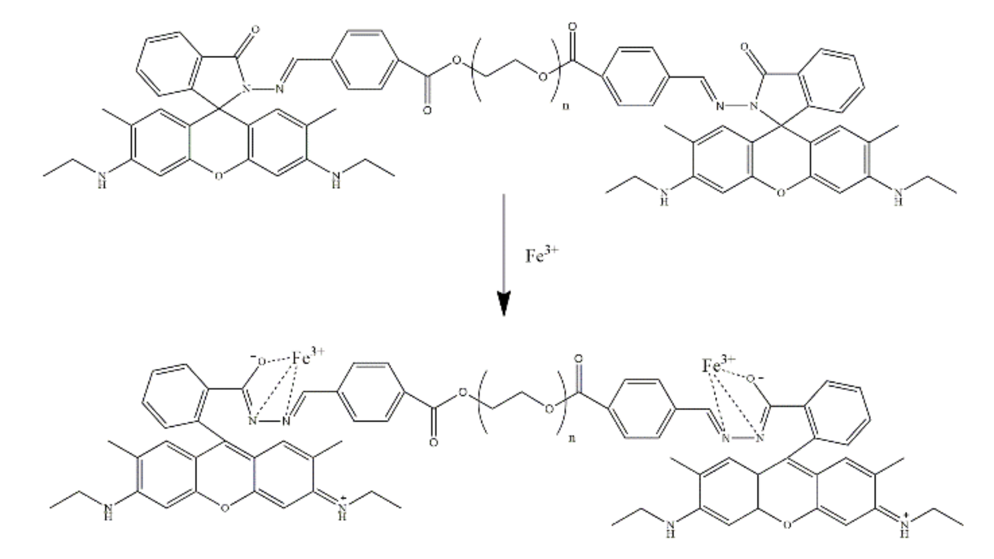
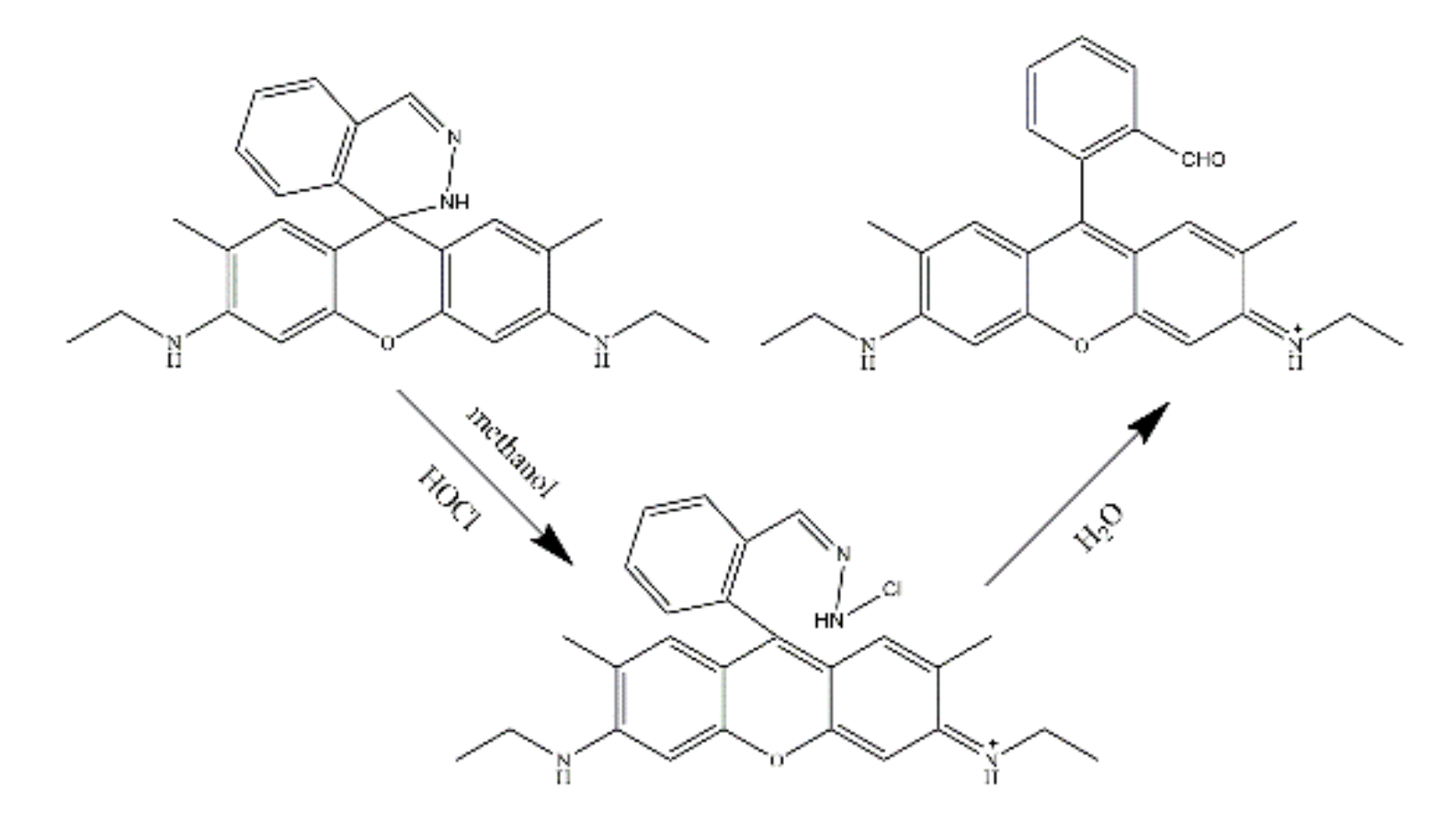
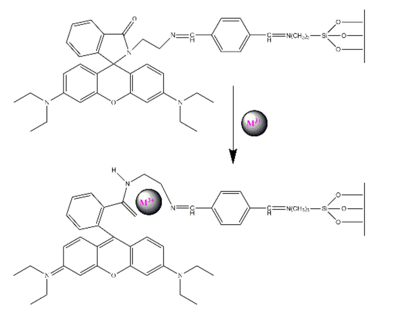
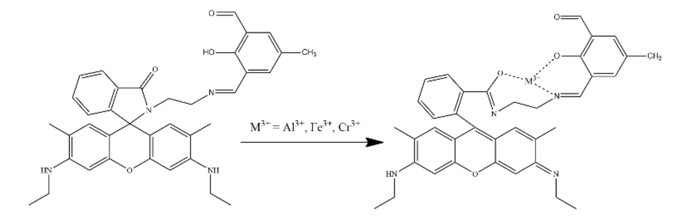
2.3. Rhodamine-Based Sensors for Fe3+
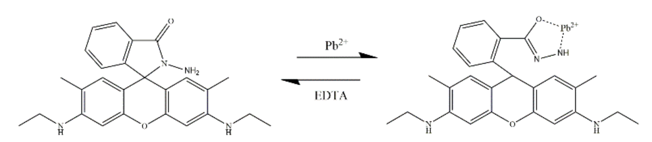
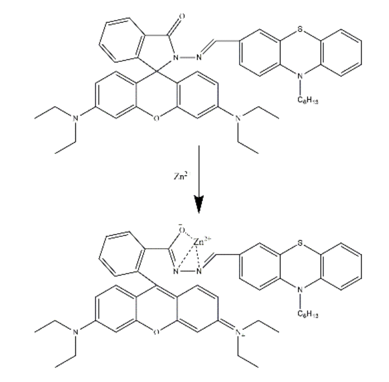
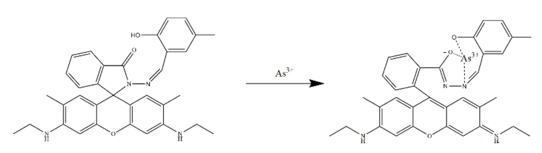
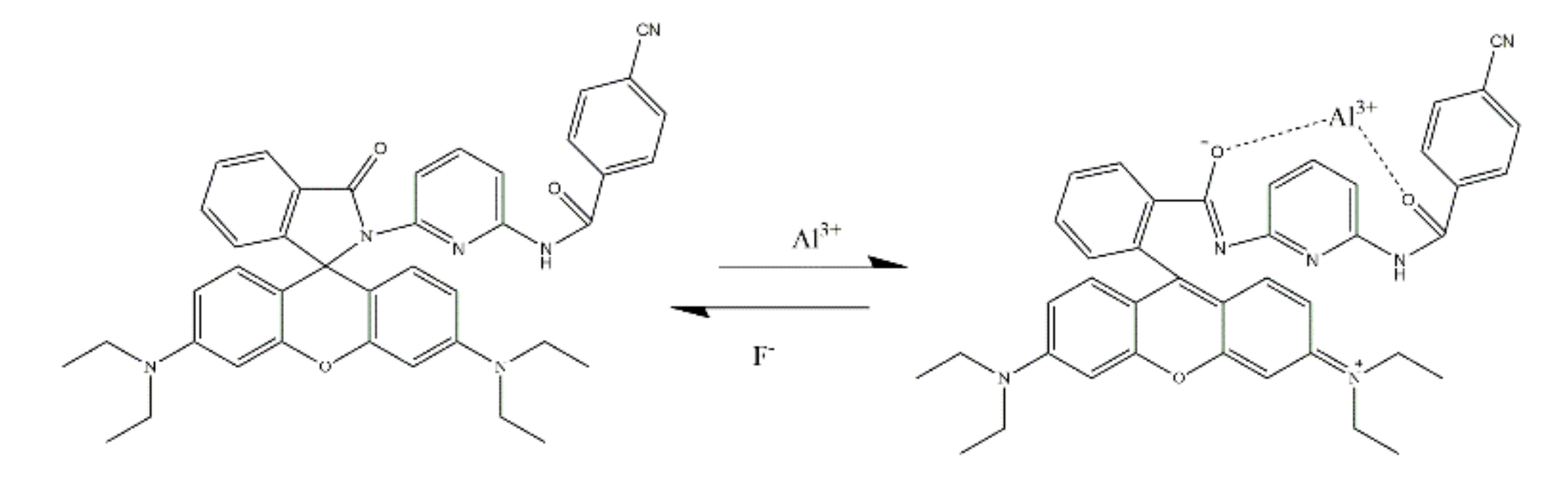
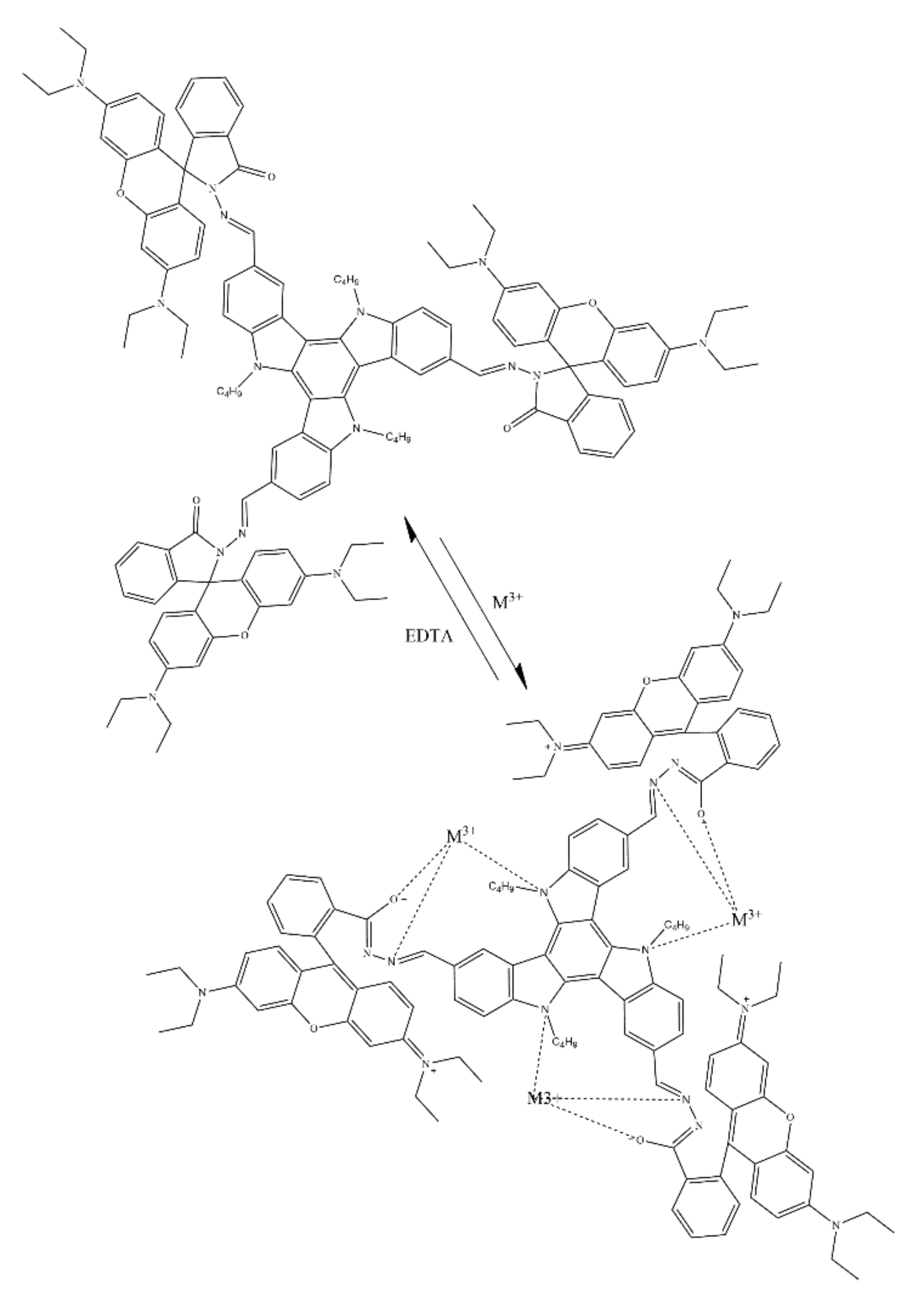
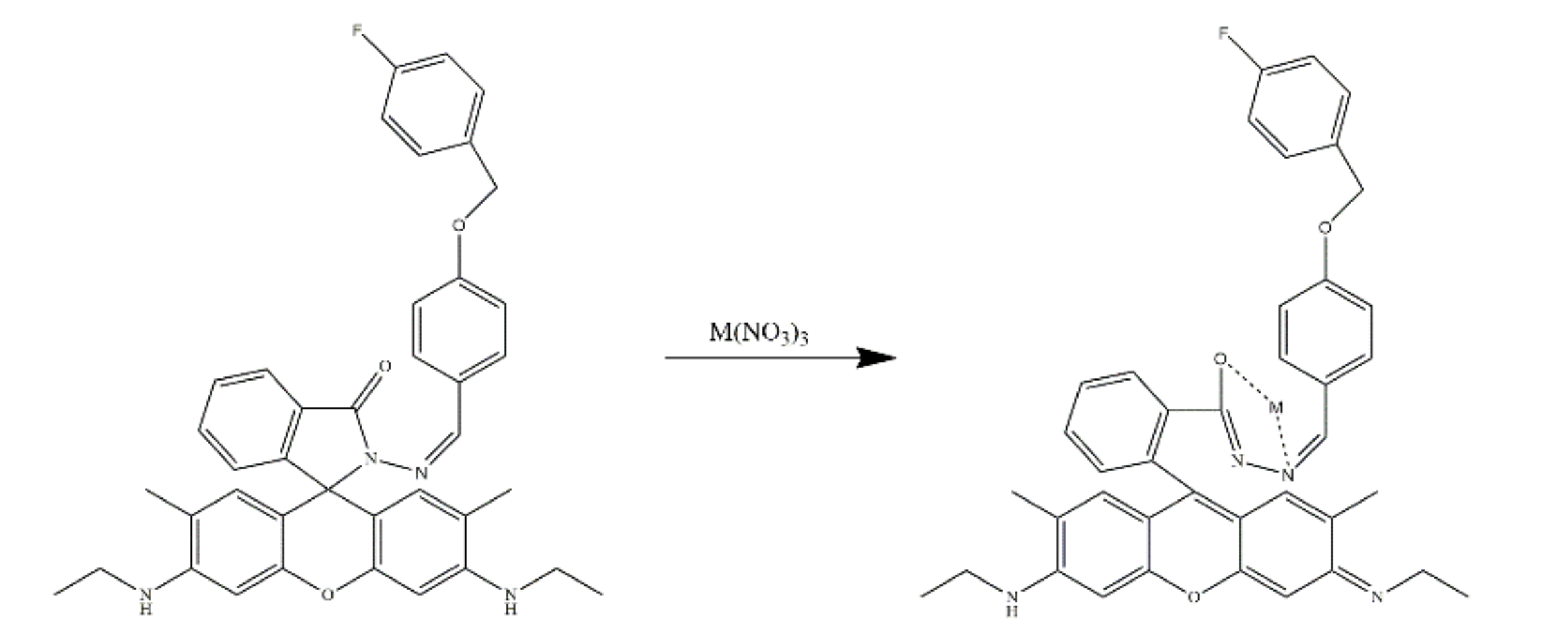
2.4. Rhodamine-Based Sensors for Other Metal Ions
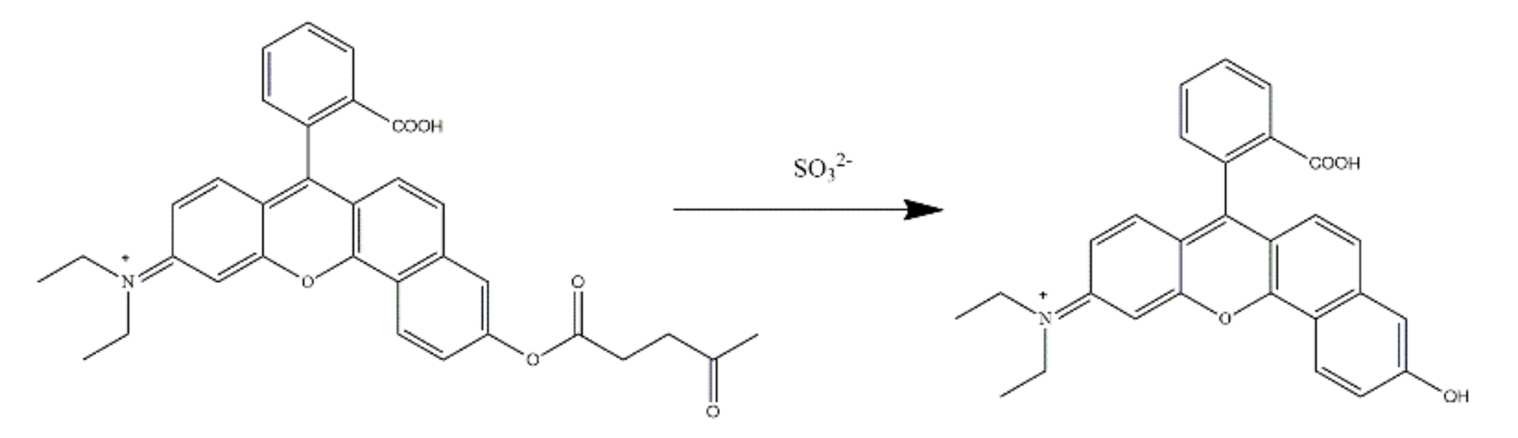
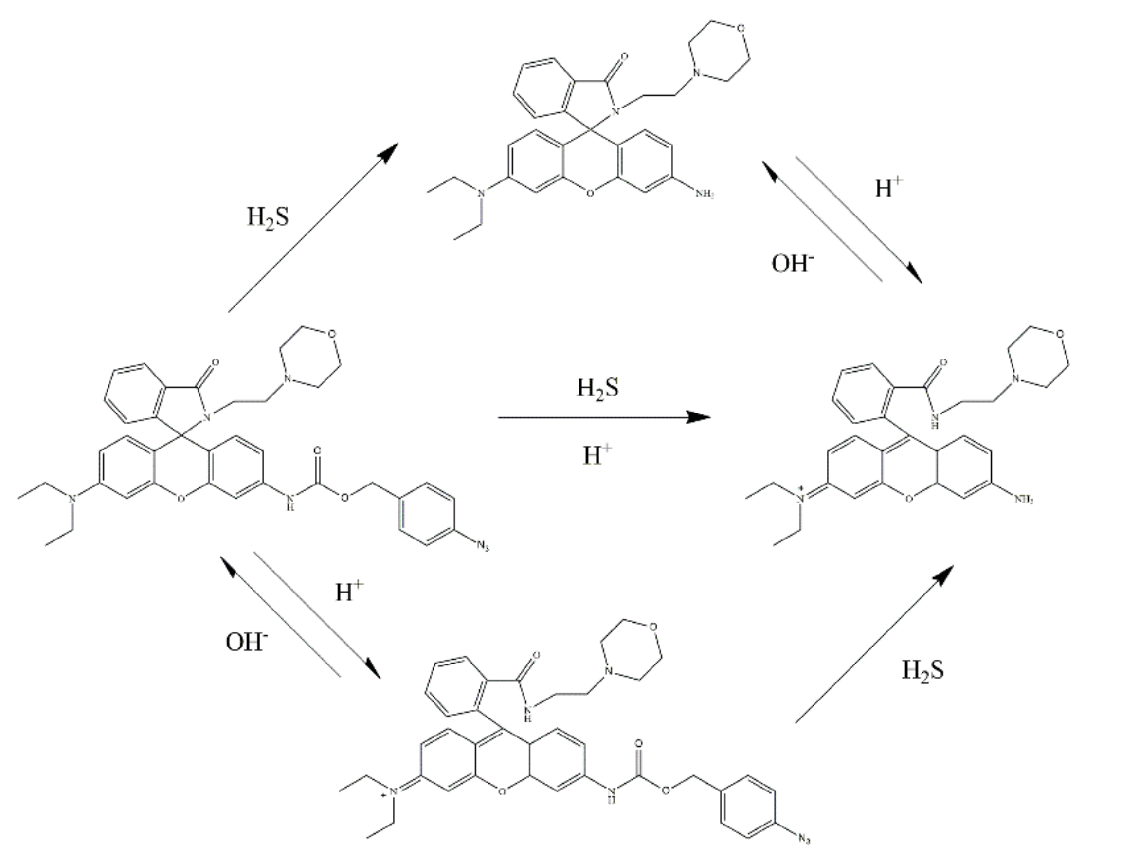
3. Rhodamine-Based Sensors for H+

| No. | Molecular Structure | Target | Sensing Mechanism | Sensor Type | Sensitive pH Range | pKa | Solution Systems | Application | Ref. |
|---|---|---|---|---|---|---|---|---|---|
| 1 |  | H+ | / | Fluorescent sensor | / | 4.63 | / | Living cells | [75] |
| 2 |  | H+ | / | Fluorescent sensor | pH = 4.0–7.0 | 4.10 | / | Living cells | [76] |
| 3 |  | H+ | / | Fluorescent sensor | / | 6.24 | NaPi buffer | Living cells | [78] |
| 4 |  | H+ | TBET and FRET | Fluorescent sensor | pH = 3.0–7.4 | 4.4 (1) 4.6 (2) 4.8 (3) | / | Living cells | [79] |
| 5 |  | H+ | FRET | Ratiometric sensor | pH = 4.0–8.0 | 7.1 | / | Living cells | [80] |
| 6 |  | H+ | FRET | Fluorescent and colorimetric sensor | / | / | DMF/H2O (v/v, 1:4) (pH = 7.2) | Living cells | [81] |
| 7 |  | H+ | PET | Fluorescent and colorimetric sensor | pH = 1.0–7.5 | 2.59 | THF/H2O (v/v, 1: 1) | / | [82] |
4. Rhodamine-Based Sensors for Anions and Others
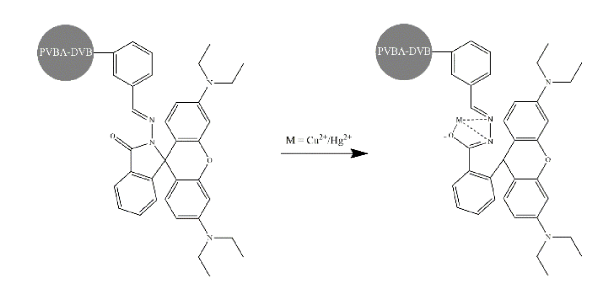
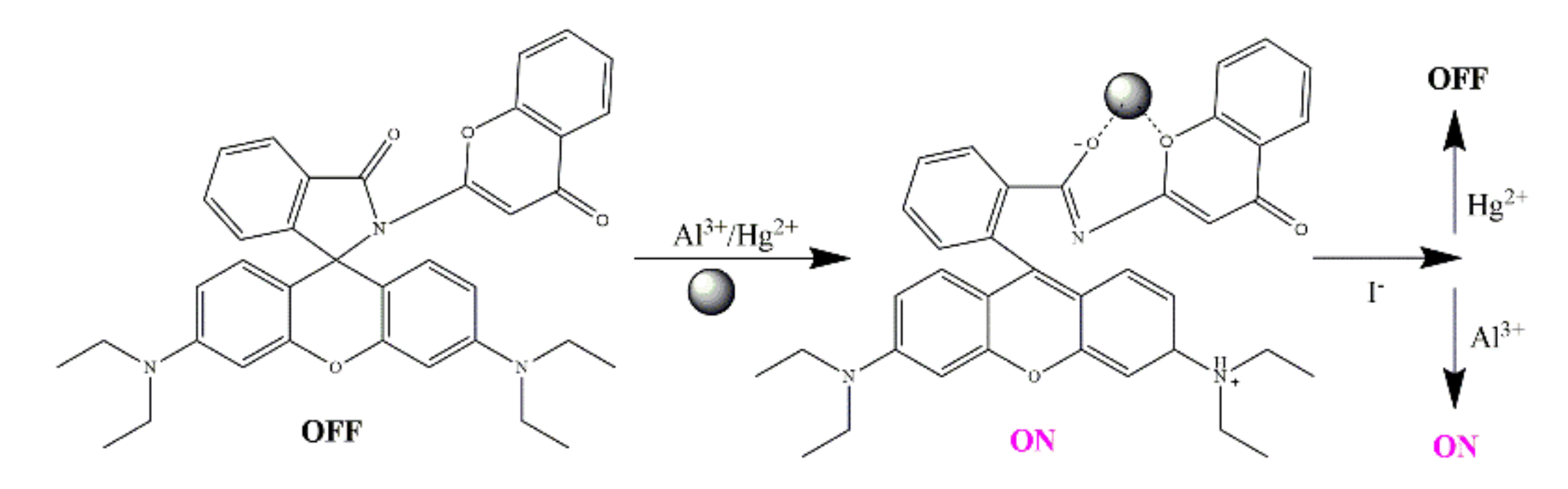
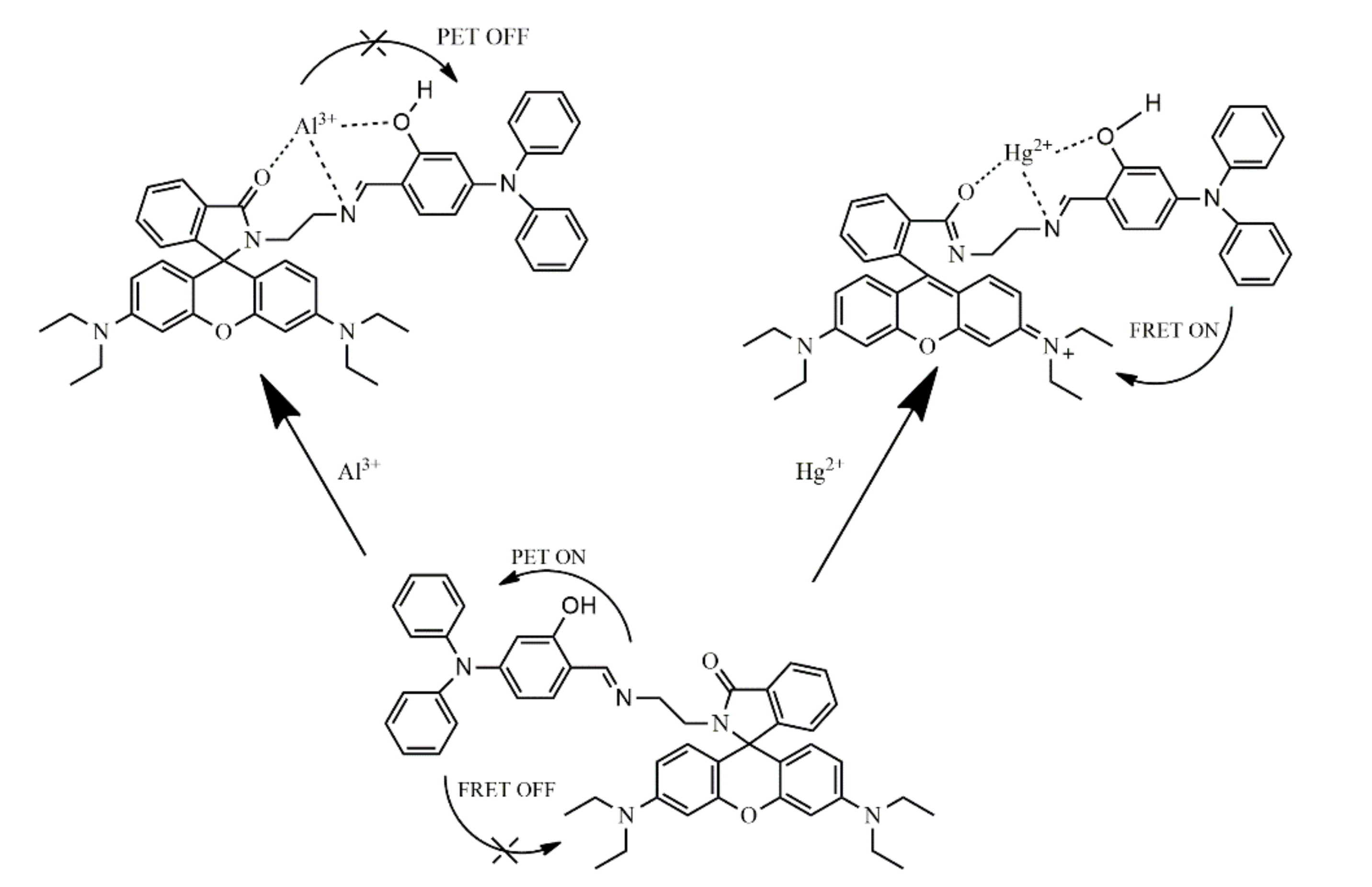
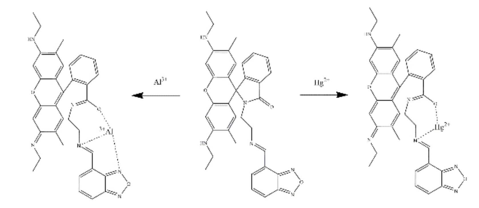
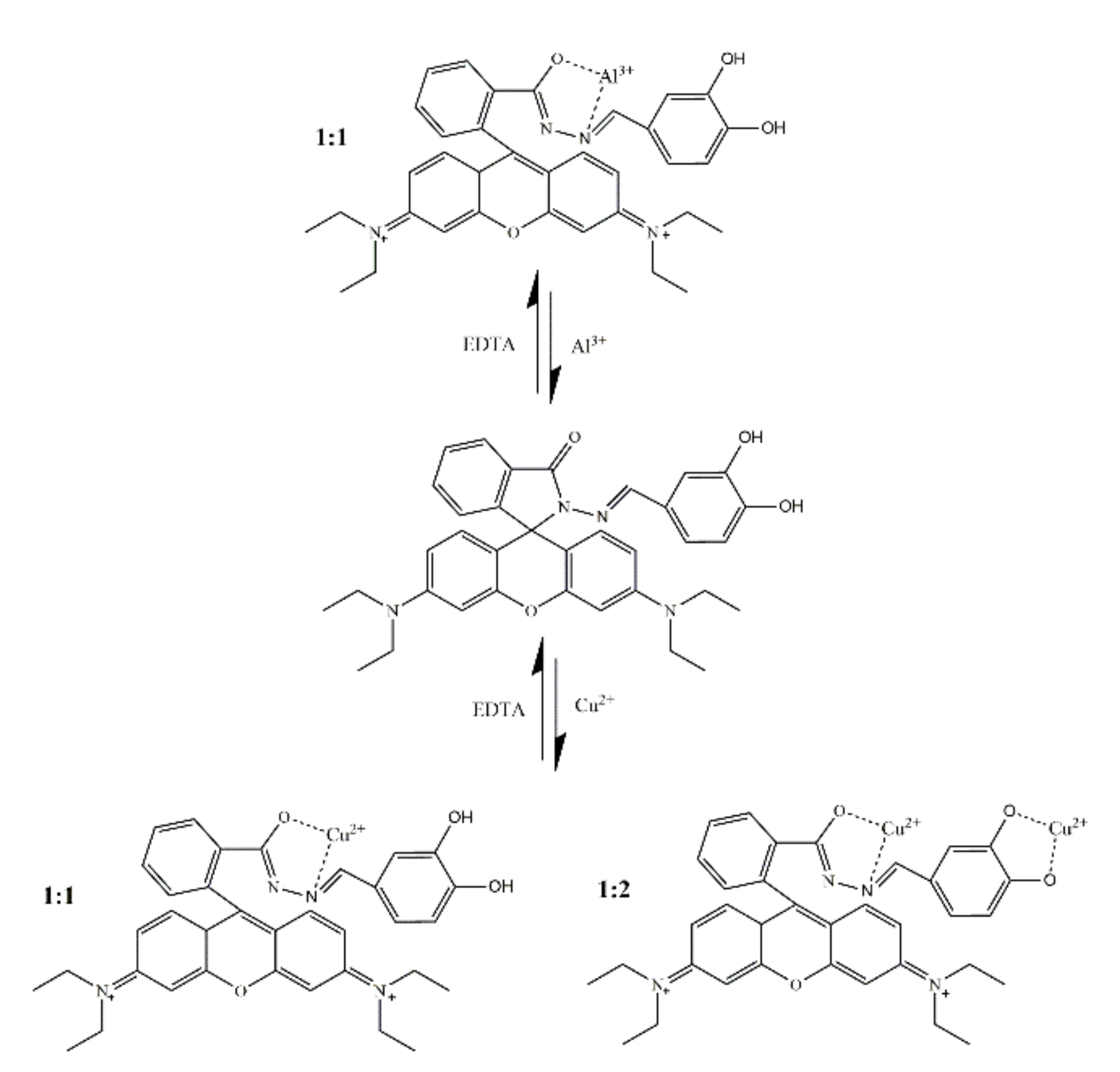
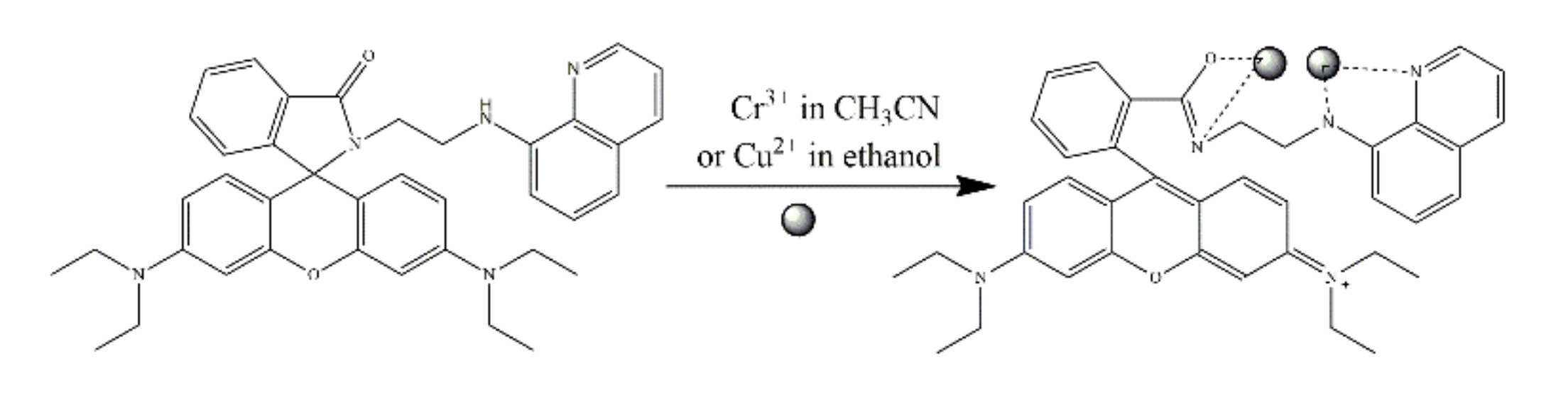
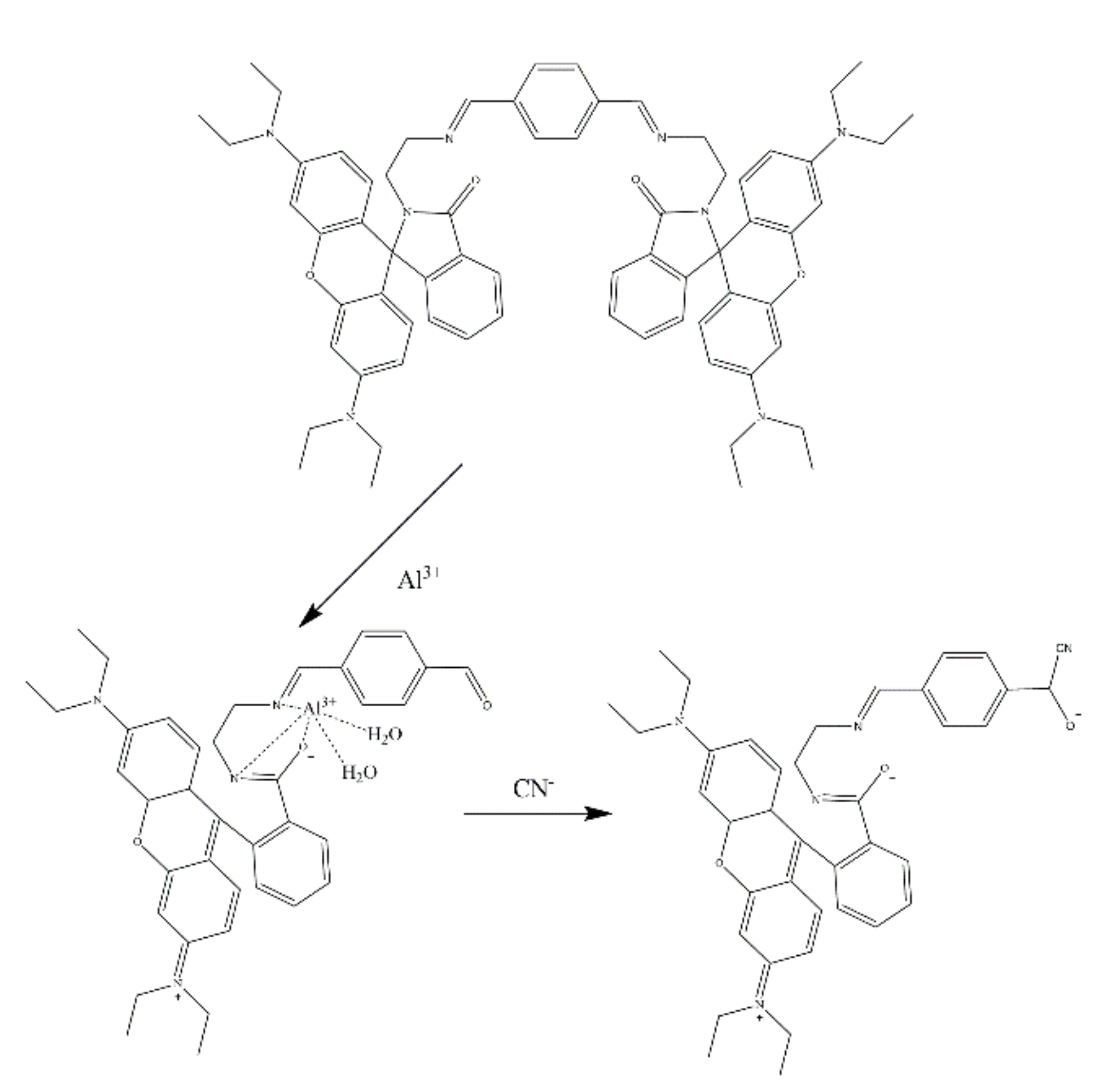
5. Rhodamine-Based Sensors for Multi-Targets
6. Conclusions and Perspective
Author Contributions
Funding
Conflicts of Interest
References
- Kaur, B.; Kaur, N.; Kumar, S. Colorimetric metal ion sensors—A comprehensive review of the years 2011–2016. Coord. Chem. Rev. 2018, 358, 13–69. [Google Scholar] [CrossRef]
- Park, S.-H.; Kwon, N.; Lee, J.-H.; Yoon, J.; Shin, I. Synthetic ratiometric fluorescent probes for detection of ions. Chem. Soc. Rev. 2020, 49, 143–179. [Google Scholar] [CrossRef] [PubMed]
- Shree, G.J.; Sivaraman, G.; Siva, A.; Chellappa, D. Anthracene-and pyrene-bearing imidazoles as turn-on fluorescent chemosensor for aluminum ion in living cells. Dye. Pigment. 2019, 163, 204–212. [Google Scholar] [CrossRef]
- Wu, D.; Sedgwick, A.C.; Gunnlaugsson, T.; Akkaya, E.U.; Yoon, J.; James, T.D. Fluorescent chemosensors: The past, present and future. Chem. Soc. Rev. 2017, 46, 7105–7123. [Google Scholar] [CrossRef] [PubMed] [Green Version]
- Rasheed, T.; Li, C.; Bilal, M.; Yu, C.; Iqbal, H.M.N. Potentially toxic elements and environmentally-related pollutants recognition using colorimetric and ratiometric fluorescent probes. Sci. Total Environ. 2018, 640, 174–193. [Google Scholar] [CrossRef] [PubMed]
- Chen, S.-H.; Jiang, K.; Xiao, Y.; Cao, X.-Y.; Arulkumar, M.; Wang, Z.-Y. Recent endeavors on design, synthesis, fluorescence mechanisms and applications of benzazole-based molecular probes toward miscellaneous species. Dye. Pigment. 2020, 175, 108157. [Google Scholar] [CrossRef]
- Kwon, N.; Hu, Y.; Yoon, J. Fluorescent Chemosensors for Various Analytes Including Reactive Oxygen Species, Biothiol, Metal Ions, and Toxic Gases. ACS Omega 2018, 3, 13731–13751. [Google Scholar] [CrossRef]
- Wu, D.; Chen, L.; Xu, Q.; Chen, X.; Yoon, J. Design Principles, Sensing Mechanisms, and Applications of Highly Specific Fluorescent Probes for HOCl/OCl(). Acc. Chem. Res. 2019, 52, 2158–2168. [Google Scholar] [CrossRef]
- State Administration for Market Regulation; National Standardization Administration. GB 5749-2022; Standards for Drinking Water Quality: National Standards of the People’s Republic of China. National Health Commission of the People’s Republic of China: Beijing, China, 2022.
- Han, B.; Hu, X.; Yan, Q.; Jiang, J.; He, G. Ag-location-based color-tunable fluorescent AuAg nanoclusters for “turn-on” and “turn-off” detection of l-cysteine. Sens. Actuators B Chem. 2019, 284, 695–703. [Google Scholar] [CrossRef]
- Tan, X.; Liu, S.; Shen, Y.; He, Y.; Yang, J. Quantum dots (QDs) based fluorescence probe for the sensitive determination of kaempferol. Spectrochim. Acta Part A Mol. Biomol. Spectrosc. 2014, 133, 66–72. [Google Scholar] [CrossRef]
- Vikrant, K.; Kumar, V.; Ok, Y.S.; Kim, K.-H.; Deep, A. Metal-organic framework (MOF)-based advanced sensing platforms for the detection of hydrogen sulfide. TrAC Trends Anal. Chem. 2018, 105, 263–281. [Google Scholar] [CrossRef]
- Li, Z.; Zhi, Y.; Shao, P.; Xia, H.; Li, G.; Feng, X.; Chen, X.; Shi, Z.; Liu, X. Covalent organic framework as an efficient, metal-free, heterogeneous photocatalyst for organic transformations under visible light. Appl. Catal. B Environ. 2019, 245, 334–342. [Google Scholar] [CrossRef]
- Chen, H.; Lin, W.; Jiang, W.; Dong, B.; Cui, H.; Tang, Y. Locked-flavylium fluorescent dyes with tunable emission wavelengths based on intramolecular charge transfer for multi-color ratiometric fluorescence imaging. Chem. Commun. 2015, 51, 6968–6971. [Google Scholar] [CrossRef] [PubMed]
- Dsouza, R.N.; Pischel, U.; Nau, W.M. Fluorescent Dyes and Their Supramolecular Host/Guest Complexes with Macrocycles in Aqueous Solution. Chem. Rev. 2011, 111, 7941–7980. [Google Scholar] [CrossRef] [PubMed]
- Noelting, E.; Dziewonsky, K. Ber. Dtsch. Chem. Ges 1905, 38, 3516. [Google Scholar] [CrossRef] [Green Version]
- Kim, H.N.; Lee, M.H.; Kim, H.J.; Kim, J.S.; Yoon, J. A new trend in rhodamine-based chemosensors: Application of spirolactam ring-opening to sensing ions. Chem. Soc. Rev. 2008, 37, 1465–1472. [Google Scholar] [CrossRef]
- Dujols, V.; Ford, F.; Czarnik, A.W. A long-wavelength fluorescent chemodosimeter selective for Cu (II) ion in water. J. Am. Chem. Soc. 1997, 119, 7386–7387. [Google Scholar] [CrossRef]
- Yang, Y.-K.; Yook, K.-J.; Tae, J. A rhodamine-based fluorescent and colorimetric chemodosimeter for the rapid detection of Hg2+ ions in aqueous media. J. Am. Chem. Soc. 2005, 127, 16760–16761. [Google Scholar] [CrossRef]
- Chen, G.; Guo, Z.; Zeng, G.; Tang, L. Fluorescent and colorimetric sensors for environmental mercury detection. Analist 2015, 140, 5400–5443. [Google Scholar] [CrossRef]
- Roy, P. Recent advances in the development of fluorescent chemosensors for Al(3). Dalton Trans. 2021, 50, 7156–7165. [Google Scholar] [CrossRef]
- Jain, N.; Kaur, N. A comprehensive compendium of literature of 1,8-Naphthalimide based chemosensors from 2017 to 2021. Coord. Chem. Rev. 2022, 459, 214454. [Google Scholar] [CrossRef]
- Zhu, H.; Liu, C.; Su, M.; Rong, X.; Zhang, Y.; Wang, X.; Wang, K.; Li, X.; Yu, Y.; Zhang, X.; et al. Recent advances in 4-hydroxy-1,8-naphthalimide-based small-molecule fluorescent probes. Coord. Chem. Rev. 2021, 448, 214153. [Google Scholar] [CrossRef]
- Zhang, J.; Zhu, M.; Jiang, D.; Zhang, H.; Li, L.; Zhang, G.; Wang, Y.; Feng, C.; Zhao, H. A FRET-based colorimetric and ratiometric fluorescent probe for the detection of Cu2+ with a new trimethylindolin fluorophore. New J. Chem. 2019, 43, 10176–10182. [Google Scholar] [CrossRef]
- Chen, H.; Jin, X.; Zhang, W.; Lu, H.; Shen, W. A new rhodamine B-based ‘off-on’ colorimetric chemosensor for Pd2+ and its imaging in living cells. Inorg. Chim. Acta 2018, 482, 122–129. [Google Scholar] [CrossRef]
- Luo, W.; Lei, M.; Wang, Y.; Gao, H.; Wang, Y.; Zhou, Q.; Xu, Z.; Yang, F. An indole–rhodamine-based ratiometric fluorescent probe for Pd2+ determination and cell imaging. Anal. Methods 2019, 11, 1080–1086. [Google Scholar] [CrossRef]
- Hu, L.; Lin, Y.; Wang, P.; Zhang, H.; Liu, M.; Mo, S. A rhodamine derivative probe for highly selective detection of Cu(II). Front. Biosci. 2022, 27, 28. [Google Scholar] [CrossRef]
- Qiu, X.; Huang, J.; Wang, N.; Zhao, K.; Cui, J.; Hao, J. Facile Synthesis of Water-Soluble Rhodamine-Based Polymeric Chemosensors via Schiff Base Reaction for Fe(3+) Detection and Living Cell Imaging. Front. Chem. 2022, 10, 845627. [Google Scholar] [CrossRef]
- Cuc, T.T.K.; Nhien, P.Q.; Khang, T.M.; Chen, H.-Y.; Wu, C.-H.; Hue, B.T.B.; Li, Y.-K.; Wu, J.I.; Lin, H.-C. Controllable FRET processes towards ratiometric Fe3+ ion sensor of pseudo [3] rotaxane containing naphthalimide-based macrocyclic host donor and multi-stimuli responsive rhodamine-modified guest acceptor. Dye. Pigment. 2022, 197, 109907. [Google Scholar] [CrossRef]
- Lee, M.H.; Kim, J.S.; Sessler, J.L. Small molecule-based ratiometric fluorescence probes for cations, anions, and biomolecules. Chem. Soc. Rev. 2015, 44, 4185–4191. [Google Scholar] [CrossRef] [Green Version]
- Williams, D.R. Metals, ligands, and cancer. Chem. Rev. 1972, 72, 203–213. [Google Scholar] [CrossRef]
- Cheng, Z.; Zheng, L.; Xu, H.; Pang, L.; He, H. A rhodamine-based fluorescent probe for Fe3+: Synthesis, theoretical calculation and bioimaging application. Anal. Methods 2019, 11, 2565–2570. [Google Scholar] [CrossRef]
- Min, K.S.; Manivannan, R.; Son, Y.-A. Rhodamine-fluorene based dual channel probe for the detection of Hg2+ ions and its application in digital printing. Sens. Actuators B Chem. 2018, 261, 545–552. [Google Scholar] [CrossRef]
- Hong, M.; Chen, Y.; Zhang, Y.; Xu, D. A novel rhodamine-based Hg2+ sensor with a simple structure and fine performance. Analist 2019, 144, 7351–7358. [Google Scholar] [CrossRef] [PubMed]
- Cicekbilek, F.; Yilmaz, B.; Bayrakci, M.; Gezici, O. An Application of a Schiff-Base Type Reaction in the Synthesis of a New Rhodamine-Based Hg(II)-Sensing Agent. J. Fluoresc. 2019, 29, 1349–1358. [Google Scholar] [CrossRef] [PubMed]
- Patil, S.K.; Das, D. A nanomolar detection of mercury(II) ion by a chemodosimetric rhodamine-based sensor in an aqueous medium: Potential applications in real water samples and as paper strips. Spectrochim. Acta Part A Mol. Biomol. Spectrosc. 2019, 210, 44–51. [Google Scholar] [CrossRef]
- Kan, C.; Shao, X.; Song, F.; Xu, J.; Zhu, J.; Du, L. Bioimaging of a fluorescence rhodamine-based probe for reversible detection of Hg (II) and its application in real water environment. Microchem. J. 2019, 150, 104142. [Google Scholar] [CrossRef]
- Dewangan, S.; Barik, T.; Mishra, S.; Mawatwal, S.; Kumari, S.; Giri, S.; Das, S.; Dhiman, R.; Wölper, C.; Chatterjee, S. Half sandwich based rhodamine-hydrazone single molecule probe: Light responsive, metal sensing and imaging properties. Appl. Organomet. Chem. 2018, 32, e4612. [Google Scholar] [CrossRef]
- Wang, Q.; Jin, L.; Wang, W.; Hu, T.; Chen, C. Rhodamine derivatives as selective“naked-eye” colorimetric and fluorescence off-on sensor for Hg2+ in aqueous solution and its applications in bioimaging. J. Lumin. 2019, 209, 411–419. [Google Scholar] [CrossRef]
- Dewangan, S.; Barik, T.; Parida, R.; Mawatwal, S.; Dhiman, R.; Giri, S.; Chatterjee, S. Solvent free synthesis of ferrocene based rhodamine–hydrazone molecular probe with improved bioaccumulation for sensing and imaging applications. J. Organomet. Chem. 2019, 904, 120999. [Google Scholar] [CrossRef]
- Gauthama, B.U.; Narayana, B.; Sarojini, B.K.; Suresh, N.K.; Sangappa, Y.; Kudva, A.K.; Satyanarayana, G.; Raghu, S.V. Colorimetric “off–on” fluorescent probe for selective detection of toxic Hg2+ based on rhodamine and its application for in-vivo bioimaging. Microchem. J. 2021, 166, 106233. [Google Scholar] [CrossRef]
- Singh, S.; Coulomb, B.; Boudenne, J.L.; Bonne, D.; Dumur, F.; Simon, B.; Robert-Peillard, F. Sub-ppb mercury detection in real environmental samples with an improved rhodamine-based detection system. Talanta 2021, 224, 121909. [Google Scholar] [CrossRef] [PubMed]
- Li, X.; Li, X.; Zhao, H.; Kang, H.; Fan, C.; Liu, G.; Pu, S. A Novel Diarylethene-rhodamine Unit Based Chemosensor for Fluorimetric and Colorimetric Detection of Hg(2). J. Fluoresc. 2021, 31, 1513–1523. [Google Scholar] [CrossRef] [PubMed]
- Yilmaz, B.; Keskinates, M.; Bayrakci, M. Novel integrated sensing system of calixarene and rhodamine molecules for selective colorimetric and fluorometric detection of Hg(2+) ions in living cells. Spectrochim. Acta A Mol. Biomol. Spectrosc. 2021, 245, 118904. [Google Scholar] [CrossRef] [PubMed]
- Aduroja, O.; Shaw, R.; Abebe, F. A bis(rhodamine 6G)-based fluorescent sensor for Hg2+: Microwave-assisted synthesis, photophysical properties, and computational studies. Res. Chem. Intermed. 2022, 48, 1847–1861. [Google Scholar] [CrossRef]
- Li, M.; Zhang, S.; Zhang, P.; Qin, K.; Xu, B.; Zhou, J.; Yuan, C.; Cao, Q.; Xiao, H. Rhodamine functionalized cellulose for mercury detection and removal: A strategy for providing in situ fluorimetric and colorimetric responses. Chem. Eng. J. 2022, 436, 135251. [Google Scholar] [CrossRef]
- Liang, F.; Xu, L.; Jin, D.; Dong, L.; Lin, S.; Huang, R.; Song, D.; Ma, P. A novel near-infrared fluorescence probe for detecting and imaging Hg(2+) in living cells. Luminescence 2022, 37, 161–169. [Google Scholar] [CrossRef]
- Zhang, Q.; Ding, H.; Xu, X.; Wang, H.; Liu, G.; Pu, S. Rational design of a FRET-based ratiometric fluorescent probe with large Pseudo-Stokes shift for detecting Hg(2+) in living cells based on rhodamine and anthracene fluorophores. Spectrochim. Acta A Mol. Biomol. Spectrosc. 2022, 276, 121242. [Google Scholar] [CrossRef]
- Tromp, A.; De Klerk, C. Effect of copperoxychloride on the fermentation of must and on wine quality. S. Afr. J. Enol. Vitic. 1988, 9, 31–36. [Google Scholar] [CrossRef] [Green Version]
- Doumani, N.; Bou-Maroun, E.; Maalouly, J.; Tueni, M.; Dubois, A.; Bernhard, C.; Denat, F.; Cayot, P.; Sok, N. A New pH-Dependent Macrocyclic Rhodamine B-Based Fluorescent Probe for Copper Detection in White Wine. Sensors 2019, 19, 4514. [Google Scholar] [CrossRef] [Green Version]
- Deepa, A.; Srinivasadesikan, V.; Lee, S.-L.; Padmini, V. Highly Selective and Sensitive Colorimetric and Fluorimetric Sensor for Cu2+. J. Fluoresc. 2019, 30, 3–10. [Google Scholar] [CrossRef]
- Hosseinjani-Pirdehi, H.; Allah Mahmoodi, N.O.; Taheri, A.; Asalemi, K.A.A.; Esmaeili, R. Selective immediate detection of Cu2+ by a pH-sensitive rhodamine-based fluorescence probe in breast cancer cell-line. Spectrochim. Acta Part A Mol. Biomol. Spectrosc. 2020, 229, 117989. [Google Scholar] [CrossRef] [PubMed]
- Karakuş, E. A rhodamine based fluorescent chemodosimeter for the selective and sensitive detection of copper (II) ions in aqueous media and living cells. J. Mol. Struct. 2021, 1224, 129037. [Google Scholar] [CrossRef]
- Wang, Y.; Wang, Y.; Guo, F.; Wang, Y.; Xie, P. A new naked-eye fluorescent chemosensor for Cu(II) and its practical applications. Res. Chem. Intermed. 2021, 47, 3515–3528. [Google Scholar] [CrossRef]
- Yang, L.; Zhang, X.; Yang, J.; Yuan, M.-S.; Wang, J. A rhodamine-based chemosensor and functionalized gel ball for detecting and adsorbing copper ions. Tetrahedron 2021, 80, 131893. [Google Scholar] [CrossRef]
- Liu, Y.; Zhao, C.; Zhao, X.; Liu, H.; Wang, Y.; Du, Y.; Wei, D. A selective N,N-dithenoyl-rhodamine based fluorescent probe for Fe3+ detection in aqueous and living cells. J. Environ. Sci. 2020, 90, 180–188. [Google Scholar] [CrossRef]
- Wang, K.-P.; Zheng, W.-J.; Lei, Y.; Zhang, S.-J.; Zhang, Q.; Chen, S.; Hu, Z.-Q. A thiophene-rhodamine dyad as fluorescence probe for ferric ion and its application in living cells imaging. J. Lumin. 2019, 208, 468–474. [Google Scholar] [CrossRef]
- Wang, Y.; Guo, R.; Hou, X.; Lei, M.; Zhou, Q.; Xu, Z. Highly Sensitive and Selective Fluorescent Probe for Detection of Fe3+ Based on Rhodamine Fluorophore. J. Fluoresc. 2019, 29, 645–652. [Google Scholar] [CrossRef] [PubMed]
- Zhang, M.; Shen, C.; Jia, T.; Qiu, J.; Zhu, H.; Gao, Y. One-step synthesis of rhodamine-based Fe3+ fluorescent probes via Mannich reaction and its application in living cell imaging. Spectrochim. Acta Part A Mol. Biomol. Spectrosc. 2020, 231, 118105. [Google Scholar] [CrossRef]
- Chen, Y.; Wu, Y.; Zhu, Y.; Tian, S. A fluorescent polyurethane foam based on rhodamine derivative as Fe(III) sensor in pure water. Polym. Int. 2021, 71, 169–174. [Google Scholar] [CrossRef]
- Liu, X.; Chen, Z.; Gao, R.; Kan, C.; Xu, J. Portable quantitative detection of Fe3+ by integrating a smartphone with colorimetric responses of a rhodamine-functionalized polyacrylamide hydrogel chemosensor. Sens. Actuators B Chem. 2021, 340, 129958. [Google Scholar] [CrossRef]
- Qin, Z.; Su, W.; Liu, P.; Ma, J.; Zhang, Y.; Jiao, T. Facile Preparation of a Rhodamine B Derivative-Based Fluorescent Probe for Visual Detection of Iron Ions. ACS Omega 2021, 6, 25040–25048. [Google Scholar] [CrossRef] [PubMed]
- Zhang, J.; Bai, C.B.; Chen, M.Y.; Yue, S.Y.; Qin, Y.X.; Liu, X.Y.; Xu, M.Y.; Zheng, Q.J.; Zhang, L.; Li, R.Q.; et al. Novel Fluorescent Probe toward Fe(3+) Based on Rhodamine 6G Derivatives and Its Bioimaging in Adult Mice, Caenorhabditis elegans, and Plant Tissues. ACS Omega 2021, 6, 8616–8624. [Google Scholar] [CrossRef] [PubMed]
- Liu, X.; Chen, Z.; Gao, R.; Kan, C.; Xu, J. A paper-based fluorescent and colorimetric portable device with smartphone application for Fe3+ sensing. J. Environ. Chem. Eng. 2022, 10, 107650. [Google Scholar] [CrossRef]
- Xie, X.; Pan, M.; Hong, L.; Liu, K.; Yang, J.; Wang, S.; Wang, S. An “Off-On” Rhodamine 6G Hydrazide-Based Output Platform for Fluorescence and Visual Dual-Mode Detection of Lead(II). J. Agric. Food Chem. 2021, 69, 7209–7217. [Google Scholar] [CrossRef]
- Karmegam, M.V.; Karuppannan, S.; Christopher Leslee, D.B.; Subramanian, S.; Gandhi, S. Phenothiazine–rhodamine-based colorimetric and fluorogenic ‘turn-on’ sensor for Zn2+ and bioimaging studies in live cells. Luminescence 2019, 35, 90–97. [Google Scholar] [CrossRef]
- Zhang, X.; Jin, G.; Chen, Z.; Wu, Y.; Li, Q.; Liu, P.; Xi, G. An efficient turn-on fluorescence chemosensor system for Zn(II) ions detection and imaging in mitochondria. J. Photochem. Photobiol. B 2022, 234, 112485. [Google Scholar] [CrossRef] [PubMed]
- Paul, S.; Bhuyan, S.; Mukhopadhyay, S.K.; Murmu, N.C.; Banerjee, P. Sensitive and Selective in Vitro Recognition of Biologically Toxic As(III) by Rhodamine Based Chemoreceptor. ACS Sustain. Chem. Eng. 2019, 7, 13687–13697. [Google Scholar] [CrossRef]
- Kan, C.; Wu, L.; Wang, X.; Shao, X.; Qiu, S.; Zhu, J. Rhodamine B-based chemiluminescence sensor for aluminum ion monitoring and bioimaging applications. Tetrahedron 2021, 85, 132054. [Google Scholar] [CrossRef]
- Jiang, T.; Bian, W.; Kan, J.; Sun, Y.; Ding, N.; Li, W.; Zhou, J. Sensitive and rapid detection of Cr(3+) in live cells by a red turn-on fluorescent probe. Spectrochim. Acta A Mol. Biomol. Spectrosc. 2021, 245, 118903. [Google Scholar] [CrossRef]
- Rathinam, B.; Murugesan, V.; Liu, B.-T. Fluorescent “OFF–ON” Sensors for the Detection of Sn2+ Ions Based on Amine-Functionalized Rhodamine 6G. Chemosensors 2022, 10, 69. [Google Scholar] [CrossRef]
- Fukuda, T.; Ewan, L.; Bauer, M.; Mattaliano, R.J.; Zaal, K.; Ralston, E.; Plotz, P.H.; Raben, N. Dysfunction of endocytic and autophagic pathways in a lysosomal storage disease. Ann. Neurol. Off. J. Am. Neurol. Assoc. Child Neurol. Soc. 2006, 59, 700–708. [Google Scholar] [CrossRef] [PubMed]
- Yu, M.; Wu, X.; Lin, B.; Han, J.; Yang, L.; Han, S. Lysosomal pH decrease in inflammatory cells used to enable activatable imaging of inflammation with a sialic acid conjugated profluorophore. Anal. Chem. 2015, 87, 6688–6695. [Google Scholar] [CrossRef] [PubMed]
- Webb, B.A.; Chimenti, M.; Jacobson, M.P.; Barber, D.L. Dysregulated pH: A perfect storm for cancer progression. Nat. Rev. Cancer 2011, 11, 671–677. [Google Scholar] [CrossRef] [PubMed]
- Mao, G.-J.; Liang, Z.-Z.; Gao, G.-Q.; Wang, Y.-Y.; Guo, X.-Y.; Su, L.; Zhang, H.; Ma, Q.-J.; Zhang, G. A photostable Si-rhodamine-based near-infrared fluorescent probe for monitoring lysosomal pH during heat stroke. Anal. Chim. Acta 2019, 1092, 117–125. [Google Scholar] [CrossRef]
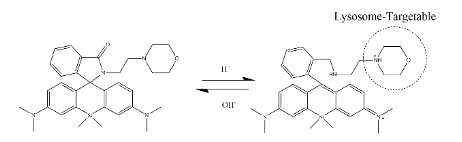
- Lee, D.; Swamy, K.M.K.; Hong, J.; Lee, S.; Yoon, J. A rhodamine-based fluorescent probe for the detection of lysosomal pH changes in living cells. Sens. Actuators B Chem. 2018, 266, 416–421. [Google Scholar] [CrossRef]
- Urano, Y.; Asanuma, D.; Hama, Y.; Koyama, Y.; Barrett, T.; Kamiya, M.; Nagano, T.; Watanabe, T.; Hasegawa, A.; Choyke, P.L. Selective molecular imaging of viable cancer cells with pH-activatable fluorescence probes. Nat. Med. 2009, 15, 104. [Google Scholar] [CrossRef] [Green Version]
- Koide, Y.; Kojima, R.; Hanaoka, K.; Numasawa, K.; Komatsu, T.; Nagano, T.; Kobayashi, H.; Urano, Y. Design strategy for germanium-rhodamine based pH-activatable near-infrared fluorescence probes suitable for biological applications. Commun. Chem. 2019, 2, 94. [Google Scholar] [CrossRef] [Green Version]
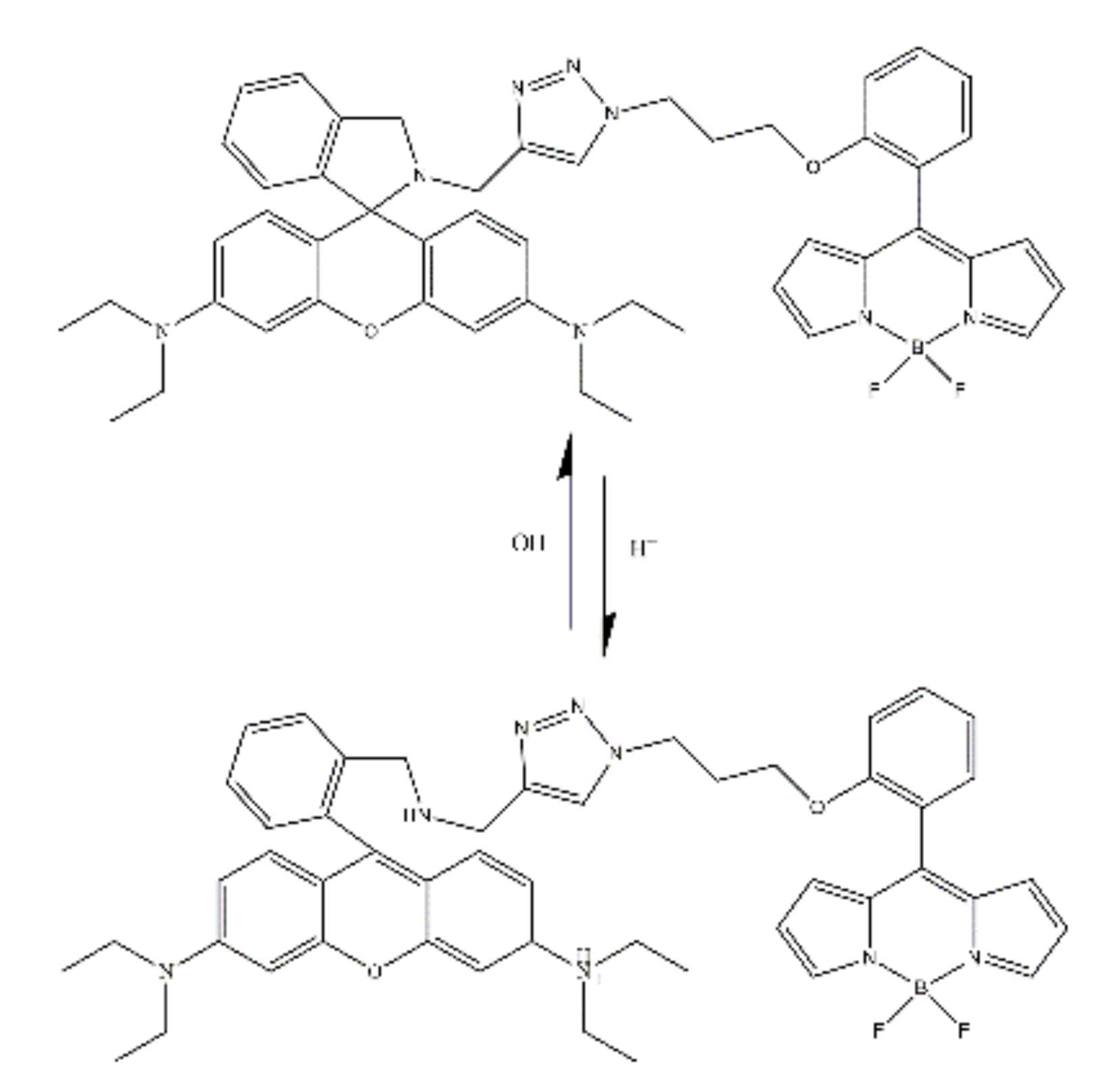
- Wang, J.; Xia, S.; Bi, J.; Zhang, Y.; Fang, M.; Luck, R.L.; Zeng, Y.; Chen, T.-H.; Lee, H.-M.; Liu, H. Near-infrared fluorescent probes based on TBET and FRET rhodamine acceptors with different pKa values for sensitive ratiometric visualization of pH changes in live cells. J. Mater. Chem. B 2019, 7, 198–209. [Google Scholar] [CrossRef]
- Yan, Y.; Zhang, X.; Zhang, X.; Li, N.; Man, H.; Chen, L.; Xiao, Y. Ratiometric sensing lysosomal pH in inflammatory macrophages by a BODIPY-rhodamine dyad with restrained FRET. Chin. Chem. Lett. 2019, 31, 1091–1094. [Google Scholar] [CrossRef]
- Liu, Z.; Wang, L.; Zhu, W.; Ding, Y.; Liu, S.; Wang, Q.; Chen, Y. A versatile color and fluorescence pH sensor based on AIE and open-loop synergy effect: Crystal structure and its application in cell imaging. Dye. Pigment. 2021, 190, 109310. [Google Scholar] [CrossRef]
- Srivastava, P.; Fürstenwerth, P.C.; Witte, J.F.; Resch-Genger, U. Synthesis and spectroscopic characterization of a fluorescent phenanthrene-rhodamine dyad for ratiometric measurements of acid pH values. New J. Chem. 2021, 45, 13755–13762. [Google Scholar] [CrossRef]
- Kimura, H. Hydrogen sulfide: Its production, release and functions. Amino Acids 2011, 41, 113–121. [Google Scholar] [CrossRef] [PubMed]
- Blackstone, E.; Morrison, M.; Roth, M.B. H2S induces a suspended animation–like state in mice. Science 2005, 308, 518. [Google Scholar] [CrossRef] [PubMed] [Green Version]
- Xu, F.; Gao, X.; Li, H.; Xu, S.; Li, X.; Hu, X.; Li, Z.; Xu, J.; Hua, H.; Li, D. Hydrogen sulfide releasing enmein-type diterpenoid derivatives as apoptosis inducers through mitochondria-related pathways. Bioorganic Chem. 2019, 82, 192–203. [Google Scholar] [CrossRef]
- Hu, X.; Jiao, R.; Li, H.; Wang, X.; Lyu, H.; Gao, X.; Xu, F.; Li, Z.; Hua, H.; Li, D. Antiproliferative hydrogen sulfide releasing evodiamine derivatives and their apoptosis inducing properties. Eur. J. Med. Chem. 2018, 151, 376–388. [Google Scholar] [CrossRef]
- Gadalla, M.M.; Snyder, S.H. Hydrogen sulfide as a gasotransmitter. J. Neurochem. 2010, 113, 14–26. [Google Scholar] [CrossRef] [Green Version]
- de Mel, A.; Murad, F.; Seifalian, A.M. Nitric oxide: A guardian for vascular grafts? Chem. Rev. 2011, 111, 5742–5767. [Google Scholar] [CrossRef]
- Pacher, P.; Beckman, J.S.; Liaudet, L. Nitric oxide and peroxynitrite in health and disease. Physiol. Rev. 2007, 87, 315–424. [Google Scholar] [CrossRef] [Green Version]
- Wang, Q.; Jiao, X.; Liu, C.; He, S.; Zhao, L.; Zeng, X. A rhodamine-based fast and selective fluorescent probe for monitoring exogenous and endogenous nitric oxide in live cells. J. Mater. Chem. B 2018, 6, 4096–4103. [Google Scholar] [CrossRef]
- Alam, R.; Islam, A.S.M.; Sasmal, M.; Katarkar, A.; Ali, M. A rhodamine-based turn-on nitric oxide sensor in aqueous medium with endogenous cell imaging: An unusual formation of nitrosohydroxylamine. Org. Biomol. Chem. 2018, 16, 3910–3920. [Google Scholar] [CrossRef]
- Podder, A.; Koo, S.; Lee, J.; Mun, S.; Khatun, S.; Kang, H.-G.; Bhuniya, S.; Kim, J.S. A rhodamine based fluorescent probe validates substrate and cellular hypoxia specific NADH expression. Chem. Commun. 2019, 55, 537–540. [Google Scholar] [CrossRef] [PubMed]
- Gu, T.; Mo, S.; Mu, Y.; Huang, X.; Hu, L. Detection of endogenous hydrogen peroxide in living cells with para-nitrophenyl oxoacetyl rhodamine as turn-on mitochondria-targeted fluorescent probe. Sens. Actuators B Chem. 2020, 309, 127731. [Google Scholar] [CrossRef]
- Liu, Y.; Lee, D.; Wu, D.; Swamy, K.M.K.; Yoon, J. A new kind of rhodamine-based fluorescence turn-on probe for monitoring ATP in mitochondria. Sens. Actuators B Chem. 2018, 265, 429–434. [Google Scholar] [CrossRef]
- Tikum, A.F.; Kim, G.; Nasirian, A.; Ko, J.W.; Yoon, J.; Kim, J. Rhodamine-based near-infrared probe for emission detection of ATP in lysosomes in living cells. Sens. Actuators B Chem. 2019, 292, 40–47. [Google Scholar] [CrossRef]
- Ran, F.; Ma, C.; Xiang, Y.; Xu, Y.; Liu, X.; Zhang, H. A fluorescent and colorimetric dual-channel sensor based on acid phosphatase-triggered blocking of internal filtration effect. Mikrochim. Acta 2021, 188, 282. [Google Scholar] [CrossRef]
- Tang, J.; Li, Q.; Guo, Z.; Zhu, W. A fast-response and highly specific Si-Rhodamine probe for endogenous peroxynitrite detection in living cells. Org. Biomol. Chem. 2019, 17, 1875–1880. [Google Scholar] [CrossRef]
- Wu, J.; Ye, Z.; Wu, F.; Wang, H.; Zeng, L.; Bao, G.-M. A rhodamine-based fluorescent probe for colorimetric and fluorescence lighting-up determination of toxic thiophenols in environmental water and living cells. Talanta 2018, 181, 239–247. [Google Scholar] [CrossRef]
- Wang, Z.; Zhang, Q.; Liu, J.; Sui, R.; Li, Y.; Li, Y.; Zhang, X.; Yu, H.; Jing, K.; Zhang, M.; et al. A twist six-membered rhodamine-based fluorescent probe for hypochlorite detection in water and lysosomes of living cells. Anal. Chim. Acta 2019, 1082, 116–125. [Google Scholar] [CrossRef]
- Zhang, D.; Ma, Z.; Wang, Y.; Yin, H.; Li, M.; Wang, Y.; Wang, H.; Jia, B.; Liu, J. Dual-binding benzene and rhodamine B conjugate derivatives as fluorescent chemodosimeter for hypochlorite in living cell imaging. Spectrochim. Acta Part A Mol. Biomol. Spectrosc. 2020, 229, 117908. [Google Scholar] [CrossRef]
- Wang, K.; Wang, W.; Chen, S.-Y.; Guo, J.-C.; Li, J.-H.; Yang, Y.-S.; Wang, X.-M.; Xu, C.; Zhu, H.-L. A novel Near-Infrared rhodamine-derivated turn-on fluorescence probe for sensing SO32− detection and their bio-imaging in vitro and in vivo. Dye. Pigment. 2021, 188, 109229. [Google Scholar] [CrossRef]
- Shang, H.; Chen, H.; Tang, Y.; Ma, Y.; Lin, W. Development of a two-photon fluorescent turn-on probe with far-red emission for thiophenols and its bioimaging application in living tissues. Biosens. Bioelectron. 2017, 95, 81–86. [Google Scholar] [CrossRef] [PubMed]
- Khandare, D.G.; Banerjee, M.; Gupta, R.; Kumar, N.; Ganguly, A.; Singh, D.; Chatterjee, A. Green synthesis of a benzothiazole based ‘turn-on’type fluorimetric probe and its use for the selective detection of thiophenols in environmental samples and living cells. RSC Adv. 2016, 6, 52790–52797. [Google Scholar] [CrossRef]
- Stanford, B.D.; Pisarenko, A.N.; Snyder, S.A.; Gordon, G. Perchlorate, bromate, and chlorate in hypochlorite solutions: Guidelines for utilities. J. -Am. Water Work. Assoc. 2011, 103, 71–83. [Google Scholar] [CrossRef]
- Bruch, M.K. Toxicity and safety of topical sodium hypochlorite. In Disinfection by Sodium Hypochlorite: Dialysis Applications; Karger Publishers: Berlin, Germany, 2007; Volume 154, pp. 24–38. [Google Scholar]
- Chang, L.L.; Gao, Q.; Liu, S.; Hu, C.C.; Zhou, W.J.; Zheng, M.M. Selective and differential detection of Hg2+ and Cu2+ with use of a single rhodamine hydrazone-type probe in the absence and presence of UV irradiation. Dye. Pigment. 2018, 153, 117–124. [Google Scholar] [CrossRef]
- Wang, K.; Kong, Q.; Chen, X.; Yoon, J.; Swamy, K.M.K.; Wang, F. A bifunctional rhodamine derivative as chemosensor for recognizing Cu2+ and Hg2+ ions via different spectra. Chin. Chem. Lett. 2019, 31, 1087–1090. [Google Scholar] [CrossRef]
- Ozmen, P.; Demir, Z.; Karagoz, B. An easy way to prepare reusable rhodamine-based chemosensor for selective detection of Cu2+ and Hg2+ ions. Eur. Polym. J. 2022, 162, 110922. [Google Scholar] [CrossRef]
- Mondal, S.; Bandyopadhyay, C.; Ghosh, K. Chromenone-rhodamine conjugate for naked eye detection of Al3+ and Hg2+ ions in semi aqueous medium. Supramol. Chem. 2018, 31, 1–8. [Google Scholar] [CrossRef]
- Erdemir, S. Fluorometric dual sensing of Hg2+ and Al3+ by novel triphenylamine appended rhodamine derivative in aqueous media. Sens. Actuators B Chem. 2019, 290, 558–564. [Google Scholar] [CrossRef]
- Hazra, A.; Ghosh, P.; Roy, P. A rhodamine based dual chemosensor for Al(3+) and Hg(2+): Application in the construction of advanced logic gates. Spectrochim. Acta A Mol. Biomol. Spectrosc. 2022, 271, 120905. [Google Scholar] [CrossRef]
- Chan, W.C.; Saad, H.M.; Sim, K.S.; Lee, V.S.; Tan, K.W. A rapid turn-on dual functional rhodamine B probe for aluminum (III) and copper (II) that can be utilised as a molecular logic gate and in water analysis. J. Mol. Struct. 2022, 1254, 132337. [Google Scholar] [CrossRef]
- Zhang, Z.; Deng, C.; Song, H. A novel rhodamine-based turn-on fluorescent probe for dual detection of Cr3+ and Cu2+ with solvent-dependent binding properties. Inorg. Chem. Commun. 2018, 95, 56–60. [Google Scholar] [CrossRef]
- Mehta, R.; Kaur, P.; Choudhury, D.; Paul, K.; Luxami, V. Al3+ induced hydrolysis of rhodamine-based Schiff-base: Applications in cell imaging and ensemble as CN-sensor in 100% aqueous medium. J. Photochem. Photobiol. A Chem. 2019, 380, 111851. [Google Scholar] [CrossRef]
- Zhu, L.; Liao, W.; Chang, H.; Liu, X.; Miao, S. A Novel Fluorescent Probe for Detection of Hydrogen Sulfide and Its Bioimaging Applications in Living Cells. ChemistrySelect 2020, 5, 829–833. [Google Scholar] [CrossRef]
- Das, D.; Alam, R.; Katarkar, A.; Ali, M. A differentially selective probe for trivalent chemosensor upon single excitation with cell imaging application: Potential applications in combinatorial logic circuit and memory devices. Photochem. Photobiol. Sci. 2019, 18, 242–252. [Google Scholar] [CrossRef] [PubMed]
- Singha, D.; Das, T.; Satyanarayana, L.; Roy, P.; Nandi, M. Rhodamine functionalized mesoporous silica as a chemosensor for the efficient sensing of Al3+, Cr3+ and Fe3+ ions and their removal from aqueous media. New J. Chem. 2019, 43, 15563–15574. [Google Scholar] [CrossRef]
- Roy, A.; Das, S.; Sacher, S.; Mandal, S.K.; Roy, P. A rhodamine based biocompatible chemosensor for Al3+, Cr3+ and Fe3+ ions: Extraordinary fluorescence enhancement and a precursor for future chemosensors. Dalton Trans. 2019, 48, 17594–17604. [Google Scholar] [CrossRef]
- Banerjee, M.; Ghosh, M.; Ta, S.; Das, J.; Das, D. A smart optical probe for detection and discrimination of Zn2+, Cd2+ and Hg2+ at nano-molar level in real samples. J. Photochem. Photobiol. A Chem. 2019, 377, 286–297. [Google Scholar] [CrossRef]
- Sadak, A.E.; Karakuş, E. Triazatruxene–Rhodamine-Based Ratiometric Fluorescent Chemosensor for the Sensitive, Rapid Detection of Trivalent Metal Ions: Aluminium (III), Iron (III) and Chromium (III). J. Fluoresc. 2020, 30, 213–220. [Google Scholar] [CrossRef]
- Wang, Y.; Wu, H.; Wu, W.N.; Mao, X.J.; Zhao, X.L.; Xu, Z.Q.; Xu, Z.H.; Fan, Y.C. Novel rhodamine-based colorimetric and fluorescent sensor for the dual-channel detection of Cu(2+) and Co(2+)/trivalent metal ions and its AIRE activities. Spectrochim. Acta A Mol. Biomol. Spectrosc. 2019, 212, 1–9. [Google Scholar] [CrossRef]
- Pungut, N.A.S.; Heng, M.P.; Saad, H.M.; Sim, K.S.; Lee, V.S.; Tan, K.W. From one to three, modifications of sensing behavior with solvent system: DFT calculations and real-life application in detection of multianalytes (Cu2+, Ni2+ and Co2+) based on a colorimetric Schiff base probe. J. Mol. Struct. 2021, 1238, 130453. [Google Scholar] [CrossRef]
- Hazra, A.; Roy, P. A rhodamine based dye for sensing of Group 13 metal ions. Anal. Chim. Acta 2022, 1193, 339378. [Google Scholar] [CrossRef] [PubMed]
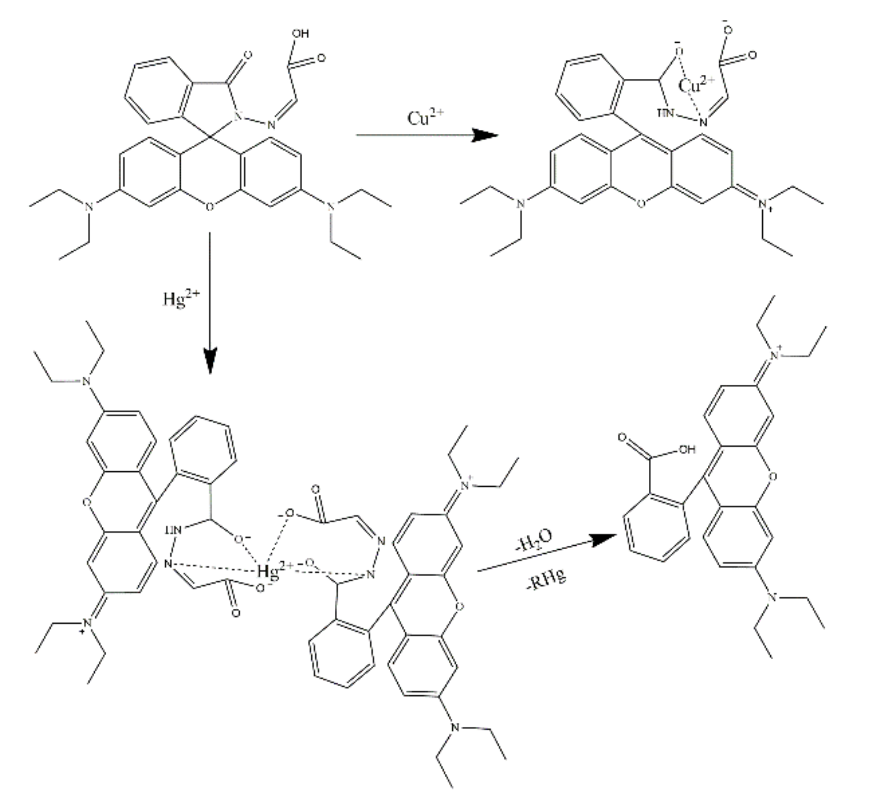
- Podshibyakin, V.A.; Shepelenko, E.N.; Karlutova, O.Y.; Dubonosova, I.V.; Borodkin, G.S.; Popova, O.S.; Zaichenko, S.B.; Dubonosov, A.D.; Bren, V.A.; Minkin, V.I. Solvent-dependent selective “naked eye” chromofluorogenic multifunctional rhodamine-based probe for Al3+, Cu2+, Hg2+, S2− and CN− ions. Tetrahedron 2022, 110, 132710. [Google Scholar] [CrossRef]
- Kilic, H.; Bozkurt, E. A rhodamine-based novel turn on trivalent ions sensor. J. Photochem. Photobiol. A Chem. 2018, 363, 23–30. [Google Scholar] [CrossRef]


| No. | Molecular Structure | Target | Sensing Mechanism | Sensor Type | Detection Limit(M) | Bonding Ratio * | Solution Systems | Application | Ref. |
|---|---|---|---|---|---|---|---|---|---|
| 1 |  | Hg2+ | / | Colorimetric and fluorescent probe | 7.48 × 10⁻⁹ | 1:2 | 100% H2O | Test strip | [33] |
| 2 |  | Hg2+ | / | Colorimetric sensor | / | 1:1 | CH3CN/H2O (1/99, v/v) HEPES buffer (pH = 7.05) | Actual water samples, living cells | [34] |
| 3 |  | Hg2+ | / | Fluorescent and colorimetric sensor | 8.73 × 10⁻⁷ | / | DMF | Living cells | [35] |
| 4 |  | Hg2+ | / | Fluorescent and colorimetric sensor | 1.36 × 10⁻⁷ | 1:1 | CH3CN/H2O (3/7, v/v) | Actual water samples and test paper | [36] |
| 5 |  | Hg2+ | / | Fluorescent and colorimetric sensor | 0.11 × 10⁻⁹ | 1:1 | EtOH/H2O (2/1, v/v) | Actual water samples, MCF-7 cells, zebrafish and soybean rhizome tissues | [37] |
| 6 |  | Hg2+ | / | Fluorescent sensor | / | 1:1 | / | Living cells | [38] |
| 7 |  | Hg2+ | / | Fluorescent and colorimetric sensor | 1.8 × 10⁻⁸ 1.6 × 10⁻⁸ 5.6 × 10⁻⁸ | 1:1 | Tris-HCl/ C2H5OH (3/7, v/v, pH = 7.2) | Living cells | [39] |
| 8 |  | Hg2+ | Fluorescent sensor | / | 1:1 | C2H5OH/ PBS buffer (2/8, v/v, pH = 7.4) | Living cells | [40] | |
| 9 |  | Hg2+ | / | Fluoric-metric sensor | 2.57 × 10⁻⁶ | 1:1 | EtOH/HEPES buffer (1:1, v/v) | Test paler and living cells | [41] |
| 10 |  | Hg2+ | FRET | Fluorescent and colorimetric sensor | 2.46 × 10⁻⁷ | 1:1 | / | Water sample | [42] |
| 11 |  | Hg2+ | / | Fluorescent sensor | 0.27 μg/L | 1:1 | / | Water sample | [43] |
| 12 |  | Hg2+ | / | Fluorescent and colorimetric sensor | 3.4 × 10⁻⁹ | 1:1 | HEPES/DMF (1/1, v/v) | Living cells | [44] |
| 13 |  | Hg2+ | / | Fluorescent and colorimetric sensor | 1.2 × 10⁻⁸ | 1:1 | CH3CN/H2O (9:1v/v) | Test strip | [45] |
| 14 |  | Hg2+ | / | Fluorescent and colorimetric sensor | 9.18 × 10⁻⁷ | 1:1 | / | Tap water, colorimetric card | [46] |
| 15 |  | Hg2+ | / | Fluorescent sensor | / | 2:1 | H2O/EtOH (3/2, v/v, pH = 7.3); | Living cells | [47] |
| 16 |  | Hg2+ | FRET | Colorimetric and ratiometric fluorescent sensor | 0.81 × 10⁻⁶ | 1:1 | CH3CN/H2O (1/1, v/v) | Living cells | [48] |
| 17 |  | Cu2+ | FRET | Colorimetric and ratiometric probe | 1.168 × 10⁻⁸ | 1:1 | CH3CN/HEPES buffer (4/1, v/v) (pH = 7.3) | Actual water samples | [24] |
| 18 |  | Cu2+ | / | Fluorescent sensor | 4.38 × 10⁻⁸ | 1:1 | H2O | White wine samples | [49] |
| 19 |  | Cu2+ | PET | Fluorescent and colorimetric sensor | 7.4 × 10⁻⁸ | 1:1 | H2O/DMSO (9/1, v/v) | Test paper | [50] |
| 20 |  | Cu2+ | / | Fluorescent and colorimetric sensor | 0.69 × 10⁻⁸ | 1:2 | Acetone/H2O (1/2, v/v) | Living cells | [51] |
| 21 |  | Cu2+ | / | Fluorescent and colorimetric sensor | / | 1:1 | HEPES /CH3CN (4/1, v/v) (pH = 7.0) | Living cells | [52] |
| 22 |  | Cu2+ | / | Fluorescent and colorimetric sensor | 8.99 × 10⁻⁸ | 1:2 | CH3CN/H2O (1/2, v/v) (pH = 7.2, 50 mM tris–HCl) | HepG2 living cells | [53] |
| 23 |  | Cu2+ | / | Fluorescent and colorimetric sensor | 3.4 × 10⁻⁸ | 1:2 | CH3OH/H2O (4:1, v/v) | Gel balls | [54] |
| 24 |  | Cu2+ | ICT | Fluorescent sensor | 6 × 10⁻⁶ | 2:1 | PBS/EtOH (1/1, v/v) | / | [27] |
| 25 |  | Fe3+ | / | Colorimetric sensor | 2.54 × 10⁻⁸ | 1:1 | EtOH/H2O (5/5, v/v) Tris–HCl (pH = 7.4) | Living cells | [32] |
| 26 |  | Fe3+ | / | Fluorescent and colorimetric sensor | 3.76 × 10⁻⁹ | 1:1 | MeCN/H2O (1/1, v/v) (pH = 7.2) | Living cells | [55] |
| 27 |  | Fe3+ | / | Fluorescent and colorimetric sensor | 2.7 × 10⁻⁷ | 2:1 | EtOH/H2O (2/3, v/v) (pH = 7.2) | Living cells | [56] |
| 28 |  | Fe3+ | / | Fluorescent and colorimetric sensor | 7.4 × 10⁻⁶ | 2:1 | CH3CN/ Tris–HCl buffer (9/1, v/v; pH = 7.00) | Actual water samples | [57] |
| 29 |  | Fe3+ | / | Fluorescent sensor | 5.2 × 10⁻⁶ | 1:1 | Acetonitrile/ Tris-HCl buffer (3/7, v/v; pH = 7.4) | Living cells | [58] |
| 30 |  | Fe3+ | / | Fluorescent sensor | 1.64 × 10⁻⁶ | 1:1 | Pure water | Water | [59] |
| 31 |  | Fe3+ | / | Colorimetric sensor | 0.88 × 10⁻⁷ | / | Polyacrylamide | Water | [60] |
| 32 |  | Fe3+ | / | Fluorescent and colorimetric sensor | / | 1:1 | / | / | [61] |
| 33 |  | Fe3+ | / | Fluorescent sensor | 3.47 × 10⁻⁹ | / | HEPES/ CH3CN (2:3, v/v) (10 mM, pH = 7.4) | Caenorhabditis elegans, adult mice, plant tissue | [62] |
| 34 |  | Fe3+ | / | Fluorescent and colorimetric sensor | 0.19 × 10⁻⁹ | / | Rhodamine-grafted paper | Environmental water | [63] |
| 35 |  | Fe3+ | FRET | Fluorescent sensor | 3.90 × 10⁻⁸ | / | Dioxane/H2O (4:6, v/v) | Paper test strips, water samples | [29] |
| 36 |  | Fe3+ | ICT | Fluorescent and colorimetric sensor | 3.16 × 10⁻⁶ | 1:2 | H2O (pH = 6.5) | Living cells, fetal bovine serum samples | [28] |
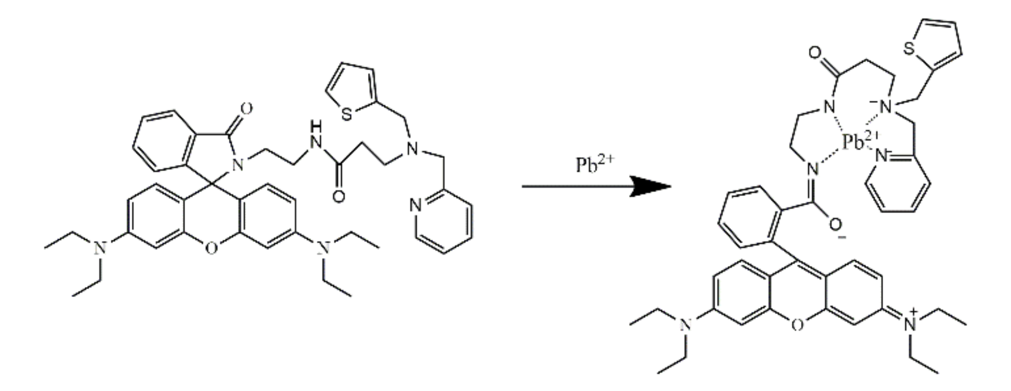
| 37 |  | Pb2+ | PET | Colorimetric sensor | 4.2 × 10⁻⁹ | 1:1 | EtOH/H2O (3/2, v/v) HEPES Buffer (pH = 7.4) | Living cells, test paper | [25] |
| 38 |  | Pb2+ | PET | Fluorescent and colorimetric sensor | 0.17 × 10⁻⁹ | 1:1 | CH3OH/PBS buffer (1/1, v/v) pH = 7.40) | Living cells | [26] |
| 39 |  | Pb2+ | / | Fluorescent and colorimetric sensor | 2.5 × 10⁻⁶ | 1:1 | / | Tap water, soil, fish, shrimp | [64] |
| 40 |  | Zn2+ | FRET | Fluorescent sensor | 2.89 × 10⁻⁸ | 1:1 | H2O /Acetonitrile (8/2, v/v) | Living cells | [65] |
| 41 |  | Zn2+ | ICT | Fluorescent sensor | 1.8–1.9 × 10⁻⁹ | / | 50 mM, HEPES buffer, pH = 7.54 | Living cells, mitochondria | [66] |
| 42 |  | As3+ | / | Fluorescent and colorimetric sensor | 0.164 μg/L | / | Acetonitrile/ HEPES buffer (4/1, v/v, pH = 7.4) | Living cells | [67] |
| 43 |  | Al3+ | / | Fluorescent sensor | 0.146 × 10⁻⁶ | 1:1 | / | Living cells, plant cells and living bodies | [68] |
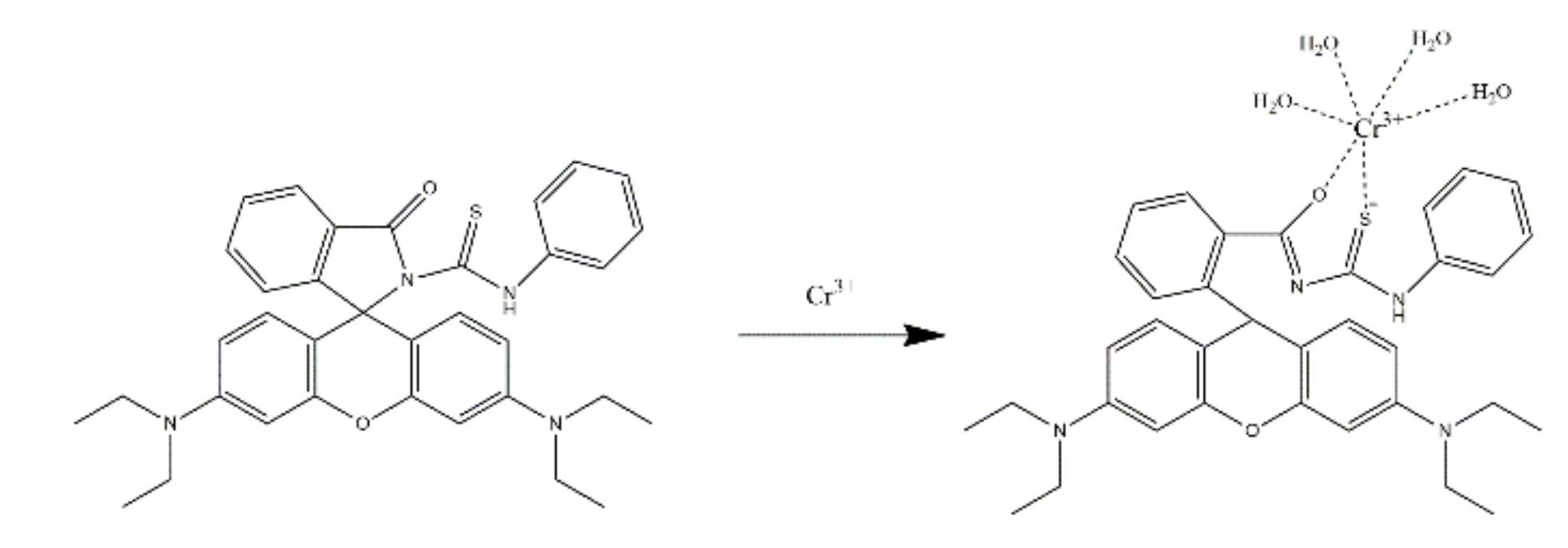
| 44 |  | Cr3+ | / | Fluorescent sensor | / | 1:1 | / | Living cells | [69] |
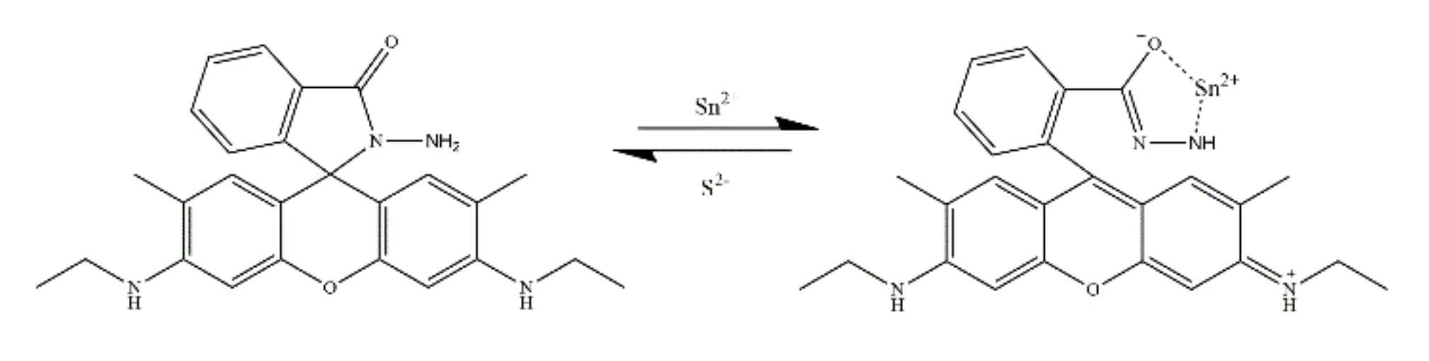
| 45 |  | Sn2+ | ICT | Fluorescent and colorimetric sensor | 1.62 × 10⁻⁷ | 1:1 | EtOH/H2O (8/2, v/v) | Living cells | [70] |
| No. | Molecular Structure | Target | Probe Type | Sensing Mechanism | Sensor Type | Solution Systems | Application | Ref. |
|---|---|---|---|---|---|---|---|---|
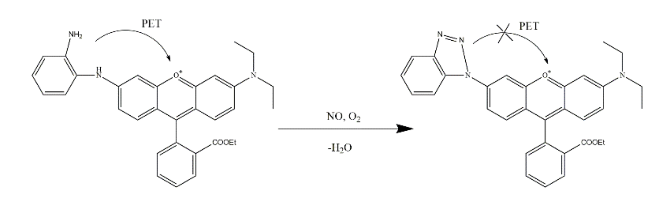
| 1 |  | NO | Turn-on | PET blocked | Fluorescent probe | PBS buffers (pH = 7.4) | Living cells | [90] |
| 2 |  | NO | Turn-on | / | Fluorescent probe | HEPES buffers (pH = 7.4) | Living cells and zebrafish | [91] |
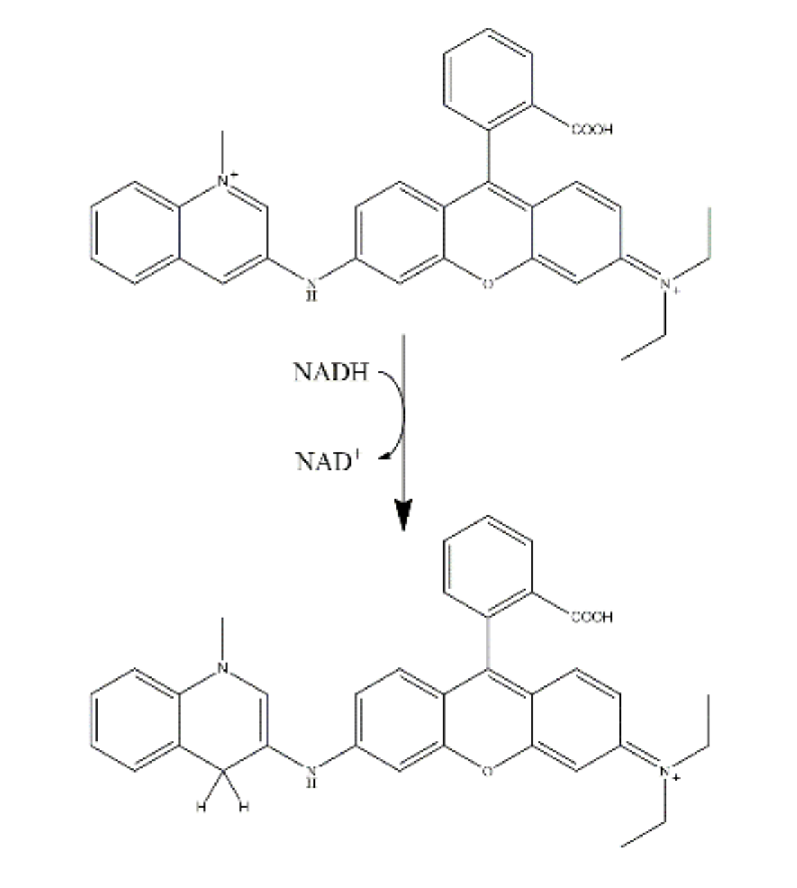
| 3 |  | NADH | Turn-on | / | Fluorescent probe | PBS buffers (pH = 7.4) | Living cells | [92] |
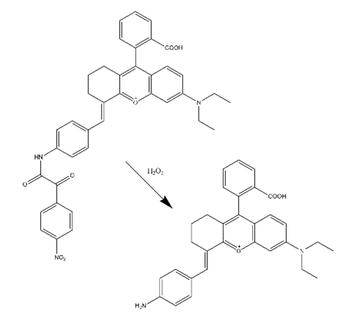
| 4 |  | H2O2 | Turn-on | PET blocked | Fluorescent sensor | PBS with 0.1% DMSO (pH = 7.4,) | Living cells | [93] |
| 5 |  | ATP | Turn-on | / | Fluorescent and colorimetric sensor | PBS solution (containing 40% DMSO) | Living cells | [94] |
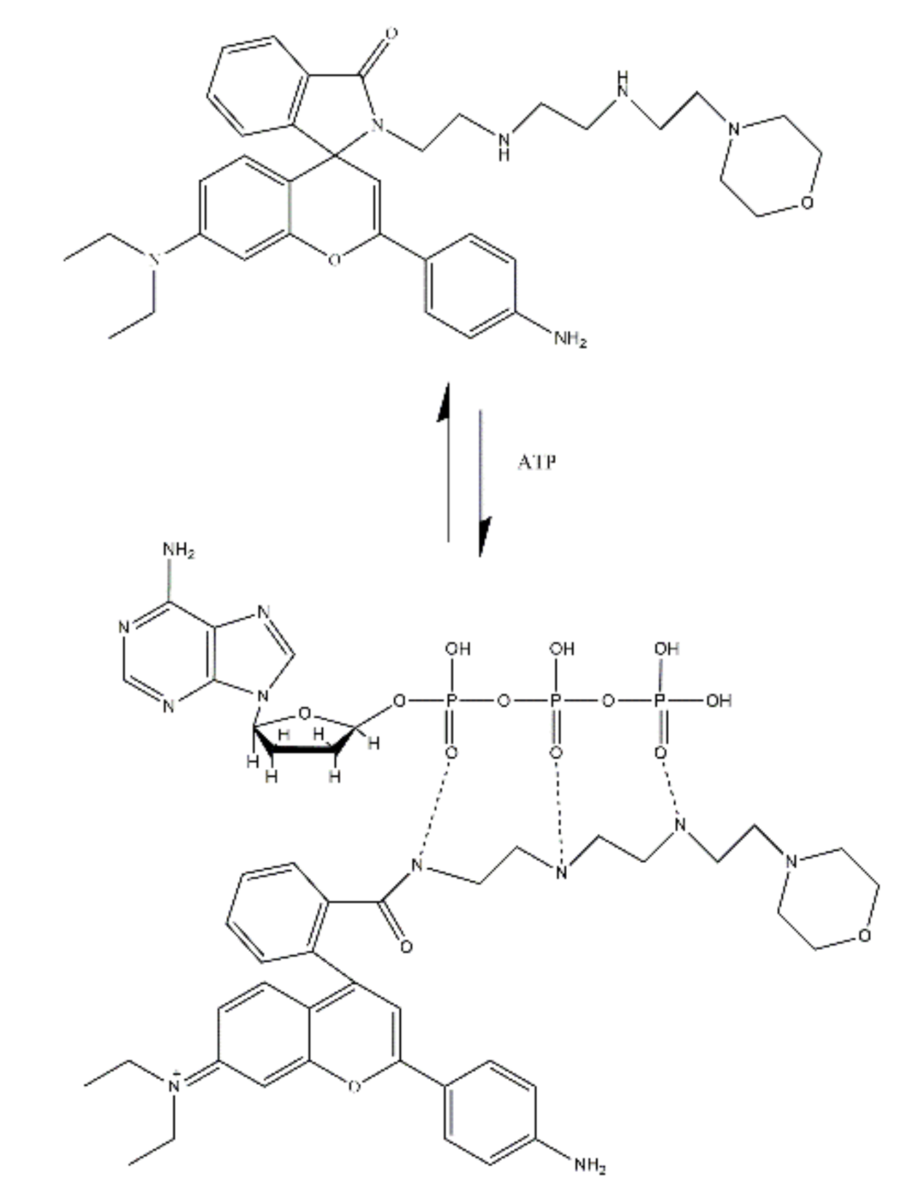
| 6 |  | ATP | Turn-on | / | Fluorescent and colorimetric sensor | EtOH/ PBS buffer (1/99, v/v; pH = 7.4) | Living lysosomes | [95] |
| 7 |  | ACP | Turn-on | IFE | Fluorescent and colorimetric sensor | / | Human serum | [96] |
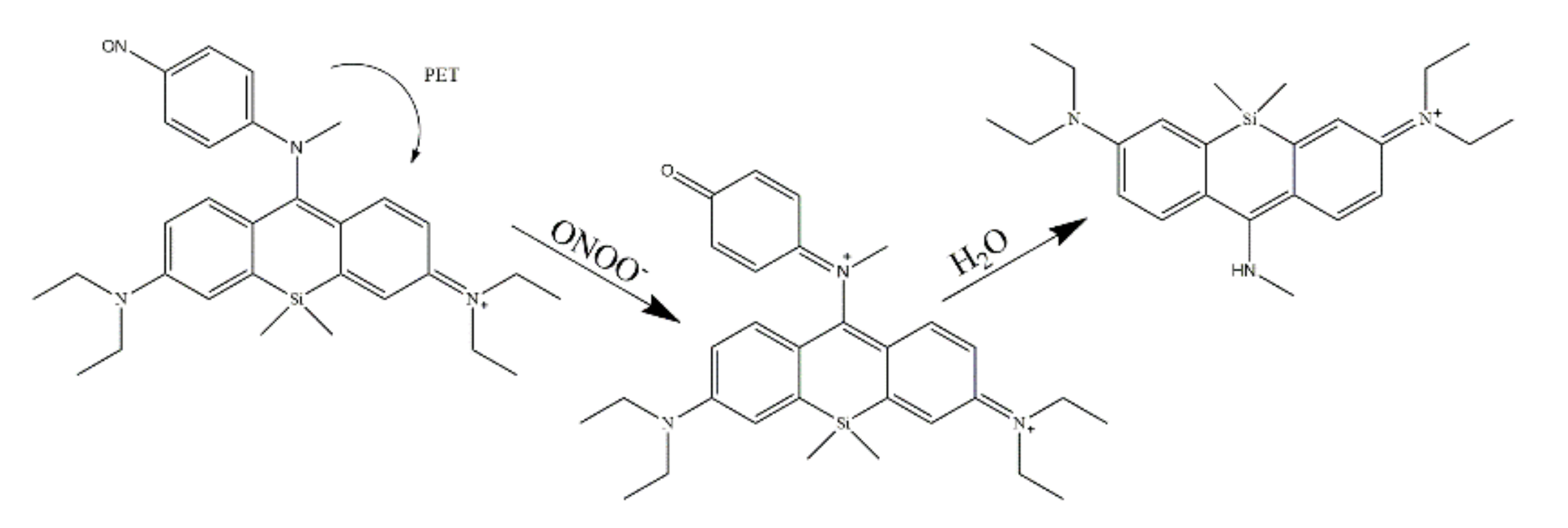
| 8 |  | Peroxynitrite (ONOO−) | Turn-on | PET blocked | Fluorescent probe | H2O/CH3CN (7:3, v/v) HEPES buffers (pH = 7.4) | Living cells | [97] |
| 9 |  | Thiophenols (-SH) | Turn-on | PET blocked | Fluorescent probe | DMSO/HEPES solution (1:1, v/v) (pH = 7.4) | Water and living cells | [98] |
| 10 |  | ClO− | Turn-on | / | Fluorescent and colorimetric sensor | C2H5OH/H2O (1:1, v/v) | Living lysosomes and real water samples | [99] |
| 11 |  | ClO− | Turn-on | / | Fluorescent and colorimetric sensor | CH3CH2OH-Tris (7/3, v/v, pH = 7.4) | Living cells | [100] |
| 12 |  | SO32− | Turn-on | ICT | Fluorescent probe | PBS buffer (10 mM, pH 7.4, 1% DMSO) | Living cells and zebrafish | [101] |
| No. | Molecular Structure | Target | Sensing Mechanism | Sensor Type | LOD | Solution Systems | Application | Ref. |
|---|---|---|---|---|---|---|---|---|
| 1 |  | Hg2+ Cu2+ | / | Colorimetric and fluorescent probe | 7.1 × 10⁻⁸ 1.5 × 10⁻⁷ | IPA/H2O (8/1, v/v) (Iso-Propyl Alcohol) | / | [106] |
| 2 |  | Cu2+ Hg2+ | / | Fluorescent and colorimetric sensor | / | CH3CN/ HEPES buffer (1/9, v/v; pH = 7.4) | / | [107] |
| 3 |  | Cu2+ Hg2+ | / | Fluorescent and colorimetric sensor | 5.06 × 10⁻⁷ 3.90 × 10⁻⁷ | CH3CN/H2O (8/2, v/v) (pH = 7.2) | / | [108] |
| 4 |  | Hg2+ Al3+ | FRET | Fluorescent and colorimetric sensor | 1.26 × 10⁻⁷ 1.9 × 10⁻⁷ | MeCN/H2O (3/1, v/v) HEPES buffer (pH = 6.85) | / | [109] |
| 5 |  | Hg2+ Al3+ | FRET PET | Fluorescent probe | 0.48 × 10⁻⁶ 7.18 × 10⁻⁸ | MeCN/H2O (9/1, v/v) | Test paper | [110] |
| 6 |  | Hg2+ Al3+ | / | Fluorescent sensor | 1.60 × 10⁻⁵ 6.54 × 10⁻⁶ | H2O/EtOH (1:9, v/v) (pH = 7.4) | River water, paper test | [111] |
| 7 |  | Cu2+ Al3+ | / | Fluorescent and colorimetric sensor | 9.90 × 10⁻⁹ 5.63 × 10⁻⁹ | MeCN/H2O (6:4, v/v) | Living cells, natural mineral water, tap water | [112] |
| 8 |  | Cr3+ Cu2+ | / | Colorimetric sensor | 4.7 × 10⁻⁸ 7.6 × 10⁻⁸ | CH3CN solution Ethanol solution | / | [113] |
| 9 |  | Al3+ CN− | PET blocked | Fluorescent and colorimetric sensor | 1.68 × 10⁻⁹ 0.82 × 10⁻⁹ | HEPES buffer (pH = 7.04) | Living cells | [114] |
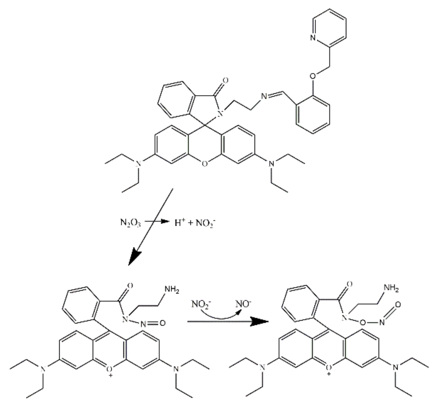
| 10 |  | H+ H2S | ICT (Intramolecular charge transfer) | Fluorescent probe | / | Living cells | [115] | |
| 11 |  | Cr3+ Al3+ Fe3+ | / | Fluorescent and colorimetric sensor | 3.16 × 10⁻⁹ 1.17 × 10⁻⁹ 2.50 × 10⁻⁹ | H2O/EtOH (1/1, v/v) | / | [116] |
| 12 |  | Cr3+ Al3+ Fe3+ | / | Colorimetric sensor | 2.28 × 10⁻⁶ 1.28 × 10⁻⁶ 1.34 × 10⁻⁶ | H2O/CH3CN (4/1, v/v, pH = 7.2) | Living cells | [117] |
| 13 |  | Cr3+ Al3+ Fe3+ | / | Fluorescent and colorimetric sensor | 9.18 × 10⁻⁹ 5.40 × 10⁻⁹ 7.23 × 10⁻⁷ | H2O/EtOH (14/1, v/v) | / | [118] |
| 14 |  | Cr3+ Al3+ Fe3+ | / | Fluorescent and colorimetric sensor | 1.58 × 10⁻⁸ 6.97 × 10⁻⁹ 1.4 × 10⁻⁸ | CH3OH/H2O (9/1, v/v) HEPES buffer (pH = 7.4) | Living cells | [119] |
| 15 |  | Cr3+ Al3+ Fe3+ | TBET | Ratiometric fluorescent and colorimetric sensor | 1.70 × 10⁻⁴ 2.3 × 10⁻⁵ 2.5 × 10⁻⁵ | MeOH | Test strips | [120] |
| 16 |  | Zn2+ Cd2+ Hg2+ | PET-CHEF PET-CHEF PET-FRET | Fluorescent and colorimetric sensor | 1.2 × 10⁻⁹ 9.6 × 10⁻⁹ 1.5 × 10⁻¹⁰ | MeOH/H2O, (4/1, v/v) HEPES buffer (pH = 7.4) | Sea fish and water samples | [121] |
| 17 |  | Cu2+ Co2+ Al3+ | / | Fluorescent sensor | 8.68 × 10⁻⁸ / 1.06 × 10⁻⁹ | CH3OH CH3OH/H2O H2O | Living cells | [122] |
| 18 |  | Cu2+ Ni2+ Co2+ | / | Colorimetric sensor | 4.0 × 10⁻⁸ 3.2 × 10⁻⁷ 4.8 × 10⁻⁷ | DMSO/TN (2:1, v/v) (pH = 7.5) | Test strips | [123] |
| 19 |  | Al3+ Ga3+ In3+ Tl3+ | / | Fluorescent and colorimetric sensor | 2.66 × 10⁻⁸ 1.04 × 10⁻⁷ 8.19 × 10⁻⁸ 3.10 × 10⁻⁸ | 10 mM HEPES buffer (pH = 7.4) in water/ethanol (1:9, v/v) | River water | [124] |
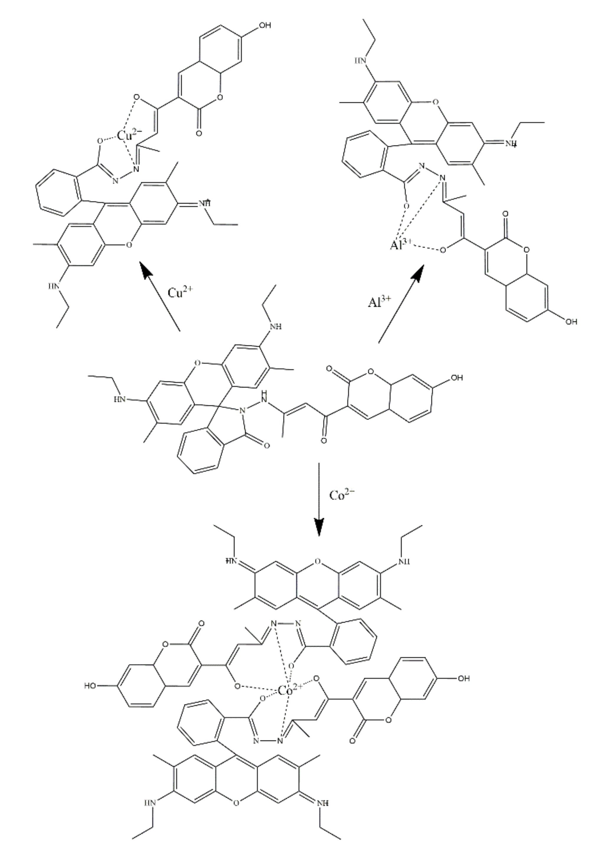
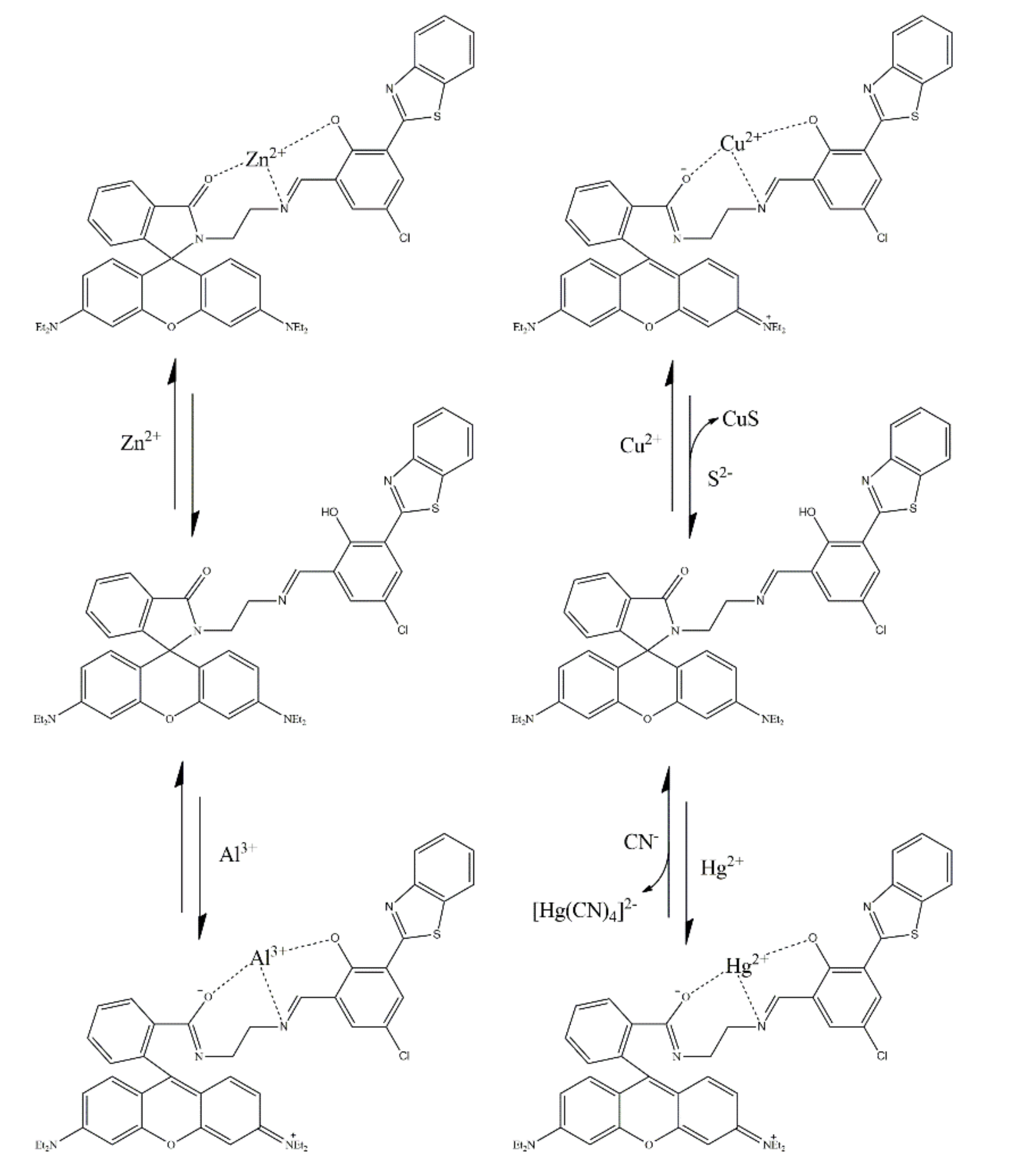
| 20 |  | Al3+ Cu2+ Hg2+ S2− CN− | ICT | Fluorescent and colorimetric sensor | 2.1 × 10⁻⁷ 3.2 × 10⁻⁷ 4.0 × 10⁻⁷ / / | CH3CN (5.0 × 10⁻⁵ mol/L) | Logic gate | [125] |
Publisher’s Note: MDPI stays neutral with regard to jurisdictional claims in published maps and institutional affiliations. |
© 2022 by the authors. Licensee MDPI, Basel, Switzerland. This article is an open access article distributed under the terms and conditions of the Creative Commons Attribution (CC BY) license (https://creativecommons.org/licenses/by/4.0/).
Share and Cite
Wang, Y.; Wang, X.; Ma, W.; Lu, R.; Zhou, W.; Gao, H. Recent Developments in Rhodamine-Based Chemosensors: A Review of the Years 2018–2022. Chemosensors 2022, 10, 399. https://doi.org/10.3390/chemosensors10100399
Wang Y, Wang X, Ma W, Lu R, Zhou W, Gao H. Recent Developments in Rhodamine-Based Chemosensors: A Review of the Years 2018–2022. Chemosensors. 2022; 10(10):399. https://doi.org/10.3390/chemosensors10100399
Chicago/Turabian StyleWang, Yujiao, Xiaojun Wang, Wenyu Ma, Runhua Lu, Wenfeng Zhou, and Haixiang Gao. 2022. "Recent Developments in Rhodamine-Based Chemosensors: A Review of the Years 2018–2022" Chemosensors 10, no. 10: 399. https://doi.org/10.3390/chemosensors10100399