

Pilot Study: The Effectiveness of Hyperbaric Oxygen Therapy in the Treatment of Periodontitis in Patients with Type 2 Diabetes
 , , and
Abstract
:1. Introduction
2. Materials and Methods
2.1. Sampling and Sample Size
2.2. Participant Recruitment
2.3. Study Design
2.4. Diagnosis and Assessment of Periodontal Disease
- presence of CAL on the proximal surfaces of ≥2 non-adjacent teeth;
- presence of CAL ≥ 3 mm and PD pocket > 3 mm on the vestibular or lingual surfaces of ≥2 teeth, and the observed CAL was not due to other phenomena, i.e., recessions of traumatic origin, misalignment of the last molar, vertical root fracture (VRF), and endodontic lesion.
- stage I—initial periodontitis;
- stage II—moderate periodontitis;
- stage III—severe periodontitis with possible additional tooth loss;
- stage IV—severe periodontitis with extensive tooth loss with the possibility of further tooth loss.
- grade A—slow rate of progression;
- grade B—medium rate of progression;
- grade C—fast rate of progression [10].
2.5. Data Analysis
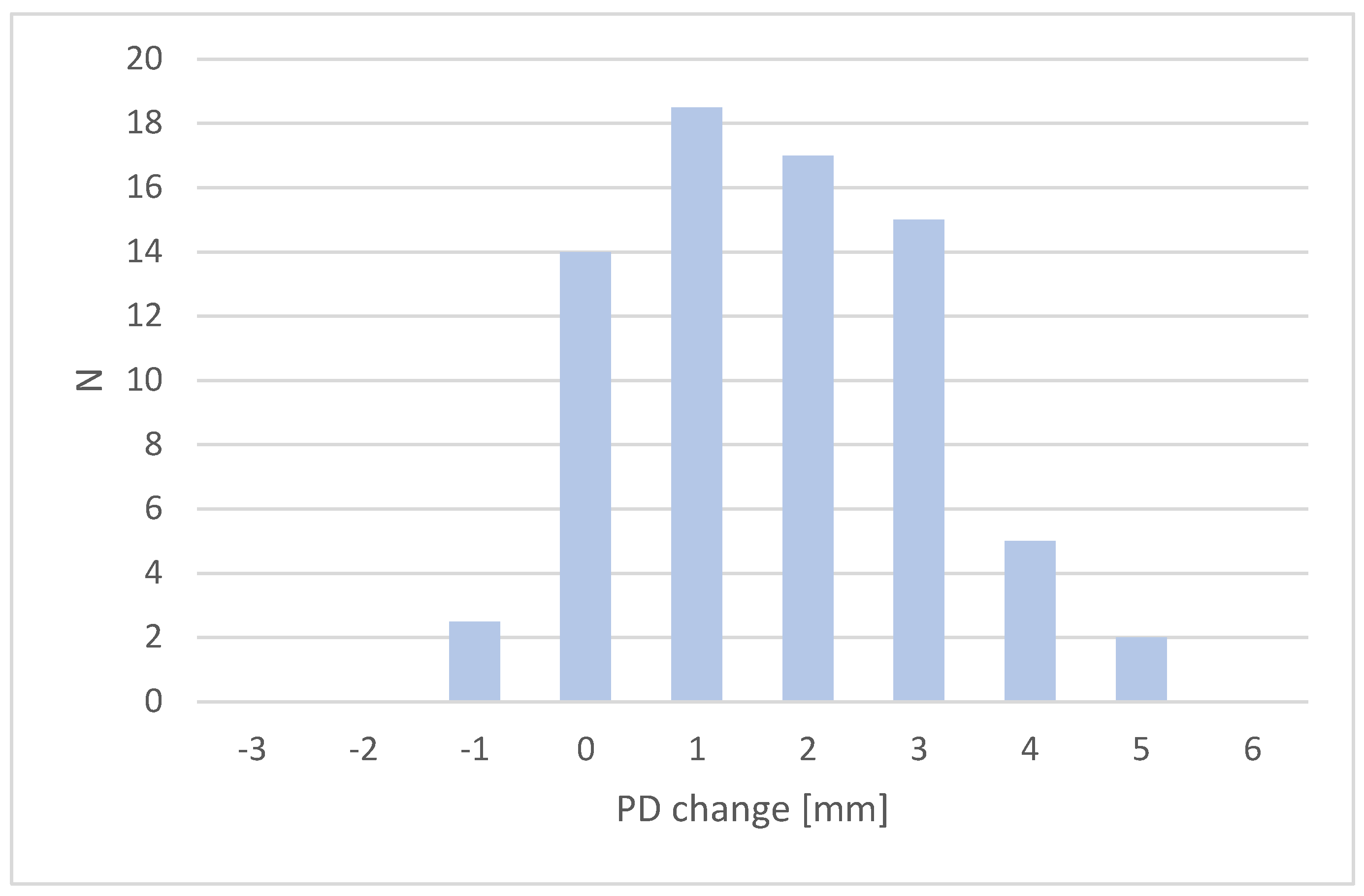
3. Results
- control group—average CAL gain = 0.56 mm (X1);
- study group—average CAL gain = 0.92 mm (X2).
4. Discussion
4.1. Impact of Diabetes on Periodontal Disease
- increased production of free oxygen radicals;
- gingival epithelial barrier modifications;
- shifts in the dental plaque microbiome.
4.2. Impact of Periodontal Disease on Diabetes
4.3. Hyperbaric Oxygen Therapy-Medical Applications
4.4. Hyperbaric Oxygen Therapy-Study Rewiev
5. Conclusions
- taking a history of periodontal disease;
- making patients aware of the relationship between the two diseases;
- referring newly diagnosed patients for periodontal evaluation;
- cooperating with the dentist;
- motivating patients to make regular dental visits.
- inform patients about the relationship between the two conditions;
- carry out regular periodontal check-ups;
- ask about HbA1c levels;
- cooperate with the general practitioner (GP), internist, or diabetologist and consider assessing the risk of diabetes in patients with suspected diabetes (for example, using a screening questionnaire) [29].
Author Contributions
Funding
Institutional Review Board Statement
Informed Consent Statement
Data Availability Statement
Conflicts of Interest
References
- The State of Oral Health in Europe. Available online: http://www.oralhealthplatform.eu/our-work/the-state-of-oral-health-in-europe (accessed on 2 January 2023).
- Vos, T.; Abajobir, A.A.; Abate, K.H.; Abbafati, C.; Abbas, K.M.; Abd-Allah, F.; Abera, S.F. Global, regional, and national incidence, prevalence, and years lived with disability for 328 diseases and injuries for 195 countries, 1990–2016: A systematic analysis for the Global Burden of Disease Study 2016. Lancet 2017, 390, 1211–1259. [Google Scholar] [CrossRef] [PubMed]
- Alawaji, Y.N.; Alshammari, A.; Mostafa, N.; Carvalho, R.M.; Aleksejuniene, J. Periodontal disease prevalence, extent, and risk associations in untreated individuals. Clin. Exp. Dent. Res. 2022, 8, 380–394. [Google Scholar] [CrossRef] [PubMed]
- Relvas, M.; López-Jarana, P.; Monteiro, L.; Pacheco, J.J.; Braga, A.C.; Salazar, F. Study of prevalence, severity and risk factors of periodontal disease in a Portuguese population. J. Clin. Med. 2022, 11, 3728. [Google Scholar] [CrossRef] [PubMed]
- Janakiram, C.; Dye, B.A. A public health approach for prevention of periodontal disease. Periodontol 2000 2020, 84, 202–214. [Google Scholar] [CrossRef] [PubMed]
- WHO. Available online: https://www.who.int/health-topics/oral-health#tab=tab_1 (accessed on 2 January 2023).
- IDF Diabetes Atlas. Available online: https://diabetesatlas.org/atlas/ninth-edition;2019 (accessed on 2 January 2023).
- Shinomiya, N. Molecular Mechanisms of Hyperbaric Oxygen Therapy. In Hyperbaric Oxygenation Therapy; Shinomiya, N., Asai, Y., Eds.; Springe: Singapore, 2020; Volume 1, pp. 3–20. [Google Scholar]
- Resanović, I.; Zarić, B.; Radovanović, J.; Sudar-Milovanović, E.; Gluvić, Z.; Jevremović, D.; Isenović, E.R. Hyperbaric Oxygen Therapy and Vascular Complications in Diabetes Mellitus. Angiology 2020, 71, 876–885. [Google Scholar] [CrossRef]
- Caton, J.G.; Armitage, G.; Berglundh, T.; Chapple, I.L.; Jepsen, S.; Kornman, K.S.; Mealey, B.L.; Papapanou, P.N.; Sanz, M.; Tonetti, M.S.; et al. A new classification scheme for periodontal and peri—implant diseases and conditions—Introduction and key changes from the 1999 classification. J. Periodontol. 2018, 89 (Suppl. S1), S1–S8. [Google Scholar] [CrossRef]
- Available online: https://www.periodontalchart-online.com/uk/ (accessed on 2 January 2023).
- Gottrup, F.; Dissemond, J.; Baines, C.; Frykberg, R.; Jensen, P.; Kot, J.; Kröger, K.; Longobardi, P. Use of Oxygen Therapies in Wound Healing. J. Wound Care 2017, 26 (Suppl. S5), S1–S43. [Google Scholar] [CrossRef]
- Rekomendacje Grupy Roboczej Polskiego Towarzystwa Stomatologicznego i Narodowego Programu Ochrony Antybiotyków w Zakresie Stosowania Antybiotyków w Stomatologii. Available online: https://stomatologia-zalecenia-25_01-net.pdf(antybiotyki.edu.pl) (accessed on 20 April 2023).
- Eley, B.; Soory, M.; Manson, J.D. Periodontics, 6th ed.; Churchill Livingstone: Livingstone, UK, 2010; Volume 15, p. 233. [Google Scholar]
- Papapanou, P.N.; Sanz, M.; Buduneli, N.; Dietrich, T.; Feres, M.; Fine, D.H.; Flemmig, T.F.; Garcia, R.; Giannobile, W.V.; Graziani, F.; et al. Periodontitis: Consensus report of workgroup 2 of the 2017 World Workshop on the Classification of Periodontal and Peri—Implant Diseases and Conditions. J. Periodontol. 2018, 89 (Suppl. S1), S173–S182. [Google Scholar] [CrossRef]
- Genco, R.J.; Borgnakke, W.S. Diabetes as a potential risk for periodontitis: Association studies. Periodontol 2000 2020, 83, 40–45. [Google Scholar] [CrossRef]
- Löe, H. Periodontal disease. The sixth complication of diabetes mellitus. Diabetes Care 1993, 16, 329–334. [Google Scholar] [CrossRef]
- Tonetti, M.S.; Greenwell, H.; Kornman, K.S. Staging and grading of periodontitis: Framework and proposal of a new classification and case definition. J. Periodontol. 2018, 89, S159–S172. [Google Scholar] [CrossRef]
- Nascimento, G.G.; Leite, F.R.M.; Vestergaard, P.; Scheutz, F.; López, R. Does diabetes increase the risk of periodontitis? A systematic review and meta-regression analysis of longitudinal prospective studies. Acta Diabetol. 2018, 55, 653–667. [Google Scholar] [CrossRef]
- Pacios, S.; Andriankaja, O.; Kang, J.; Alnammary, M.; Bae, J.; Bezerra, B.D.B.; Schreiner, H.; Fine, D.H.; Graves, D.T. Bacterial infection increases periodontal bone loss in diabetic rats through enhanced apoptosis. Am. J. Pathol. 2013, 183, 1928–1935. [Google Scholar] [CrossRef]
- Graves, D.T.; Ding, Z.; Yang, Y. The impact of diabetes on periodontal diseases. Periodontol 2000 2020, 82, 214–224. [Google Scholar] [CrossRef]
- Mesia, R.; Gholami, F.; Huang, H.; Clare-Salzler, M.; Aukhil, I.; Wallet, S.M.; Shaddox, L.M. Systemic inflammatory responses in patients with type 2 diabetes with chronic periodontitis. BMJ Open Diabetes Res. Care 2016, 4, e000260. [Google Scholar] [CrossRef]
- Preshaw, P.M.; Bissett, S.M. Periodontitis and diabetes. Br. Dent. J. 2019, 227, 577–584. [Google Scholar] [CrossRef]
- Simpson, T.C.; Weldon, J.C.; Worthington, H.V.; Needleman, I.; Wild, S.H.; Moles, D.R.; Stevenson, B.; Furness, S.; Iheozor-Ejiofor, Z. Treatment of periodontal disease for glycaemic control in people with diabetes mellitus. Cochrane Database Syst. Rev. 2015, 11, CD004714. [Google Scholar] [CrossRef]
- Devaraj, D.; Srisakthi, D. Hyperbaric oxygen therapy—Can it be the new era in dentistry? J. Clin. Diagn. Res. 2014, 8, 263–265. [Google Scholar] [CrossRef]
- Latusek, K.; Slotwinska, A.; Michniak, A.; Orzechowska-Wylegala, B. Effects of hyperbaric oxygen therapy on periodontal disease: A literature review. Undersea Hyperb. Med. 2023, 50, 17–27. [Google Scholar] [CrossRef]
- Mulawarmanti, D.; Parisihni, K. The Effect of sticopus hermanii—hyperbaric oxygen therapy to inflammatory response of diabetic periodontitis. In IOP Conference Series: Earth and Environmental Science; IOP Publishing: Bristol, UK, 2019; Volume 217, p. 12060. [Google Scholar]
- Giacon, T.A.; Giancola, F.; Paganini, M.; Tiengo, C.; Camporesi, E.M.; Bosco, G. Hyperbaric Oxygen Therapy and A-PRF Pre-Treated Implants in Severe Periodontitis: A Case Report. Int. J. Environ. Res. Public Health 2021, 18, 413. [Google Scholar] [CrossRef]
- Sanz, M.; Ceriello, A.; Buysschaert, M.; Chapple, I.; Demmer, R.T.; Graziani, F.; Herrera, D.; Jepsen, S.; Lione, L.; Madianos, P.; et al. Scientific evidence on the links between periodontal diseases and diabetes: Consensus report and guidelines of the joint workshop on periodontal diseases and diabetes by the International Diabetes Federation and the European Federation of Periodontology. J. Clin. Periodontol. 2018, 45, 138–149. [Google Scholar] [CrossRef] [PubMed]


| Parameter | Pockets | SD1 | SD2 | F-S |
|---|---|---|---|---|
| PD | 4—5 mm | 0.93 | 0.83 | 1.11 |
| ≥6 mm | 1.37 | 1.43 | 1.04 | |
| CAL | 4—5 mm | 1.01 | 1.07 | 1.06 |
| ≥6 mm | 1.34 | 1.68 | 1.25 |
| Group | Pockets | N | E | Chi-Square |
|---|---|---|---|---|
| Control Group | 4–5 mm | 357 | 337.5 | 0.48 |
| ≥6 mm | 74 | 62.5 | 0.17 | |
| Study Group | 4–5 mm | 318 | 337.5 | 0.48 |
| ≥6 mm | 51 | 62.5 | 0.17 | |
| Sum | 1.28 |
| Parameter | Pockets | X1 (SD1) | X2 (SD2) | T-Student |
|---|---|---|---|---|
| PD | 4–5 mm | 0.79 (0.93) | 0.99 (0.83) | 2.88 |
| ≥6 mm | 1.70 (1.37) | 2.79 (1.43) | 4.27 | |
| CAL | 4–5 mm | 0.56 (1.01) | 0.92 (1.07) | 4.41 |
| ≥6 mm | 1.09 (1.34) | 1.87 (1.68) | 2.90 |
| Group | BOP1 | BOP2 |
|---|---|---|
| Control Group | 75.4% | 63.7% |
| Study Group | 32.0% | 28.8% |
Disclaimer/Publisher’s Note: The statements, opinions and data contained in all publications are solely those of the individual author(s) and contributor(s) and not of MDPI and/or the editor(s). MDPI and/or the editor(s) disclaim responsibility for any injury to people or property resulting from any ideas, methods, instructions or products referred to in the content. |
© 2023 by the authors. Licensee MDPI, Basel, Switzerland. This article is an open access article distributed under the terms and conditions of the Creative Commons Attribution (CC BY) license (https://creativecommons.org/licenses/by/4.0/).
Share and Cite
Latusek, K.; Słotwińska-Pawlaczyk, A.; Warakomska, A.; Kubicka-Musiał, M.; Wiench, R.; Orzechowska-Wylęgała, B. Pilot Study: The Effectiveness of Hyperbaric Oxygen Therapy in the Treatment of Periodontitis in Patients with Type 2 Diabetes. Healthcare 2023, 11, 1344. https://doi.org/10.3390/healthcare11091344
Latusek K, Słotwińska-Pawlaczyk A, Warakomska A, Kubicka-Musiał M, Wiench R, Orzechowska-Wylęgała B. Pilot Study: The Effectiveness of Hyperbaric Oxygen Therapy in the Treatment of Periodontitis in Patients with Type 2 Diabetes. Healthcare. 2023; 11(9):1344. https://doi.org/10.3390/healthcare11091344
Chicago/Turabian StyleLatusek, Katarzyna, Adrianna Słotwińska-Pawlaczyk, Aleksandra Warakomska, Magdalena Kubicka-Musiał, Rafał Wiench, and Bogusława Orzechowska-Wylęgała. 2023. "Pilot Study: The Effectiveness of Hyperbaric Oxygen Therapy in the Treatment of Periodontitis in Patients with Type 2 Diabetes" Healthcare 11, no. 9: 1344. https://doi.org/10.3390/healthcare11091344