
Robust Zero-Watermarking Algorithm for Medical Images Using Double-Tree Complex Wavelet Transform and Hessenberg Decomposition


Abstract
:1. Introduction
- (1)
- We proposed a novel zero-watermarking algorithm for medical images using DTCWT, MDCT, and Hessenberg decomposition.
- (2)
- The paper used DTCWT extract multi-directional and multi-scale features to better describe the feature information of medical images. In addition, when medical images are attacked, the proposed algorithm can show better orientation optionality and translation invariance and effectively improve the robustness against translation attacks.
- (3)
- The authors utilized the MDCT to take full advantage of the energy concentration characteristics of DCT, giving the algorithm fastness and accurate feature extraction capabilities.
- (4)
- The proposed algorithm used Hessenberg decomposition to effectively improve the execution efficiency and has good rotational invariance, which exhibits strong robustness against geometric attacks.
2. Basic Theory
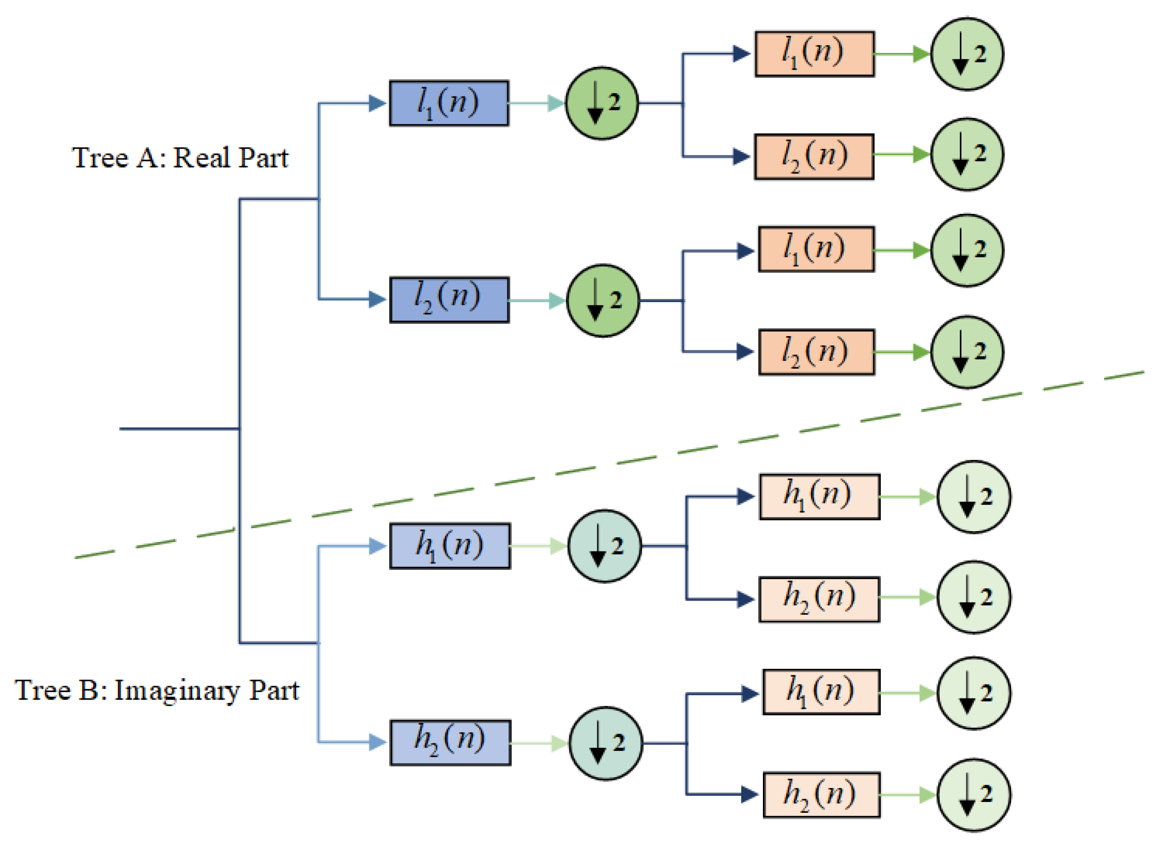
2.1. DTCWT
2.2. DCT
2.3. Hessenberg Decomposition
3. Zero-Watermarking Algorithm
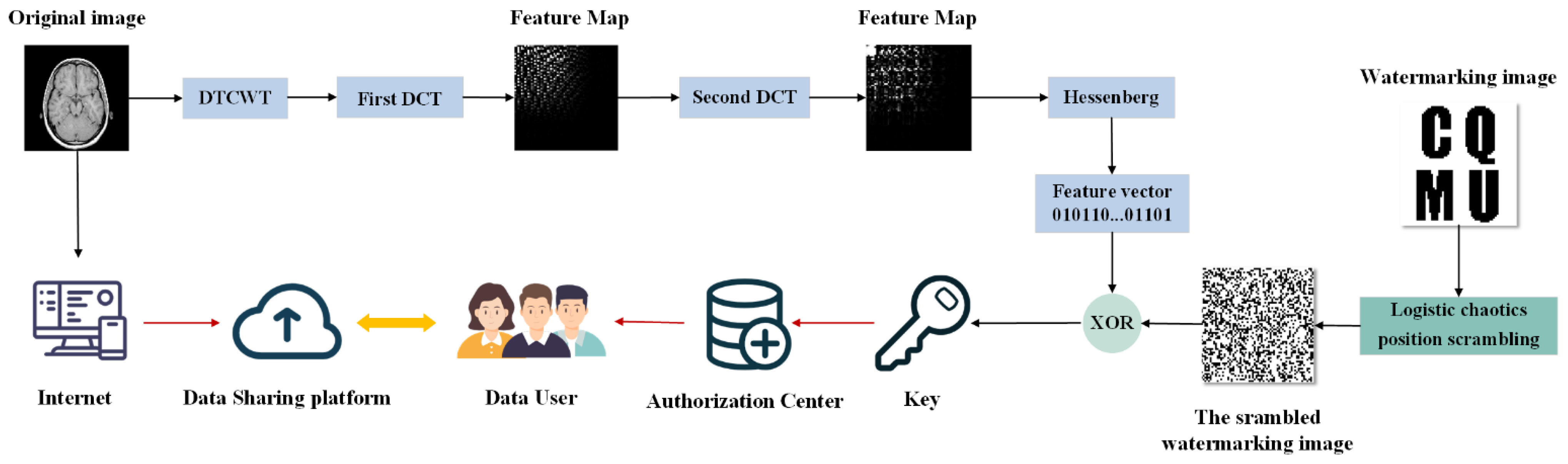
3.1. Watermarking-Generation Algorithm
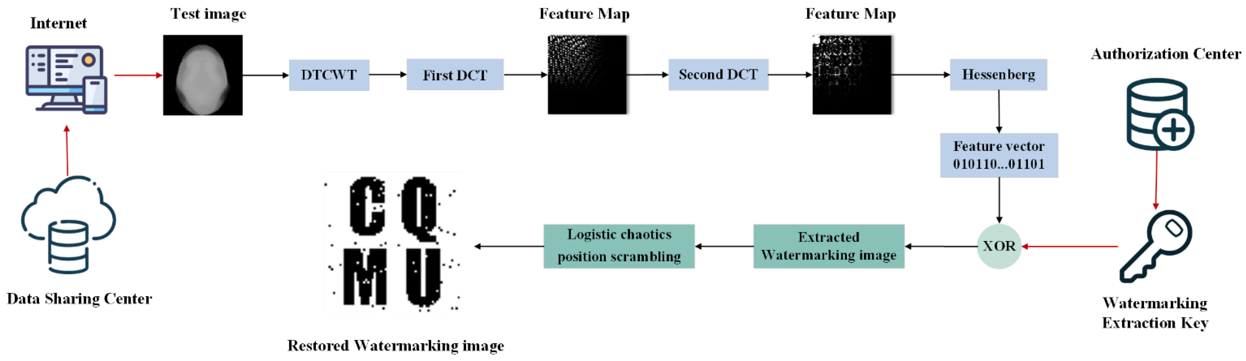
3.2. Watermarking-Extraction Algorithm
4. Experiments and Results
4.1. The Experimental Description
4.2. Robustness Experiment
4.2.1. Common Attacks
- Gaussian Noise Attacks
- JPEG Compression Attacks
- Median Filter Attacks
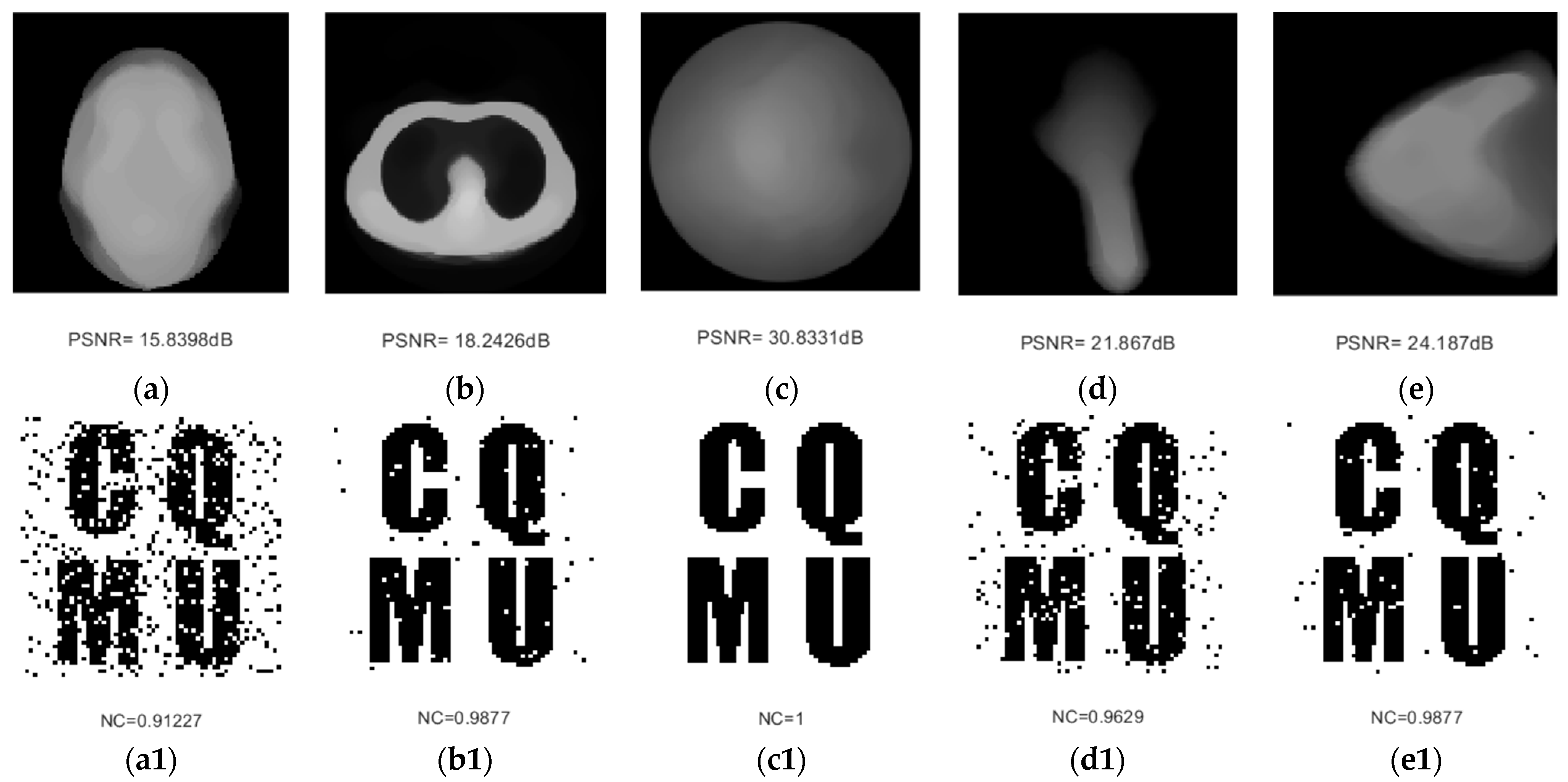
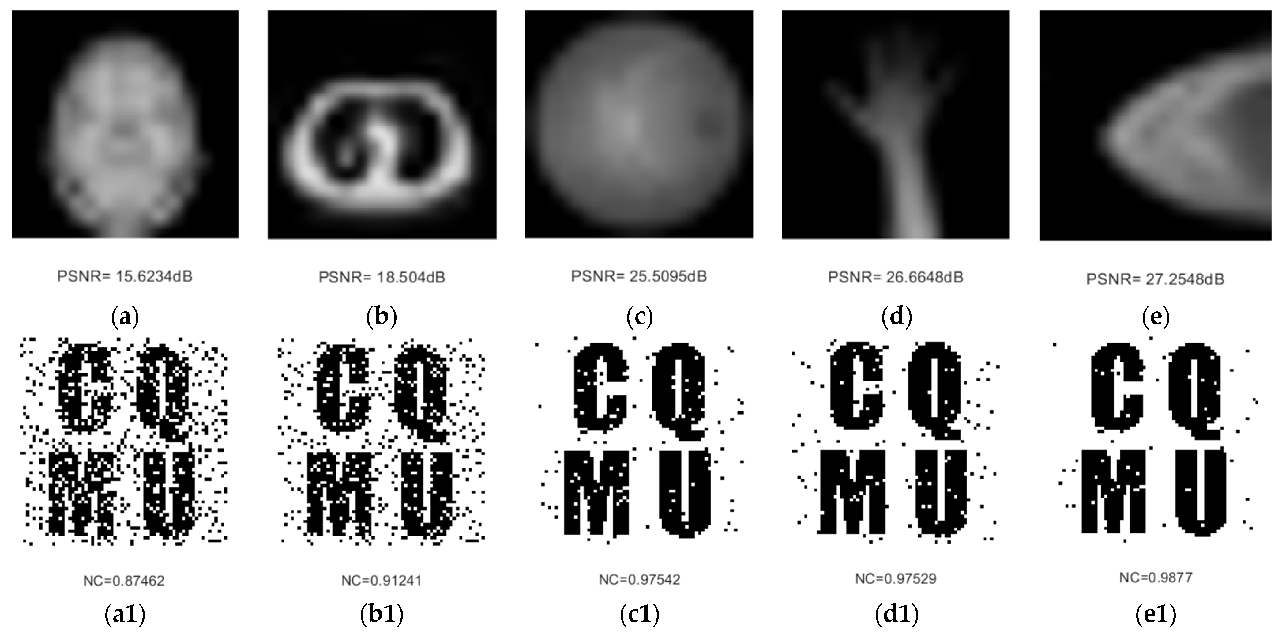
4.2.2. Geometric Attacks
- Rotation Attacks
- Scaling Attacks
- Cropping Attacks
- Translation Attacks
4.3. Comparison Experiments
- Robustness comparison
5. Conclusions
Author Contributions
Funding
Institutional Review Board Statement
Informed Consent Statement
Data Availability Statement
Conflicts of Interest
References
- Manuel, C.H.; Antonio, C.H.; Garcia-Ugalde, F.J. Improving DFT-Based Image Watermarking Using Particle Swarm Optimization Algorithm. Mathematics 2021, 9, 1795. [Google Scholar]
- Rani, A.; Bhullar, A.K.; Dangwal, D.; Kumar, S. A Zero-Watermarking Scheme using Discrete Wavelet Transform. Procedia Comput. Sci. 2015, 70, 603–609. [Google Scholar] [CrossRef] [Green Version]
- Anand, A.; Singh, A.K. An improved DWT-SVD domain watermarking for medical information security. Comput. Commun. 2020, 152, 72–80. [Google Scholar] [CrossRef]
- Malayil, M.V.; Vedhanayagam, M. A novel image scaling based reversible watermarking scheme for secure medical image transmission. ISA Trans. 2021, 108, 269–281. [Google Scholar] [CrossRef] [PubMed]
- Li, Y.; Wang, J.W.; Jia, H.Y. A Robust and Reversible Watermarking Algorithm for a Relational Database Based on Continuous Columns in Histogram. Mathematics 2020, 8, 1994. [Google Scholar] [CrossRef]
- Wang, B.W.; Zhao, P. An Adaptive Image Watermarking Method Combining SVD and Wang-Landau Sampling in DWT Domain. Mathematics 2020, 8, 691. [Google Scholar] [CrossRef]
- Gangadhar, Y.; Giridhar Akula, V.S.; Reddy, P.C. An evolutionary programming approach for securing medical images using watermarking scheme in invariant discrete wavelet transformation. Biomed. Signal Process. Control. 2018, 43, 31–40. [Google Scholar] [CrossRef]
- Balasamy, K.; Suganyadevi, S. A fuzzy based ROI selection for encryption and watermarking in medical image using DWT and SVD. Multimed. Tools Appl. 2020, 80, 7167–7186. [Google Scholar]
- Fares, K.; Amine, K.; Redouane, K.; Salah, E. DCT & DWT based watermarking scheme for medical information security. Biomed. Signal Process. Control 2021, 66, 102403. [Google Scholar]
- Cedillo-Hernandez, M.; Cedillo-Hernandez, A.; Nakano-Miyatake, M.; Perez-Meana, H. Improving the management of medical imaging by using robust and secure dual watermarking. Biomed. Signal Process. Control. 2020, 56, 101695. [Google Scholar] [CrossRef]
- Kavitha, C.; Sakthivel, S. An effective mechanism for medical images authentication using quick response code. Clust. Comput. 2019, 22, 4375–4382. [Google Scholar] [CrossRef]
- Wen, Q.; Sun, T.F.; Wang, S.X. Concept and application of zero-watermarking. Acta Electron. Sin. 2003, 31, 214–216. [Google Scholar]
- Xiao, Z.J.; Zhang, H.; Chen, H.; Gao, T. Zero-watermarking based on boost normed singular value decomposition and cellular neural network. J. Image Graph. 2017, 22, 288–296. [Google Scholar]
- Wu, X.Q.; Li, J.B.; Tu, R.; Cheng, J.R.; Bhatti, U.A.; Ma, J.X. Contourlet-DCT based multiple robust watermarkings for medical images. Multimed. Tools Appl. 2019, 78, 8463–8480. [Google Scholar] [CrossRef]
- Qin, F.M.; Li, J.B.; Li, H.; Liu, J.; Nawaz, S.A.; Liu, Y.L. A robust zero-watermarking algorithm for medical images using curvelet-dct and RSA pseudo-random sequences. In The International Conference on Artificial Intelligence and Security; Springer: Cham, Switzerland, 2020; Volume 12240, pp. 179–190. [Google Scholar]
- Wu, D.Y.; Tang, Y.; Zhao, W.; Wan, Y.C.; Qu, C.B. Zero-watermarking algorithm based on Curvelet-DWT-SVD. J. Yanshan Univ. 2020, 44, 42–52. [Google Scholar]
- Xue, H.F.; Chi, W.D.; Liu, X.X. Zero-Watermark Algorithm based on NSST and Hessenberg Decomposition. Mod. Comput. 2020, 10, 89–93. [Google Scholar]
- Liu, J.; Li, J.B.; Zhang, K.; Bhatti, U.A.; Ai, Y. Zero-watermarking algorithm for medical images based on dual-tree complex wavelet transform and discrete cosine transform. J. Med. Imaging Health Inform. 2019, 9, 188–194. [Google Scholar] [CrossRef]
- Xia, Z.Q.; Wang, X.Y.; Wang, C.P.; Wang, C.X.; Ma, B.; Li, Q.; Wang, M.X.; Zhao, T.T. A robust zero-watermarking algorithm for lossless copyright protection of medical images. Appl. Intell. 2022, 52, 607–621. [Google Scholar] [CrossRef]
- Vaidya, S.P. Fingerprint-based robust medical image watermarking in hybrid transform. Vis. Comput. 2022, 38, 1–16. [Google Scholar] [CrossRef] [PubMed]
- Fang, Y.X.; Liu, J.; Li, J.B.; Cheng, J.R.; Hu, J.B.; Yi, D.; Xiao, X.L.; Bhatti, U.A. Robust zero-watermarking algorithm for medical images based on SIFT and Bandelet-DCT. Multimed. Tools Appl. 2022, 81, 1–17. [Google Scholar] [CrossRef]
- Liu, J.; Ma, J.X.; Li, J.B.; Huang, M.X.; Sadiq, N.; Ai, Y. Robust watermarking algorithm for medical volume data in internet of medical things. IEEE Access 2020, 8, 93939–93961. [Google Scholar] [CrossRef]
- Nick, K. Image processing with complex wavelets. Philos. Trans. Math. Phys. Eng. Sci. 1999, 357, 2527–2542. [Google Scholar]
- Nick, K. Complex wavelets for shift invariant analysis and filtering of signals. Appl. Comput. Harmon. Anal. 2001, 10, 234–253. [Google Scholar]
- Nouioua, A.; Seddiki, N.; Ghaz, A. Blind digital watermarking framework based on DTCWT and NSCT for telemedicine application. Traitement Du Signal 2020, 37, 955–964. [Google Scholar] [CrossRef]
- Li, Z.Y.; Zhang, H.; Liu, X.L.; Wang, C.P.; Wang, X.Y. Blind and safety-enhanced dual watermarking algorithm with chaotic system encryption based on RHFM and DWT-DCT. Digit. Signal Process. 2021, 115, 103062. [Google Scholar] [CrossRef]
- Mahbuba, B.; Shorif, U.M. Multiple image watermarking with discrete cosine transform. J. Comput. Commun. 2021, 9, 88–94. [Google Scholar]
- Zhou, K.; Zhang, Y.M.; Li, J.; Zhan, Y.T.; Wan, W.B. Spatial-Perceptual Embedding with Robust Just Noticeable Difference Model for Color Image Watermarking. Mathematics 2020, 8, 1506. [Google Scholar] [CrossRef]
- Qu, C.B.; Yan, Y. A robust digital watermarking algorithm based on multiple level DCT and SVD. Comput. Appl. Softw. 2012, 29, 288–291. [Google Scholar]
- Su, Q.T.; Chen, B.J. A novel blind color image watermarking using upper Hessenberg matrix. AEU—Int. J. Electron. Commun. 2017, 78, 64–71. [Google Scholar] [CrossRef]
- Cheng, Y.Y. Image watermarking algorithm based on discrete cosine transform and Hessenberg composition. J. Jinggangshan Univ. Nat. Sci. Ed. 2019, 40, 45–51. [Google Scholar]
- Huang, T.Y.; Xu, J.; Yang, Y.L.; Tu, S.X.; Han, B.R. Zero-Watermarking Algorithm for Medical Images Based on Nonsubsampled Contourlet Transform and Double Singular Value Decomposition. In Proceedings of the 2021 5th Asian Conference on Artificial Intelligence Technology (ACAIT), Haikou, China, 29–31 October 2021; pp. 65–76. [Google Scholar]


| Name | Noise 1% | Noise 5% | Noise 15% | Noise 30% | Noise 50% | |||||
|---|---|---|---|---|---|---|---|---|---|---|
| PSNR/dB | NC | PSNR/dB | NC | PSNR/dB | NC | PSNR/dB | NC | PSNR/dB | NC | |
| Medical image A | 21.3809 | 1.0000 | 14.6856 | 1.0000 | 10.7088 | 0.9877 | 8.6252 | 0.9753 | 7.9512 | 0.9754 |
| Medical image B | 21.8515 | 1.0000 | 15.2301 | 1.0000 | 10.9588 | 0.9753 | 8.7182 | 0.9753 | 7.4081 | 0.9753 |
| Medical image C | 17.7054 | 1.0000 | 11.0908 | 1.0000 | 7.3532 | 0.9877 | 5.6276 | 0.9753 | 4.6856 | 0.9754 |
| Medical image D | 19.4994 | 1.0000 | 12.8283 | 1.0000 | 8.4299 | 0.9753 | 6.0647 | 0.9753 | 4.7484 | 0.9754 |
| Medical image E | 18.1251 | 1.0000 | 11.5601 | 1.0000 | 7.4808 | 0.9877 | 5.4692 | 0.9753 | 4.3108 | 0.9754 |
| Name | Compress 2% | Compress 5% | Compress 9% | Compress 15% | Compress 25% | |||||
|---|---|---|---|---|---|---|---|---|---|---|
| PSNR/dB | NC | PSNR/dB | NC | PSNR/dB | NC | PSNR/dB | NC | PSNR/dB | NC | |
| Medical image A | 20.707 | 0.9628 | 22.0045 | 0.9877 | 23.6216 | 0.9503 | 24.9075 | 0.9629 | 26.7087 | 0.9628 |
| Medical image B | 22.557 | 0.9877 | 24.6316 | 0.9753 | 26.9914 | 0.9754 | 28.9971 | 0.9628 | 31.0536 | 0.9754 |
| Medical image C | 23.2354 | 0.9877 | 25.2333 | 0.9877 | 28.5905 | 0.9877 | 30.9748 | 0.9877 | 33.1253 | 0.9753 |
| Medical image D | 26.1048 | 0.9877 | 26.4586 | 0.9753 | 32.8437 | 1.0000 | 34.6839 | 1.0000 | 35.0334 | 1.0000 |
| Medical image E | 24.0996 | 0.9877 | 24.4521 | 0.9753 | 29.9548 | 1.0000 | 32.1558 | 0.9877 | 34.9622 | 1.0000 |
| Name | Parameter (3 × 3) | Parameter (5 × 5) | Parameter (7 × 7) | |||||||
|---|---|---|---|---|---|---|---|---|---|---|
| 10 Times | 20 Times | 10 Times | 20 Times | 10 Times | ||||||
| PSNR/dB | NC | PSNR/dB | NC | PSNR/dB | NC | PSNR/dB | NC | PSNR/dB | NC | |
| Medical image A | 20.91 | 0.9504 | 20.3341 | 0.9377 | 17.2479 | 0.9126 | 16.6834 | 0.9123 | 15.917 | 0.9124 |
| Medical image B | 26.5736 | 0.9754 | 26.02 | 0.9629 | 22.609 | 0.9753 | 22.1802 | 0.9754 | 19.9116 | 0.9753 |
| Medical image C | 35.49 | 1.0000 | 35.2661 | 1.0000 | 32.6347 | 0.9877 | 32.3348 | 0.9877 | 31.4819 | 0.9877 |
| Medical image D | 38.0348 | 1.0000 | 37.2862 | 1.0000 | 28.0059 | 0.9629 | 26.3181 | 0.9753 | 24.288 | 0.9629 |
| Medical image E | 35.173 | 1.0000 | 34.9371 | 1.0000 | 29.2758 | 0.9877 | 28.4422 | 0.9877 | 26.391 | 0.9877 |
| Name | Rotation 5% | Rotation 10% | Rotation 20% | Rotation 25% | Rotation 40% | |||||
|---|---|---|---|---|---|---|---|---|---|---|
| PSNR/dB | NC | PSNR/dB | NC | PSNR/dB | NC | PSNR/dB | NC | PSNR/dB | NC | |
| Medical image A | 15.5831 | 0.9752 | 12.9944 | 0.9377 | 12.068 | 0.9378 | 11.9703 | 0.9628 | 11.232 | 0.9753 |
| Medical image B | 18.1468 | 0.9379 | 14.5409 | 0.9629 | 11.5648 | 0.9503 | 10.7143 | 0.9504 | 9.448 | 0.9378 |
| Medical image C | 30.0359 | 0.9877 | 28.5605 | 0.9877 | 27.0817 | 0.9877 | 26.5903 | 0.9877 | 25.7494 | 0.9877 |
| Medical image D | 20.8156 | 0.9877 | 17.0125 | 0.9877 | 13.8992 | 0.9877 | 13.0057 | 0.9753 | 12.16 | 0.9377 |
| Medical image E | 20.5585 | 0.9877 | 16.717 | 0.9754 | 13.4887 | 0.9877 | 12.7267 | 0.9753 | 11.4282 | 0.9877 |
| Name | Zoom Factor 0.125 then 8 | Zoom Factor 0.25 then 4 | Zoom Factor 0.5 then 2 | Zoom Factor 2 then 0.5 | Zoom Factor 4 then 0.25 | |||||
|---|---|---|---|---|---|---|---|---|---|---|
| PSNR/dB | NC | PSNR/dB | NC | PSNR/dB | NC | PSNR/dB | NC | PSNR/dB | NC | |
| Medical image A | 15.6234 | 0.8746 | 17.8888 | 0.9628 | 21.5809 | 0.9753 | 30.2404 | 1.0000 | 30.5191 | 1.0000 |
| Medical image B | 18.504 | 0.9124 | 21.7819 | 0.9753 | 27.4683 | 1.0000 | 38.8449 | 1.0000 | 39.1239 | 1.0000 |
| Medical image C | 25.5095 | 0.9754 | 29.7844 | 0.9877 | 35.0533 | 1.0000 | 45.9477 | 1.0000 | 46.6227 | 1.0000 |
| Medical image D | 26.6648 | 0.9753 | 33.0577 | 0.9877 | 41.1167 | 1.0000 | 52.6998 | 1.0000 | 53.3732 | 1.0000 |
| Medical image E | 27.2548 | 0.9877 | 31.7092 | 0.9877 | 37.6185 | 1.0000 | 48.3699 | 1.0000 | 49.124 | 1.0000 |
| Name | X-Axis Crop 5% | X-Axis Crop 15% | X-Axis Crop 40% | Y-Axis Crop 10% | Y-Axis Crop 25% | |||||
|---|---|---|---|---|---|---|---|---|---|---|
| PSNR/dB | NC | PSNR/dB | NC | PSNR/dB | NC | PSNR/dB | NC | PSNR/dB | NC | |
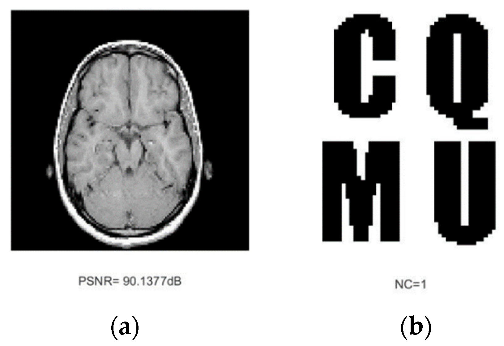
| Medical image A | 90.1377 | 1.0000 | 56.9175 | 1.0000 | 12.9247 | 0.9753 | 18.7664 | 0.9377 | 13.59 | 0.9377 |
| Medical image B | 50.2622 | 1.0000 | 23.43 | 0.9630 | 14.5185 | 0.9629 | 28.6074 | 0.9753 | 14.3028 | 0.9754 |
| Medical image C | 32.7365 | 0.9877 | 20.7318 | 0.9877 | 12.3005 | 0.9629 | 24.811 | 0.9877 | 16.2596 | 0.9753 |
| Medical image D | 87.581 | 1.0000 | 87.581 | 1.0000 | 19.3996 | 0.9877 | 19.4777 | 0.9629 | 16.4589 | 0.9629 |
| Medical image E | 20.3824 | 0.9877 | 15.3823 | 0.9754 | 9.7896 | 0.9630 | 32.9291 | 0.9877 | 16.7177 | 0.9877 |
| Name | Left 5% | Left 17% | Left 41% | Down 13% | Down 40% | |||||
|---|---|---|---|---|---|---|---|---|---|---|
| PSNR/dB | NC | PSNR/dB | NC | PSNR/dB | NC | PSNR/dB | NC | PSNR/dB | NC | |
| Medical image A | 11.1507 | 0.9753 | 8.6638 | 0.9377 | 6.6029 | 0.9503 | 11.2107 | 0.9250 | 8.8705 | 0.9376 |
| Medical image B | 11.8073 | 0.9752 | 8.9485 | 0.9504 | 8.7963 | 0.9628 | 9.1991 | 0.9628 | 9.0726 | 0.9378 |
| Medical image C | 17.8609 | 0.9754 | 12.6764 | 0.9877 | 8.654 | 0.9877 | 13.9912 | 0.9753 | 8.4723 | 0.9753 |
| Medical image D | 17.481 | 0.9877 | 12.2204 | 0.9877 | 10.4476 | 0.9630 | 19.1956 | 0.9877 | 15.9374 | 0.9753 |
| Medical image E | 16.7173 | 0.9877 | 10.358 | 0.9877 | 6.3622 | 0.9380 | 11.2122 | 0.9877 | 7.67 | 0.9877 |
Publisher’s Note: MDPI stays neutral with regard to jurisdictional claims in published maps and institutional affiliations. |
© 2022 by the authors. Licensee MDPI, Basel, Switzerland. This article is an open access article distributed under the terms and conditions of the Creative Commons Attribution (CC BY) license (https://creativecommons.org/licenses/by/4.0/).
Share and Cite
Huang, T.; Xu, J.; Yang, Y.; Han, B. Robust Zero-Watermarking Algorithm for Medical Images Using Double-Tree Complex Wavelet Transform and Hessenberg Decomposition. Mathematics 2022, 10, 1154. https://doi.org/10.3390/math10071154
Huang T, Xu J, Yang Y, Han B. Robust Zero-Watermarking Algorithm for Medical Images Using Double-Tree Complex Wavelet Transform and Hessenberg Decomposition. Mathematics. 2022; 10(7):1154. https://doi.org/10.3390/math10071154
Chicago/Turabian StyleHuang, Tongyuan, Jia Xu, Yuling Yang, and Baoru Han. 2022. "Robust Zero-Watermarking Algorithm for Medical Images Using Double-Tree Complex Wavelet Transform and Hessenberg Decomposition" Mathematics 10, no. 7: 1154. https://doi.org/10.3390/math10071154