
Identification of Systemic Sclerosis through Machine Learning Algorithms and Gene Expression

Abstract
:1. Introduction
2. Aims
3. Materials and Methods
3.1. Algorithm
- 1.
- The first step entails dividing the data into the control and patient subsets.
- 2.
- Estimating the mean values for each gene in each subset
- 3.
- Compare the expression value for each gene on both sets
- 4.
- If (with a predefined threshold) then eliminate the gene from both subsets.Hence:with . This process results in a reduction in the number of genes taken into consideration. The data can now be consolidated into a matrix and a vector containing both control and patients.
- 5.
- Divide the data into a testing and a training datasets with both containing control and patients.
- 6.
- Choose a classification technique , such as an artificial neural network.
- 7.
- Train the classification technique with the training data .
- 8.
- Estimate the classification forecast using the trained algorithm.
- 9.
- Compare the classification forecasts with the the actual values .
- 10.
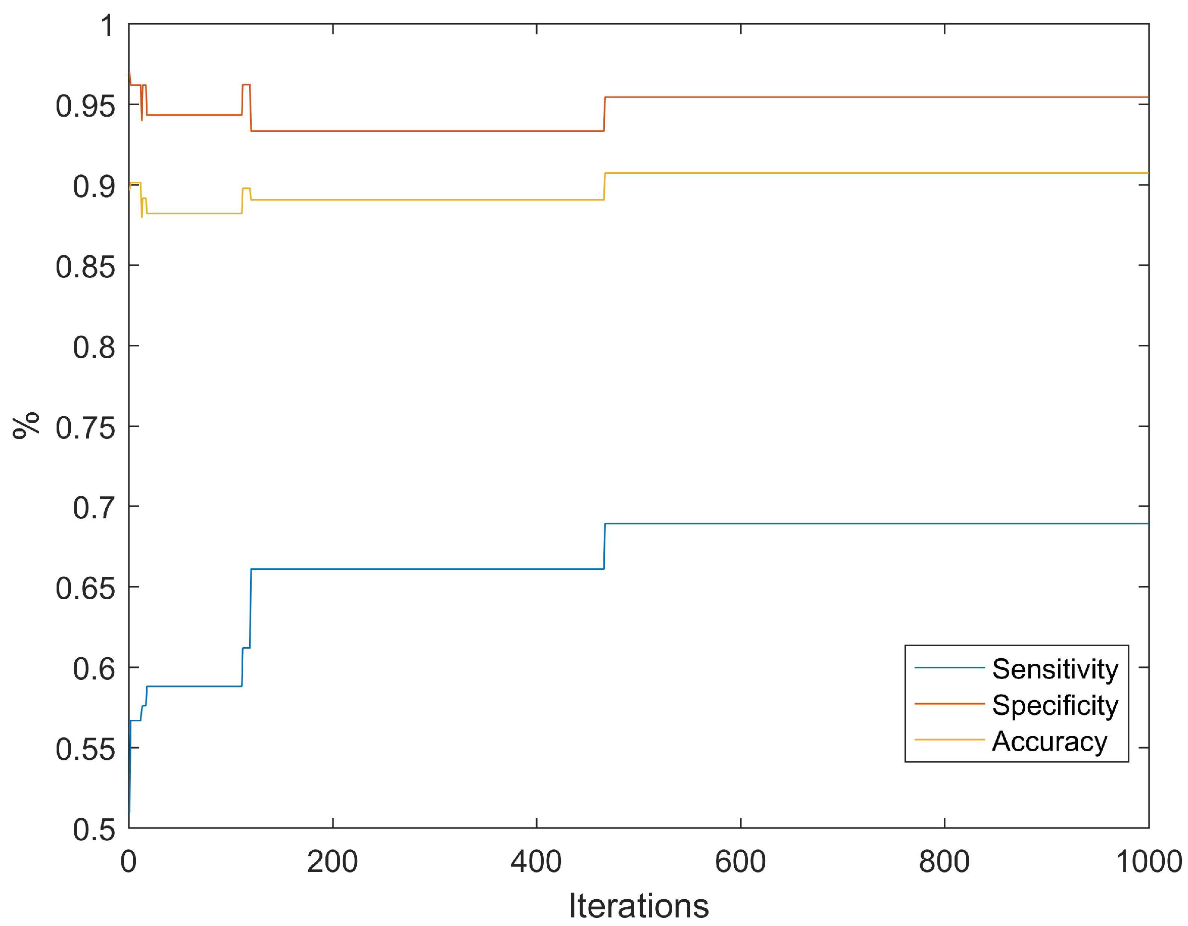
- If then otherwise . Estimate mean accuracy.Similarly estimate the sensitivity .
- 11.
- This is the first iteration
- 12.
- Then, define an integer with .
- 13.
- Eliminate genes randomly chosen from the previous group of genes.
- 14.
- Repeat steps 7 to 11, estimating the new sensitivity . If then the new configuration (group of genes) is accepted, else and revert to the previous configuration.
- 15.
- Repeat until the maximum number of iterations is reached.
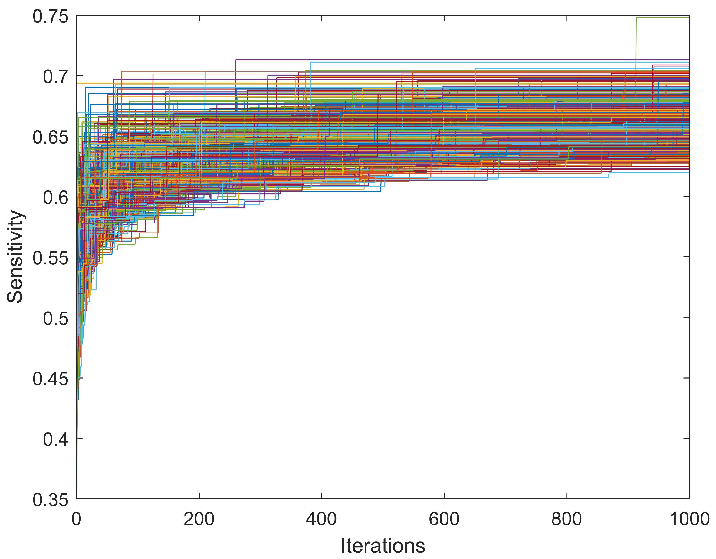
- 16.
- Repeat entire process times.
- 17.
- Select the configuration with the highest sensitivity.
3.2. Data
3.3. Classification Algorithm
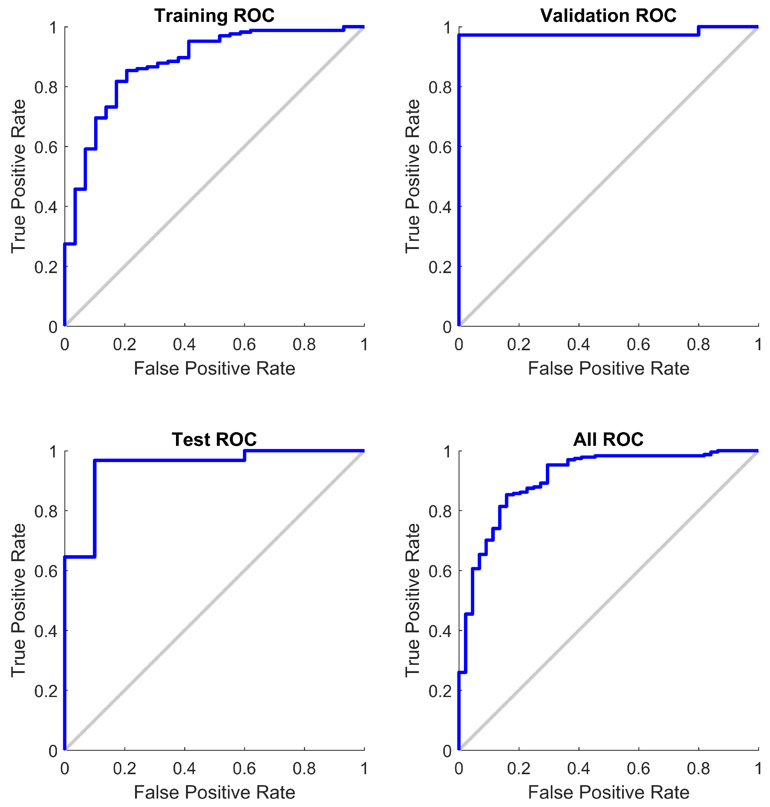
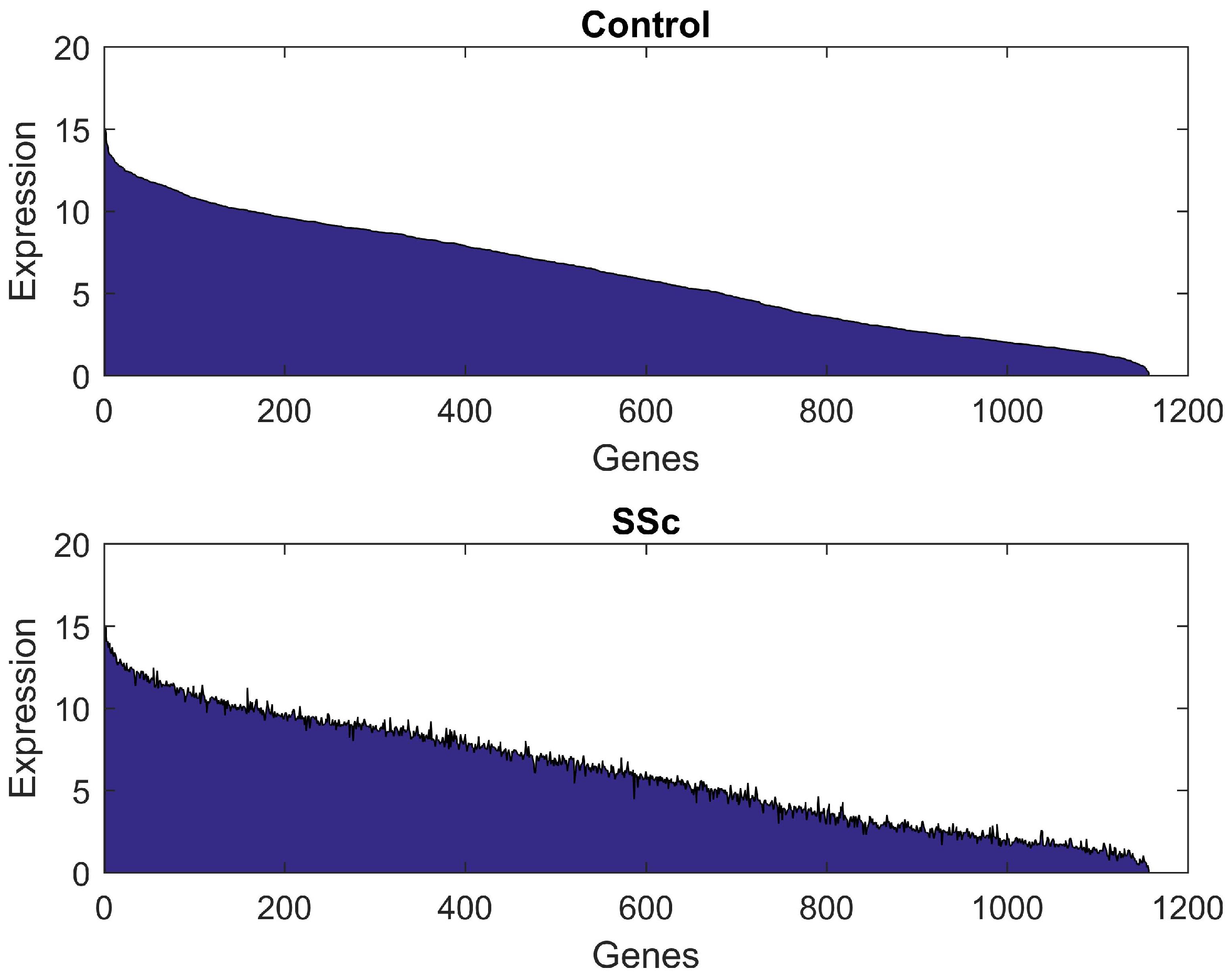
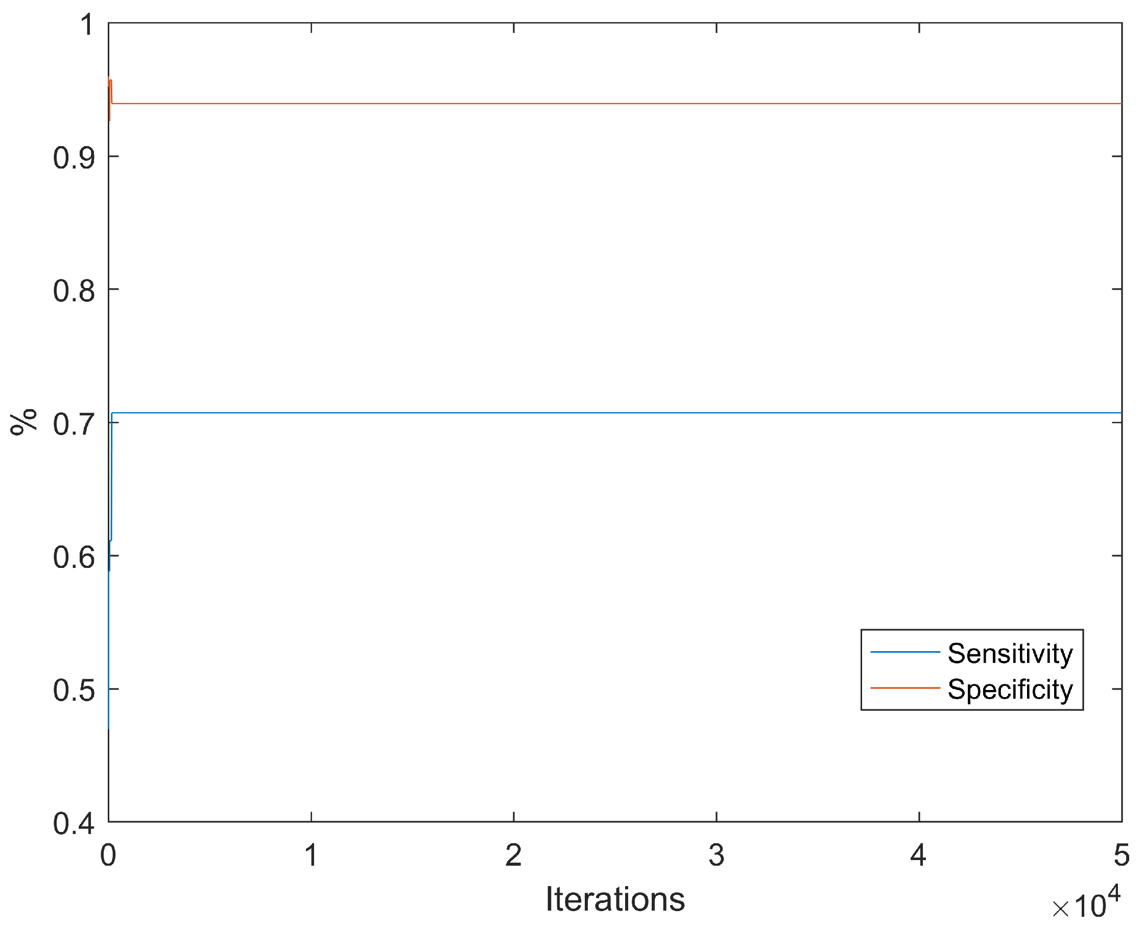
4. Results
5. Discussion
Supplementary Materials
Author Contributions
Funding
Data Availability Statement
Conflicts of Interest
References
- Sapadin, A.N.; Fleischmajer, R. Treatment of scleroderma. Arch. Dermatol. 2002, 138, 99–105. [Google Scholar] [CrossRef] [PubMed]
- Pattanaik, D.; Brown, M.; Postlethwaite, B.C.; Postlethwaite, A.E. Pathogenesis of systemic sclerosis. Front. Immunol. 2015, 6, 272. [Google Scholar] [CrossRef] [PubMed] [Green Version]
- Domsic, R.; Fasanella, K.; Bielefeldt, K. Gastrointestinal manifestations of systemic sclerosis. Dig. Dis. Sci. 2008, 53, 1163–1174. [Google Scholar] [CrossRef]
- Denton, C.P.; Khanna, D. Systemic sclerosis. Lancet 2017, 390, 1685–1699. [Google Scholar] [CrossRef]
- Yen, E.Y.; Singh, D.R.; Singh, R.R. Trends in systemic sclerosis mortality over Forty-Eight years, 1968–2015: A US Population–Based study. Arthritis Care Res. 2021, 73, 1502–1510. [Google Scholar] [CrossRef]
- Allanore, Y.; Simms, R.; Distler, O.; Trojanowska, M.; Pope, J.; Denton, C.P.; Varga, J. Systemic sclerosis. Nat. Rev. Dis. Prim. 2015, 1, 15002. [Google Scholar] [CrossRef] [PubMed]
- Moore, S.C.; Hermes, E.R. Systemic sclerosis. Treat. Complicat. Assoc. Syst. Scler. 2008, 65, 315–321. [Google Scholar]
- Godard, D. The needs of patients with systemic sclerosis—What are the difficulties encountered? Autoimmun. Rev. 2011, 10, 291–294. [Google Scholar] [CrossRef] [PubMed]
- Cheng, H.; Yu, Z.; Yan, C.; Yang, H.; Gao, C.; Wen, H. Long-term efficacy and low adverse events of methylprednisolone pulses combined to low-dose glucocorticoids for systemic sclerosis: A retrospective clinical study of 10 years’ follow-up. J. Inflamm. Res. 2022, 15, 4421–4433. [Google Scholar] [CrossRef] [PubMed]
- Almeida, C.; Almeida, I.; Vasconcelos, C. Autoimmunity reviews. Autoimmun. Rev. 2015, 14, 1087–1096. [Google Scholar] [CrossRef] [PubMed]
- Green, E.W.; Kahl, L.; Jou, J.H. Systemic sclerosis and the liver. Clin. Liver Dis. 2021, 18, 76–80. [Google Scholar] [CrossRef] [PubMed]
- Zhong, L.; Pope, M.; Shen, Y.; Hernandez, J.J.; Wu, L. Prevalence and incidence of systemic sclerosis: A systematic review and meta-analysis. Int. J. Rheum. Dis. 2019, 22, 2096–2107. [Google Scholar] [CrossRef]
- Englert, H.; Small-McMahon, J.; Davis, K.; O’Connor, H.J.; Chambers, P.; Brooks, P. Systemic sclerosis prevalence and mortality in Sydney 1974-88. Aust. N. Z. J. Med. 1999, 29, 42–50. [Google Scholar] [CrossRef] [PubMed] [Green Version]
- Mayes, M.D.; Lacey, J.V.; Beebe-Dimmer, J.; Gillespie, B.W.; Cooper, B.; Brooks, P.; Laing, T.J.; Schottenfeld, D. Prevalence, incidence, survival, and disease characteristics of systemic sclerosis in a large US population. Arthritis Rheum. Off. J. Am. Coll. Rheumatol. 2003, 48, 2246–2255. [Google Scholar] [CrossRef] [PubMed]
- Barnabe, C.; Joseph, L.; Belisle, P.; Labrecque, J.; Edworthy, S.; Barr, S.G.; Fritzler, M.; Fritzler, M.; Svenson, L.W.; Hemmelgarn, B.; et al. Prevalence of systemic lupus erythematosus and systemic sclerosis in the First Nations population of Alberta, Canada. Arthritis Care Res. 2012, 64, 138–143. [Google Scholar] [CrossRef]
- Hoffmann-Vold, A.; Midtvedt, O.; Molberg, O.; Garen, T.; Gran, J.T. Prevalence of systemic sclerosis in south-east Norway. Rheumatology 2012, 51, 1600–1605. [Google Scholar] [CrossRef] [PubMed] [Green Version]
- Gu, Y.S.; Kong, J.; Cheema, G.S.; Keen, C.L.; Wick, G.; Gershwin, M.E. The immunobiology of systemic sclerosis. Semin. Arthritis Rheum. 2015, 38, 132–160. [Google Scholar] [CrossRef]
- Ngian, G.; Sahhar, J.; Proudman, S.M.; Stevens, W.; Wicks, I.P.; Van Doornum, S. Prevalence of coronary heart disease and cardiovascular risk factors in a national cross-sectional cohort study of systemic sclerosis. Ann. Rheum. Dis. 2012, 71, 1980–1983. [Google Scholar] [CrossRef] [PubMed]
- Hughes, M.; Zanatta, E.; Sandler, R.D.; Avouac, J.; Allanore, Y. Improvement with time of vascular outcomes in systemic sclerosis: A systematic review and meta-analysis study. Rheumatology 2022, 61, 2755–2769. [Google Scholar] [CrossRef] [PubMed]
- Ingegnoli, F.; Ughi, N.; Mihai, C. Update on the epidemiology, risk factors, and disease outcomes of systemic sclerosis. Best Pract. Res. Clin. Rheumatol. 2018, 32, 223–240. [Google Scholar] [CrossRef]
- Marie, I. Systemic sclerosis and exposure to heavy metals. Autoimmun. Rev. 2019, 18, 62–72. [Google Scholar] [CrossRef] [PubMed]
- Ota, Y.; Kuwana, M. Updates on genetics in systemic sclerosis. Inflamm. Regen. 2021, 41, 17. [Google Scholar] [CrossRef]
- Varga, J.; Abraham, D. Systemic sclerosis: A prototypic multisystem fibrotic disorder Systemic sclerosis. J. Clin. Investig. 2007, 117, 557–567. [Google Scholar] [CrossRef] [PubMed]
- Cowie, R.L. Silica-dust-exposed mine workers with scleroderma (systemic sclerosis). Chest 1987, 92, 260–262. [Google Scholar] [CrossRef] [Green Version]
- Mora, G.F. High serum levels of silica nanoparticles in systemic sclerosis patients with occupational exposure: Possible pathogenetic role in disease phenotypes. Semin. Arthritis Rheum. 2009, 48, 475–481. [Google Scholar]
- Ouchene, L.; Muntyanu, A.; Lavoue, J.; Baron, M.; Litvinov, I.V.; Netchiporouk, E. Toward Understanding of Environmental Risk Factors in Systemic Sclerosis. J. Cutan. Med. Surg. 2021, 25, 188–204. [Google Scholar] [CrossRef] [PubMed]
- Andreussi, R.; Silva, L.; Carrico, H.; Luppino-Asad, A.P.; Andrade, D.C.; Sampaio-Barros, P.D. systemic sclerosis induced by the use of cocaine: Is there an association? Rheumatol. Int. 2019, 39, 387–393. [Google Scholar] [CrossRef]
- Dolcino, M.; Pelosi, A.; Fiore, P.F.; Patuzzo, G.; Tinazzi, E.; Lunardi, C.; Puccetti, A. Gene Profiling in Patients with Systemic Sclerosis Reveals the Presence of Oncogenic Gene Signatures. Front. Immunol. 2018, 9, 449. [Google Scholar] [CrossRef] [PubMed] [Green Version]
- Bertsch, C. CREST syndrome: A variant of systemic sclerosis Orthop. Nurs. 1995, 14, 53–60. [Google Scholar] [CrossRef]
- Velayos, E.E.; Masi, A.T.; Stevens, M.B.; Shulman, L.E. The ‘CREST’ syndrome: Comparison with systemic sclerosis (scleroderma). Arch. Intern. Med. 1979, 11, 1240–1244. [Google Scholar] [CrossRef]
- Rodnan, G.P.; Lipinski, E.; Luksick, J. Skin thickness and collagen content in progressive systemic sclerosis and localized scleroderma. Arthritis Rheum. Off. J. Am. Coll. Rheumatol. 1979, 2, 130–140. [Google Scholar] [CrossRef]
- Bobeica, C.; Niculet, E.; Craescu, M.; Parapiru, E.; Musat, C.L.; Dinu, C.; Chiscop, I.; Nechita, L.; Debita, M.; Stefanescu, V. CREST Syndrome in Systemic Sclerosis Patients–Is Dystrophic Calcinosis a Key Element to a Positive Diagnosis? J. Inflamm. Res. 2022, 15, 3387–3394. [Google Scholar] [CrossRef]
- Schoenfeld, S.R.; Castelino, F.V. Interstitial lung disease in scleroderma. Rheum. Dis. Clin. N. Am. 2015, 41, 237–248. [Google Scholar] [CrossRef] [Green Version]
- Woodworth, T.G.; Suliman, Y.A.; Li, W.; Furst, D.E.; Clements, P. Scleroderma renal crisis and renal involvement in systemic sclerosis. Nat. Rev. Nephrol. 2016, 12, 678–691. [Google Scholar] [CrossRef]
- Steen, V.D.; Medsger, T.A. Changes in causes of death in systemic sclerosis. Ann. Rheum. Dis. 2007, 66, 1972–2002. [Google Scholar] [CrossRef] [PubMed] [Green Version]
- Steen, V.D.; Medsger, T.A. Severe organ involvement in systemic sclerosis with diffuse scleroderma. Arthritis Rheum. Off. J. Am. Coll. Rheumatol. 2000, 43, 2437–2444. [Google Scholar] [CrossRef]
- Al-Dhaher, F.F.; Pope, J.E.; Ouimet, J.M. Determinants of Morbidity and Mortality of Systemic Sclerosis in Canada. Semin. Arthritis Rheum. 2010, 39, 269–277. [Google Scholar] [CrossRef] [PubMed]
- Bussone, G.; Mouthon, L. Interstitial lung disease in systemic sclerosis. Autoimmun. Rev. 2011, 10, 248–255. [Google Scholar] [CrossRef]
- Goh, N.S.L.; Desai, S.R.; Veeraraghavan, S.; Hansell, D.M.; Copley, S.J.; Maher, T.M.; Corte, T.J.; Sander, C.R.; Ratoff, J.; Devaraj, A. Interstitial lung disease in systemic sclerosis. Am. J. Respir. Crit. Care Med. 2008, 177, 1248–1254. [Google Scholar] [CrossRef]
- Lynch, D.A.; Godwin, J.D.; Safrin, S.; Starko, K.M.; Hormel, P.; Brown, K.K.; Raghu, G.; King, T.E.; Bradford, W.Z.; Schwartz, D.A. High-resolution computed tomography in idiopathic pulmonary fibrosis: Diagnosis and prognosis. Am. J. Respir. Crit. Care Med. 2005, 172, 488–493. [Google Scholar] [CrossRef] [Green Version]
- Hoffmann-Vold, A.; Maher, T.M.; Philpot, E.E.; Ashrafzadeh, A.; Barake, R.; Barsotti, S.; Bruni, C.; Carducci, P.; Carreira, P.E.; Castellvi, I. The identification and management of interstitial lung disease in systemic sclerosis: Evidence-based European consensus statement. Lancet Rheumatol. 2020, 2, 71–83. [Google Scholar] [CrossRef]
- Giacomelli, R.; Liakouli, V.; Berardicurti, O.; Ruscitti, P.; Di Benedetto, P.; Carubbi, F.; Guggino, G.; Di Bartolomeo, S.; Ciccia, F.; Triolo, G. Interstitial lung disease in systemic sclerosis: Current and future treatment. Lancet Rheumatol. 2017, 37, 853–863. [Google Scholar] [CrossRef] [PubMed]
- Luo, Y.; Wang, Y.; Wang, Q.; Xiao, R.; Lu, Q. Systemic sclerosis: Genetics and epigenetics. J. Autoimmun. 2013, 41, 161–167. [Google Scholar] [CrossRef] [PubMed]
- Romano, E.; Manetti, M.; Guiducci, S.; Ceccarelli, C.; Allanore, Y.; Matucci-Cerinic, M. The genetics of systemic sclerosis: An update. Clin. Exp.-Rheumatol.-Incl Suppl. 2011, 29, S75. [Google Scholar]
- Murdaca, G.; Contatore, M.; Gulli, R.; Mandich, P.; Puppo, F. Genetic factors and systemic sclerosis. Autoimmun. Rev. 2016, 15, 427–432. [Google Scholar] [CrossRef] [PubMed]
- Jamian, L.; Wheless, L.; Crofford, L.J.; Barnado, A. Rule-based and machine learning algorithms identify patients with systemic sclerosis accurately in the electronic health record. Arthritis Res. Ther. 2019, 21, 305. [Google Scholar] [CrossRef] [Green Version]
- Akay, M.; Du, Y.; Sershen, C.L.; Wu, M.; Chen, T.Y.; Assassi, S.; Mohan, C.; Akay, Y.M. Deep learning classification of systemic sclerosis skin using the MobileNetV2 model. IEEE Open J. Eng. Med. Biol. 2021, 2, 104–110. [Google Scholar] [CrossRef] [PubMed]
- Assassi, S.; Volkmann, E.R.; Zheng, W.J.; Wang, X.; Wilhalme, H.; Lyons, M.A.; Roth, M.D.; Tashkin, D.P. Peripheral blood gene expression profiling shows predictive significance for response to mycophenolate in systemic sclerosis-related interstitial lung disease. Ann. Rheum. Dis. 2022, 81, 854–860. [Google Scholar] [CrossRef] [PubMed]
- Sen, P.C.; Hajra, M.; Ghosh, M. Emerging Technology in Modelling and Graphics; Springer: Singapore, 2020. [Google Scholar]
- Li, L.; Liu, X.; Yang, F.; Xu, W.; Wang, J.; Shu, R. A review of artificial neural network based chemometrics applied in laser-induced breakdown spectroscopy analysis. Spectrochim. Acta Part B At. Spectrosc. 2021, 180, 106183. [Google Scholar] [CrossRef]
- Jawad, J.; Hawari, A.H.; Zaidi, S.J. Artificial neural network modeling of wastewater treatment and desalination using membrane processes: A review. Chem. Eng. J. 2021, 419, 129540. [Google Scholar] [CrossRef]
- Jena, P.R.; Majhi, R.; Kalli, R.; Managi, S.; Majhi, B. Impact of COVID-19 on GDP of major economies: Application of the artificial neural network forecaster. Econ. Anal. Policy 2021, 69, 324–339. [Google Scholar] [CrossRef]
- Norimatsu, Y.; Yoshizaki, A.; Kabeya, Y.; Fukasawa, T.; Omatsu, J.; Fukayama, M.; Kuzumi, A.; Ebata, S.; Yoshizaki-Ogawa, A.; Asano, Y.; et al. Expert-Level Distinction of Systemic Sclerosis from Hand Photographs Using Deep Convolutional Neural Networks. J. Investig. Dermatol. 2021, 141, 2536–2539. [Google Scholar] [CrossRef] [PubMed]
- Chassagnon, G.; Vakalopoulou, M.; Regent, A.; Zacharaki, E.I.; Aviram, G.; Martin, C.; Marini, R.; Bus, N.; Jerjir, N.; Mekinian, A. Deep learning–based approach for automated assessment of interstitial lung disease in systemic sclerosis on CT images. Radiol. Artif. Intell. 2020, 2, e190006. [Google Scholar] [CrossRef] [PubMed]
- Chandrasekaran, A.C.; Fu, Z.; Kraniski, R.; Wilson, F.P.; Teaw, S.; Cheng, M.; Wang, A.; Ren, S.; Omar, I.M.; Hinchcliff, M.E. Computer vision applied to dual-energy computed tomography images for precise calcinosis cutis quantification in patients with systemic sclerosis. Arthritis Res. Ther. 2021, 23, 6. [Google Scholar] [CrossRef] [PubMed]
- Karsoliya, S. Approximating number of hidden layer neurons in multiple hidden layer BPNN architecture. Int. J. Eng. Trends Technol. 2012, 3, 714–717. [Google Scholar]
- Deng, Y.; Zhou, X.; Shen, J.; Xiao, G.; Hong, H.; Lin, H.; Wu, F.; Liao, B. New methods based on back propagation (BP) and radial basis function (RBF) artificial neural networks (ANNs) for predicting the occurrence of haloketones in tap water. Sci. Total Environ. 2021, 772, 145534. [Google Scholar] [CrossRef]
- Rahman, A.; Chandren, M.R.; Albashish, D.; Rahman, M.; Usman, O.L. Artificial neural network with Taguchi method for robust classification model to improve classification accuracy of breast cancer. PeerJ Comput. Sci. 2021, 7, e344. [Google Scholar] [CrossRef]
- Cervantes, J.; Garcia-Lamont, F.; Rodríguez-Mazahua, L.; Lopez, A. A comprehensive survey on support vector machine classification: Applications, challenges and trends. Neurocomputing 2020, 408, 189–215. [Google Scholar] [CrossRef]
- Pisner, D.A.; Schnyer, D.M. Support vector machine. In Machine Learning; Elsevier: Amsterdam, The Netherlands, 2020. [Google Scholar]
- Milanese, G.; Mannil, M.; Martini, K.; Maurer, B.; Alkadhi, H.; Frauenfelder, T. Quantitative CT texture analysis for diagnosing systemic sclerosis: Effect of iterative reconstructions and radiation doses. Medicine 2019, 98, e16423. [Google Scholar] [CrossRef]
- Filippini, C.; Cardone, D.; Perpetuini, D.; Chiarelli, A.M.; Gualdi, G.; Amerio, P.; Merla, A. Convolutional neural networks for differential diagnosis of raynaud’s phenomenon based on hands thermal patterns. Appl. Sci. 2021, 11, 3614. [Google Scholar] [CrossRef]
- Nitkunanantharajah, S.; Haedicke, K.; Moore, T.B.; Manning, J.B.; Dinsdale, G.; Berks, M.; Taylor, C.; Dickinson, M.R.; Justel, D.; Ntziachristos, V. Three-dimensional optoacoustic imaging of nailfold capillaries in systemic sclerosis and its potential for disease differentiation using deep learning. Sci. Rep. 2020, 10, 16444. [Google Scholar] [CrossRef] [PubMed]


| Category | Value |
|---|---|
| Age | 52.4 |
| Male | 36 |
| Female | 98 |
| White | 93 |
| African American | 29 |
| Asian | 9 |
| Native American | 3 |
| dc | non-dc | |
|---|---|---|
| Baseline | 79 | 55 |
| 12 months | 59 | 38 |
| Metric | SSc (Model) | SSc (Base) | Variant (Model) | Variant (Base) |
|---|---|---|---|---|
| Avg. Sensitivity | 0.7478 | 0.5146 | 0.7241 | 0.5152 |
| Avg. Specificity | 0.9533 | 0.8664 | 0.7000 | 0.5833 |
| Avg. Accuracy | 0.9217 | 0.8060 | 0.7101 | 0.5507 |
| Avg. ROC | 0.8632 | 0.6907 | 0.6962 | 0.5549 |
Publisher’s Note: MDPI stays neutral with regard to jurisdictional claims in published maps and institutional affiliations. |
© 2022 by the authors. Licensee MDPI, Basel, Switzerland. This article is an open access article distributed under the terms and conditions of the Creative Commons Attribution (CC BY) license (https://creativecommons.org/licenses/by/4.0/).
Share and Cite
Alfonso Perez, G.; Castillo, R. Identification of Systemic Sclerosis through Machine Learning Algorithms and Gene Expression. Mathematics 2022, 10, 4632. https://doi.org/10.3390/math10244632
Alfonso Perez G, Castillo R. Identification of Systemic Sclerosis through Machine Learning Algorithms and Gene Expression. Mathematics. 2022; 10(24):4632. https://doi.org/10.3390/math10244632
Chicago/Turabian StyleAlfonso Perez, Gerardo, and Raquel Castillo. 2022. "Identification of Systemic Sclerosis through Machine Learning Algorithms and Gene Expression" Mathematics 10, no. 24: 4632. https://doi.org/10.3390/math10244632