Article

Contribution of Root Hair Development to Sulfate Uptake in Arabidopsis

1
Department of Bioscience and Biotechnology, Faculty of Agriculture, Kyushu University, 744 Motooka, Nishi-ku, Fukuoka 819-0395, Japan
2
Graduate School of Biosphere Sciences, Hiroshima University, 1-4-4 Kagamiyama, Higashi Hiroshima, Hiroshima 739-8528, Japan
*
Author to whom correspondence should be addressed.
Plants 2019, 8(4), 106; https://doi.org/10.3390/plants8040106
Submission received: 26 February 2019 / Revised: 15 April 2019 / Accepted: 17 April 2019 / Published: 19 April 2019
(This article belongs to the Special Issue Advances in Plant Sulfur Research)

Abstract

:
Root hairs often contribute to nutrient uptake from environments, but the contribution varies among nutrients. In Arabidopsis, two high-affinity sulfate transporters, SULTR1;1 and SULTR1;2, are responsible for sulfate uptake by roots. Their increased expression under sulfur deficiency (−S) stimulates sulfate uptake. Inspired by the higher and lower expression, respectively, of SULTR1;1 in mutants with more (werwolf [wer]) and fewer (caprice [cpc]) root hairs, we examined the contribution of root hairs to sulfate uptake. Sulfate uptake rates were similar among plant lines under both sulfur sufficiency (+S) and −S. Under −S, the expression of SULTR1;1 and SULTR1;2 was negatively correlated with the number of root hairs. These results suggest that both −S-induced SULTR expression and sulfate uptake rates were independent of the number of root hairs. In addition, we observed (1) a negative correlation between primary root lengths and number of root hairs and (2) a greater number of root hairs under −S than under +S. These observations suggested that under both +S and −S, sulfate uptake was influenced by the root biomass rather than the number of root hairs.

1. Introduction

Plant nutrients are generally absorbed from the roots in appropriate chemical forms. From the root surface to cortex, nutrients are transported via the symplasmic or apoplastic routes, but have to take the symplasmic route when they have reached the endodermis. At the endodermis, nutrients are subsequently transported to the xylem parenchyma cells and loaded into the xylem stream for further transport to shoot tissues [1,2]. The root surface cell layers, epidermis, and cortex all play important roles in nutrient uptake by expressing high-affinity transporters for most essential nutrients [2,3,4,5]. It has been suggested that increasing the root surface area exposed to the soil environments enables root hairs to also contribute to nutrient uptake [6,7,8]. Root hairs have been shown to increase the uptake of some nutrients, including phosphate and ammonium [8,9,10], but not others, such as iron and silicon [11,12]. Root hair growth is stimulated by the deficiency of phosphorous, manganese, iron, and some other nutrients in Arabidopsis, whereas deficiencies of other nutrients fail to produce a similar phenotype [3,9,13,14,15,16,17]. Thus, the importance of root hairs in nutrient uptake varies among nutrients.
Plants absorb sulfate as the main source of sulfur through the activity of sulfate transporters (SULTRs) [5,18]. Sulfate taken up from the soil is transported to the shoots and primarily metabolized through reductive assimilation to form cysteine (Cys). Then, methionine and glutathione (GSH) are synthesized from Cys [5,18,19,20]. In Arabidopsis, two high-affinity sulfate transporters, SULTR1;1 and SULTR1;2, which are localized to the root epidermis and cortex, are responsible for the initial uptake of sulfate from the soil environment [21,22,23,24,25]. The transcript levels of both SULTR1;1 and SULTR1;2 are increased by sulfur deficiency (−S), which increases the sulfate uptake rate [21,22,23,24,25]. The −S-induced the expression of SULTR1;1 and SULTR1;2 is controlled by the promoter activities of their 5′-upstream regions [26,27,28,29].
Although SULTR1;1 and SULTR1;2 both contribute to sulfate uptake from the roots and their expression is stimulated by −S, they differ with regard to several features related to transcriptional regulation. When sulfate is sufficiently supplied, the transcript levels of SULTR1;2 are higher than those of SULTR1;1 are [24,25]. Under–S conditions, the transcript levels of SULTR1;2 are still higher than those of SULTR1;1, but the rate of induction by −S is less in SULTR1;2 than that in SULTR1;1 [23,24,25,30,31]. The sulfate uptake rate and the levels of Cys and GSH in SULTR1;1 and SULTR1;2 knockout lines indicate that SULTR1;2 is the main contributor to sulfate uptake under sulfur sufficiency (+S) and −S conditions [23,24,25,30]. In addition, the tissue specificity of SULTR1;1 and SULTR1;2 expression in root surface cell layers is not identical: SULTR1;1 is mainly expressed in the epidermis, including root hairs, whereas SULTR1;2 is mainly expressed in the cortex [21,24,26].
The genetic control of root hair development has been extensively studied in Arabidopsis [6,7,9,32,33,34,35]. Root epidermal cells are separated into root hair and non-hair cells in Arabidopsis. Many transcription factors function in the early events of epidermal cell differentiation. In non-hair cells, a core transcriptional complex consisting of TRANSPARENT TESTA GLABRA1 (TTG1, WD40 repeat protein, [36]), GLABRA3 (GL3, bHLH transcription factor, [37]), ENHANCER of GLABRA3 (EGL3, bHLH transcription factor, [38]), and WERWOLF (WER, R2R3 MYB transcription factor, [39]) promotes the expression of the transcription factor GLABRA2 (GL2, homeobox-leucine zipper protein, [40]) which negatively regulates root hair cell-specific genes and positively regulates non-hair cell specific genes to determine non-hair cell fate. In root hair cells, a different protein complex, comprising R3 MYB transcription factors CAPRICE (CPC, [41]), TRIPTYCHON (TRY, [42]), and ENHANCER of TRY and CPC1 (ETC1, [43,44]), inhibits the association of the WER protein with the transcriptional complex described above, thereby repressing GL2 [45,46]. Therefore, given their functions in root hair development, mutation in WER results in an increase in the number of root hairs and mutation in CPC results in a decrease in the number of root hairs.
In addition to these transcription factors, which function as cell fate determinants, many other factors involved in root hair growth have been identified [35]. Among them, a basic Helix-Loop-Helix (bHLH) transcription factor, ROOT HAIR DEFECTIVE 6 (RHD6), plays key roles in determining root hair identity under the control of CPC and GL2 [35,47,48,49]. RHD6 promotes the expression of other bHLH transcription factors including RHD6-LIKE4 (RSL4), which positively regulates root hair growth [50]. The expression of RSL4 is also induced by ethylene through direct induction by the transcription factors ETHYLENE INSENSITIVE 3 (EIN3) and its homolog EIN3-LIKE1 (EIL1) [17]. EIN3 and EIL1 physically interact with RHD6 to synergistically induce RSL4 expression [17]. As phosphate starvation increases the level of EIN3 protein [16], a phosphate starvation-responsive increase in the number of root hairs would also be mediated by RSL4 and the MYB transcription factor PHOSPHATE STARVATION RESPONSE 1 (PHR1) which plays a central role in phosphate starvation responses [51,52].
In the present study, we investigated the role of root hairs in sulfate uptake using werwolf (wer) and caprice (cpc) Arabidopsis mutants, as CPC and WER control epidermal cell fate upstream of the signal transduction system of root hair development [6,7,9,32,33,34,35]. Our results indicated that the fate of root epidermal cells influenced the expression of SULTR1;1 and SULTR1;2. However, the –S-induced expression of SULTR1;1 and SULTR1;2, and sulfate uptake were independent of root epidermal cell development. Overall, the sulfate uptake rate was likely influenced by the total root biomass rather than the number of root hairs under both +S and –S conditions.

2. Results

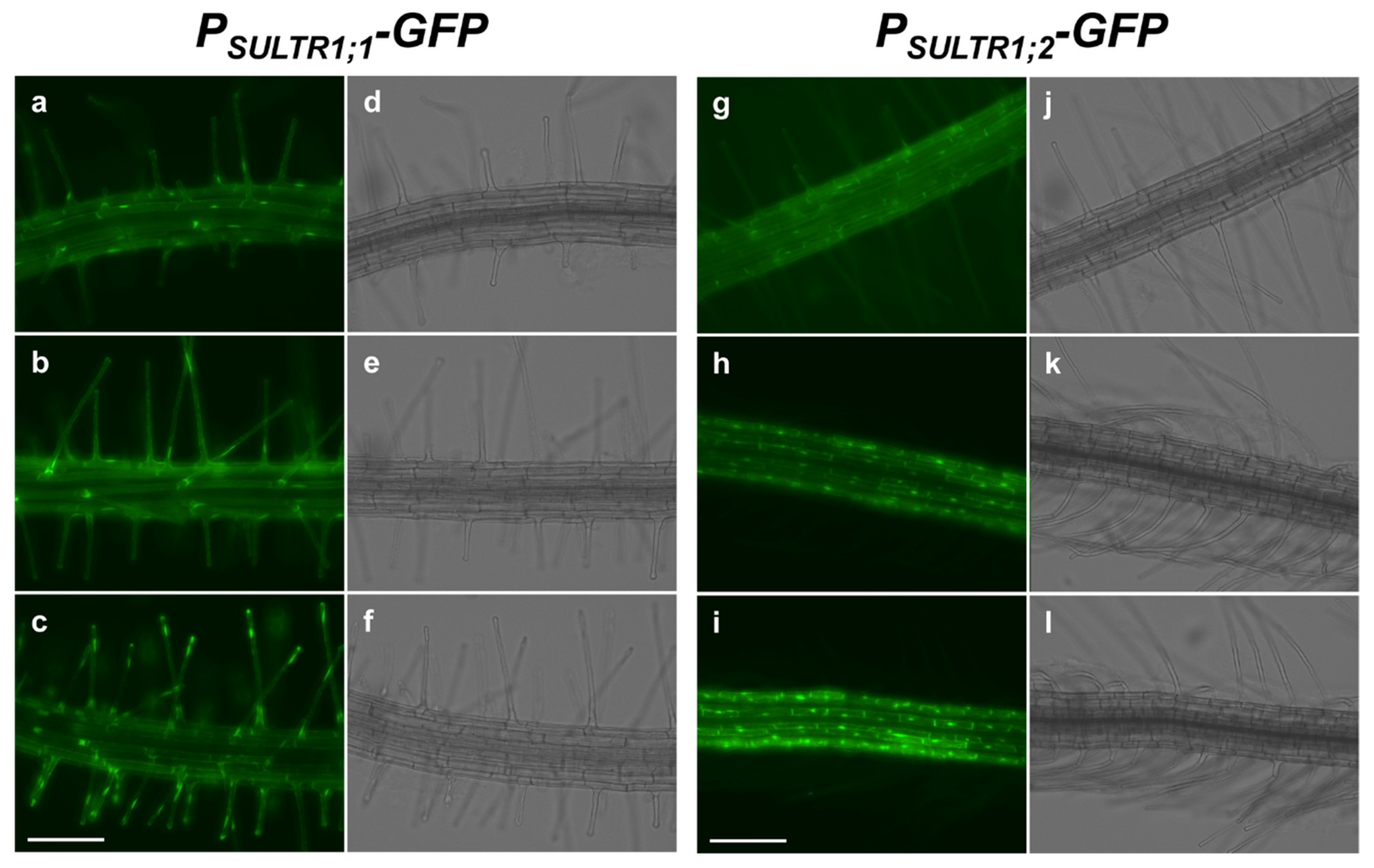
2.1. SULTR1;1 and SULTR1;2 Were Differentially Expressed in the Root Epidermal Cells under −S

The difference in tissue-specific expression between SULTR1;1 and SULTR1;2 in the root surface was investigated by assessing green fluorescent protein (GFP) fluorescence in the root elongation zones of PSULTR1;1-GFP [26] and PSULTR1;2-GFP [27] plants. When the plants were grown under +S sulfur sufficient (S1500) conditions, GFP fluorescence was not detected in either plant line (data not shown). Under −S sulfur deficient (S15) conditions, GFP fluorescence was clearly visible in root elongation zones (Figure 1). In PSULTR1;1-GFP plants, GFP fluorescence was mainly detected in root hair cells (Figure 1a–c), whereas it was detected in non-hair cells in PSULTR1;2-GFP plants (Figure 1g–i). Interestingly, GFP fluorescence was detected in a striped pattern in the root hair cells and non-hair cells in PSULTR1;1-GFP and PSULTR1;2-GFP plants, respectively, suggesting that they were differentially expressed in these cell lines and played different roles in sulfate uptake from the root surface.

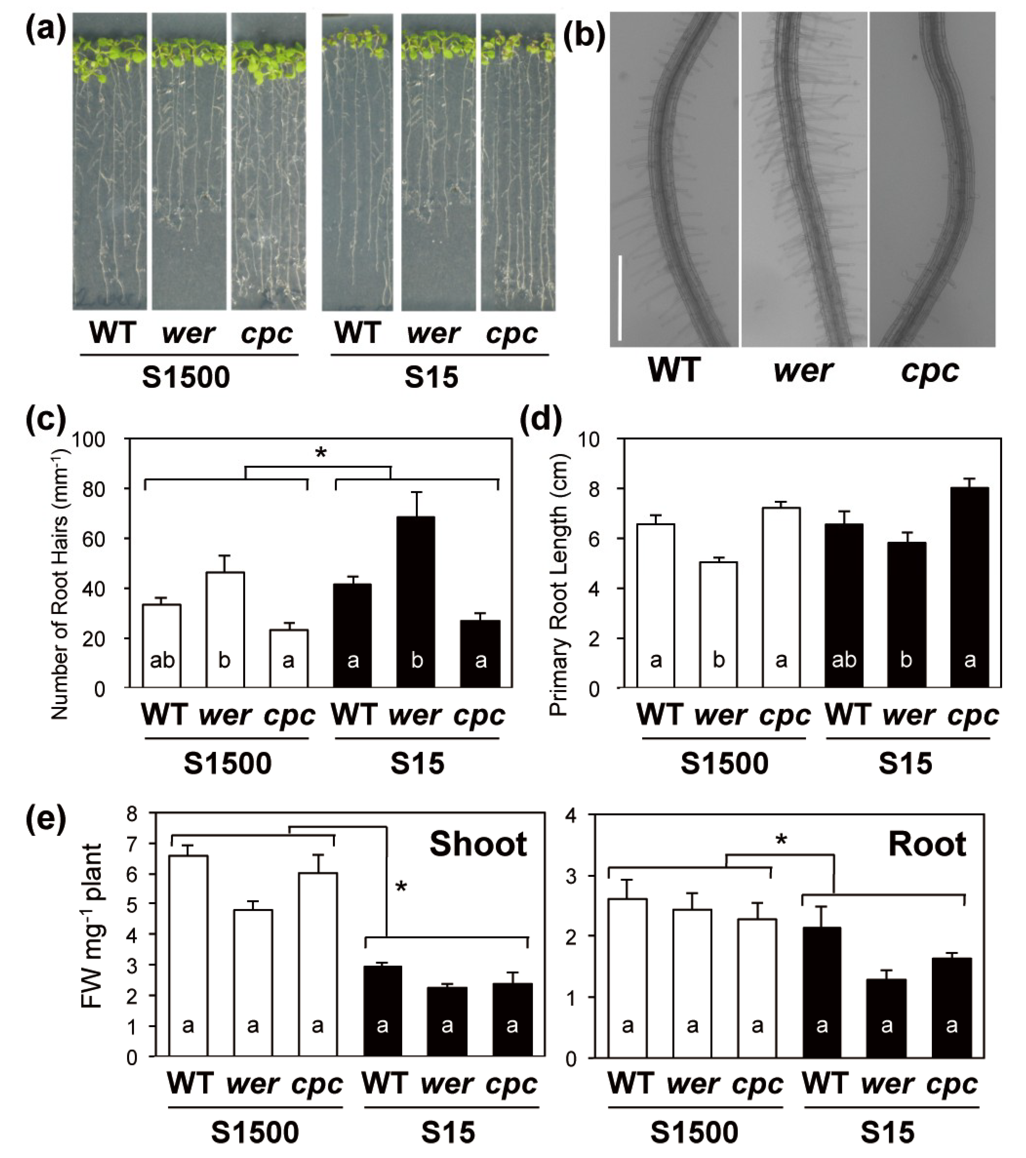
2.2. Primary Root Length and Numbers of Root Hairs Compensated for Each Other in wer and cpc

To assess the relationship between sulfate uptake and root hair development, root phenotypes of wer, cpc and wild-type Columbia plants (WT) under S1500 and S15 conditions were observed (Figure 2). The number of root hairs 5 mm from the root tip were counted in plants grown under S1500 and S15 conditions (Figure 2a–c). The number of root hairs was significantly higher under S15 conditions than under S1500 conditions (Figure 2c), suggesting that plants increased the number of root hairs for more efficient sulfate uptake under –S. Under both sulfur conditions, wer had more root hairs than the WT, and cpc tended to have less root hairs than the WT, as reported previously [39,41] (Figure 2c).
Primary root lengths were not influenced by sulfate concentrations (Figure 2d). Interestingly, primary root lengths were influenced in the opposite direction as the number of root hairs in the two mutants as compared to the WT. That is, primary root lengths tended to be shorter in wer and longer in cpc than they were in WT under both the sulfur conditions. Significant negative correlations between the number of root hairs and primary root lengths were detected using Sperman’s Rank correlation coefficient (R = 0.047, correlation constant = −0.89). These phenotypes suggested that plants adjusted their root biomass by balancing the inverse relationship between primary root lengths and root hair densities.
Both shoot and root fresh weights were decreased under –S conditions (Figure 2e). In wer, shoot fresh weights under S1500 and S15 conditions tended to be less than those in the WT (Figure 2e). When plants were grown under the S15 condition, the root fresh weights of wer and cpc tended to be less than those in the WT (Figure 2e).

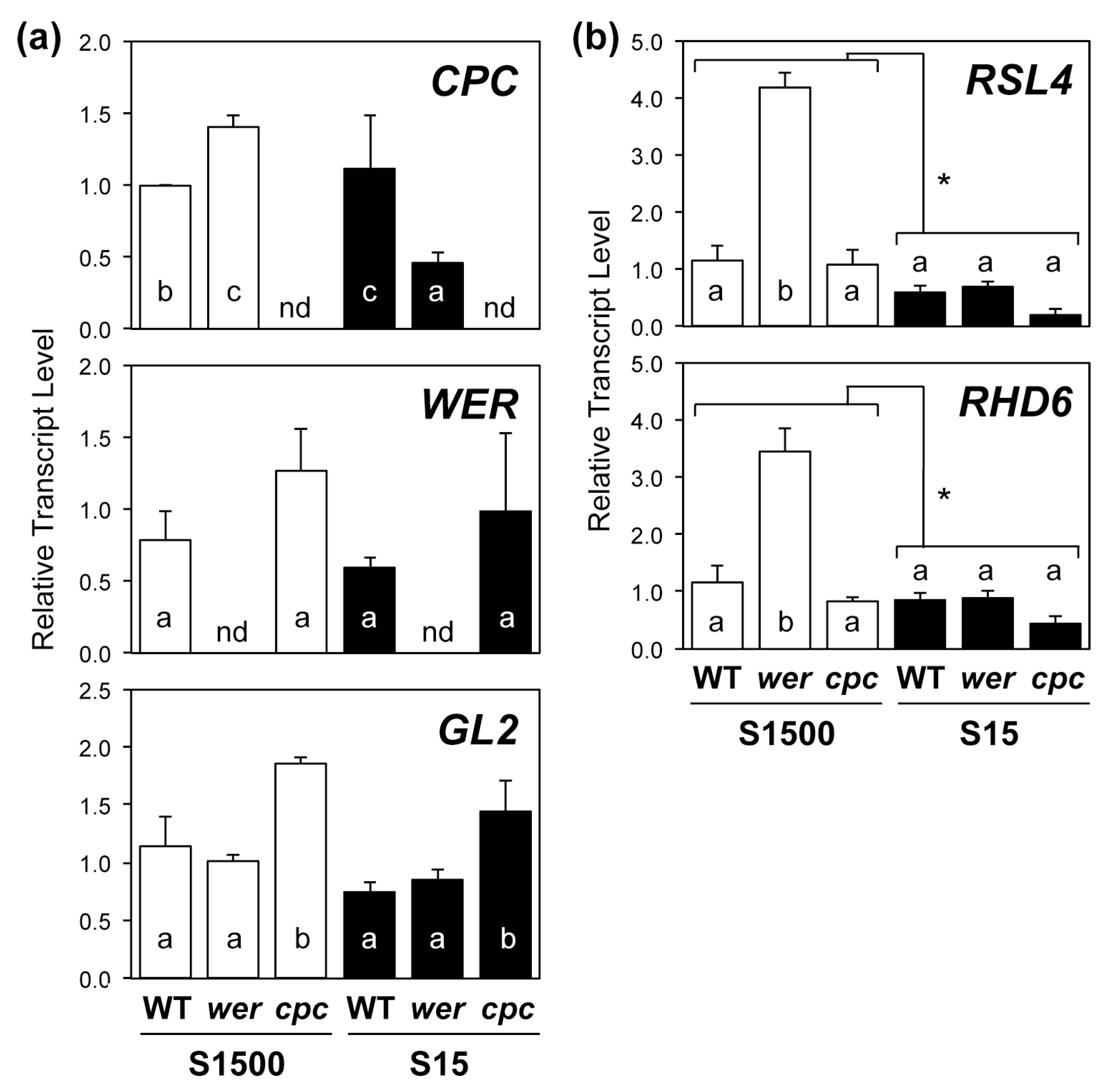
2.3. Regulatory Gene Expression in Root Hair Development Was Consistent with Root Hair Numbers under +S but Not –S Conditions

The number of root hairs was increased under –S conditions and, therefore, we analyzed the expression of transcription factors CPC, WER, GL2, RHD6, and RSL4, which regulate root hair development (Figure 3).
The expression of CPC in wer was higher under the S1500 condition but lower under the S15 condition than that in the WT, whereas the expression of WER was not influenced in cpc (Figure 3a). The expression of GL2 was higher in cpc under both conditions than that in the WT.
The transcript levels of root hair-specific transcription factors, RHD6 and RSL4, were higher in wer under the S1500 condition than they were in WT but were similar among the genotypes under S15 (Figure 3b). Their transcript levels tended to be lower in cpc under both conditions than they were in the WT, which was likely due to the increased expression of GL2 in cpc as GL2 suppressed the expression of RHD6 [49]. With regard to the expression of RHD6 and RSL4, an interaction was found between sulfur conditions and genotypes using a two-way ANOVA, suggesting their expressions were differentially influenced by the disruption of WER or the number of root hairs under +S and −S.

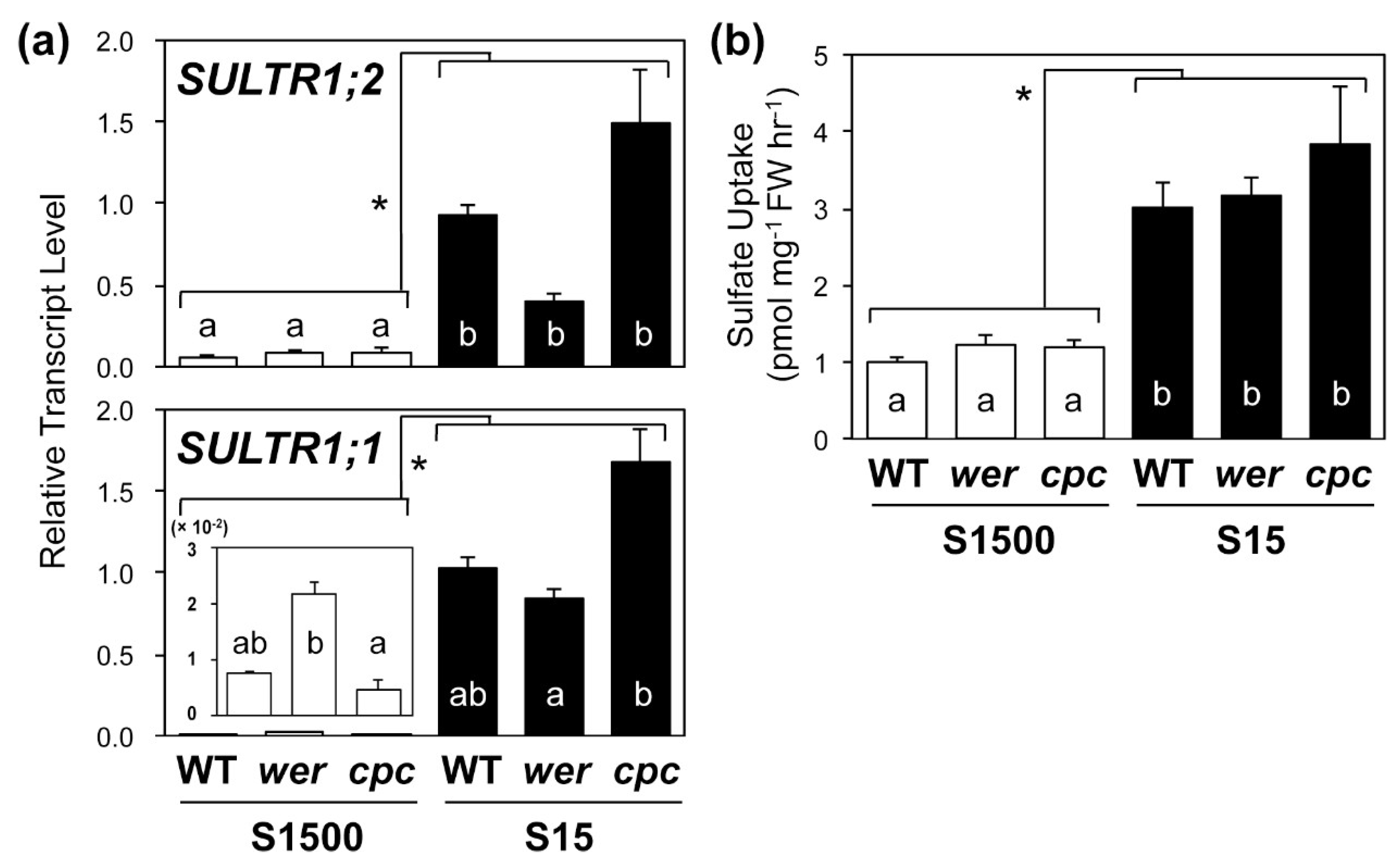
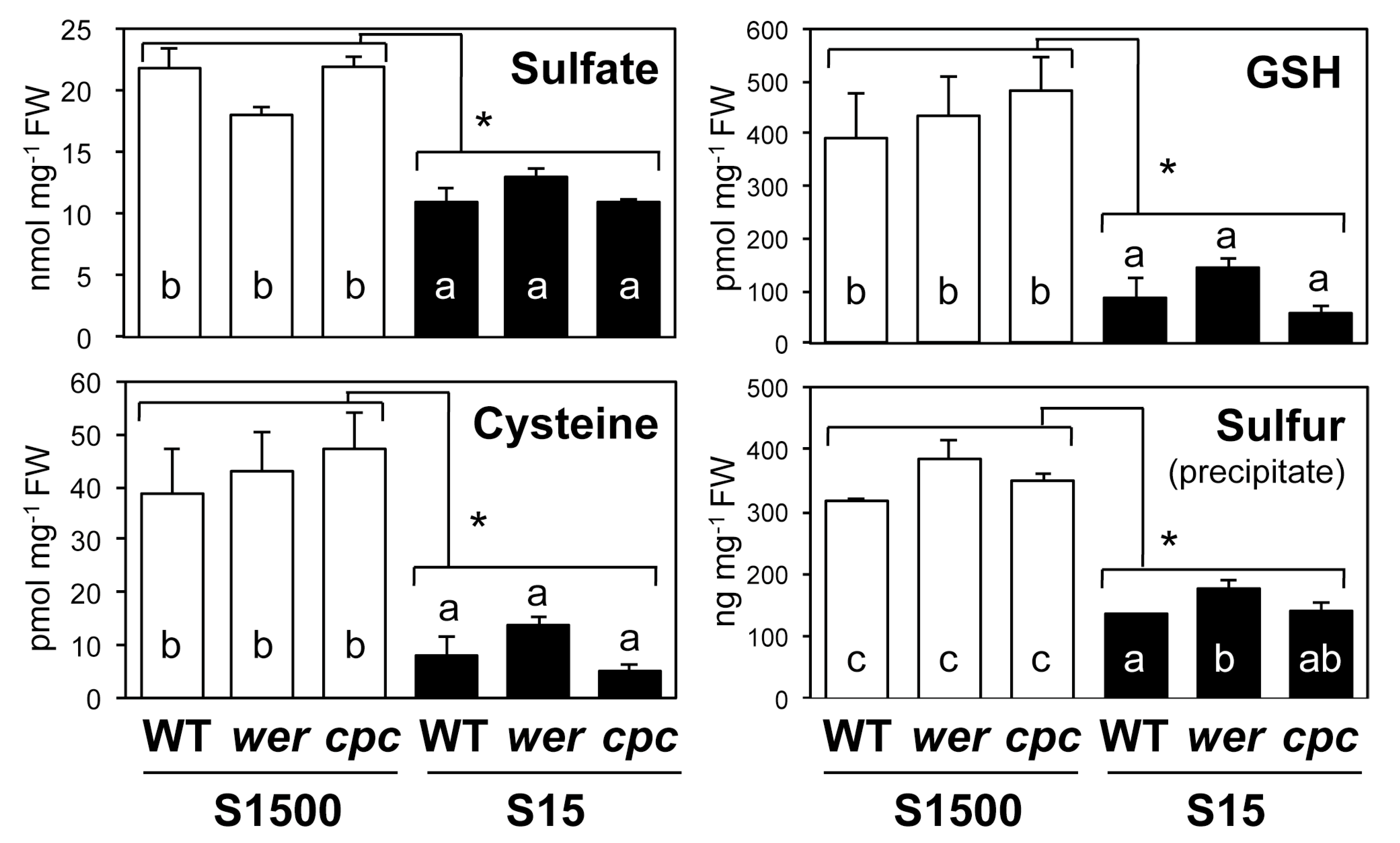
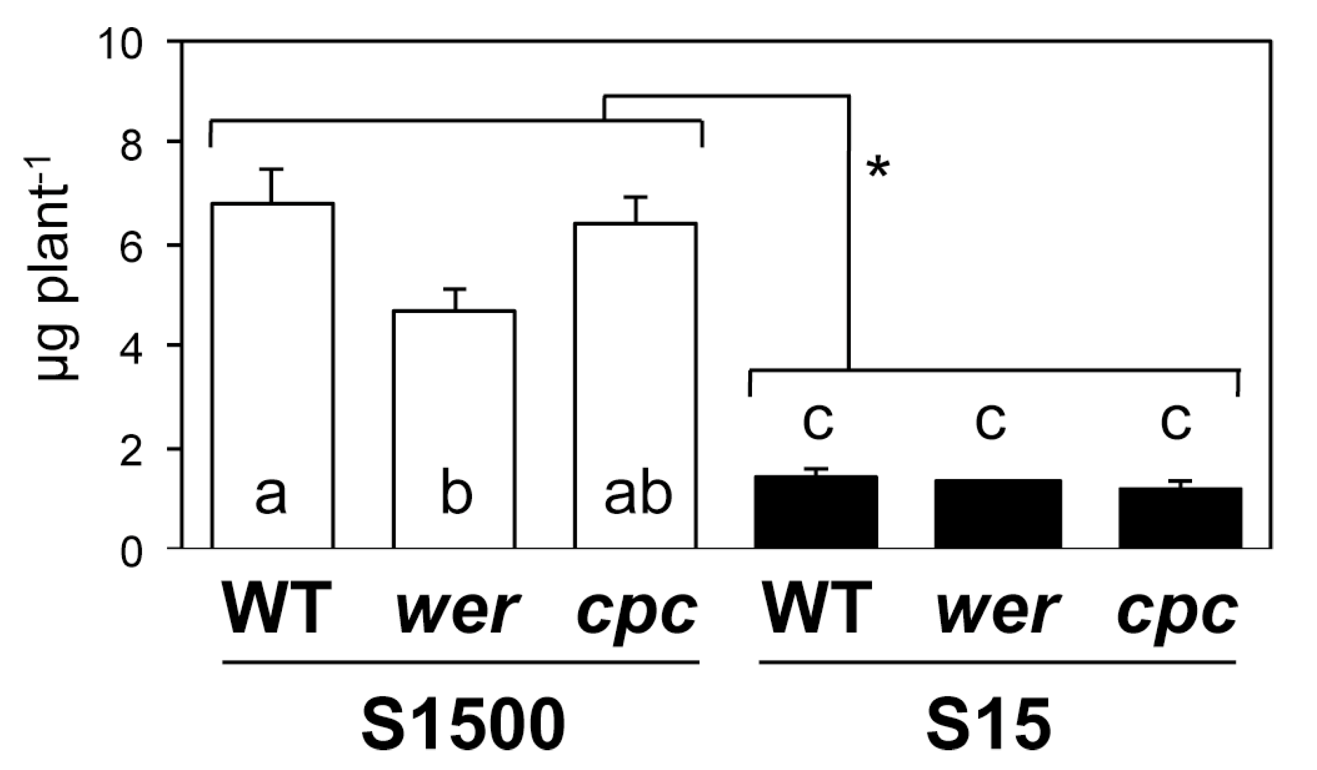
2.4. Sulfate Uptake Was Constant among WT, wer, and cpc

We analyzed the transcript levels of SULTR1;1 and SULTR1;2 in the roots and the sulfate uptake activity in WT, wer, and cpc plants grown under S1500 and S15 conditions (Figure 4). Sulfur deficiency increased the transcript levels of SULTR1;1 and SULTR1;2 in all plant lines (Figure 4a). Under the S1500 condition, transcript levels of SULTR1;2 were similar between the mutants and WT, whereas those of SULTR1;1 were higher in wer and lower in cpc than they were in WT (Figure 3a), which is consistent with previous reports [53,54]. These results indicated that the expression of SULTR1;1 was correlated with the number of root hairs when there was sufficient sulfate for plant growth (Figure 2b,c and Figure 4a).
However, under –S, transcript levels of SULTR1;1 were lower in wer than they were in cpc, indicating a negative correlation with the number of root hairs (Figure 2b,c and Figure 4a). Supporting this observation, interactive effects between sulfur conditions and genotypes on the expression of SULTR1;1 and SULTR1;2 were detected using a two-way ANOVA, which indicates that the effects of the number of root hairs on their expression would be switched from positive to negative with environmental changes from +S to −S.
The sulfate uptake rate was stimulated under the S15 condition, which was consistent with previous reports [23,24,25,27,55,56] (Figure 4b). Despite the differences in SULTR1;1 expression between the plant lines, sulfate uptake rates were relatively constant between the two lines under both S1500 and S15 conditions (Figure 4b).
The levels of sulfur-containing metabolites, sulfate, Cys, GSH and total sulfur in the precipitate after extraction with 10 mM HCl were decreased under the S15 condition in all plant lines (Figure 5). Similar to the sulfate uptake rate, the content of sulfate, Cys, and GSH in shoots was generally not influenced by mutations in WER and CPC under both S1500 and S15 conditions, except for slightly higher sulfur levels in the precipitate of wer under the S15 condition than in the WT.
The sum of sulfur levels analyzed in this study was converted to sulfur content in shoot per plant (Figure 6). Concentrations of sulfate, Cys, and GSH (Figure 5) were converted to sulfur content, and added to the sulfur content of the precipitate, and then the total was multiplied by the shoot FW per plant (Figure 2e). The sum of sulfur levels was lower in wer under the S1500 condition than it was in the WT and was similar among the genotypes under the S15 condition (Figure 6). Interaction between sulfur conditions and genotypes was detected using a two-way ANOVA, suggesting that sulfur levels were differentially controlled by the number of root hairs under +S and −S conditions.

3. Discussion

The mechanisms by which cellular differentiation influences the cellular biochemical or physiological functions and, vice versa, how cellular function influences cell fate, have both long been topics of discussion. In the case of sulfate uptake, two high-affinity sulfate transporters, SULTR1;1 and SULTR1;2, show different spatial expression patterns in root epidermal cells [21,24,26] (Figure 1). Specifically, we found that the expression of SULTR1;1 was induced preferentially in the root-hair cells in a −S condition [57], whereas SULTR1;2 was preferentially expressed in non-hair cells (Figure 1). Additionally, the expression of SULTR1;1 was reported to be higher in wer and lower in cpc than in WT, whereas the expression of SULTR1;2 was not influenced in the mutants [53,54]. Based on this information, we postulated that a decrease in the number of root hairs would reduce sulfate uptake activity. However, an increase or decrease in the number of root hairs did not influence the sulfate uptake rate in plants under either +S or −S conditions (Figure 4b). These results indicated that the number of root hairs does not contribute to the sulfate uptake rate, at least in the comparison among WT, wer and cpc.
Although we observed no difference in sulfate uptake between the WT and mutants, the contribution of root hairs to sulfate uptake cannot be completely ruled out because there was a negative correlation between the number of root hairs and primary root length [58] (Figure 2). The negative correlation suggested that there might be a mechanism to maintain a constant root biomass. A similar negative correlation was observed when plants were exposed to phosphate-deficient conditions, ethylene, or both [16,59], suggesting the involvement of general homeostatic mechanisms. The molecular machinery coordinating the negative interaction between root hair development and primary root length would be an interesting subject to investigate as a potential tool to improve the understanding of nutrient acquisition by plants. The S15 condition also induced a slight but significant increase in the number of root hairs (Figure 2c), which could contribute to the increase in sulfate uptake rate under –S conditions. These results suggested that the sulfate uptake rate was influenced by the total root biomass rather than the number of root hairs under both +S and –S conditions.
The expression of both SULTRs was correlated with the number of root hairs negatively under the S15 condition, but positively under the S1500 condition (Figure 2c and Figure 4a). A similar tendency was observed for the expression of RHD6 and RSL4 (Figure 3b). Under the S1500 condition, the expression levels of RHD6 and RSL4 were higher in wer and tended to be lower in cpc than they were in the WT, which was consistent with the number of root hairs (Figure 2c and Figure 3b). However, under the S15 condition, the expression of RHD6 and RSL4 was similar among the plant lines, whereas the number of root hairs varied in the mutants in a manner similar to that under S1500 condition (Figure 2c and Figure 3b). Although it is not clear whether the expression of RHD6, RSL4, and SULTRs was regulated by the same mechanism under –S conditions, these results suggested that another mechanism likely increased root hair development under –S conditions.
Furthermore, phosphate deficiency induces RSL4 expression, probably by increasing the stability of EIN3 and EIL1, which both bind to cognate binding sites in the promoter of RSL4 [16,17]. In another root hair-less line, NR23, several events are induced by phosphate deficiency, including increased the expression of phosphate transporter genes and secretion of acid phosphatases and organic acids, compared to the WT [8]. This observation also supports the differing contribution of root hairs to the uptake of phosphate and sulfate, which varies with the chemical forms of nutrients in the soil [3,4].
Although there were negative or positive correlations between the expression of SULTRs and the number of root hairs under both +S and −S conditions, the sulfate uptake rate did not fluctuate among the plant lines (Figure 4). This finding suggests that the transcript levels of SULTR1;1 and SULTR1;2 were not the only determinants of sulfate uptake rate. Further studies of the relationship between root architecture and sulfate uptake may shed light on the as yet unknown determinants of this process.

4. Materials and Methods

4.1. Plant Materials and Growth Conditions

Arabidopsis thaliana plants, ecotype “Columbia” (Col-0), were used as the wild-type (WT) while cpc [41] (cpc-1) and wer [39] (wer-1) mutants were obtained from the Arabidopsis Biological Resource Center (ABRC). Plants were grown at 22 °C under continuous light (40 µmol m−1 s−1) conditions on mineral nutrient media containing 1% sucrose [60,61]. For the preparation of the agar medium, agar was washed twice with 1 L de-ionized water and vacuum filtered to remove the sulfate. Sulfur sufficient (S1500) agar medium was supplemented with 1500 µM MgSO4. Sulfur-deficient (S15) agar medium was supplemented with 15 µM MgSO4 and Mg concentration was adjusted to 1500 µM by adding MgCl2. After the indicated shown in each figure, the shoot and root tissues were harvested separately, rinsed with distilled water, and subsequently subjected to various analyses.

4.2. Observation of GFP Fluorescence

The tissue-specific expression of GFP in PSULTR1;1-GFP and PSULTR1;2-GFP transgenic plants was visualized in whole mounts of 7-day-old plants using a fluorescent microscope system (EVOS FL Auto 2 Imaging System) equipped with the EVOS Light Cube, GFP (Ex: 470/22, Em: 525/50) (Thermo Fisher Scientific, USA).

4.3. Observation of Root Development

The primary root lengths of plants were analyzed using images captured with a STAGE2000-BG system (AMZ System Science, Japan). The number of root hairs in 5 mm from the root tip was analyzed from the image captured using a CCD camera (WRAYCAM G500, WRAYMER, Japan) connected to a stereoscopic microscope (SW-700TD, WRAYMER). The free software package, ImageJ [62,63] was used for the analysis.

4.4. Quantitative Real-Time RT-PCR Analysis

RNA preparation and RT were performed as reported previously [27,54,55]. Real-time PCR was carried out using a SYBR Green Perfect real-time kit (Takara, Japan) and Thermal Cycler Dice real-time system (Takara) using the gene-specific primers for CPC, CPC-F (5′-GGATGTATAAACTCGTTGGCGACAG-3′) and CPC-R (5′-GCCGTGTTTCATAAGCCAATATCTC-3′) [64]; for WER, WER-F (5′-TGGTAATAGGTATAACTTCATTTGC-3′) and WER-R (5′-TTTGATTCCGAGTTTCTTACTAAGGATG-3′); for GL2, GL2-F (5′-TCGGATCACTGAGACCACAA-3′) and GL2-R (5′-GTGTATCCCGGAACCAGTGT-3′) [64]; for RHD6, RHD6-real-F (5′-TGATTTGGTGACAATGCTTGA-3′) and RHD6-real-R (5′-GGAGAGAATGGCATCAATGG-3′) [49]; for RSL4, RSL4_q-PCR f new (5′-AACCTTGTGCCAAACGGGAC-3′) and RSL4_q-PCR r new (5′-CCAGGCCGTTGTAAGCCAAT-3′) [17]; and, for SULTR1;2, SULTR1;2-1854F (5′-GGATCCAGAGATGGCTACATGA-3′) and SULTR1;2-1956R (5′-TCGATGTCCGTAACAGGTGAC-3′) [27]; for SULTR1;1, SULTR1;1-625F (5′-GCCATCACAATCGCTCTCCAA-3′) and SULTR1;1-750R (5′-TTGCCAATTCCACCCATGC-3′) [30]; and for ubiquitin, UBQ2-144F (5′-CCAAGATCCAGGACAAAGAAGGA-3′) and UBQ2-372R (5′-TGGAGACGAGCATAACACTTGC-3′) [30]. The relative mRNA levels were calculated using ubiquitin2 as an internal standard.

4.5. Sulfate Uptake Assay

Plants were vertically grown for 10 days on S1500 and S15 agar media. The roots were submerged in S1500 medium containing 15 µM [35S] sodium sulfate (American Radiolabeled Chemicals, USA) and incubated for 1 h. Washing and measurement were carried out as described previously [25,27,54,55,65,66].

4.6. Measurement of Sulfate, Cysteine and Glutathione, and Total Sulfur Levels

Plant tissues were frozen in liquid nitrogen and homogenized in 5 volumes of 10 mM HCl using a Tissue Lyser (Retsch, Germany). After homogenization, the cell debris was removed by centrifugation, and the supernatant was subsequently analyzed.
For sulfate measurement, the extracts were diluted 100-fold with extra pure water and analyzed using ion chromatography (IC-2001, TOSOH, Japan). Using serial 30-µL injections, the diluted extracts were separated at 40 °C using a TSK SuperIC-AZ column (TOSOH) at a flow rate of 0.8 mL min−1 with an eluent containing 1.9 mM NaHCO3 and 3.2 mM Na2CO3. Anion mixture standard solution 1 (Wako Pure Chemicals, Japan) was used as a standard.
Cys and GSH contents were determined using monobromobimane (Invitrogen, USA) labeling of the thiols after reduction of the plant extracts with dithiothreitol (DTT). The labeled products were then separated using HPLC (JASCO, Japan) using the TSKgel ODS-120T column (150 × 4.6 mm, TOSOH) and detected using a fluorescence detector, FP-920 (JASCO), monitoring for fluorescence of thiol-bimane adducts at 482 nm under excitation at 390 nm. Cys and GSH (Nacalai Tesque, Japan) were used as standards.
The total sulfur content was determined using inductively coupled plasma-mass spectroscopy (ICP-MS; Agilent7700, Agilent Technologies, USA). The precipitates obtained from the extraction described above were digested in 200 µL HNO3 at 95 °C for 30 min and then 115 °C for 90 min. After cooling to room temperature, the digested samples were diluted to 1 mL with extra pure water, filtered using 0.45 µm filters (DISMIC-03CP, ADVANTEC, Japan). The filtered samples were diluted 10 times with a solution consisting of 0.1 M HNO3 and 10 µg L−1 gallium (KANTO CHEMICAL, Japan) as an internal standard, before being subjected to ICP-MS. Quantification was performed using the standard curve obtained via serial dilutions of the sulfur standard solution (KANTO CHEMICAL).

4.7. Statistical Analysis

Two-way ANOVA was used to detect the effects of sulfur conditions, genotypes, and their interactions (Figure 2, Figure 3, Figure 4, Figure 5 and Figure 6). Significant differences between S1500 and S15 conditions detected using a two-way ANOVA are indicated with asterisks (P < 0.05). Furthermore, where an interaction was detected between sulfur conditions and genotypes, the Tukey–Kramer test was applied to all experimental conditions (Figure 3 and Figure 4a). In addition, where interaction was not detected between the values, the Tukey–Kramer test was used to analyze all genotypes grown under the same growth condition. Significant differences detected by the Tukey–Kramer test were shown with different letters (P < 0.05). Statcel4 software (OMS Publishing Inc., Tokyo, Japan) was used for all statistical analysis with the Microsoft Excel program.

Author Contributions

A.M.N., R.T.W. and T.W. designed the research. Y.K., T.U., A.S. and A.M.N. performed the experiments and analyzed the data. A.M.N. wrote the manuscript.

Funding

This work was supported by JSPS KAKENHI Grant Number 24380040 and 17H03785 (for A.M.N.).

Acknowledgments

We thank Yuki Mori (Kyushu University, Japan) for instructing the total sulfur analysis with ICP-MS. We thank Yasuo Niwa (University of Shizuoka, Japan) for providing the sGFP(S65T) vector. We thank Chihiro Iwaya and Yukiko Okuo for the technical support.

Conflicts of Interest

The authors declare no conflict of interest.

References

  1. White, P. Ion uptake mechanisms of individual cells and roots: Short-distance transport. In Mineral Nutrition of Higher Plants, 3rd ed.; Marschner, P., Ed.; Academic Press: Oxford, UK, 2012; pp. 7–48. [Google Scholar]
  2. Patrick, J.W.; Tyerman, S.D.; van Bel, A.J.E. Long-Distance Transport. In Biochemistry & Molecular Biology of Plants, 2nd ed.; Buchana, B.B., Gruissem, W., Jones, R.L., Eds.; Wiley Blackwell: Oxford, UK, 2015; pp. 658–710. [Google Scholar]
  3. Lopez-Bucio, J.; Cruz-Ramirez, A.; Herrera-Estrella, L. The role of nutrient availability in regulating root architecture. Curr. Opin. Plant Biol. 2003, 6, 280–287. [Google Scholar] [CrossRef]
  4. Delhaize, E.; Schachtman, D.; Kochian, L.; Ryan, P.R. Mineral nutrient acquisition, transport, and utilization. In Biochemistry & Molecular Biology of Plants, 2nd ed.; Buchana, B.B., Gruissem, W., Jones, R.L., Eds.; Wiley Blackwell: Oxford, UK, 2015; pp. 1101–1131. [Google Scholar]
  5. Long, S.R.; Kahn, M.; Seefeldt, L.; Tsay, Y.F.; Kopriva, S. Nitrogen and Sulfur. In Biochemistry & Molecular Biology of Plants, 2nd ed.; Buchana, B.B., Gruissem, W., Jones, R.L., Eds.; Wiley Blackwell: Oxford, UK, 2015; pp. 746–768. [Google Scholar]
  6. Schiefelbein, J.; Kwak, S.-H.; Wieckowski, Y.; Barron, C.; Bruex, A. The gene regulatory network for root epidermal cell-type pattern formation in Arabidopsis. J. Exp. Bot. 2009, 60, 1515–1521. [Google Scholar] [CrossRef] [PubMed]
  7. Libault, M.; Brechenmacher, L.; Cheng, J.; Xu, D.; Stacey, G. Root hair systems biology. Trends Plant Sci. 2010, 15, 641–650. [Google Scholar] [CrossRef] [PubMed]
  8. Tanaka, N.; Kato, M.; Tomioka, R.; Kurata, R.; Fukao, Y.; Aoyama, T.; Maeshima, M. Characteristics of a root hair-less line of Arabidopsis thaliana under physiological stresses. J. Exp. Bot. 2014, 65, 1497–1512. [Google Scholar] [CrossRef] [PubMed]
  9. Gilroy, S.; Jones, D.L. Through form to function: Root hair development and nutrient uptake. Trends Plant Sci. 2000, 5, 56–60. [Google Scholar] [CrossRef]
  10. Gahoonia, T.S.; Nielsen, N.E.; Joshi, P.A.; Jahoor, A. A root hairless barley mutant for elucidating genetic of root hairs and phosphorus uptake. Plant Soil 2001, 235, 211–219. [Google Scholar] [CrossRef]
  11. Schmidt, W.; Tittel, J.; Schikora, A. Role of hormones in the induction of iron deficiency responses in Arabidopsis roots. Plant Physiol. 2000, 122, 1109–1118. [Google Scholar] [CrossRef]
  12. Ma, J.F.; Goto, S.; Tamai, K.; Ichii, M. Role of root hairs and lateral roots in silicon uptake by rice. Plant Phys. 2001, 127, 1773–1780. [Google Scholar] [CrossRef]
  13. Ma, Z.; Bielenberg, D.G.; Brown, K.M.; Lynch, J.P. Regulation of root hair density by phosphorus availability in Arabidopsis thaliana. Plant Cell Environ. 2001, 24, 459–467. [Google Scholar] [CrossRef]
  14. Mueller, M.; Schmidt, W. Environmentally induced plasticity of root hair development in Arabidopsis. Plant Phys. 2004, 134, 409–419. [Google Scholar] [CrossRef] [PubMed]
  15. Yang, T.J.W.; Perry, P.J.; Ciani, S.; Pandian, S.; Schmidt, W. Manganese deficiency alters the patterning and development of root hairs in Arabidopsis. J. Exp. Bot. 2008, 59, 3453–3464. [Google Scholar] [CrossRef] [PubMed]
  16. Song, L.; Yu, H.; Dong, J.; Che, X.; Jiao, Y.; Liu, D. The molecular mechanism of ethylene-mediated root hair development induced by phosphate starvation. PLoS Genet. 2016, 12, e1006194. [Google Scholar] [CrossRef]
  17. Feng, Y.; Xu, P.; Li, B.; Li, P.; Wen, X.; An, F.; Gong, Y.; Xin, Y.; Zhu, Z.; Wang, Y.; et al. Ethylene promotes root hair growth through coordinated EIN3/EIL1 and RHD6/RSL1 activity in Arabidopsis. Proc. Natl. Acad. Sci. USA 2017, 114, 13834–13839. [Google Scholar] [CrossRef]
  18. Takahashi, H.; Kopriva, S.; Giordano, M.; Saito, K.; Hell, R. Sulfur assimilation in photosynthetic organisms: Molecular functions and regulations of transporters and assimilatory enzymes. Annu. Rev. Plant Biol. 2011, 62, 157–184. [Google Scholar] [CrossRef]
  19. Leustek, T.; Martin, M.N.; Bick, J.; Davies, J.P. Pathways and regulation of sulfur metabolism revealed through molecular genetic studies. Annu. Rev. Plant Physiol. Plant Mol. Biol. 2000, 51, 141–166. [Google Scholar] [CrossRef]
  20. Saito, K. Sulfur assimilatory metabolism. The long and smelling road. Plant Physiol. 2004, 136, 2443–2450. [Google Scholar] [CrossRef]
  21. Takahashi, H.; Watanabe-Takahashi, A.; Smith, F.W.; Blake-Kalff, M.; Hawkesford, M.J.; Saito, K. The roles of three functional sulfate transporters involved in uptake and translocation of sulfate in Arabidopsis thaliana. Plant J. 2000, 23, 171–182. [Google Scholar] [CrossRef] [PubMed]
  22. Vidmar, J.J.; Tagmount, A.; Cathala, N.; Touraine, B.; Davidian, J.C.E. Cloning and characterization of a root specific high-affinity sulfate transporter from Arabidopsis thaliana. FEBS Lett. 2000, 475, 65–69. [Google Scholar] [CrossRef]
  23. Shibagaki, N.; Rose, A.; McDermott, J.P.; Fujiwara, T.; Hayashi, H.; Yoneyama, T.; Davies, J.P. Selenate-resistant mutants of Arabidopsis thaliana identify Sultr1;2, a sulfate transporter required for efficient transport of sulfate into roots. Plant J. 2002, 29, 475–486. [Google Scholar] [CrossRef] [PubMed]
  24. Yoshimoto, N.; Takahashi, H.; Smith, F.W.; Yamaya, T.; Saito, K. Two distinct high-affinity sulfate transporters with different inducibilities mediate uptake of sulfate in Arabidopsis roots. Plant J. 2002, 29, 465–473. [Google Scholar] [CrossRef] [PubMed]
  25. Yoshimoto, N.; Inoue, E.; Watanabe-Takahashi, A.; Saito, K.; Takahashi, H. Posttranscriptional regulation of high-affinity sulfate transporters in Arabidopsis by sulfur nutrition. Plant Physiol. 2007, 145, 378–388. [Google Scholar] [CrossRef] [PubMed]
  26. Maruyama-Nakashita, A.; Nakamura, Y.; Watanabe-Takahashi, A.; Yamaya, T.; Takahashi, H. Induction of SULTR1;1 sulfate transporter in Arabidopsis roots involves protein phosphorylation/dephosphorylation circuit for transcriptional regulation. Plant Cell Physiol. 2004, 45, 340–345. [Google Scholar] [CrossRef]
  27. Maruyama-Nakashita, A.; Nakamura, Y.; Yamaya, T.; Takahashi, H. A novel regulatory pathway of sulfate uptake in Arabidopsis roots: Implication of CRE1/WOL/AHK4-mediated cytokinin-dependent regulation. Plant J. 2004, 38, 779–789. [Google Scholar] [CrossRef]
  28. Maruyama-Nakashita, A.; Nakamura, Y.; Watanabe, A.; Inoue, E.; Yamaya, T.; Takahashi, H. Identification of a novel cis-acting element conferring sulfur deficiency response in Arabidopsis roots. Plant J. 2005, 42, 305–314. [Google Scholar] [CrossRef] [PubMed]
  29. Maruyama-Nakashita, A. Metabolic changes sustain the plant life in low-sulfur environments. Curr. Opin. Plant Biol. 2017, 39, 144–151. [Google Scholar] [CrossRef]
  30. Maruyama-Nakashita, A.; Inoue, E.; Watanabe-Takahashi, A.; Yamaya, T.; Takahashi, H. Transcriptome profiling of sulfur-responsive genes in Arabidopsis reveals global effect on sulfur nutrition on multiple metabolic pathways. Plant Physiol. 2003, 132, 597–605. [Google Scholar] [CrossRef]
  31. Rouached, H.; Wirtz, M.; Alary, R.; Hell, R.; Arpat, A.B.; Davidian, J.C.; Fourcroy, P.; Berthomieu, P. Differential regulation of the expression of two high-affinity sulfate transporters, SULTR1.1 and SULTR1.2, in Arabidopsis. Plant Physiol. 2008, 147, 897–911. [Google Scholar] [CrossRef]
  32. Grebe, M. The patterning of epidermal hairs in Arabidopsis—updated. Curr. Opin. Plant Biol. 2012, 15, 31–37. [Google Scholar] [CrossRef]
  33. Salazar-Henao, J.E.; Veélez-Bermuédez, I.C.; Schmidt, W. The regulation and plasticity of root hair patterning and morphogenesis. Development 2016, 143, 1848–1858. [Google Scholar] [CrossRef]
  34. Ishida, T.; Kurata, T.; Okada, K.; Wada, T. A genetic regulatory network in the development of trichomes and root hairs. Annu. Rev. Plant Biol. 2008, 59, 365–386. [Google Scholar] [CrossRef] [PubMed]
  35. Shibata, M.; Sugimoto, K. A gene regulatory network for root hair development. J. Plant Res. 2019. [Google Scholar] [CrossRef] [PubMed]
  36. Walker, A.R.; Davison, P.A.; Bolognesi-Winfield, A.C.; James, C.M.; Srinivasan, N.; Blundell, T.L.; Esch, J.J.; Marks, M.D.; Gray, J.C. The TRANSPARENT TESTA GLABRA1 Locus, which regulates trichome differentiation and anthocyanin biosynthesis in Arabidopsis, encodes a WD40 repeat protein. Plant Cell 1999, 11, 1337–1349. [Google Scholar] [CrossRef]
  37. Payne, C.T.; Zhang, F.; Lloyd, A.M. GL3 encodes a bHLH protein that regulates trichome development in Arabidopsis through interaction with GL1 and TTG1. Genetics 2000, 156, 1349–1362. [Google Scholar]
  38. Bernhardt, C.; Lee, M.M.; Gonzalez, A.; Zhang, F.; Lloyd, A.; Schiefelbein, J. The bHLH genes GLABRA3 (GL3) and ENHANCER OF GLABRA3 (EGL3) specify epidermal cell fate in the Arabidopsis root. Development 2003, 130, 6431–6439. [Google Scholar] [CrossRef]
  39. Lee, M.M.; Schiefelbein, J. WERWOLF, a MYB-related protein in Arabidopsis, is a position-dependent regulator of epidermal cell patterning. Cell 1999, 99, 473–483. [Google Scholar] [CrossRef]
  40. Rerie, W.G.; Feldmann, K.A.; Marks, M.D. The GLABRA2 gene encodes a homeo domain protein required for normal trichome development in Arabidopsis. Genes Dev. 1994, 8, 1388–1399. [Google Scholar] [CrossRef] [PubMed]
  41. Wada, T.; Tachibana, T.; Shimura, Y.; Okada, K. Epidermal cell differentiation in Arabidopsis determined by a Myb-homolog, CPC. Science 1997, 277, 1113–1116. [Google Scholar] [CrossRef] [PubMed]
  42. Schellmann, S.; Schnittger, A.; Kirik, V.; Wada, T.; Okada, K.; Beermann, A.; Thumfahrt, J.; Jürgens, G.; Hülskamp, M. TRIPTYCHON and CAPRICE mediate lateral inhibition during trichome and root hair patterning in Arabidopsis. EMBO J. 2002, 21, 5036–5046. [Google Scholar] [CrossRef] [PubMed]
  43. Kirik, V.; Simon, M.; Huelskamp, M.; Schiefelbein, J. The ENHANCER OF TRY AND CPC1 gene acts redundantly with TRIPTYCHON and CAPRICE in trichome and root hair cell patterning in Arabidopsis. Dev. Biol. 2004, 268, 506–513. [Google Scholar] [CrossRef]
  44. Tominaga, R.; Iwata, M.; Sano, R.; Okada, K.; Wada, T. Arabidopsis CAPRICE-like Myb 3 (CPL3) controls endoreduplication and flowering development in addition to trichome and root-hair formation. Development 2008, 135, 1335–1345. [Google Scholar] [CrossRef]
  45. Tominaga, R.; Iwata, M.; Okada, K.; Wada, T. Functional analysis of the epidermal-specific MYB genes CAPRICE and WEREWOLF in Arabidopsis. Plant Cell 2007, 19, 2264–2277. [Google Scholar] [CrossRef] [PubMed]
  46. Song, S.K.; Ryu, K.H.; Kang, Y.H.; Song, J.H.; Cho, Y.H.; Yoo, S.D.; Schiefelbein, J.; Lee, M.M. Cell fate in the Arabidopsis root epidermis is determined by competition between WEREWOLF and CAPRICE. Plant Physiol. 2011, 157, 1196–1208. [Google Scholar] [CrossRef]
  47. Masucci, J.D.; Schiefelbein, J.W. The rhd6 mutation of Arabidopsis thaliana alters root-hair initiation through an auxin- and ethylene-associated process. Plant Physiol. 1994, 106, 1335–1346. [Google Scholar] [CrossRef]
  48. Menand, B.; Yi, K.; Jouannic, S.; Hoffmann, L.; Ryan, E.; Linstead, P.; Schaefer, D.G.; Dolan, L. An ancient mechanism controls the development of cells with a rooting function in land plants. Science 2007, 316, 1477–1480. [Google Scholar] [CrossRef] [PubMed]
  49. Lin, Q.; Ohashi, Y.; Kato, M.; Tsuge, T.; Gu, H.; Qu, L.-J.; Aoyama, T. GLABRA2 directly suppresses basic helix-loop-helix transcription factor genes with diverse functions in root hair development. Plant Cell 2015, 27, 2894–2906. [Google Scholar] [CrossRef]
  50. Yi, K.; Menand, B.; Bell, E.; Dolan, L. A basic helix-loop-helix transcription factor controls cell growth and size in root hairs. Nat. Genet. 2010, 42, 264–267. [Google Scholar] [CrossRef]
  51. Rubio, V.; Linhares, F.; Solano, R.; Martin, A.C.; Iglesias, J.; Leyva, A.; Paz-Ares, J. A conserved MYB transcription factor involved in phosphate starvation signaling both in vascular plants and in unicellular algae. Genes Dev. 2001, 15, 2122–2133. [Google Scholar] [CrossRef] [PubMed]
  52. Bustos, R.; Castrillo, G.; Linhares, F.; Puga, M.I.; Rubio, V.; Pérez-Pérez, J.; Solano, R.; Leyva, A.; Paz-Ares, J. A central regulatory system largely controls transcriptional activation and repression responses to phosphate starvation in Arabidopsis. PLoS Genet. 2010, 6, e1001102. [Google Scholar] [CrossRef]
  53. Bruex, A.; Kainkaryam, R.M.; Wieckowski, Y.; Kang, Y.H.; Bernhardt, C.; Xia, Y.; Zheng, X.; Wang, J.Y.; Lee, M.M.; Benfey, P.; et al. A gene regulatory network for root epidermis cell differentiation in Arabidopsis. PLoS Genet. 2012, 8, e1002446. [Google Scholar] [CrossRef] [PubMed]
  54. Simon, M.; Bruex, A.; Kainkaryam, R.M.; Zheng, X.; Huang, L.; Woolf, P.J.; Schiefelbein, J. Tissue-specific profiling reveals transcriptome alterations in Arabidopsis mutants lacking morphological phenotypes. Plant Cell 2013, 25, 3175–3185. [Google Scholar] [CrossRef] [PubMed]
  55. Maruyama-Nakashita, A.; Nakamura, Y.; Tohge, T.; Saito, K.; Takahashi, H. Arabidopsis SLIM1 is a central transcriptional regulator of plant sulfur response and metabolism. Plant Cell 2006, 18, 3235–3251. [Google Scholar] [CrossRef]
  56. Maruyama-Nakashita, A.; Watanabe-Takahashi, A.; Inoue, E.; Yamaya, T.; Saito, K.; Takahashi, H. Sulfur-responsive elements in the 3’-nontranscribed intergenic region are essential for the induction of SULFATE TRANSPORTER 2;1 gene expression in Arabidopsis roots under sulfur deficiency. Plant Cell 2015, 27, 1279–1296. [Google Scholar] [CrossRef] [PubMed]
  57. Lan, P.; Li, W.; Lin, W.-D.; Santi, S.; Schmidt, W. Mapping gene activity of Arabidopsis root hairs. Genome Biol. 2013, 14, R67. [Google Scholar] [CrossRef]
  58. Tominaga-Wada, R.; Wada, T. Relationship between root hair formation and primary root length in Arabidopsis thaliana. Res. Rev. Biosci. 2017, 12, 128. [Google Scholar]
  59. Negi, S.; Ivanchenko, M.G.; Muday, G.K. Ethylene regulates lateral root formation and auxin transport in Arabidopsis thaliana. Plant J. 2008, 55, 175–187. [Google Scholar] [CrossRef]
  60. Fujiwara, T.; Hirai, M.Y.; Chino, M.; Komeda, Y.; Naito, S. Effects of sulfur nutrition on expression of the soybean seed storage protein genes in transgenic petunia. Plant Physiol. 1992, 99, 263–268. [Google Scholar] [CrossRef] [PubMed]
  61. Hirai, M.Y.; Fujiwara, T.; Chino, M.; Naito, S. Effects of sulfate concentrations on the expression of a soybean seed storage protein gene and its reversibility in transgenic Arabidopsis thaliana. Plant Cell Physiol. 1995, 36, 1331–1339. [Google Scholar] [PubMed]
  62. Schneider, C.A.; Rasband, W.S.; Eliceiri, K.W. NIH image to ImageJ: 25 years of image analysis. Nat. Methods 2012, 9, 671–675. [Google Scholar] [CrossRef] [PubMed]
  63. Hartig, S.M. Basic image analysis and manipulation in ImageJ. Curr. Protoc. Mol. Biol. 2013, 102, 14–15. [Google Scholar] [CrossRef]
  64. Hayashi, N.; Tetsumura, T.; Sawa, S.; Wada, T.; Tominaga-Wada, R. CLE14 peptide signaling in Arabidopsis root hair cell fate determination. Plant Biotechnol. 2018, 35, 17–22. [Google Scholar] [CrossRef]
  65. Kataoka, T.; Hayashi, N.; Yamaya, T.; Takahashi, H. Root-to-shoot transport of sulfate in Arabidopsis: Evidence for the role of SULTR3; 5 as a component of low-affinity sulfate transport system in the root vasculature. Plant Physiol. 2004, 136, 4198–4204. [Google Scholar] [CrossRef] [PubMed]
  66. Yoshimoto, N.; Kataoka, T.; Maruyama-Nakashita, A.; Takahashi, H. Measurement of uptake and root-to-shoot distribution of sulfate in Arabidopsis seedlings. Bio-Protocol 2016, 6, e1700. [Google Scholar] [CrossRef]
Figure 1. Expression of green fluorescent protein (GFP) in the root elongation zones of PSULTR1;1-GFP plants (af, [26]) and PSULTR1;2-GFP plants (gl, [27]). Three T2 generation lines carrying each construct were grown for 7 d on agar media supplied with 15 μM sulfate and observed using fluorescent microscopy (EVOS FL Auto 2). GFP images (ac,gi) and bright field images (df,jl) are presented. Scale bars, 100 µm.
Plants 08 00106 g001
Figure 2. Plant growth and root development of wild-type (WT), and werwolf (wer), and caprice (cpc) mutants grown under sulfur-sufficient and -deficient conditions. Plants were vertically grown for 10 d on agar media supplied with 1500 μM sulfate (sulfur sufficient, S1500, white bar) or 15 μM sulfate (sulfur-deficient, S15, black bar). (a) Images of plants on their media. (b) Typical root image of plants grown under the S15 condition. Scale bar, 500 µm. (c) Number of root hairs in root elongation zone. Number of root hairs 5 mm from root tip was counted. (d) Primary root lengths. (e) Shoot and root fresh weights (FW) per plant. Values and error bars indicate means ± SEM [n = 5 in (c), n = 8 to 12 in (d), and n = 3 in (e)]. Two-way ANOVA was used to detect the effects of sulfur conditions, genotypes, and their interactions. Asterisks indicate significant differences between the S1500 and S15 conditions (two-way analysis of vatiance [ANOVA], * P < 0.05). As all comparisons in (ce) did not detect interaction, the Tukey–Kramer test was used to analyze significant differences among mutants and the WT under the same growth condition. Different letters indicate significant differences (P < 0.05).
Plants 08 00106 g002
Figure 3. Expression of transcription factors regulating root hair development in wild-type (WT), werwolf (wer), and caprice (cpc) mutants grown under S1500 and S15 conditions. (a) Relative transcript levels of CPC, WER, and GL2, and (b) those of RHD6, and RSL4, in root tissues were determined using quantitative reverse transcription-polymerase chain reaction (qRT-PCR). Plants were vertically grown for 10 d on S1500 (white bars) and S15 (black bars) agar media. Values and error bars indicate means ± SEM (n = 3). Two-way analysis of variance (ANOVA) was used to detect the effects of sulfur conditions, genotypes, and their interactions. In (a), all comparisons did not detect interactions, so the Tukey–Kramer test was applied to the three genotypes under the same growth conditions. In (b), interactions between sulfur conditions and genotypes were detected, so the Tukey–Kramer test was applied to all six experimental conditions. Different letters indicate significant differences (P < 0.05).
Plants 08 00106 g003
Figure 4. Transcript levels of SULTR1;1 and SULTR1;2, and sulfate uptake activity in wild-type (WT), werwolf (wer), and caprice (cpc) mutants grown under S1500 and S15 conditions. (a) Relative transcript levels of SULTR1;1 and SULTR1;2 in root tissues determined using quantitative reverse transcription-polymerase chain reaction (qRT-PCR). (b) Sulfate uptake activity of plants. Absolute values of [35S] sulfate uptake rates are presented. Plants were vertically grown for 10 d on S1500 (white bars) and S15 (black bars) agar media. Values and error bars indicate means ± SEM [n = 3 in (a), n = 6 in (b)]. Two-way analysis of variance (ANOVA) was used to detect the effects of sulfur conditions, genotypes, and their interactions. Asterisks indicate significant differences between S1500 and S15 conditions (two-way ANOVA; * P < 0.05). In (a), interactions between sulfur conditions and genotypes were detected and, so the Tukey–Kramer test was applied to all six experimental conditions. In (b), no interaction was detected and, so the Tukey–Kramer test was applied to the three genotypes grown under the same condition. Different letters indicate significant differences (P < 0.05).
Plants 08 00106 g004
Figure 5. Accumulation of sulfate, cysteine (Cys), glutathione (GSH), and sulfur in shoots of wild-type (WT), werwolf (wer), and caprice (cpc) mutants grown under S1500 and S15 conditions. Plants were vertically grown for 10 d on S1500 (white bars) and S15 (black bars) agar media. Sulfate content in shoot tissues was determined using ion chromatography. Cys and GSH content of shoot tissues was analyzed using a high-performance liquid chromatography (HPLC)-fluorescent detection system, after labeling thiol bases with monobromobimane. Total sulfur content in precipitates was analyzed using inductively coupled plasma-mass spectroscopy (ICP-MS) after nitric acid digestion. Values and error bars indicate means ± SEM (n = 3). Two-way analysis of variance (ANOVA) was used to detect effects of sulfur conditions, genotypes, and their interactions. Asterisks indicate significant differences between S1500 and S15 conditions (two-way ANOVA; * P < 0.05). As all comparisons did not detect interactions, the Tukey–Kramer test was applied to detect significant differences among genotypes grown under similar conditions. Different letters indicate significant differences (P < 0.05).
Plants 08 00106 g005
Figure 6. Sum of sulfur levels in shoots of WT, wer, and cpc mutants grown under S1500 and S15 conditions. Plants were vertically grown for 10 d on S1500 (white bars) and S15 (black bars) agar media. Sulfate, Cys, GSH, and sulfur contents in precipitate of shoot tissues were calculated to µg sulfur per plant. The values and error bars indicate means ± SEM (n = 3). As two-way ANOVA detected the interaction between sulfur conditions and genotypes, the Tukey–Kramer test was applied to all six experimental conditions. Different letters mean significant differences (P < 0.05). Asterisks indicate significant differences between S1500 and S15 conditions (2-way ANOVA; * P < 0.05).
Plants 08 00106 g006

