Communication

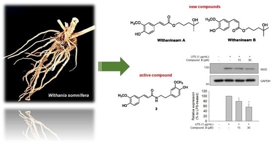
Withaninsams A and B: Phenylpropanoid Esters from the Roots of Indian Ginseng (Withania somnifera)

1
School of Pharmacy, Sungkyunkwan University, Suwon 16419, Korea
2
College of Pharmacy, Chonnam National University, Gwangju 61186, Korea
3
Laboratory of Nuclear Magnetic Resonance, National Center for Inter-University Research Facilities (NCIRF), Seoul National University, Gwanak-gu, Seoul 08826, Korea
*
Author to whom correspondence should be addressed.
Plants 2019, 8(12), 527; https://doi.org/10.3390/plants8120527
Submission received: 30 October 2019 / Revised: 16 November 2019 / Accepted: 19 November 2019 / Published: 20 November 2019
(This article belongs to the Special Issue Bioactive Compounds in Plants)

Abstract

:
Withania somnifera (L.) Dunal (Solanaceae), known as Indian ginseng or ashwagandha, has been used in Indian Ayurveda for the treatment of a variety of disorders, such as diabetes and reproductive and nervous system disorders. It is particularly used as a general health tonic, analgesic, and sedative. As part of continuing projects to discover unique bioactive natural products from medicinal plants, phytochemical investigation of the roots of W. somnifera combined with a liquid chromatography–mass spectrometry (LC/MS)-based analysis has led to the isolation of two novel phenylpropanoid esters, Withaninsams A (1) and B (2), as an inseparable mixture, along with three known phenolic compounds (3, 4, and 6) and a pyrazole alkaloid (5). The structures of the new compounds were elucidated using a combination of spectroscopic methods, including one-dimensional (1D) and two-dimensional (2D) nuclear magnetic resonance (NMR) and high-resolution electrospray ionization mass spectroscopy (HR-ESIMS). Withaninsams A (1) and B (2) are phenylpropanoid esters that contain a side chain, 4-methyl-1,4-pentanediol unit. To the best of our knowledge, the present study is the first to report on phenylpropanoid esters with 4-methyl-1,4-pentanediol unit. The anti-inflammatory activity of the isolated compounds (16) was evaluated by determining their inhibitory effects on nitric oxide (NO) production in lipopolysaccharide (LPS)-stimulated RAW 264.7 macrophages, where compound 3 inhibited LPS-induced NO production (IC50 = 33.3 μM) and TNF-α production, a pro-inflammatory cytokine (IC50 = 40.9 μM). The anti-inflammatory mechanism through the inhibition of transcriptional iNOS protein expression was confirmed by western blotting experiments for the active compound 3, which showed decreased iNOS protein expression.

Graphical Abstract

1. Introduction

Withania somnifera (L.) Dunal (Solanaceae), commonly known as Indian ginseng or ashwagandha, is a perennial shrub distributed in India, Morocco, Egypt, Israel, Jordan, South Africa, and the Mediterranean region. Currently, it is cultivated on a small scale in South Korea as well [1,2,3]. This plant has been used in Indian Ayurveda for over 3000 years for the treatment of a variety of disorders, such as diabetes and nervous and reproductive disorders. It is particularly used as a general health tonic, analgesic, and sedative [4]. The name, Indian ginseng is botanically not related to Korean ginseng (Panax ginseng). The similarity in the name arises from its similar bioactivity [5]. Roots of W. somnifera have recently gained popularity as a functional food that promotes longevity through delaying aging, increasing immunity against extrinsic factors, and strengthening the body [1,6]. In practice, extracts of the roots are consumed in several forms, such as powder, liquid, tablets, and capsules. Moreover, roots are consumed as a dietary supplement.
Previous pharmacological studies on W. somnifera have revealed that extracts of W. somnifera exhibit a protective role against bromobenzene-induced oxidative damage in the rat liver [7], as well as increase the exercise performance [8]. Similarly, a recent study on W. somnifera extracts reported its therapeutic role in stroke repair through its anti-apoptotic and anti-oxidant properties [9]. Chemically, it is a rich source of withanolides, which possess diverse pharmacological properties, including anti-inflammatory, anti-microbial, anti-tumor, hepatoprotective, and immunosuppressive effects [10,11,12,13,14,15,16]. In addition, alkaloids, steroidal saponins, lignanamides [17], and phenolics [18] have also been reported, and some of them were found to have anti-tumor activities [19].
As part of a continuing program to determine structurally and/or biologically novel natural products from medicinal plants [20,21,22,23], we conducted a chemical investigation of the methanol (MeOH) extract of roots of W. somnifera. In our recent study, chemical analysis of the MeOH extract, combined with a liquid chromatography/mass spectroscopy (LC/MS)-based analysis, we identified six new withanolides, namely withasilolides A to F, and seven known withanolides [3], some of which exhibited cytotoxicity against several human cancer cell lines (A549, SK-OV-3, SK-MEL-2, and HCT-15). In the present study, we focused on other constituents of W. somnifera rather than withanolides. The phytochemical analysis of the MeOH extract led to the isolation of two new phenylpropanoid esters (1 and 2) as an inseparable mixture, along with four known compounds (36) (Figure 1). The structures of the new compounds (1 and 2) were elucidated using a combination of one-dimensional (1D) and two-dimensional (2D) nuclear magnetic resonance (NMR) spectroscopy and high-resolution electrospray ionization mass spectroscopy (HR-ESIMS) data. Further, we evaluated the inhibitory effects of the isolates on nitric oxide (NO) production in lipopolysaccharide (LPS)-activated RAW 264.7 macrophages. In the present study, we describe the isolation and structural characterization of isolated compounds, as well as the evaluation of their NO inhibitory effects on LPS-activated RAW264.7 macrophages.

2. Results and Discussion

2.1. Isolation of Compounds

The dried roots of W. somnifera were extracted with 80% MeOH under reflux to yield the methanol extract, which was sequentially applied to solvent-partitioning with hexane, dichloromethane, ethyl acetate, and n-butanol to obtain each solvent fraction. Chemical analysis of hexane-soluble and dichloromethane-soluble fractions was performed using repeated column chromatography and high-performance liquid chromatography (HPLC) along with LC/MS-based analysis. These techniques were combined with our house-built UV library to determine other types of minor constituents, rather than withanolides. The analysis led to the isolation of two new phenylpropanoid esters (1 and 2) as an inseparable mixture, along with three known phenolic compounds (3, 4, and 6) and a pyrazole alkaloid (5) (Figure 1). The novel compounds resulted in only one peak in HPLC performed using Phenomenex Luna C18 column (MeOH/H2O, 7:3 to 1:0).

2.2. Structure Elucidation of Compounds

Compounds 1 and 2 were obtained as a trans/cis inseparable mixture in the ratio of approximately 5:4 (as calculated from the 1H NMR integral). They were isolated as an amorphous powder, and their HR-ESIMS (Figure S1) showed the molecular ion peak [M + Na]+ at m/z 317.1375 (calculated for C16H22O5Na, 317.1365) in the positive mode, compatible with the molecular formula of C16H22O5. The IR spectrum exhibited characteristic absorptions of hydroxy (3305 cm−1) and ester groups (1755 cm−1). The 1H NMR spectrum (Table 1, Figure S2) showed complicated aromatic proton signals in the narrow region between δH 6.77 and 7.08. Most of the 13C NMR signals, which were assigned by 1H-1H correlation spectroscopy (COSY) (Figure S3), heteronuclear single quantum coherence (HSQC) (Figure S4), and heteronuclear multiple bond correlation (HMBC) (Figure S5) experiments, were split into pairs of narrowly separated signals (Table 1). Based on the assigned molecular formula (C16H22O5) by HR-ESIMS, we deduced that the two compounds could be a mixture of two closely related compounds, 1 and 2. The detailed inspection of the 1H NMR spectrum (Table 1) revealed the presence of two sets of 1,3,4-trisubstituted aromatic protons, one at δH 6.89 (1H, d, J = 8.0 Hz), 7.01 (1H, d, J = 1.5 Hz), and 7.05 (1H, dd, J = 8.0, 1.5 Hz) and another at δH 6.86 (1H, d, J = 8.0 Hz), 7.08 (1H, dd, J = 8.0, 2.0 Hz), and 7.74 (1H, d, J = 2.0 Hz), as well as two methoxy groups at δH 3.91 (6H, s). Interpretation of the 1H NMR and 1H-1H COSY spectra (Figure S3) led to the following structural units: C-5 to C-6 and C-7 to C-8 (Figure 2). Connectivities of the partial structure of phenylpropanoid were established on the HMBC cross-peaks of H-2/C-4, C-6, H-5/C-1, C-3, H-6/C-2, C-4, H-7/C-2, C-6, C-9, and H-8/C-1, C-9 (Figure 2). The HMBC correlation of the methoxy group (δH 3.91)/C-3 provided evidence of the presence of the methoxy group at C-3. The presence of 4-methyl-1,4-pentanediol was established on the HMBC cross-peaks of H-1’/C-3’, H-3’/C-1’, C-5’, C-6’, H-5’/C-3’, C-6’, and H-6’/C-3’, C-5’, together with 1H-1H COSY correlations from H-1’ to H-3’ (Figure 2). The linkage between the phenylpropanoid and 4-methyl-1,4-pentanediol unit was established using the HMBC correlation from H-1’ to C-9 (Figure 2).
The geometry of the di-substituted olefin Δ7/8 in compounds 1 and 2 was determined on the basis of the vicinal 1H coupling constant (J7,8 = 16.0 Hz in 1; J7,8 = 13.0 Hz in 2) [24], which indicated that compound 1 had trans-conformation, whereas compound 2 had cis-conformation. Moreover, this assignment is supported by the typical NMR chemical shifts exhibiting δH6.27 (H-8) and δH7.58 (H-7) in compound 1 (trans) and δH5.79 (H-8) and δH6.77 (H-7) in compound 2 (cis) [24]. Therefore, structures of compounds 1 and 2 were determined as shown in Figure 1, with their trivial names designated as Withaninsams A (1) and B (2), respectively. Withaninsams A (1) and B (2) are phenylpropanoid esters containing a side chain, 4-methyl-1,4-pentanediol unit, which is a rare example of a natural product. Although phenylpropanoid esters with similar side chains, such as tert-butyl trans-ferulate and tert-butyl (2E)-3-(3,4-dimethoxyphenyl)-2-propenoate, have been reported [25,26], to the best of our knowledge, the present study is the first to report on phenylpropanoid esters with 4-methyl-1,4-pentanediol unit. Finally, we attempted to separate the mixture of 1 and 2 using the chiral HPLC column, Phenomenex Lux Cellulose-1; however, we failed to resolve the mixture into a pair of pure enantiomers under different conditions.
The known compounds were identified as N-trans-feruloyl methoxytyramine (3) [27], N-trans-feruloyltyramine (4) [28], withasomnine (5) [29], and acetosyringone (6) [30] by comparing their NMR spectroscopic data with reported values as well as using the LC/MS analysis results.

2.3. Inhibitory Effects of Compounds 16 on LPS-Induced NO Production in RAW 264.7 Cells

To determine whether the isolated compounds 1 to 6 had anti-inflammatory properties, we performed NO assay using supernatants from LPS-stimulated RAW 264.7 cells. All compounds tested exhibited no cytotoxicity up to each highest concentration (Figure 3A). Of these, compound 3 exhibited an inhibitory effect on NO production in LPS-stimulated RAW 264.7 cells (IC50 = 33.3 μM) (Figure 3B). The effect of compound 3 on the LPS-induced TNF-α production showed similar patterns to those of NO production (IC50 = 40.9 μM) (Figure 3C). To confirm whether NO inhibitory effect of 3 was related to reduced expression of nitric oxide synthase (iNOS), enzyme involved in the synthesis of NO, we evaluated the effect of compound 3 on iNOS expression. Similar to the results of NO production, compound 3 reduced iNOS expression in LPS-stimulated RAW 264.7 cells (Figure 4). These results indicated that compound 3 exerted anti-inflammatory effect on macrophages by reducing LPS-induced NO production through transcriptional inhibition of iNOS.

3. Materials and Methods

3.1. Plant Material

The roots of W. somnifera were purchased from Seonggeosan Farm, Cheonan, Korea, in October 2016, and the plant was identified by one of the authors (K.H.K.). A voucher specimen of the material (IDG-2016) was deposited in the herbarium of the School of Pharmacy, Sungkyunkwan University, Suwon, Republic of Korea.

3.2. Extraction and Isolation

Dried roots of W. somnifera (1.3 kg) were extracted with 80% aqueous MeOH (each 3.0 L × 3 days) under reflux and filtered. The filtrate was combined and concentrated under vacuum using a rotary evaporator to obtain a MeOH extract (189.6 g). The extract was suspended in distilled water (700 mL) and successively solvent-partitioned using hexane, dichloromethane (CH2Cl2), ethyl acetate (EtOAc), and n-butanol (n-BuOH). Four fractions with increasing polarity were obtained: hexane-soluble (3.4 g), CH2Cl2-soluble (4.5 g), EtOAc-soluble (2.0 g), and n-BuOH-soluble (18.6 g). The hexane-soluble fraction (3.2 g) was fractionated using silica gel column chromatography with a gradient solvent system of hexane/EtOAc (20:1 to 1:1). The column was washed with CH2Cl2/MeOH (1:1) to obtain seven hexane fractions (H1–H7). Fraction H6 (300.1 mg) was separated by preparative reversed-phase HPLC using an Agilent Eclipse C18 column with gradient solvent system MeOH/H2O (7:3 to 1:0) to obtain seven subfractions (H6a–H6g). Subfraction H6c (134.6 mg) was purified by semi-preparative HPLC (38% MeCN) on Phenomenex Luna C18 column to yield compound 5 (24.3 mg). Subfraction H6g (22.9 mg) was purified by semi-preparative HPLC with gradient solvent system MeOH/H2O (7:3 to 1:0) on Phenomenex Luna C18 column to yield a mixture of compounds 1 and 2 (1.1 mg). The CH2Cl2-soluble fraction (4.5 g) was fractionated using silica gel column chromatography with gradient solvent system of CH2Cl2/MeOH (50:1 to 1:1) to obtain seven fractions (C1–C7). Fraction C1 (73.6 mg) was separated by preparative reversed-phase HPLC on an Agilent Eclipse C18 column with gradient solvent system MeOH/H2O (3:7 to 1:0) to obtain five subfractions (C1a–C1e). Subfraction C1c (11.8 mg) was purified using semi-preparative HPLC (33% MeOH) on Phenomenex Luna C18 column to yield compound 6 (7.2 mg). Fraction C3 (401.6 mg) was fractionated using preparative reversed-phase HPLC on Agilent Eclipse C18 column with gradient solvent system MeOH/H2O (3:7 to 1:0) to obtain six subfractions (C3a–C3f). Subfraction C3d (33.8 mg) was purified using semi-preparative HPLC on Phenomenex Luna C18 column (47% MeOH) to yield compound 3 (2.4 mg). Fraction C4 (166.0 mg) was separated by reversed-phase preparative HPLC using an Agilent Eclipse C18 column with gradient solvent system MeOH/H2O (4:6 to 9:1) to obtain six subfractions (C4a–C4f). Subfraction C4c (19.2 mg) was purified by semi-preparative HPLC (40% MeOH) on Phenomenex Luna C18 column to yield compound 4 (3.3 mg).

Withaninsams A (1) and B (2)

White, amorphous powder; ESIMS (positive mode) m/z:317 [M + Na]+; HRESIMS (positive mode) m/z:317.1375 [M + Na]+, calcd for C16H22O5Na, 317.1365; UV (MeOH) λmax nm (log ε): 220 (2.29), 240 (2.10), 290 (2.54), 325 (3.42); IR (KBr) νmax cm−1: 3305, 3126, 1755, 1585, 1492, 1042; 1H (CD3OD, 800 MHz) and 13C (CD3OD, 200 MHz) NMR spectroscopic data, see Table 1.

3.3. Cell Viability Assay

RAW 264.7 cells (6.0 × 10⁴ cells/well) were seeded into a 96-well plate and incubated overnight for adhesion. Following incubation, the cells were treated with compounds for 24 h. Next, Ez-CytoX solution (1/10 volume of the culture medium, Daeil Lab., Seoul, Korea) was added to each well and cells were further incubated for 1 h. The cell viability was assessed by measuring the absorbance at 450 nm.

3.4. NO Production Assay

RAW 264.7 cells (6.0 × 10⁴ cells/well) were seeded into a 96-well plate and incubated overnight for adhesion. Following incubation, the cells were treated with compounds and LPS. After 24 h incubation, supernatants were collected and treated with Griess reagent for evaluating NO concentration in the reactants. The absorbance was measured at 540 nm and NO production was calculated by referring to the nitrite standard curve.

3.5. TNF-α ELISA

Culture supernatants were applied to ELISA experiments for measuring the production of TNF-α in LPS-stimulated RAW 264.7 cells. ELISA was performed by manufacturer’s instructions (Ebioscience, San Diego, CA, USA). Each step was followed by washing with 1 × PBST 5 times. Briefly, plate was coated with coating antibody solution overnight at 4 °C and then blocked with 1 × assay diluent for 1 h at room temperature (RT). Supernatants were applied to the plate for 2 h at RT and the plate was incubated with biotinylated secondary antibody for 1 h at RT. After reacting the plate with horseradish peroxidase (HRP)-streptavidin for 40 min at RT, the plate was reacted with 4-nitrophenyl phosphate disodium salt in diethanolamine buffer as a substrate for 10 min at dark condition and the reaction was stopped by adding 1 N NaOH. Absorbances of each well at 405 nm were applied to standard curve for calculating the quantity of TNF-α at supernatants.

3.6. Western Blotting

RAW 264.7 cells (2.0 × 10⁵ cells/well) were seeded into 6-well plates and incubated overnight for adhesion. Following incubation, the cells were treated with compounds 3 and 4 in the presence of LPS for 24 h. Total cell lysates were obtained and loaded onto sodium dodecyl sulfate-polyacrylamide gel electrophoresis (SDS-PAGE). Proteins were subsequently transferred onto nitrocellulose (NC) membrane. Membranes were incubated with primary antibodies against iNOS and GAPDH, following which the membranes were incubated with appropriate secondary antibodies. Finally, the membranes were blotted and protein band intensities were analyzed using Imager 680 (GE Healthcare; Chicago, IL, USA).

3.7. Statistical Analysis

The data were statistically analyzed using Student’s t-test. To prove statistical significance, experiments were conducted in replicates as follows: nine for cell viability, NO assay, TNF-α ELISA and three for western blotting. A p-value < 0.05 was considered statistically significant.

4. Conclusions

In the present study, phytochemical analysis of the MeOH extracts of roots of W. somnifera led to the isolation of two novel phenylpropanoid esters, namely Withaninsams A (1) and B (2) as an inseparable mixture, along with three known phenolic compounds (3, 4, and 6) and a pyrazole alkaloid (5). Withaninsams A (1) and B (2) are phenylpropanoid esters that contain a side chain, 4-methyl-1,4-pentanediol unit. To the best of our knowledge, the present study is the first to report on phenylpropanoid esters with 4-methyl-1,4-pentanediol unit. All isolated compounds were evaluated for their anti-inflammatory effects on nitric oxide (NO) production in LPS-stimulated RAW 264.7 macrophages. Compound 3 exhibited NO and TNF-α inhibitory properties without cytotoxicity. The active compound 3 inhibited NO production by reducing iNOS protein expression.

Supplementary Materials

The following are available online at https://www.mdpi.com/2223-7747/8/12/527/s1, Figure S1: The HRESIMS data of compounds 1 and 2, Figure S2: The 1H NMR spectrum of compounds 1 and 2, Figure S3: The 1H-1H COSY spectrum of compounds 1 and 2, Figure S4: The HSQC spectrum of compounds 1 and 2, Figure S5: The HMBC spectrum of compounds 1 and 2.

Author Contributions

Conceptualization, Y.-C.C. and K.H.K.; formal analysis, S.C.B., S.L., S.K., Y.-J.K., and M.S.J.; investigation, S.C.B., S.L., S.K., M.S.J., J.S.Y., and Y.-C.C.; writing—original draft preparation, S.C.B., Y.-C.C., and K.H.K.; writing—review and editing, S.C.B. and K.H.K.; supervision, K.H.K.; project administration, Y.-C.C. and K.H.K.; funding acquisition, K.H.K.

Funding

This work was supported by a National Research Foundation of Korea (NRF) grant funded by the Korean government (MSIT) (2018R1A2B2006879 and 2019R1A5A2027340).

Conflicts of Interest

The authors declare no conflict of interest.

References

  1. Khare, C.P. Encyclopedia of Indian Medicinal Plants; Springer Verlag: New York, NY, USA, 2004; pp. 480–483. [Google Scholar]
  2. Gupta, G.L.; Rana, A.C. Withaniasomnifera (Ashwagandha): A review. Pharmacogn. Rev. 2007, 1, 129–136. [Google Scholar]
  3. Kim, S.; Yu, J.S.; Lee, J.Y.; Choi, S.U.; Lee, J.; Kim, K.H. Cytotoxic Withanoildes from the Roots of Indian Ginseng (Withaniasomnifera). J. Nat. Prod. 2019, 82, 765–773. [Google Scholar] [CrossRef] [PubMed]
  4. Kulkarni, S.K.; Dhir, A. Withaniasomnifera: An Indian ginseng. Prog. Neuro-Psychopharmacol. Biol. Psychiatry 2008, 32, 1093–1105. [Google Scholar] [CrossRef] [PubMed]
  5. Singh, B.; Saxena, A.K.; Chandan, B.K.; Gupta, D.K.; Bhutani, K.K.; Anand, K.K. Adaptogenic activity of novel, withanolide-free aqueous fraction from the roots of Withaniasomnifera Dun. Phytother. Res. 2001, 15, 311–318. [Google Scholar] [CrossRef] [PubMed]
  6. Weiner, M.A.; Weiner, J. Herbs That Heal: Prescription for Herbal Healing; Atrium Publishers Group: Mill Valley, CA, USA, 1994; p. 70. [Google Scholar]
  7. Vedi, M.; Rasool, M.; Sabina, E.P. Amelioration of bromobenzene hepatotoxicity by Withaniasomnifera pretreatment: Role of mitochondrial oxidative stress. Toxicol. Rep. 2014, 1, 629–638. [Google Scholar] [CrossRef] [PubMed]
  8. Ziegenfuss, T.N.; Kedia, A.W.; Sandrock, J.E.; Raub, B.J.; Kerksick, C.M.; Lopez, H.L. Effects of an Aqueous Extract if Withaniasomnifera on Strength Training Adaptations and Recovery: The STAR Trial. Nurtrients 2018, 10, 1807. [Google Scholar] [CrossRef]
  9. Raghavan, A.; Shah, Z.A. Withaniasomnifera Improves Ischemic Stroke Outcomes by Attenuating PARP1-AIF-Mediated Caspase-Independent Apoptosis. Mol. Neurobiol. 2015, 52, 1093–1105. [Google Scholar] [CrossRef]
  10. Ma, T.; Zhang, W.-N.; Yang, L.; Zhang, C.; Lin, R.; Shan, S.-M.; Zhu, M.-D.; Luo, J.-G.; Kong, L.-Y. Cytotoxic withanolides from Physalis angulata var. villosa and the apoptosis-inducing effect via ROS generation and the activation of MAPK in human osteosarcoma cells. RSC Adv. 2016, 6, 53089–53100. [Google Scholar] [CrossRef]
  11. Habtemariam, S. Cytotoxicity and Immunosuppressive Activity of Withanoliedes from Discopodiumpenninervium. Planta Med. 1997, 63, 15–17. [Google Scholar] [CrossRef]
  12. Budhiraja, R.D.; Sudhir, S.; Garg, K.N. Antiinflmmatory Activity of 3 β-Hydroxy-2,3-dihydro-withanolide F. Planta Med. 1984, 50, 134–136. [Google Scholar] [CrossRef]
  13. Chakraborti, S.K.; De Barun, K.; Bandyopadhyay, T. Variations in the antitumor constituents of Withaniasomnifera dunal. Experientia 1974, 30, 852–853. [Google Scholar] [CrossRef] [PubMed]
  14. Mareggiani, G.; Picollo, M.I.; Zerba, E.; Burton, G.; Tettamanzi, M.C.; Benedetti-Doctorovich, M.O.V.; Veleiro, A.S. Antifeedant Activity of Withanolides from Salpichroaoriganifolia on Musca Domestica. J. Nat. Prod. 2000, 63, 1113–1116. [Google Scholar] [CrossRef] [PubMed]
  15. Basso, A.V.; Leiva Gonzalez, S.; Barboza, G.E.; Careaga, V.P.; Calvo, J.C.; Sacca, P.A.; Nicotra, V.E. Phytochemical Study of the Genus Salpichroa (Solanaceae), Chemotaxonomic Considerations, and Biological Evaluation in Prostate and Breast Cancer Cells. Chem. Biodivers. 2017, 14, e1700118. [Google Scholar] [CrossRef]
  16. Zhang, H.; Cao, C.-M.; Gallagher, R.J.; Timmermann, B.N. Antiproliferative withanolides from several solanaceous spicies. Nat. Prod. Res. 2014, 28, 1941–1951. [Google Scholar] [CrossRef]
  17. Bolleddula, J.; Fitch, W.; Vareed, S.K.; Nair, M.G. Identification of metabolies in Withaniasomnifera fruits by liquid chromatography and high-resolution mass spectrometry. Rapid Commun. Mass Spectrom. 2012, 26, 1277–1290. [Google Scholar] [CrossRef]
  18. Elsakka, M.; Grigorescu, E.; Stănescu, U.; Dorneanu, V. New data referring to chemistry of Withaniasomnifera species. Rev. Med. Chir. Soc. Med. Nat. IASI 1990, 94, 385–387. [Google Scholar]
  19. Jayaprakasam, B.; Zhang, Y.; Seeram, N.P.; Nair, M.G. Growth inhibition of human tumor cell lines by withanolides from Withaniasomnifera leaves. Life Sci. 2003, 74, 125–132. [Google Scholar] [CrossRef]
  20. So, H.M.; Eom, H.J.; Lee, D.; Kim, S.; Kang, K.S.; Lee, I.K.; Baek, K.-H.; Park, J.Y.; Kim, K.H. Bioactivity evaluations of betulin identified from the bark of Betula platyphylla var. japonica for cancer therapy. Arch. Pharm. Res. 2018, 41, 815–822. [Google Scholar] [CrossRef]
  21. Yu, J.S.; Roh, H.-S.; Baek, K.-H.; Lee, S.; Kim, S.; So, H.M.; Moon, E.; Pang, C.; Jang, T.S.; Kim, K.H. Bioactivity-guided isolation of ginsenosides from Korean Red Ginsengwith cytotoxic activity against human lung adenocarcinoma cells. J. Ginseng Res. 2018, 42, 562–570. [Google Scholar] [CrossRef]
  22. Baek, S.C.; Choi, E.; Eom, H.J.; Jo, M.S.; Kim, S.; So, H.M.; Kim, S.H.; Kang, K.S.; Kim, K.H. LC/MS-based analysis of bioactive compounds from the bark of Betula platyphylla var. japonica and their effects on regulation of adipocyte and osteoblast differentiation. Nat. Prod. Sci. 2018, 24, 235–240. [Google Scholar] [CrossRef]
  23. Yu, J.S.; Lee, D.; Lee, S.R.; Lee, J.W.; Choi, C.-I.; Jang, T.S.; Kang, K.S.; Kim, K.H. Chemical characterization of cytotoxic indole acetic acid derivative from Mulberry fruit (Morus alba L.) against human cervical cancer. Bioorg. Chem. 2018, 76, 28–36. [Google Scholar] [CrossRef] [PubMed]
  24. Pavia, D.L.; Lampman, G.M.; Kriz, G.S.; Vyvyan, J.R. Introduction to Spectroscopy, 4th ed.; Cengage Learning: Stamford, CT, USA, 2008; pp. 237–297. [Google Scholar]
  25. Faig, J.J.; Klein, S.; Ouimet, M.A.; Yu, W.; Uhrich, K.E. Attenuating Oxidative Stress Via Oxalate Ester-Containing Ferulic Acid-Based Poly(anhydride-esters) that Scavenge Hydrogen Peroxide. Macromol. Chem. Phys. 2016, 217, 108–114. [Google Scholar] [CrossRef]
  26. Liu, J.; Zhao, Y.; Zhou, Y.; Li, L.; Zhang, T.Y.; Zhang, H. Imidazolylidene carbene ligated palladium catalysis of the Heck reaction in the presence of air. Org. Biomol. Chem. 2003, 1, 3227–3231. [Google Scholar] [CrossRef] [PubMed]
  27. Kim, H.R.; Min, H.; Jeong, Y.H.; Lee, S.K.; Lee, N.S.; Seo, E. Cytotoxic Constituents from the Whole Plant of Corydalis pallida. Arch. Pharm. Res. 2005, 28, 122–1227. [Google Scholar]
  28. Ma, J.; Jones, S.H.; Hecht, S.M. Phenolic acid amides: A new type of DNA strand scission agent from Piper caninum. Bioorg. Med. Chem. 2004, 12, 3885–3889. [Google Scholar] [CrossRef]
  29. Majumder, S.; Gipson, K.R.; Staples, R.J.; Odom, A.L. Pyrazole Synthesis Using a Titanium-Catalyzed multicomponent Coupling Reaction and Synthesis of Withasomnine. Adv. Synth. Catal. 2009, 351, 2013–2023. [Google Scholar] [CrossRef]
  30. Bao, K.; Fan, A.; Zhang, L.; Zhang, W.; Cheng, M.; Yao, X. Selective demethylation and debenzylation of aryl ethers by magnesium iodide under solvent-free conditions and its application to the total synthesis of natural products. Org. Biomol. Chem. 2009, 7, 5084–5090. [Google Scholar] [CrossRef]
Figure 1. Chemical structures of compounds 16.
Plants 08 00527 g001
Figure 2. 1H-1H correlation spectroscopy (COSY) ( Plants 08 00527 i001) and key heteronuclear multiple bond correlation (HMBC) ( Plants 08 00527 i002) correlations for compounds 1 and 2.
Plants 08 00527 g002
Figure 3. Inhibitory effects of compounds 16 on the production of pro-inflammatory mediators in macrophages. RAW 264.7 cells were treated with compounds 16 in the absence or presence of lipopolysaccharide (LPS) and then (A) cell viability, (B) nitric oxide (NO) production, and (C) TNF-α production in RAW 264.7 cells were determined. Quercetin was used as a positive control. (A) Cell viability data represent relative cell viability compared with that of the untreated group (100%, white bar). (B,C) NO and TNF-α production data represent actual production levels calculated by applying absorbance values to each standard curve. (white bar, untreated control group; grey and black bar, LPS-treated control group). p < 0.05 relative to the LPS-treated control group.
Plants 08 00527 g003
Figure 4. Inhibitory effect of compound 3 on LPS-induced nitric oxide synthase (iNOS) expression in RAW 264.7 cells. Data represent relative iNOS protein expression levels compared with those in the LPS-treated group (100%). p < 0.05 relative to the LPS-treated control group.
Plants 08 00527 g004
Table 1. 1H and 13C NMR data of compounds 1 and 2 in CD3OD (δ in ppm, 800 MHz for 1H and 200 MHz for 13C) a.
Position12
δHδCδHδC
1 127.0 127.3
27.01 d (1.5)109.27.74 d (2.0)112.6
3 146.5 145.9
4 147.9 147.0
56.89 d (8.0)114.66.86 d (8.0)113.7
67.05 dd (1.5, 8.0)123.17.08 dd (2.0, 8.0)125.5
77.58 d (16.0)114.66.77 d (13.0)143.7
86.27 d (16.0)115.65.79 d (13.0)116.8
9 167.3 166.7
1′4.16 t (7.0)64.44.09 t (7.0)64.4
2′1.37 m28.61.30 m28.5
3′1.67 m25.91.62 m25.8
4′ n.d.b n.d. b
5′1.23 s29.51.23 s29.5
6′1.23 s29.51.23 s29.5
–OCH33.91 s55.63.91 s55.6
aJ values are in parentheses and reported in Hz; 13C NMR assignments are based on 1H-1H COSY, HSQC, and HMBC experiments; b not detected.

