
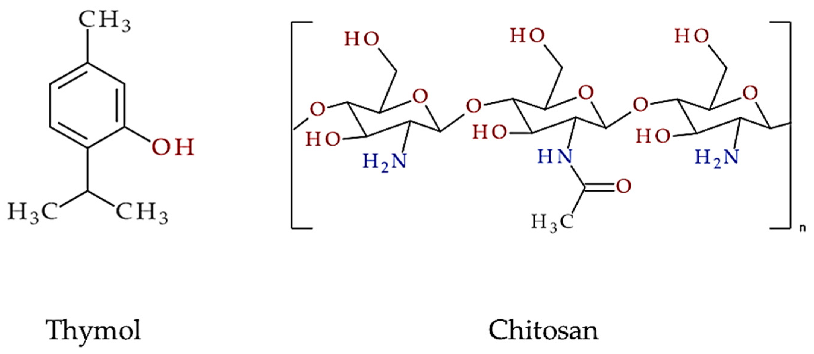
Thymol as a Component of Chitosan Systems—Several New Applications in Medicine: A Comprehensive Review
 ,
,  and
and
Abstract
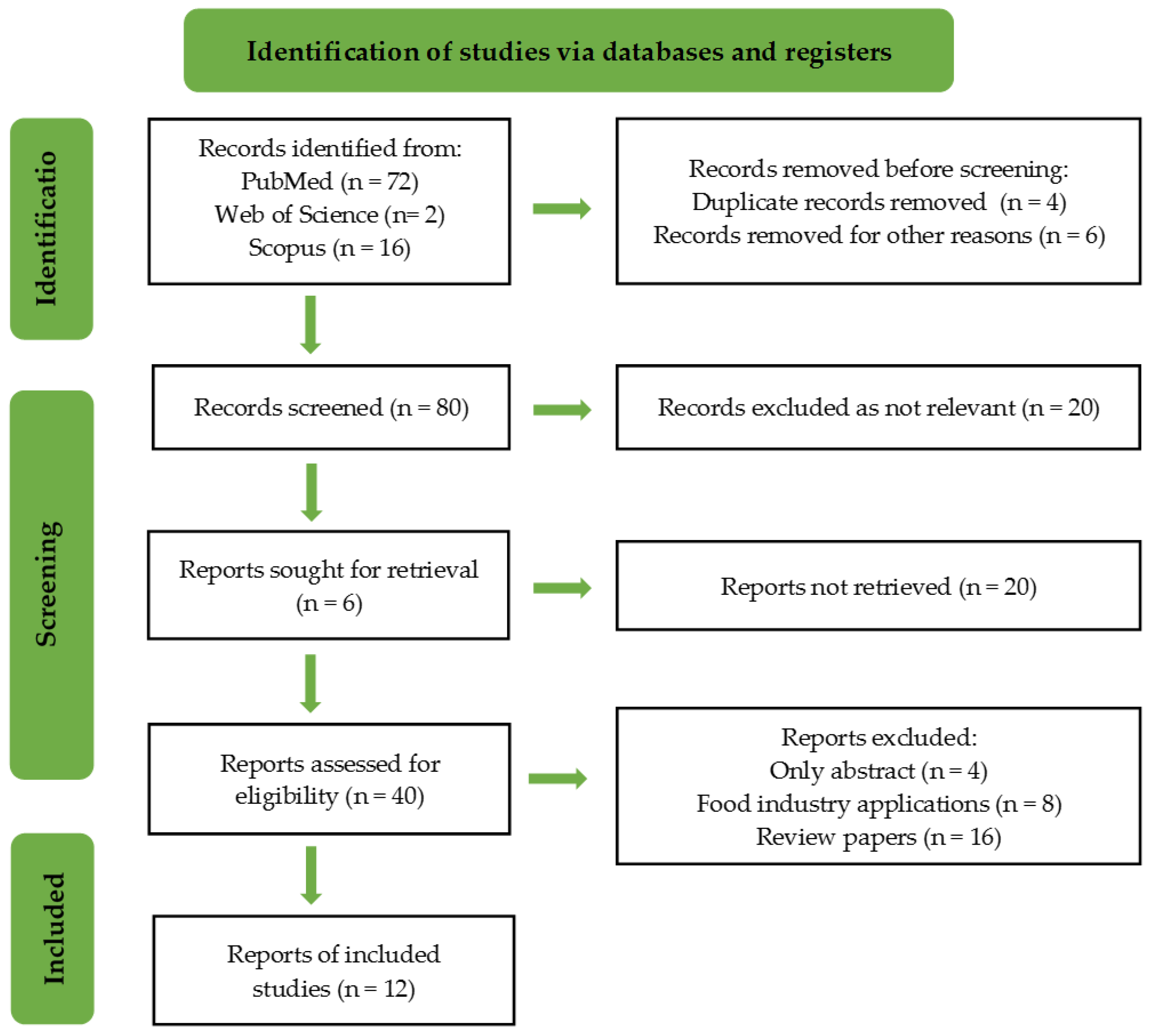
:1. Introduction
- -
- Studies on the use of thymol as an ingredient in chitosan-based functional biomaterials.
- -
- Research published in English between 2018 and 2023.
- -
- Research on biomedical applications, such as drug delivery and antimicrobial materials.
- -
- Research did not focus on thymol-based biomaterials.
- -
- Studies published before 2018 or in languages other than English.
- -
- Studies not related to biomedical applications.
2. Thymol and Chitosan in Nanogel Formulations
3. Tragacanth/Chitosan Nanocarriers for Efficient Thymol Delivery
4. Light-Controlled Chitosan Micelles for Thymol Delivery
5. Chitosan-Thymol Films Modified with Various Carriers/Compounds
6. Optimizing the Synthesis of Thymol-Embedded Chitosan Nanoparticle
7. Thymol-Induced Enhancement of Apoptotic Potential in Adenocarcinomic Human Alveolar Basal Epithelial Cells (A549)
8. Advancing Bone Regeneration through Thymol-Loaded Polymeric Hydrogels
9. Chitosan/Thymol Materials for Oral Administration/Periodontal Treatment
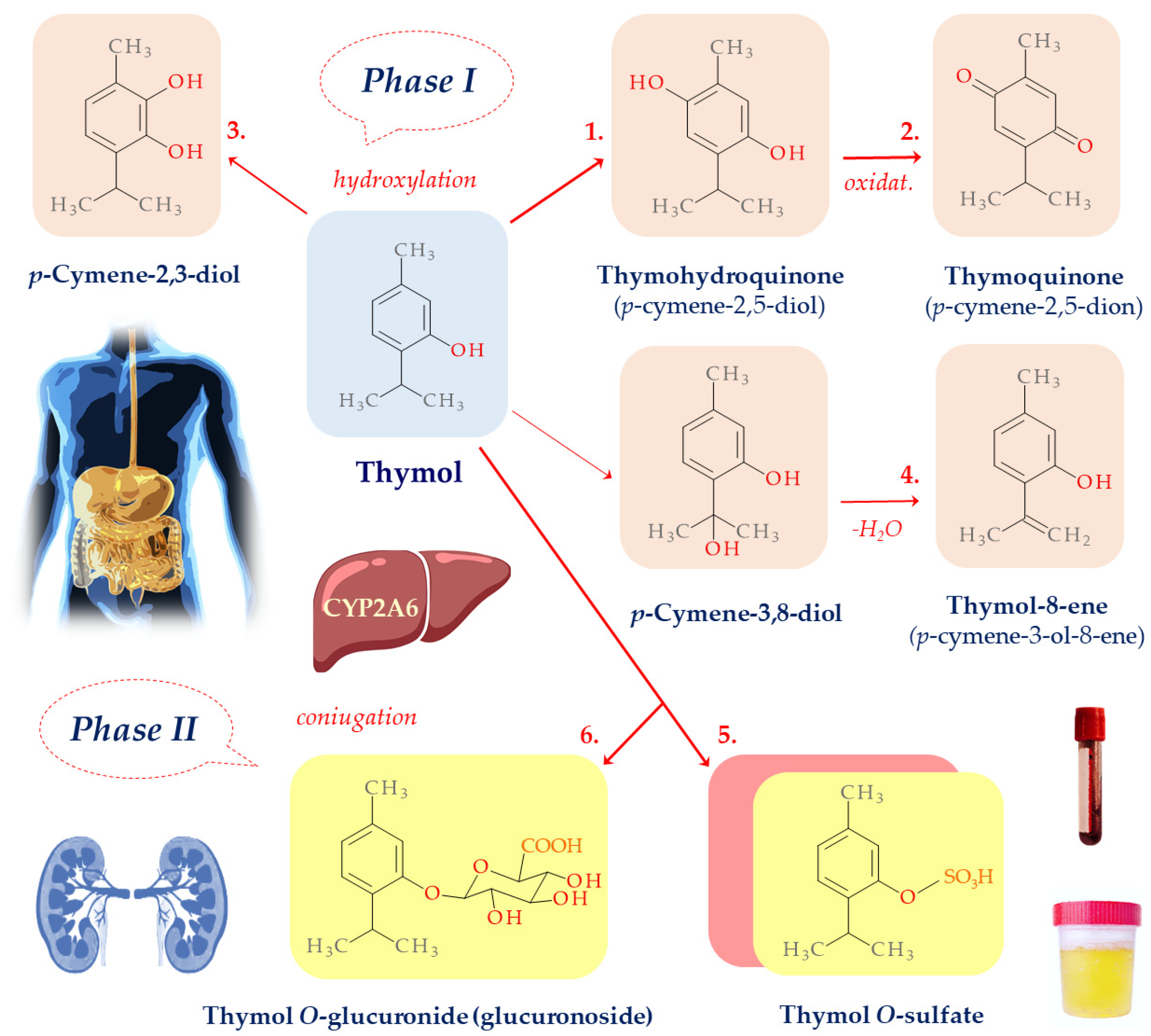
10. Thymol Biotransformation
11. Thymol Antimicrobial Activity and Cell Membrane Interactions
12. Limitations of Presented Studies and Future Directions
13. Conclusions
Author Contributions
Funding
Data Availability Statement
Conflicts of Interest
References
- Cui, W.; Santos, H.A.; Zhang, B.; Zhang, Y.S. Functional biomaterials. APL Bioeng. 2022, 6, 2020–2022. [Google Scholar] [CrossRef]
- Da Silva, J.; Leal, E.C.; Carvalho, E.; Silva, E.A. Innovative functional biomaterials as therapeutic wound dressings for chronic diabetic foot ulcers. Int. J. Mol. Sci. 2023, 24, 9900. [Google Scholar] [CrossRef]
- Luan, J.; Li, R.; Xu, W.; Sun, H.; Li, Q.; Wang, D.; Dong, S.; Ding, J. Functional biomaterials for comprehensive periodontitis therapy. Acta Pharm. Sin. B 2023, 13, 2310–2333. [Google Scholar] [CrossRef]
- Ahmed, T. Functional biomaterials for biomimetic 3D in vitro tumor microenvironment modeling. Vitr. Model. 2023, 2, 1–23. [Google Scholar] [CrossRef]
- Lin, Y.; Fu, M.L.; Harb, I.; Ma, L.X.; Tran, S.D. Functional biomaterials for local control of orthodontic tooth movement. J. Funct. Biomater. 2023, 14, 294. [Google Scholar] [CrossRef]
- Wang, Y.; Chen, G.; Zhang, H.; Zhao, C.; Sun, L.; Zhao, Y. Emerging functional biomaterials as medical patches. ACS Nano 2021, 15, 5977–6007. [Google Scholar] [CrossRef]
- Rubežić, M.; Krstić, A.; Stanković, H.; Ljupković, R.; Ranđelović, M.; Zarubica, A. Different types of biomaterials: Structure and application: A short review. Adv. Technol. 2020, 9, 69–79. [Google Scholar] [CrossRef]
- Kim, Y.; Zharkinbekov, Z.; Raziyeva, K.; Tabyldiyeva, L.; Berikova, K.; Zhumagul, D.; Temirkhanova, K.; Saparov, A. Chitosan-based biomaterials for tissue regeneration. Pharmaceutics 2023, 15, 807. [Google Scholar] [CrossRef]
- Liu, W.; Xu, B.; Zhao, S.; Han, S.; Quan, R.; Liu, W.; Ji, C.; Chen, B.; Xiao, Z.; Yin, M.; et al. Spinal cord tissue engineering via covalent interaction between biomaterials and cells. Sci. Adv. 2023, 9, eade8829. [Google Scholar] [CrossRef]
- Kang, Y. Cell biological techniques and cell-biomaterial interactions. Cells 2020, 9, 8–10. [Google Scholar] [CrossRef]
- Petaroudi, M.; Rodrigo-Navarro, A.; Dobre, O.; Dalby, M.J.; Salmeron-Sanchez, M. Living biomaterials to engineer hematopoietic stem cell niches. Adv. Healthc. Mater. 2022, 11, 2200964. [Google Scholar] [CrossRef]
- Andriani, Y.; Pratama, R.I.; In, I. Chitosan application in aquatic feed and its impact on fish and shrimp productivity. Asian J. Biol. 2023, 19, 25–30. [Google Scholar] [CrossRef]
- Wang, W.; Meng, Q.; Li, Q.; Liu, J.; Zhou, M.; Jin, Z.; Zhao, K. Chitosan derivatives and their application in biomedicine. Int. J. Mol. Sci. 2020, 21, 4532–4546. [Google Scholar] [CrossRef] [PubMed]
- Geng, Y.; Xue, H.; Zhang, Z.; Panayi, A.C.; Knoedler, S.; Zhou, W.; Mi, B.; Liu, G. Recent advances in carboxymethyl chitosan-based materials for biomedical applications. Carbohydr. Polym. 2023, 305, 120555. [Google Scholar] [CrossRef] [PubMed]
- Victor, R.d.S.; Santos, A.M.d.C.; de Sousa, B.V.; Neves, G.d.A.; Santana, L.N.d.L.; Menezes, R.R. A review on chitosan’s uses as biomaterial: Tissue engineering, drug delivery systems and cancer treatment. Materials 2020, 13, 4995. [Google Scholar] [CrossRef] [PubMed]
- Notario-Pérez, F.; Martín-Illana, A.; Cazorla-Luna, R.; Ruiz-Caro, R.; Veiga, M.D. Applications of chitosan in surgical and post-surgical materials. Mar. Drugs 2022, 20, 365. [Google Scholar] [CrossRef] [PubMed]
- Gozali, D.; Hudaya, A.R.; Suharyani, I.N.E.; Wathoni, N. A review on chitosan-based materials as potential wound dressing materials. Int. J. App. Pharm. 2022, 14, 27–32. [Google Scholar] [CrossRef]
- Escobar, A.; Pérez, M.; Romanelli, G.; Blustein, G. Thymol Bioactivity: A review focusing on practical applications. Arab. J. Chem. 2020, 13, 9243–9269. [Google Scholar] [CrossRef]
- Nieto, G. A review on applications and uses of Thymus in the food industry. Plants 2020, 9, 961. [Google Scholar] [CrossRef]
- Mollarafie, P.; Khadiv Parsi, P.; Zarghami, R.; Amini Fazl, M.; Ghafarzadegan, R. Antibacterial and wound healing properties of thymol (Thymus vulgaris oil) and its application in a novel wound dressing. J. Med. Plants 2015, 14, 69–81. [Google Scholar]
- Othman, S.H.; Othman, N.F.L.; Shapi’i, R.A.; Ariffin, S.H.; Yunos, K.F. Corn starch /chitosan nanoparticles/thymol packaging applications. Polymers 2021, 13, 390. [Google Scholar] [CrossRef] [PubMed]
- Feng, P.; Luo, Y.; Ke, C.; Qiu, H.; Wang, W.; Zhu, Y.; Hou, R.; Xu, L.; Wu, S. Chitosan-based functional materials for skin wound repair: Mechanisms and applications. Front. Bioeng. Biotechnol. 2021, 9, 650598. [Google Scholar] [CrossRef] [PubMed]
- Xu, J.; Guan, W.; Kong, Y.; Liu, F.; Zhao, Y.; Li, G.; Yang, Y. Regulation of macrophage behavior by chitosan scaffolds with different elastic modulus. Coatings 2022, 12, 7142. [Google Scholar] [CrossRef]
- Wang, W.; Xue, C.; Mao, X. Chitosan: Structural modification, biological activity and application. Int. J. Biol. Macromol. 2020, 164, 4532–4546. [Google Scholar] [CrossRef] [PubMed]
- Nagoor Meeran, M.F.; Javed, H.; Al Taee, H.; Azimullah, S.; Ojha, S.K. Pharmacological properties and molecular mechanisms of thymol: Prospects for its therapeutic potential and pharmaceutical development. Front. Pharmacol. 2017, 8, 380. [Google Scholar] [CrossRef] [PubMed]
- Islam, M.M.; Shahruzzaman, M.; Biswas, S.; Nurus Sakib, M.; Rashid, T.U. Chitosan based bioactive materials in tissue engineering applications-a review. Bioact. Mater. 2020, 5, 164–183. [Google Scholar] [CrossRef]
- Aranaz, I.; Alcántara, A.R.; Civera, M.C.; Arias, C.; Elorza, B.; Caballero, A.H.; Acosta, N. Chitosan: An overview of its properties and applications. Polymers 2021, 13, 3256. [Google Scholar] [CrossRef]
- Piri-Gharaghie, T.; Beiranvand, S.; Riahi, A.; Shirin, N.J.; Badmasti, F.; Mirzaie, A.; Elahianfar, Y.; Ghahari, S.; Ghahari, S.; Pasban, K.; et al. Fabrication and characterization of thymol-loaded chitosan nanogels: Improved antibacterial and anti-biofilm activities with negligible cytotoxicity. Chem. Biodivers. 2022, 19, e20210042. [Google Scholar] [CrossRef]
- Sheorain, J.; Mehra, M.; Thakur, R.; Grewal, S.; Kumari, S. In vitro anti-inflammatory and antioxidant potential of thymol loaded bipolymeric (Tragacanth Gum/Chitosan) Nanocarrier. Int. J. Biol. Macromol. 2019, 125, 1069–1074. [Google Scholar] [CrossRef]
- Wang, Z.; Bai, H.; Lu, C.; Hou, C.; Qiu, Y.; Zhang, P.; Duan, J.; Mu, H. Light controllable chitosan micelles with ROS generation and essential oil release for the treatment of bacterial biofilm. Carbohydr. Polym. 2019, 205, 533–539. [Google Scholar] [CrossRef]
- Ahmady, A.R.; Razmjooee, K.; Saber-Samandari, S.; Toghraie, D. Fabrication of chitosan-gelatin films incorporated with thymol-loaded alginate microparticles for controlled drug delivery, antibacterial activity and wound healing: In-vitro and in-vivo studies. Int. J. Biol. Macromol. 2022, 223, 567–582. [Google Scholar] [CrossRef] [PubMed]
- Pires, A.L.R.; de Azevedo Motta, L.; Dias, A.M.A.; de Sousa, H.C.; Moraes, Â.M.; Braga, M.E.M. Towards wound dressings with improved properties: Effects of poly(dimethylsiloxane) on chitosan-alginate films loaded with thymol and beta-carotene. Mater. Sci. Eng. C 2018, 93, 595–605. [Google Scholar] [CrossRef]
- Sharma, K.; Mehak Munjal, M.; Sharma, R.K.; Meenakshi Sharma, M. Thymol encapsulated chitosan-Aloe vera films for antimicrobial infection. Int. J. Biol. Macromol. 2023, 235, 123897. [Google Scholar] [CrossRef] [PubMed]
- Çakır, M.A.; Icyer, N.C.; Tornuk, F. Optimization of production parameters for fabrication of thymol-loaded chitosan nanoparticles. Int. J. Biol. Macromol. 2020, 151, 230–238. [Google Scholar] [CrossRef] [PubMed]
- Balan, D.J.; Das, M.; Sathya, S.; Kiruthiga, C.; Jeyakumar, M.; Antoniraj, M.G.; Devi, K.P. Chitosan based encapsulation increased the apoptotic efficacy of thymol on a549 cells and exhibited non toxic response in swiss albino mice. Int. J. Biol. Macromol. 2022, 202, 620–631. [Google Scholar] [CrossRef] [PubMed]
- Lavanya, K.; Balagangadharan, K.; Chandran, S.V.; Selvamurugan, N. Chitosan-coated and thymol-loaded polymeric semi-interpenetrating hydrogels: An effective platform for bioactive molecule delivery and bone regeneration in vivo. Biomater. Adv. 2023, 146, 213305. [Google Scholar] [CrossRef] [PubMed]
- Kordestani, M.; Rashidipour, M.; Mahmoudvand, H.; Jalali, S.; Kooshki, F. Antibacterial and cytotoxicity of chitosan nanocomposite loaded with thymol against some cariogenic bacteria. J. Herbmed Pharmacol. 2023, 12, 223–227. [Google Scholar] [CrossRef]
- Chittratan, P.; Chalitangkoon, J.; Wongsariya, K.; Mathaweesansurn, A.; Detsri, E.; Monvisade, P.N. New chitosan-grafted thymol coated on gold nanoparticles for control of cariogenic bacteria in the oral cavity. ACS Omega 2022, 7, 26582–26590. [Google Scholar] [CrossRef]
- Alvarez Echazú, M.I.; Olivetti, C.E.; Anesini, C.; Perez, C.J.; Alvarez, G.S.; Desimone, M.F. Development and evaluation of thymol-chitosan hydrogels with antimicrobial-antioxidant activity for oral local delivery. Mater. Sci. Eng. C 2017, 81, 588–596. [Google Scholar] [CrossRef]
- Sreelatha, S.; Kumar, N.; Si Yin, T.; Rajani, S. Evaluating the antibacterial activity and mode of action of thymol-loaded chitosan nanoparticles against plant bacterial pathogen Xanthomonas campestris pv. campestris. Front. Microbiol. 2021, 12, 792737. [Google Scholar] [CrossRef]
- Bernela, M.; Ahuja, M.; Thakur, R. Enhancement of anti-inflammatory activity of glycyrrhizic acid by encapsulation in chitosan-katira gum nanoparticles. Eur. J. Pharm. Biopharm. 2016, 105, 141–147. [Google Scholar] [CrossRef] [PubMed]
- Zolfaghari, P.S.; Packer, S.; Singer, M.; Nair, S.P.; Bennett, J.; Street, C.; Wilson, M. In vivo killing of Staphylococcus aureus using a light-activated antimicrobial agent. BMC Microbiol. 2009, 4, 27. [Google Scholar] [CrossRef]
- Kim, K.; Lee, C.S.; Na, K. Light-controlled reactive oxygen species (ROS)-producible polymeric micelles with simultaneous drug-release triggering and endo/lysosomal escape. Chem. Comm. 2016, 52, 2839–2842. [Google Scholar] [CrossRef] [PubMed]
- Zhu, Z.; Min, T.; Zhang, X.; Wen, Y. Microencapsulation of thymol in poly(lactide-co-glycolide) (PLGA): Physical and antibacterial properties. Materials 2019, 12, 1133. [Google Scholar] [CrossRef] [PubMed]
- Maciel, M.V.d.O.B.; da Rosa, C.G.; da Rosa, A.; Nunes, M.R.; Noronha, C.M.; Jumenes, B. Thymol loaded zein microparticles obtained by spray-drying: Physical-Chemical Characterization. Biocatal. Agric. Biotechnol. 2021, 37, 102177. [Google Scholar] [CrossRef]
- Bombaldi de Souza, F.C.; Bombaldi de Souza, R.F.; Moraes, A.M. Incorporation and release kinetics of alpha-bisabolol from pcl and chitosan/guar gum membranes. Braz. J. Chem. Eng. 2016, 33, 453–467. [Google Scholar] [CrossRef]
- Rafieian, S.; Mahdavi, H.; Masoumi, M.E. Improved mechanical, physical and biological properties of chitosan films using Aloe vera and electrospun PVA nanofibers for wound dressing applications. J. Ind. Text. 2021, 50, 1456–1474. [Google Scholar] [CrossRef]
- Woranuch, S.; Yoksan, R. Eugenol-loaded chitosan nanoparticles: I. thermal stability improvement of eugenol through encapsulation. Carbohydr. Polym. 2013, 96, 578–585. [Google Scholar] [CrossRef]
- Keawchaoon, L.; Yoksan, R. Preparation, characterization and in vitro release study of carvacrol-loaded chitosan nanoparticles. Colloids Surf. B Biointerfaces 2011, 84, 163–171. [Google Scholar] [CrossRef]
- Sukhanova, A.; Bozrova, S.; Sokolov, P.; Berestovoy, M.; Karaulov, A.; Igor Nabiev, I. Dependence of nanoparticle toxicity on their physical and chemical properties. Nanoscale Res. Lett. 2018, 13, 44. [Google Scholar] [CrossRef]
- Liang, Y.; Wang, Y.; Wang, L.; Liang, Z.; Li, D.; Xu, X.; Chen, Y.; Yang, X.; Zhang, H.; Niu, H. Self-crosslinkable chitosan-hyaluronic acid dialdehyde nanoparticles for CD44-targeted siRNA delivery to treat bladder cancer. Bioact. Mater. 2020, 6, 433–446. [Google Scholar] [CrossRef] [PubMed]
- Islam, M.; Khalipha, A.; Bagchi, R.; Mondal, M.; Smrity, S.Z.; Uddin, S.J.; Shilpi, J.A.; Rouf, R. Anticancer activity of thymol: A literature-based review and docking study with Emphasis on its anticancer mechanisms. IUBMB Life 2019, 71, 9–19. [Google Scholar] [CrossRef] [PubMed]
- Jurczak, P.; Lach, S. Hydrogels as scaffolds in bone-related tissue engineering and regeneration. Macromol. Biosci. 2023, 23, e2300152. [Google Scholar] [CrossRef] [PubMed]
- Guillén-Carvajal, K.; Valdez-Salas, B.; Beltrán-Partida, E.; Salomón-Carlos, J.; Cheng, N. Chitosan, gelatin, and collagen hydrogels for bone regeneration. Polymers 2023, 15, 2762. [Google Scholar] [CrossRef] [PubMed]
- Ghandforoushan, P.; Morteza Alehosseini, M.; Golafshan, N.; Castilho, M.; Dolatshahi-Pirouz, A.; Jalal Hanaee, J.; Soodabeh Davaran, S.; Gorka Orive, G. Injectable hydrogels for cartilage and bone tissue regeneration: A review. Int. J. Biol. Macromol. 2023, 246, 125674. [Google Scholar] [CrossRef] [PubMed]
- Ressler, A. Chitosan-based biomaterials for bone tissue engineering applications: A short review. Polymers 2022, 14, 3430. [Google Scholar] [CrossRef]
- Dong, R.H.; Fang, Z.Z.E.; Zhu, L.L.; Ge, G.B.; Cao, Y.F.; Li, X.B.; Hu, C.M.; Yang, L.; Liu, Z.Y. Identification of CYP isoforms involved in the metabolism of thymol and carvacrol in human liver microsomes (HLMs). Pharmazie 2012, 67, 1002–1006. [Google Scholar]
- Raunio, H.; Rahnasto-Rilla, M. CYP2A6: Genetics, structure, regulation, and function. Drug Metabol. Drug Interact. 2012, 27, 73–88. [Google Scholar] [CrossRef]
- Thalhamer, B.; Buchberger, W.M.W. Identification of thymol phase I metabolites in human urine by headspace sorptive extraction combined with thermal desorption and gas chromatography mass spectrometry. J. Pharm. Biomed. Anal. 2011, 56, 64–69. [Google Scholar] [CrossRef]
- Takada, M.; Agata, I.; Sakamoto, M.; Yagi, N.H. On the metabolic detoxication of thymol in rabbit and man. J. Toxicol. Sci. 1979, 4, 341–350. [Google Scholar] [CrossRef]
- Austgulen, L.T.; Solheim, R.R.S. Metabolism in rats of p-cymene derivatives: Carvacrol and thymol. Pharmacol. Toxicol. 1987, 61, 98–102. [Google Scholar] [CrossRef]
- Kohlert, C.; Schindler, G.; März, R.W.; Abel, G.; Brinkhaus, B.; Derendorf, H.; Gräfe, E.U.; Veit, M. Systemic availability and pharmacokinetics of thymol in humans. J. Clin. Pharmacol. 2002, 42, 731–737. [Google Scholar] [CrossRef] [PubMed]
- Abid, A.; Bouchon, I.; Siest, G.; Sabolovic, N. Glucuronidation in the Caco-2 human intestinal cell line: Induction of UDP-glucuronosyltransferase 1*6. Biochem. Pharmacol. 1995, 50, 557–561. [Google Scholar] [CrossRef] [PubMed]
- Williams, R.T. Detoxication Mechanisms: The Metabolism and Detoxication of Drugs, Toxic Substances and Other Organic Compounds, 2nd ed.; Wiley, Ed.; Chapman and Hall: London, UK, 1959. [Google Scholar]
- Mosele, J.I.; Martín-Peláez, S.; Macià, A.; Farràs, M.; Valls, R.M.; Catalán, Ú.; Motilva, M.J. Study of the catabolism of thyme phenols combining in vitro fermentation and human intervention. J. Agric. Food Chem. 2014, 62, 10954–10961. [Google Scholar] [CrossRef] [PubMed]
- Nourbakhsh, F.; Lotfalizadeh, M.; Badpeyma, M.; Shakeri, A.; Soheili, V. From plants to antimicrobials: Natural products against bacterial membranes. Phytother. Res. 2022, 36, 33–52. [Google Scholar] [CrossRef] [PubMed]
- Sharma, P.; Parthasarathi, S.; Patil, N.; Waskar, M.; Raut, J.S.; Puranik, M.; Ayappa, K.G.; Basu, J.K. Assessing barriers for antimicrobial penetration in complex asymmetric bacterial membranes: A case study with thymol. Langmuir 2020, 36, 8800–8814. [Google Scholar] [CrossRef] [PubMed]
- Wu, Y.; Chen, J.; Wei, W.; Miao, Y.; Liang, C.; Wu, J.; Huang, X.; Yin, L.; Geng, Y.; Chen, D.; et al. A study of the antibacterial mechanism of pinocembrin against multidrug-resistant Aeromonas hydrophila. Int. Microbiol. 2022, 25, 605–613. [Google Scholar] [CrossRef] [PubMed]
- Li, Q.; Huang, K.X.; Pan, S.; Su, C.; Bi, J.; Lu, X. Thymol disrupts cell homeostasis and inhibits the growth of Staphylococcus aureus. Contrast Media Mol. Imaging 2022, 2022, 8743096. [Google Scholar] [CrossRef] [PubMed]
- Marchese, A.; Erdogan Orhan, E.I.; Daglia, M.; Barbieri, R.; Di Lorenzo, A.; Nabavi, S.F.; Gortzi, O.; Izadi, M.; Nabavi, S.M. Antibacterial and antifungal activities of thymol: A brief review of the literaturę. Food Chem. 2016, 210, 402–414. [Google Scholar] [CrossRef]
- Cometa, S.; Bonifacio, M.; Bellissimo, A.; Pinto, L.; Petrella, A.; De Vietro, N.; Iannaccone, G.; Baruzzi, F.; De Giglio, E. A green approach to develop zeolite-thymol antimicrobial composites: Analytical characterization and antimicrobial activity evaluation. Heliyon 2022, 8, e09551. [Google Scholar]
- Elshamy, S.; Khadizatul, K.; Uemura, K.; Nakajima, M.; Neves, M.A. Chitosan-based film incorporated with essential oil nanoemulsion foreseeing enhanced antimicrobial effect. J. Food Sci. Technol. 2021, 58, 3314–3327. [Google Scholar] [CrossRef] [PubMed]
- Moghtaderi, M.; Bazzazan, S.; Sorourian, G.; Sorourian, M.; Akhavanzanjani, Y.; Noorbazargan, H.; Ren, Q. Encapsulation of thymol in gelatin methacryloyl (GelMa)-based nanoniosome enables enhanced antibiofilm activity and wound healing. Pharmaceutics 2023, 15, 1699. [Google Scholar] [CrossRef] [PubMed]


| TLCBS | Synopsis | Main Findings | Refs. | |
|---|---|---|---|---|
| 1. | Thymol-loaded chitosan nanogels | Nanogels for delivering thymol | - Effective against multidrug-resistant bacterial strains - Reduction in biofilm formation - Negligible cytotoxicity | [28] |
| 2. | Tragacanth gum and chitosan as a nanocarrier for thymol | Bipolymer nanocarriers to enhance thymol’s therapeutic properties | - Two-phase release kinetics - Enhanced anti-inflammatory and antioxidant properties | [29] |
| 3. | Light-controlled chitosan micelles | Light-driven chitosan micelles for thymol delivery | - Generation of ROS upon irradiation - Effective against bacterial biofilms | [30] |
| 4. | Chitosan–gelatin films with thymol-loaded alginate microparticles | Production of chitosan–gelatin films with thymol for wound care | - Controlled and prolonged thymol release - Effective antimicrobial activity - Accelerated wound healing | [31] |
| 5. | Impact of PDMS on chitosan–alginate films | Influence of PDMS on chitosan–alginate films with thymol | - Sustained thymol release - Optimal tensile strength | [32] |
| 6. | Encapsulation of thymol within a chitosan–Aloe vera matrix | Enhance applicability in wound healing | - Encapsulation promoting the prevention of bacterial infection - Encapsulated thymol had higher biological activity | [33] |
| 7. | Fine-tuning production of thymol-loaded chitosan nanoparticles | Optimization of production parameters for thymol-containing nanoparticles | - Efficient encapsulation of thymol - Improved solubility and bioavailability | [34] |
| 8. | Apoptotic potential enhancement in A549 cells | Study of thymol’s effect on A549 cells when encapsulated in chitosan | - Pronounced apoptotic effect - Safe drug candidate for non-small cell lung cancer | [35] |
| 9. | Bone regeneration through thymol-loaded polymeric hydrogels | Study of thymol’s role in bone tissue regeneration | - Promotion of osteoblast differentiation - Enhanced bone regeneration in vivo | [36] |
| 10. | Antibacterial effect of thymol–chitosan nanocomposite | Impact on tooth decay | - The combination of chitosan and thymol may have a cumulative effect on teeth decay | [37] |
| 11. | Antimicrobial properties of chitosan-coated thymol | Enhancing antimicrobial properties against oral caries bacteria. | - The potential to control bacterial growth in the oral cavity is emphasized | [38] |
| 12. | Chitosan hydrogels for extended release of thymol with dodecenylsuccinic anhydride | Potential of hydrogels for prolonged thymol release and their properties in a model of periodontitis | - Chitosan hydrogels with thymol have a more favorable effect on periodontal tissue adhesion and better potential for treating periodontitis | [39] |
| Authors | Thymol Concentration | Methods of Analysis | Results | |
|---|---|---|---|---|
| 1. | Piri-Gharaghie et al., 2022 [28] | 0.25 mg/mL in ethanol | 1. Antibacterial and antibiofilm activities against Acinetobacter baumannii, P. aeruginosa, S. aureus 2. Assessment of the antibiofilm effect 3. Stability and encapsulation efficiency assay | 1. Ty-CsNG had the highest inhibitory effect against pathogensMIC for Acinetobacter baumannii 32–128 μg/mL; S. aureus 8–64 μg/mL; P. aeruginosa 2–32 μg/mL 2. Expression of biofilm-related genes ompA and pgaB were significantly down-regulated by Ty-CsNG 3. Ty-CsNG exhibited stability for up to 60 days at 4 °C, with an average size of 82.71 ± 9.6 nm and encapsulation efficiency of 76.54 ± 0.62% |
| 2. | Sheorain et al., 2019 [29] | 4 mg/mL in ethanol | 1. Preparation of nanoformulation using ionic complexation method 2. In vitro anti-inflammatory activity using HRBC stabilization method 3. In vitro antioxidant activity using DPPH assay | 1. The concentration of chitosan influenced the particle size and encapsulation efficiency of the thymol nanoformulation the best results—200 mg tragacanth gum and 400 mg chitosan concentration, encapsulation efficiency 98.72% with a ratio of 1:2 (tragacanth gum:chitosan) 2. In vitro anti-inflammatory activity—89. 60% membrane stabilization 3. In vitro antioxidant activity—to 90% at a thymol concentration of 0.5 mg/mL in formulation |
| 3. | Wang et al., 2019 [30] | 1 mg/mL in DMSO | 1. Antibacterial activities evaluation S. aureus and P. aeruginosa 2. Determination of thymol loading content (LC) and encapsulation efficiency EE 3. Determination of critical micelle concentration (CMC) | 1. The MIC and MBC values of TBO-CHI-PPS micelles against S. aureus were found to be 165 μg/mL and 330 μg/mL, respectively, against P. aeruginosa 330 μg/mL and 660 μg/mL, respectively 2. The LC and EE of TBO-CHI-PPS micelles were 5.2% and 52%, respectively 3. The CMC of TBO-CHI-PPS micelles 0.012 mg/mL |
| 4. | Ahmady et al., 2022 [31] | 2 mg/mL in tween 80 | 1. Antibacterial activities evaluation S. aureus and E. coli 2. Determination of thymol loading content (LC) and encapsulation efficiency (EE) | 1. The antibacterial activity CS-GEL/Thymol-ALG MPs showed a significantly higher antibacterial activity after 24 h contact, with percentage reductions of 99% and 50%, respectively 2. The EE nad LC of CS-GEL/Thymol-ALG MPs were 90.00 ± 0.3% and 8.6% ± 0.4%, respectively |
| 5. | Pires et al., 2018 [32] | 0.42 mg/mL in ethanol | 1. Ionic gelation technique wasemployed for the formulation of thymol-loaded CS nanoparticles 2. Incorporation efficiency of thymol and β-carotene 3. Hemolytic properties and thrombus formationon the ASTM E96-90D | 1. The addition of poly(dimethylsiloxane) (PDMS) to the chitosan–alginate films improved their mechanical properties, increasing their Young’s modulus and tensile strength. 2. Thy loading efficiency did not exceed 0.28% 3. The addition of PDMS to the chitosan–alginate films significantly increased their thrombogenicity compared to the control |
| 6. | Sharma et al., 2023 [33] | 1 mg/mL to 4 mg/mL in aqueous ethanol (80% v/v) | 1. Antimicrobial activity testing Bacillus, Staphylococcus, Escherichia, Pseudomonas, Klebsiella, Candida using Kirby–Bauer disc diffusion method 2. UV spectrophotometry: for the DPPH radical scavenging assay 3. Encapsulation efficiency (EE) | 1. Significant antimicrobial activity, with zones of inhibition ranging from 0.5 cm to 2.8 cm 2. Encapsulated thymol showed a bioactivity of 95.70% in DPPH assays 3. EE at 95.3% |
| 7. | Çakır et al., 2020 [34] | 3 or 6 mg/mL in Tween 80 | Encapsulation efficiency (EE) | 66.4% EE for thymol-loaded chitosan nanoparticles |
| 8. | Balan et al., 2022 [35] | 20 mg/mL in DMSO | 1. Encapsulation efficiency and drug loading capacity 2. Cytotoxicity using the MTT assay and Annexin V/FITC staining followed by flow cytometry analysis 3. Sub-acute toxicity testing on Swiss albino mice | 1. Encapsulation efficiency 74.08 ± 0.73%. and drug loading capacity 37.0 ± 0.3% 2. The IC50 values for thymol and thymol-NP in cytotoxicity assay were 111.4 μg/mL and 99.57 μg/mL, respectively 3. In the sub-acute toxicity test, the animals treated with 1000 mg/kg of thymol-NP for 28 days did not show any significant changes |
| 9. | Lavanya et al., 2023 [36] | 100, 150, 200 μM in 1 mL DMSO | 1. In vivo studies using a rat tibial bone-defect model 2. Drug release analysis using UV spectrophotometric 3. In vitro cytocompatibility assessment and cell morphology evaluation and osteoblast differentiation at the cellular and molecular levels | 1. The hydrogels promote bone regeneration in a rat tibial bone-defect model, with the SA/Pox/CS-Thy hydrogels showing the highest bone mineral density and bone volume fraction 2. Thymol release percentages at day 25 were 59.3 ± 2.8%, 71.3 ± 1.4% and 78.1 ± 1.7% for SA/Pox/CS-Thy containing 100 μM, 150 μM and 200 μM thymol, respectively 3. No significant changes in cell metabolic activity, membrane integrity, or morphology observed in vitro cytocompatibility assessment |
| 10. | Kordestani et al., 2023 [37] | not provided | 1. Minimum inhibitory concentration (MIC) activity against Streptococcus mutans and Actinomyces viscosus by microdilution in broth 2. The viability assay of cell lines of tumor cells (KB) and normal cells (HGF1-PI1) with different concentrations of the nanocomposite for 72 h at 37 °C with 5% CO2, and then the 50% cytotoxic concentration (CC50) was determined | 1. The TLCN + chlorhexidine nanocomposite showed the lowest minimum bactericidal concentration (MBC) at 2.66 μg/mL for both bacteria 2. The exposure of the tested bacteria to TLCN dose-dependently increased the protein leakage, particularly at 1/2 and 1/3 MIC, markedly elevating the protein leakage in the tested bacteria. The 50% cytotoxic concentration (CC50) of nanocomposite against normal (HGF1-PI1) and cancer (KB) cells were 149.6 and 68.4 μg/mL, respectively |
| 11. | Chittratan et al., 2022 [38] | 79.32 mg/mL indimethylformamide | 1. Mannich reaction to graft thymol onto the chitosan side chain to synthesize chitosan-grafted thymol (CST) 2. Agar well diffusion assay for the antimicrobial activity against S. mutans and S. sobrinus. Minimum inhibitory concentration (MIC) and minimum bactericidal concentration (MBC) assay | 1. The Mannich reaction led to the synthesis of chitosan-grafted thymol (CST). The degree of substitution of CST was determined to be 10.0% by nuclear magnetic resonance (NMR) spectroscopy 2. MICs and MBCs for CST-AuNPs against S. mutans were 25 mg/L and 100 mg/L, respectively, and for S. sobrinus 100 mg/L and 200 mg/L, respectively |
| 12. | Alvarez Echazú et al., 2017 [39] | 0.35 ± 0.03 mg forCHI and 0.33 ± 0.03 mg for DDSA-CHI per hydrogel. | 1. Plate count method for antimicrobial activity of the hydrogels against S. aureus and P. aeruginosa. 2. Cytotoxicity of DDSA-CHI 3. Antioxidant activity using DPPH method 4. Thymol incorporation and release using Folin–Ciocalteau method | 1. The hydrogels were effective against S. aureus, with a seven log-unit decrease in colony forming units (CFU/mL) observed. For P. aeruginosa, the CFU/mL decreased from ~108 to ~107 2. Cytotoxicity studies: The DDSA-CHI hydrogel presented a ca. 40% lower cell viability when compared to unmodified CHI 3. Antioxidant studies indicate potential as an antioxidant delivery system with more 10% inhibition 4. The thymol-loaded DDSA-CHI hydrogel showed sustained release of thymol over 24 h, with a cumulative release of 60% |
Disclaimer/Publisher’s Note: The statements, opinions and data contained in all publications are solely those of the individual author(s) and contributor(s) and not of MDPI and/or the editor(s). MDPI and/or the editor(s) disclaim responsibility for any injury to people or property resulting from any ideas, methods, instructions or products referred to in the content. |
© 2024 by the authors. Licensee MDPI, Basel, Switzerland. This article is an open access article distributed under the terms and conditions of the Creative Commons Attribution (CC BY) license (https://creativecommons.org/licenses/by/4.0/).
Share and Cite
Kowalczyk, A.; Twarowski, B.; Fecka, I.; Tuberoso, C.I.G.; Jerković, I. Thymol as a Component of Chitosan Systems—Several New Applications in Medicine: A Comprehensive Review. Plants 2024, 13, 362. https://doi.org/10.3390/plants13030362
Kowalczyk A, Twarowski B, Fecka I, Tuberoso CIG, Jerković I. Thymol as a Component of Chitosan Systems—Several New Applications in Medicine: A Comprehensive Review. Plants. 2024; 13(3):362. https://doi.org/10.3390/plants13030362
Chicago/Turabian StyleKowalczyk, Adam, Bartosz Twarowski, Izabela Fecka, Carlo Ignazio Giovanni Tuberoso, and Igor Jerković. 2024. "Thymol as a Component of Chitosan Systems—Several New Applications in Medicine: A Comprehensive Review" Plants 13, no. 3: 362. https://doi.org/10.3390/plants13030362