Article

Cadmium Transport in Maize Root Segments Using a Classical Physiological Approach: Evidence of Influx Largely Exceeding Efflux in Subapical Regions

by
Alberto Rivetta
,
Michele Pesenti
,
Gian Attilio Sacchi
,
Fabio Francesco Nocito
and
Maurizio Cocucci
*
Dipartimento di Scienze Agrarie e Ambientali—Produzione, Territorio, Agroenergia (DiSAA), Università degli Studi di Milano, 20133 Milano, Italy
*
Author to whom correspondence should be addressed.
Plants 2023, 12(5), 992; https://doi.org/10.3390/plants12050992
Submission received: 24 January 2023 / Revised: 16 February 2023 / Accepted: 20 February 2023 / Published: 21 February 2023
(This article belongs to the Special Issue Plant Metal and Metalloid Homeostasis)

Abstract

:
The bidirectional fluxes of cadmium and calcium across the plasma membrane were assessed and compared in subapical maize root segments. This homogeneous material provides a simplified system for investigating ion fluxes in whole organs. The kinetic profile of cadmium influx was characterized by a combination of a saturable rectangular hyperbola (Km = 30.15) and a straight line (k = 0.0013 L h−1 g−1 fresh weight), indicating the presence of multiple transport systems. In contrast, the influx of calcium was described by a simple Michaelis–Menten function (Km = 26.57 µM). The addition of calcium to the medium reduced cadmium influx into the root segments, suggesting a competition between the two ions for the same transport system(s). The efflux of calcium from the root segments was found to be significantly higher than that of cadmium, which was extremely low under the experimental conditions used. This was further confirmed by comparing cadmium and calcium fluxes across the plasma membrane of inside-out vesicles purified from maize root cortical cells. The inability of the root cortical cells to extrude cadmium may have driven the evolution of metal chelators for detoxifying intracellular cadmium ions.

1. Introduction

Cadmium (Cd) is one of the most toxic heavy metals naturally present in the soil and it is largely released into the environment by anthropic activities [1,2,3]. Although not essential for plant nutrition, Cd2+ is rapidly taken up by plants and accumulated in vegetative and reproductive organs, which may represent the main entrance points of cadmium into the food chain [4,5].
The free hydrated ionic form of cadmium (Cd2+) present in the soil solutions is considered the main source of the element available for plant uptake [6]. Part of these Cd ions adsorbs on the negative charges of the cell wall surrounding the plasma membrane of the root cells. Others diffuse throughout the root apoplast until they move across the plasma membrane using integral membrane proteins [7].
Since Cd does not play any role in the cells, no specific transporters for this element have evolved in plants. It is generally assumed that Cd2+ uptake in plants occurs through plasma membrane transporters involved in the absorption of mineral nutrients, such as calcium, iron, zinc and manganese, which share with Cd2+ similar chemical characteristics [7,8]. Several works indicate the involvement of IRT1 (IRON-REGULATED TRANSPORTER-1)—a metal transporter with a broad substrate range—and other members of the zinc-regulated, iron-regulated transporter-like protein (ZIP-IRT) family in Cd2+ uptake [9,10,11,12,13]. Other works demonstrated that members of the NRAMP (natural resistance-associated macrophage protein) family, generally involved in the absorption of Fe and Mn ions from the soil, are the major pathways for Cd2+ uptake in rice [14,15]. The significance of these transport systems in mediating Cd2+ uptake is strongly dependent on plant nutritional status that in turn modulates the expression of each transporter on the plasma membrane of the root cells [14,15,16].
In animal cells, calcium (Ca2+) channels are implicated in cadmium uptake [17,18,19], since divalent Cd2+ and Ca2+ have very similar physicochemical properties [20,21]. In plants, several observations confirm the involvement of Ca2+ transport proteins in mediating Cd2+ uptake. In germinating radish seeds, Ca2+ reduced Cd2+ uptake and toxicity [22]. Other papers have revealed that Ca2+ and Cd2+ are antagonistic ions competing for uptake when they co-occur in the incubation media, suggesting the involvement of the Ca2+ channel in Cd2+ uptake [23,24,25].
Once inside the cells, the excess of Cd ions is mainly detoxified through specific mechanisms based on chelation and subcellular compartmentalization that allow cells to retain Cd2+ into the vacuoles [8]. Phytochelatins have largely been described as the main class of Cd2+ chelators involved in these processes [26,27], although different transporters have been described as directly involved in moving free Cd ions across the tonoplast [28,29,30,31]. Cd ions escaping these trap systems are potentially available to be exported outside the symplast [32]. Although Cd-resistant bacteria can directly extrude Cd ions from the cytosol through metal ATPases [33], in plants, similar transport activities—mediated by HMA P-Type ATPases—have been largely described as involved in controlling xylem loading in the root stele and then root-to-shoot Cd2+ translocation [32,34,35]. However, the existence of a root Cd2+ efflux was hypothesized by Costa and Morel [36] as a possible detoxification mechanism limiting Cd accumulation in the root cells of Lupinus albus. No direct evidence concerning the involvement of Ca2+ transporters in Cd2+ efflux has been reported in plants.
Notwithstanding the existence of a lot of information about the transporters potentially involved in mediating Cd2+ fluxes across the plasma membrane of the root cells, little is known about how their activities result in Cd2+ accumulation in a whole organ. Here we present a detailed characterization of both Cd2+ and Ca2+ bidirectional fluxes across the plasma membrane to provide a glimpse into the integration of different transport systems in a whole plant organ. To do this we used a classical physiological approach on non-Cd2+ stressed subapical maize root segments. Differences between Cd2+ and Ca2+ fluxes are also discussed in relation to the main mechanisms involved in Cd2+ detoxification.

2. Results

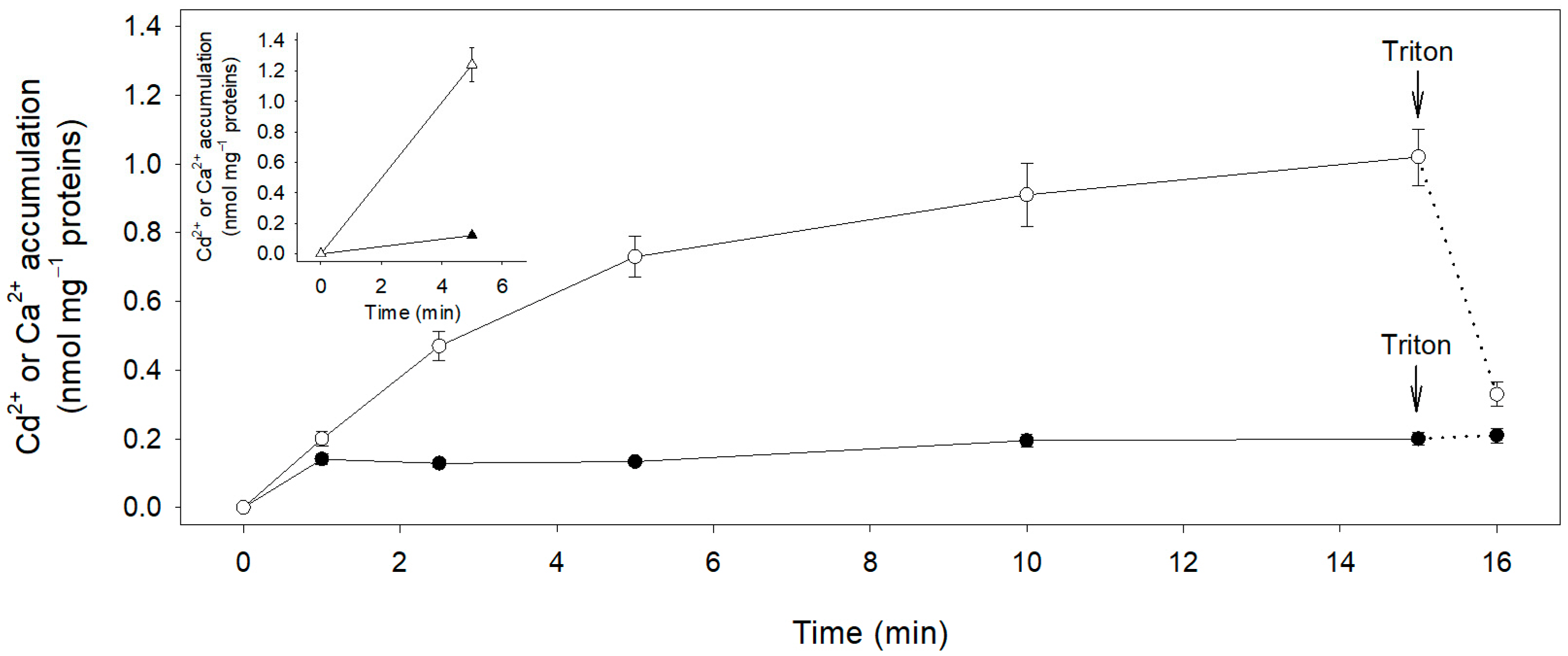
2.1. Short Term Cd2+ Influx in Maize Root Segments

Most of the experiments presented in this work were conducted using subapical maize root segments (hereafter also called “root segments”), cut between 2 and 8 mm from the tips of the main roots. This portion of root shows a differentiation which mainly consists of parenchyma tissue and epidermis, with only some protoxylem vessels already defined near the cut at 8 mm from the tip. The extremely simplified and rather homogeneous structure of the segments is thus suitable for studying ion fluxes in a whole organ, avoiding the complexity of the whole root. The choice of this material to study cadmium cell fluxes is consistent with studies on the spatial profile of Cd2+ uptake along the root axe, where the highest Cd2+ influx is sustained by the apical region of the root in wheat and rice [37,38].
Cadmium toxicity to a variety of cell types is abundantly described in the literature [39,40,41,42,43,44,45,46] and essentially depends on concentrations and time of exposure. Thus, we checked whether cadmium could induce toxicity in our experimental conditions. Relatively short-term incubations (up to 45 min) of root segments in media containing a wide range of Cd2+ concentrations (up to 500 µM) did not affect respiration (rate of O2 uptake) or cell membrane integrity measured as potassium leakage in the medium, suggesting that, at least in our conditions, Cd2+ exposure did not produce generalized toxic effects.
One complication which arises in studying cation transport in walled cells (bacteria, fungi and plants) is the presence of a cell wall surrounding the plasma membrane. In the plant cell wall compartment, ions can be free in the wall pores or electrostatically bound to the negatively charged polysaccharydes and proteins that are major components of the cell wall structure [47]. In normal plant growth conditions, negative charges on the cell wall are mostly saturated by Ca ions, but this plant nutrient can be replaced by heavy metals such as Cd when exposure occurs. In fact, binding affinity of divalent, trivalent or poly cations to the cell wall has been described to be particularly high [48,49]. To address these issues, we initially set up control experiments in which root segments were incubated in 50 µM Cd2+ for increasing lengths of time and then washed twice in cold water (2 °C) or in a cold solution (2 °C) containing LaCl3, (Figure 1a, filled and open triangles, respectively). When root segments were washed with water and then harvested for Cd2+ analysis, the rate of Cd2+ accumulation was high in the first 20 min and then decreased, becoming linear (Figure 1a, filled triangles). We could fit this time-course of Cd2+ accumulation with a non-saturating curve that could be properly described by a modified hyperbolic function—consisting in a saturable and a linear component—of the form:
y = a x b + x + c x
where y is the Cd content in the root segments, x is the time and a, b and c are the parameters of best fitting (R = 0.999). The rate of Cd2+ uptake, c, calculated from the linear component was 2.38 ± 0.09 µmol h−1 g−1 FW.
We interpreted the saturable component as Cd ions initially diffusing rapidly into the root segments’ free space and binding to the non-diffusible anions of the cell wall, and the linear component as Cd ions steadily entering the symplast after saturation of the extracellular compartments. This interpretation was validated by washing the root segments at the end of Cd2+ incubation with 1 mM with La3+ (twice at 2 °C) to exchange the Cd ions non-absorbed by the cells and retained in the root apoplast. The results are shown in Figure 1a, open triangles. Time-depended Cd2+ accumulation in the root segments appeared almost linear for at least 45 min (R = 0.996), and we defined the slope of the fitted straight line as the initial Cd2+ influx into the symplast. The rate of Cd2+ uptake was 2.21 ± 0.07 µmol h−1 g−1 FW, a value very close to that calculated on the base of the linear component observed in root segments washed only with water.
Figure 1b reports the kinetic of Cd2+ influx as a function of Cd2+ external concentrations in the range of 0 to 500 µM. The influx of Cd2+ increased markedly up to an external concentration of 50 µM; at higher concentrations, the influx increased slowly without reaching saturation. Data analysis revealed that the kinetic profile of Cd2+ influx could be best described (R = 0.999) by a function sum of a saturable rectangular hyperbola and a straight line, as follows:
v = V m a x S S + K m + k S
where v is the rate of Cd2+ influx, S is Cd2+ concentration, Vmax and Km are the Michaelis–Menten constants and k is the first-order rate coefficient. The apparent Km was 30.15 ± 3.08 µM, while the Vmax was 3.01 ± 0.16 µmol h−1 g−1 FW; the first-order rate coefficient (k) of the linear component was 0.0013 ± 0.0003 L h−1 g−1 FW. To verify that the linear component of Cd2+ influx was not an artefact of incomplete desorption of Cd ions from the apoplast at cadmium concentrations higher than 50 µM, we first disrupted the symplast by subjecting a set of root segments to three cycles of freeze-thawing and ethanol-detergent (Triton X-100) treatment; then, we used the dead root segments to run a Cd2+ uptake experiment following the same protocol used for the alive root segment. Cadmium concentration in the medium was 100 µM and incubation time was 30 min, followed by two washes in 1 mM LaCl3. Lanthanum washing removed more than 97% of Cd2+ from the dead root segments (data not shown), indicating that the linear component observed in the kinetic of Cd2+ uptake on alive root segments (Figure 1b) was due to Cd2+ influx into the cells, and probably to an integral part of the complex uptake mechanism of this heavy metal by the roots.

2.2. Short Term Ca2+ Influx in Maize Root Segments

Calcium is a well-known plant macronutrient which plays critical structural and functional roles in the plant cell, including metabolism and signal transduction [50,51,52,53]. Since Cd2+ has been shown to interfere with Ca2+ functions [10], we thought to compare the kinetic of Cd2+ influx into root segments to that of Ca2+ in the same material, to highlight possible differences in the kinetic profiles of two chemically similar divalent cations, a plant nutrient and a non-essential metal. Figure 1c shows the kinetic of Ca2+ uptake determined in a wide range of external concentrations (from 1 to 500 µM). In these experiments, radioactive 45Ca2+ was used as a tracer, and the washes to remove labelled 45Ca2+ from the apoplast were performed using non-labelled CaCl2 (see Methods). Unlike the kinetic of Cd2+ influx, Ca2+ influx isotherm could be described by a simple Michaelis–Menten function (R = 0.999), with an apparent Km value of 26.57 ± 0.80 µM and a Vmax value of 1.24 ± 0.01 µmol h−1 g−1 FW (Figure 1c).

2.3. Effect of Ca2+ on Cd2+ Influx in Maize Root Segments

The kinetic analysis was extended considering the competitions between Cd2+ and Ca2+. As shown in Table 1, the presence of Ca2+ in the incubation medium reduced Cd2+ influx into the root segments. Such an inhibition closely depended on the Ca2+/Cd2+ ratio in the incubation medium. For instance, at 10 µM Cd2+ in the external solution an equimolar amount of Ca2+ reduced Cd2+ influx by 18%; Cd2+ influx was further inhibited by 58% or 70% in the presence of 50 or 100 µM Ca2+ in the external medium, respectively. Similar trends of inhibition were observed when Cd2+ influxes at 50 or 100 µM Cd2+ were measured in the presence of 10, 50 or 100 µM Ca2+.

2.4. Cd2+ and Ca2+ Efflux from Subapical Maize Root Segments

Pulse-chase experiments were performed to evaluate possible Cd2+ efflux across the plasma membrane of the cells forming the maize root segments. These experiments require loading of the root segments with radioactive Cd2+ for a short period of time (pulse), and measurements of Cd2+ leaked from the segments over longer time periods (chase). We took the precaution of using concentrations of Cd2+ as low as possible to avoid toxicity and long-term accumulation of chelating agents that could reduce the activity of free Cd ions in the root cells [26,27,32]. In detail, root segments were pre-loaded (15 min) in a basal medium containing 10 μM (109Cd2+)Cd2+ at the specific activity of 1.68 kBq nmol−1 (pulse phase), quickly washed in a Cd-free basal medium (washing phase, 3 × 1 min) and incubated in the same basal medium supplied with unlabelled 10 μM Cd2+ (chase phase). The washing phase removed radioactivity from the external surface of the segments but not Cd ions present in the free space (cell walls and intracellular spaces), as already shown in Figure 1a. Since the amount of Cd ions present in the root segments before Cd2+ exposure was negligible (0.18 ± 0.04 nmol g−1 FW), we can reasonably assume that at the end of the washes, the specific activity of (109Cd2+)Cd2+ into the root segments was the same as that administered during the pulse phase (1.68 kBq nmol−1).
During the short pulse phase, the root segments took up (109Cd2+)Cd2+. After this phase and the wash phase, we could estimate the amount of Cd ions accumulated in the segments by using the following parameters: (i) the decrease in radioactivity present in the medium (A); (ii) the radioactivity lost during the three Cd2+ free washings (B, C, D); and (iii) the specific activity of (109Cd2+)Cd2+ (E), to solve the equation below:
C d 2 + a c c u m u l a t e d = A B + C + D E = 0.277 ± 0.001   m o l   g 1 F W
Such a value includes Cd ions accumulated in the symplast and the free spaces of the root segments and corresponds to a total radioactivity of 465 kBq g−1 FW.
Figure 2 shows the time course of (109Cd2+)Cd2+ leakage from the root segments measured during the chase. Data distribution revealed that two distinct phases characterized the chase. In the first phase (0–30 min) radioactivity was rapidly released in the medium, while in the second phase (30–60 min) the radioactivity released in the medium was partially reabsorbed by the root segments. The radioactivity released in the first 30 min corresponded to 55 kBq g−1 FW, which is less than 12% of the (109Cd2+)Cd2+ present in the root segments (symplast + free space). The overall trajectory of the chase curve was interpreted as the sum of a rectangular hyperbola and a straight line (R = 0.999) as follows:
k B q   m L 1 = a t b + t + c t
where t is time, a (1.03 ± 0.08 kBq mL−1) is the theoretical maximum radioactivity at saturation, b (10.95 ± 1.49 min) is the t1/2 of the saturable component and c (−0.005 ± 0.001 kBq min−1 mL−1) is the first-order rate coefficient.
Since the apparent (109Cd2+)Cd2+ leakage approached saturation after 30 min, we can reasonably suppose that such an event was mainly related to the equilibration of (109Cd2+)Cd2+ in the root free space with the unlabelled Cd ions in the basal medium. Conversely, the second phase, in which the radioactivity of the medium linearly decreased, mainly depended on (109Cd2+)Cd2+ re-absorption.
Considering the first-order rate coefficient, c, of the linear component of the fitting equation and the specific activity of (109Cd2+)Cd2+ measured after 30 min from the beginning of the chase (0.061 kBq nmol−1), we can calculate that during the second phase of the chase, the net uptake (influx minus efflux) of Cd2+ into the root segments was 0.52 ± 0.09 μmol h−1 g−1 FW. Since this value is close to that of Cd2+ influx (0.752 ± 0.040 μmol h−1 g−1 FW), measured at the same Cd2+ external concentration (10 μM; Figure 1b), we can reasonably consider Cd efflux as negligible.
Similar pulse-chase experiments were carried out using radioactive Ca2+ as a tracer (10 μM (45Ca2+)Ca2+ at the specific activity of 1.72 kBq nmol−1). After a pulse phase of 15 min and three washes in a Ca2+ free medium, the total radioactivity present in the root segments was 272 kBq g−1 FW. In these experiments, however, we were not able to impose Ca2+ specific activities in the different cell compartments and in the free space, since Ca ions were initially present in the root segments as natural costituents. Figure 2b shows that during the first 30 min of the chase phase, the radioactivity rapidly increased in the external medium and then remained almost constant up to the end of the experiment (60 min). The radiocatrivity released in the medium after 30 min (80 kBq g−1 FW) was 29.3% of the total radioactivity present in the root segments at the beginning of the chase phase. Such a value not only depends on the rapid equilibration of (45Ca2+)Ca2+ in the root free space with the unlabelled Ca ions in the external medium, but also on the equilibration of (45Ca2+)Ca2+ present in other cell compartments. Taken as a whole, these data suggest that Ca2+ efflux is significantly higher than Cd2+ efflux.

2.5. Cd2+ Transport in Plasma Membrane Vesicles from Maize Root Cortical Cells

To validate the results obtained from intact root, Cd2+ transport was studied on plasma membrane preparations. Plasma membrane vesicles were purified from maize root cortical cells using a sorbitol-dextran discontinuous gradient, which produced mainly right-side-out vesicles that cannot be energized with ATP in the assay medium. To address this experimental inconvenience, three freeze-thawing cycles were applied to the vesicles, producing a mixture of inside-out and right-site-out vesicles. Only the inside-out vesicles can use ATP to energize transport, as the cytoplasmic side of the vesicle membrane is accessible to ATP in the external medium [54].
In a first set of experiments we included protonophores (CCCP and ammonium) in the assay medium to exclude ion transport events dependent on the proton electrochemical gradient across the membrane vesicles generated by the activity of the Plasma Membrane H+-ATPase [55]. Figure 3 shows the time course of Cd2+ and Ca2+ accumulation into inside-out vesicles; such activities correspond to ion effluxes from the cells (Supplementary Figure S1). (109Cd2+)Cd2+ reached its maximum accumulation value in the vesicles in just the first minute of incubation, so its concentration in the vesicle was practically independent of the incubation time. The addition of Triton-X100 at the end of the incubation period did not significantly affect the level of radioactivity recovered from the vesicles. On the other hand, (45Ca2+)Ca2+ was taken up by the vesicles, and its concentration progressively increased as a function of time. When Triton X-100 was added to the incubation medium, Ca2+ level in the vesicles decreased, reaching a value similar to that measured for Cd2+. Such a finding strongly suggests that most of the divalent cations recovered from the vesicles after 1 min incubation were firmly bound to the membranes. Finally, since the amount of Cd ions accumulated in the vesicles never exceeds the amount of Cd ions bound to the membranes, we can conclude that Cd2+ efflux through the vesicle membranes is negligible. In contrast, the ATP-dependent (45Ca2+)Ca2+ accumulation in the vesicles underlines that Ca2+ efflux is an activity of the plasma membrane of the root cortical cells (Supplementary Figure S1). The presence of protonophores did not affect the accumulation of (109Cd2+)Cd2+ in the plama membrane vesicles after 5 min incubation, as the levels of (109Cd2+)Cd2+ were similar in the presence or absence of protonophores (Figure 3, inset). On the other hand, (45Ca2+)Ca2+ was 50% higher in the absence of protonophores, indicating that the transport activities of Ca2+ closely dependent on the proton electrochemical gradient cooperate with Ca2+-ATPases to facilitate Ca2+ efflux from root cortical cells (Supplementary Figure S1) [56,57,58].

3. Discussion

Results of the initial experiments shown in Figure 1a indicate that, following La3+ washing, Cd2+ accumulation into the root segments was linear for at least 45 min. In addition, since only traces of Cd2+ were present in the root segments (0.18 ± 0.04 nmol g−1 FW) before cadmium exposure, we can reasonably consider Cd2+ accumulation as the result of a unidirectional flux (influx). Such a finding allowed us to determine the kinetic isotherm of Cd2+ uptake under a wide range of Cd2+ external concentration (Figure 1b).
The kinetic analysis showed that the pattern of Cd2+ influx into root cells followed a complex isotherm, which was a result of the combination of a saturable and a first-order linear kinetic. This suggests that multiple transporters for essential elements with insufficient selectivity with respect to their substrate, such as Ca2+ channels, LCT1, ZIP-IRT and NRAMP transporters [11,14,14,38,59,60,61], were working in parallel at the plasma membrane to allow Cd2+ absorption into root cells. The apparent Km (30.15 ± 3.08 μM) of the saturable component of the kinetic was higher compared to the value measured in intact rice roots [14]; such a discrepancy may be due to the different species and/or organs used for the experiments. By contrast, kinetic of Ca2+ influx in the root segments (Figure 1c) could be described by a simple Michaelis–Menten function with an apparent Km value of 26.57 ± 0.80 μM. Such a value could be underestimated since it was calculated using the administered specific activity of (45Ca2+)Ca2+ in the medium without considering the Ca2+ present in the tissues. Nevertheless, a direct comparison of the Km values measured for Cd2+ and Ca2+ influxes does not allow us to exclude the hypothesis that the two ions may partially share the same transport system(s), as previously observed in wheat [38]. Moreover, in our experimental setup the presence of Ca2+ in the incubation medium significantly reduced Cd2+ influx in the root segments (Table 1), suggesting the existence of a competition between the two ions for the same transport system(s). Cd2+/Ca2+ competitions have also been suggested following the observation that Ca ions may alleviate Cd2+ toxicity in different plant species [22,62,63].
Cadmium concentration in the cells could be reduced by the activity of transporters mediating Cd2+ efflux across the plasma membrane [64,65,66]. Similar mechanisms have been largely described as involved in controlling Na+ [67] and Ca2+ [56] homeostasis. Mechanisms controlling Ca2+ concentration in the cells are essential to prevent potentially toxic effects due to the aggregation of proteins, nucleic acids and phosphates, as well as to allow the role of this ion as a second messenger [50,51,68].
A possible activity mediating Cd2+ efflux from the root segments has been investigated here using a pulse-chase approach with (109Cd2+)Cd2+ (Figure 2a). Notwithstanding the presence of a root free space limiting the possibility of performing a precise measurement of Cd2+ efflux, results clearly indicate that when this compartment equilibrated with the external medium (after 30 min from the beginning of the chase phase), an apparent Cd2+ re-influx occurred (30–60 min of the chase phase). Since the rate of Cd2+ net uptake measured during this last period was similar to that of Cd2+ influx measured at the same external concentration (Figure 1b), we can conclude that, in our experimental conditions, Cd2+ efflux from the root segments is minimal. On the other hand, results of similar experiments carried out with (45Ca2+)Ca2+ (Figure 2b) indicate that after the equilibration of the root free space with the external medium, 45Ca2+ was not re-absorbed by the segments, probably as the result of an equilibrium between Ca2+ influx and efflux.
The analysis of Cd2+ fluxes measured in inside-out vesicles also indicated the absence of a Cd2+ efflux activity on the plasma membrane of the root cortical cells both in the presence or in the absence of protonophores (Figure 3, Supplementary Figure S1). On the contrary, the energization of the vesicles with ATP resulted in significant Ca2+ effluxes in the presence or absence of protonophores (Figure 3, Supplementary Figure S1), suggesting the existence of both ATP- and proton-dependent activities mediating Ca2+ efflux on the plasma membrane of the root cortical cells [56,57,58].
Taken as a whole, our results indicate that Cd2+ influx into the root cells is mediated by a complex mechanism that involves different plasma membrane transport proteins, including Ca2+ transporters. Moreover, the cortical cells of the maize root are not able to extrude Cd2+ and show at the same time a highly selective activity of Ca2+ extrusion, as already suggested by Rivetta and co-workers [22].
From an evolutionary viewpoint, our data could support the heuristic hypothesis that the impossibility of extruding a large amount of Cd2+ from the root cortical cells could have imposed strong selective pressure to favour the evolution of protein or non-protein peptides (i.e., metallothioneins and phytochelatins) [69] able to bind and detoxify Cd2+ inside the cells.

4. Materials and Methods

4.1. Plant Material

Maize (Zea mays L., cv. Dekalb 300) caryopses were germinated in the dark at 26 °C on filter paper saturated with milli-Q water. After 48 h, seedlings were transferred to aerated 0.5 mM CaSO4 solutions and maintained in the dark at 26 °C for 24 h; at the end of this period, the main root was about 40–50 mm long. Subapical root segments (6 mm long) were obtained by cutting the main root at 2 and 8 mm from the tip. Batches of 20 segments (about 100 mg of fresh weight) were then washed twice for 30 min in 10 mL of a 0.1 mM Mes-BTP (pH = 6.0) basal medium, and incubated in a water bath thermoregulated at 26°C and reciprocated at 90 oscillations min−1; after these treatments (preincubation), the segments had completely recovered from wounding.

4.2. Measurement of Cd2+ Influx

After preincubation, subapical root segments were incubated at different times in 10 mL of basal medium supplemented with CdCl2 at different concentrations at 26 °C and agitated at 90 oscillations min−1. At the end of the incubation period, the segments were washed twice at 2 °C for 15 min in milli-Q water or in the basal medium supplemented with 1 mM LaCl3 and then mineralized at 100 °C in 0.5 mL of HNO3-H2SO4-HClO4 (5:1:1, v:v:v). The amount of Cd2+ in the root segments was determined by atomic absorption spectrophotometry (SpectraAA-20, Varian, Palo Alto, CA, USA). The effect of Ca2+ on Cd2+ influx was measured by incubating the root segments in solutions containing 10, 50 or 100 µM Cd2+ in the presence of variable Ca2+ concentrations (from 0 to 100 µM). Experiments of Cd2+ uptake were also carried out using segments three times frozen-thawed and then washed four times (30 min each) in absolute ethanol containing 2% (w:v) Triton X-100 (Sigma-Aldrich, Saint Louis, MI, USA) and finally extensively rinsed in the basal medium.

4.3. Measurement of Ca2+ Influx

Twenty preincubated root segments were incubated at 26 °C for 15 min in 10 mL of basal medium supplemented with CaCl2 at different concentrations and agitated at 90 oscillations min−1; solutions were labelled with 3.7 kBq mL−1 45Ca2+. At the end of the incubation period, the segments were washed twice at 2 °C for 15 min in the basal medium supplemented with 1 mM CaCl2 and then rinsed in milli-Q water. The segments were then homogenized in 0.75 mL of 0.1 N HNO3 and heated at 100 °C for 15 min. Radioactivity was measured by counting, in a Beckman LS 6000S (Fullerton, CA, USA) scintillation counter, aliquots of the supernatants dissolved in 10 mL of Ready-Solve scintillation cocktail (Backman).

4.4. Pulse and Chase Experiments with (109Cd2+)Cd2+ or (45Ca2+)Ca2+

Cd2+ and Ca2+ fluxes were measured using 10 subapical maize root segments incubated at 26 °C for 15 min in 5 mL of basal medium containing 10 μM (109Cd2+)CdCl2 or 10 μM (45Ca2+)CaCl2, at the specific activity of 1.68 kBq nmol−1 or 1.72 kBq nmol−1, respectively. At the end of the pulse phase, the root segments were washed three times for 1 min in 5 mL of Mes-BTP (pH = 6) and then incubated at 26 °C for 60 min in 5 mL of basal medium supplemented with 10 μM non-radioactive Cd2+ or Ca2+ (chase phase). Radioactivity was measured in all the liquid media (at the end of the pulse phase, at the end of the washes and at different time during the chase phase) by liquid scintillation spectrometry.

4.5. Plasma Membrane Vesicle Isolation

Plasma membrane vesicles were prepared from maize root cortex using a method based on the one described by Palmgren and co-workers [54]. The cortex was separated from the stele, using a wire stripper, and blended with a solution of 330 mM sorbitol, 50 mM Mops-BTP (pH = 7.5), 5 mM EDTA, 1 mM DTT, 0.5 mM PMSF and 1% (w:v) BSA. The resulting mixture was filtered and centrifuged at 13,000× g for 15 min, and the supernatant was subjected to further high-speed centrifugation at 100,000× g for 30 min to obtain the microsomal fraction. This fraction was then suspended in a solution of 330 mM sorbitol, 5 mM KCl, 1 mM DTT, 0.1 mM EDTA and 5 mM K-phosphate (pH = 7.8). The plasma membrane vesicles were purified using a three-step sorbitol/dextran T-500/PEG 3350 gradient [70]. The product was suspended in a solution of 330 mM sorbitol, 50 mM KCl and 2 mM Mops-BTP (pH = 7.0) to obtain a final protein concentration of about 8 µg/µL. The plasma membranes were then frozen-thawed three times to obtain a mixture of inside-out and right-side-out vesicles [54].

4.6. Measurement of Ca2+ and Cd2+ Uptake in Plasma Membrane Vesicles

The uptake of Ca2+ and Cd2+ was measured in plasma membrane vesicles using 45Ca2+ and 109Cd2+. The vesicles (100 µg protein sample−1) were pre-incubated for 60 min at 30 °C in a solution containing 0.6 mL of 250 mM sorbitol, 25 mM Tris-Mes (pH = 7.2), 1 mM ATP, 0.015% (w:v) Brij 58, and 10 µM (45Ca2+)CaCl2 (1 kBq 45Ca2+ mL−1) or 10 µM (109Cd2+)CdCl2 (1 kBq 109Cd2+ mL−1), in the presence or absence of 100 µM CCCP, 5 mM (NH4)2SO4. The uptake was initiated by adding MgSO4 to reach a final concentration of 1 mM. At various times, samples were filtered through a 0.22 µm filter (GSWP, Millipore Corp, Burlington, MA, USA) and rapidly washed with 4 mL of a solution of 25 mM Tris-Mes (pH = 7.2) containing 1 mM CaCl2 or 1 mM CdCl2. Filters were dried at 45 °C for 30 min, and radioactivity was measured by liquid scintillation spectrometry. MgATP-dependent uptake of Ca2+ or Cd2+ was determined by subtracting the radioactivity measured in the absence of Mg2+ from that measured in its presence.

4.7. Statistical Analysis

The results presented are the average of three experiments, each conduced in triplicate (n = 3). Statistical analysis was performed using SigmaPlot for Windows version 11.0 (Systat Software, Inc., Point Richmond, CA, USA), and the significance of the results was determined using ANOVA. A p value of less than 0.05 was considered significant. The Bonferroni correction was applied to account for multiple comparisons. The fitting of the data was performed using SigmaPlot for Windows version 11.0.

Supplementary Materials

The following supporting information can be downloaded at: https://www.mdpi.com/article/10.3390/plants12050992/s1, Figure S1: Ca2+ accumulation in inside-out plasma membrane vesicles in the presence (a) or absence (b) of protonophores. Cd2+ accumulation in inside-out plasma membrane vesicles in the presence (c) or absence (d) of protonophores.

Author Contributions

Conceptualization, A.R. and M.C.; methodology, A.R., F.F.N. and M.C.; validation, F.F.N., M.P., G.A.S. and M.C.; formal analysis, F.F.N. and M.C.; investigation, A.R.; writing—original draft preparation, M.C; writing—review and editing, A.R., M.P., G.A.S., F.F.N. and M.C.; supervision, M.C. All authors have read and agreed to the published version of the manuscript.

Funding

This research received no external funding.

Institutional Review Board Statement

Not applicable.

Informed Consent Statement

Not applicable.

Data Availability Statement

The raw data supporting the conclusions of this article will be made available by the authors, without undue reservation, to any qualified researcher.

Conflicts of Interest

The authors declare no conflict of interest.

References

  1. Nordberg, G.F. Historical perspectives on cadmium toxicology. Toxicol. Appl. Pharmacol. 2009, 238, 192–200. [Google Scholar] [CrossRef] [PubMed]
  2. Genchi, G.; Sinicropi, M.S.; Lauria, G.; Carocci, A.; Catalano, A. The effects of cadmium toxicity. Int. J. Environ. Res. Public Health 2020, 17, 3782. [Google Scholar] [CrossRef] [PubMed]
  3. Alloway, B.J.; Steinnes, E. Anthropogenic additions of cadmium to soils. In Cadmium in Soils and Plants; McLaughlin, M.J., Singh, B.R., Eds.; Developments in Plant and Soil Sciences; Springer Netherlands: Dordrecht, The Netherlands, 1999; pp. 97–123. ISBN 978-94-011-4473-5. [Google Scholar]
  4. Clemens, S.; Aarts, M.G.M.; Thomine, S.; Verbruggen, N. Plant Science: The key to preventing slow cadmium poisoning. Trends Plant Sci. 2013, 18, 92–99. [Google Scholar] [CrossRef]
  5. McLaughlin, M.J.; Parker, D.R.; Clarke, J.M. Metals and micronutrients—Food safety issues. Field Crops Res. 1999, 60, 143–163. [Google Scholar] [CrossRef]
  6. Welch, R.M.; Norvell, W.A. Mechanisms of cadmium uptake, translocation and deposition in plants. In Cadmium in Soils and Plants; McLaughlin, M.J., Singh, B.R., Eds.; Developments in Plant and Soil Sciences; Springer Netherlands: Dordrecht, The Netherlands, 1999; pp. 125–150. ISBN 978-94-011-4473-5. [Google Scholar]
  7. Sterckeman, T.; Thomine, S. Mechanisms of cadmium accumulation in plants. Crit. Rev. Plant Sci. 2020, 39, 322–359. [Google Scholar] [CrossRef]
  8. Clemens, S. Toxic metal accumulation, responses to exposure and mechanisms of tolerance in plants. Biochimie 2006, 88, 1707–1719. [Google Scholar] [CrossRef]
  9. Korshunova, Y.O.; Eide, D.; Clark, W.G.; Guerinot, M.L. The IRT1 protein from Arabidopsis thaliana is a metal transporter with a broad substrate range. Plant Mol. Biol. 1999, 40, 37–44. [Google Scholar] [CrossRef]
  10. Eide, D.; Broderius, M.; Fett, J.; Guerinot, M.L. A novel iron-regulated metal transporter from plants identified by functional expression in yeast. Proc. Natl. Acad. Sci. USA 1996, 93, 5624–5628. [Google Scholar] [CrossRef] [Green Version]
  11. Rogers, E.E.; Eide, D.J.; Guerinot, M.L. Altered selectivity in an Arabidopsis metal transporter. Proc. Natl. Acad. Sci. USA 2000, 97, 12356–12360. [Google Scholar] [CrossRef] [Green Version]
  12. Ajeesh Krishna, T.P.; Maharajan, T.; Victor Roch, G.; Ignacimuthu, S.; Antony Ceasar, S. Structure, function, regulation and phylogenetic relationship of ZIP family transporters of plants. Front. Plant Sci. 2020, 11, 662. [Google Scholar] [CrossRef]
  13. Ramesh, S.A.; Shin, R.; Eide, D.J.; Schachtman, D.P. Differential metal selectivity and gene expression of two zinc transporters from rice. Plant Physiol. 2003, 133, 126–134. [Google Scholar] [CrossRef] [PubMed] [Green Version]
  14. Sasaki, A.; Yamaji, N.; Yokosho, K.; Ma, J.F. Nramp5 is a major transporter responsible for manganese and cadmium uptake 478 in rice. Plant Cell 2012, 24, 2155–2167. [Google Scholar] [CrossRef] [PubMed] [Green Version]
  15. Takahashi, R.; Ishimaru, Y.; Nakanishi, H.; Nishizawa, N.K. Role of the iron transporter OsNRAMP1 in cadmium uptake and accumulation in rice. Plant Signal Behav. 2011, 6, 1813–1816. [Google Scholar] [CrossRef] [PubMed] [Green Version]
  16. Nakanishi, H.; Ogawa, I.; Ishimaru, Y.; Mori, S.; Nishizawa, N.K. Iron deficiency enhances cadmium uptake and translocation 482 mediated by the Fe2+ transporters OsIRT1 and OsIRT2 in Rice. Soil Sci. Plant Nutr. 2006, 52, 464–469. [Google Scholar] [CrossRef]
  17. Hinkle, P.; Kinsella, P.; Osterhoudt, K. Cadmium uptake and toxicity via voltage-sensitive calcium channels. J. Biol. Chem. 1987, 262, 16333–16337. [Google Scholar] [CrossRef]
  18. Usai, C.; Barberis, A.; Moccagatta, L.; Marchetti, C. Pathways of cadmium influx in mammalian neurons. J. Neurochem. 1999, 72, 2154–2156. [Google Scholar] [CrossRef] [PubMed]
  19. Souza, V.; Bucio, L.; Gutiérrez-Ruiz, M.C. Cadmium uptake by a human hepatic cell line (WRL-68 Cells). Toxicology 1997, 120, 215–220. [Google Scholar] [CrossRef]
  20. Jacobson, B.; Turner, J.E. The interaction of cadmium and certain other metal ions with proteins and nucleic acids. Toxicology 1980, 16, 1–37. [Google Scholar] [CrossRef]
  21. Choong, G.; Ying Liu, Y.; Templeton, D.M. Interplay of calcium and cadmium in mediating cadmium toxicity. Chem. Biol. Interact. 2014, 211, 54–65. [Google Scholar] [CrossRef] [PubMed]
  22. Rivetta, A.; Negrini, N.; Cocucci, M. Involvement of Ca2+-calmodulin in Cd2+ toxicity during the early phases of radish (Raphanus sativus L.) seed germination. Plant Cell Environ. 1997, 20, 600–608. [Google Scholar] [CrossRef]
  23. Jarvis, S.C.; Jones, L.H.P.; Hopper, M.J. Cadmium uptake from solution by plants and its transport from roots to shoots. Plant Soil 1976, 44, 179–191. [Google Scholar] [CrossRef]
  24. Zhao, F.; Hamon, R.E.; Lombi, E.; McLaughlin, M.J.; McGrath, S.P. Characteristics of cadmium uptake in two contrasting ecotypes of the hyperaccumulator Thlaspi caerulescens. J. Exp. Bot. 2002, 53, 535–543. [Google Scholar] [CrossRef] [Green Version]
  25. Chen, X.; Ouyang, Y.; Fan, Y.; Qiu, B.; Zhang, G.; Zeng, F. The pathway of transmembrane cadmium influx via calcium-permeable channels and its spatial characteristics along rice root. J. Exp. Bot. 2018, 69, 5279–5291. [Google Scholar] [CrossRef] [PubMed] [Green Version]
  26. Grill, E.; Winnacker, E.-L.; Zenk, M.H. Phytochelatins, a class of heavy-metal-binding peptides from plants, are functionally analogous to metallothioneins. Proc. Natl. Acad. Sci. USA 1987, 84, 439–443. [Google Scholar] [CrossRef] [Green Version]
  27. Zenk, M.H. Heavy metal detoxification in higher plants—A review. Gene 1996, 179, 21–30. [Google Scholar] [CrossRef] [PubMed]
  28. Miyadate, H.; Adachi, S.; Hiraizumi, A.; Tezuka, K.; Nakazawa, N.; Kawamoto, T.; Katou, K.; Kodama, I.; Sakurai, K.; Takahashi, H.; et al. OsHMA3, a P1B-type of ATPase affects root-to-shoot cadmium translocation in rice by mediating efflux into vacuoles. New Phytol. 2011, 189, 190–199. [Google Scholar] [CrossRef]
  29. Yan, J.; Wang, P.; Wang, P.; Yang, M.; Lian, X.; Tang, Z.; Huang, C.-F.; Salt, D.E.; Zhao, F.J. A loss-of-function allele of OsHMA3 associated with high cadmium accumulation in shoots and grain of Japonica rice cultivars. Plant Cell Environ. 2016, 39, 1941–1954. [Google Scholar] [CrossRef] [Green Version]
  30. Ueno, D.; Yamaji, N.; Kono, I.; Huang, C.F.; Ando, T.; Yano, M.; Ma, J.F. Gene limiting cadmium accumulation in rice. Proc. Natl. Acad. Sci. USA 2010, 107, 16500–16505. [Google Scholar] [CrossRef] [Green Version]
  31. Cheng, N.-H.; Pittman, J.K.; Barkla, B.J.; Shigaki, T.; Hirschi, K.D. The Arabidopsis Cax1 mutant exhibits impaired ion homeostasis, development, and hormonal responses and reveals interplay among vacuolar transporters. Plant Cell 2003, 15, 347–364. [Google Scholar] [CrossRef] [Green Version]
  32. Nocito, F.F.; Lancilli, C.; Dendena, B.; Lucchini, G.; Sacchi, G.A. Cadmium retention in rice roots is influenced by cadmium availability, chelation and translocation. Plant Cell Environ. 2011, 34, 994–1008. [Google Scholar] [CrossRef] [PubMed]
  33. Silver, S.; Phung, L.T. Bacterial heavy metal resistance: New surprises. Annu. Rev. Microbiol. 1996, 50, 753–789. [Google Scholar] [CrossRef] [PubMed]
  34. Maghrebi, M.; Baldoni, E.; Lucchini, G.; Vigani, G.; Valè, G.; Sacchi, G.A.; Nocito, F.F. Analysis of cadmium root retention for two contrasting rice accessions suggests an important role for OsHMA2. Plants 2021, 10, 806. [Google Scholar] [CrossRef] [PubMed]
  35. Wong, C.K.E.; Cobbett, C.S. HMA P-type ATPases are the major mechanism for root-to-shoot Cd translocation in Arabidopsis thaliana. New Phytol. 2009, 181, 71–78. [Google Scholar] [CrossRef] [PubMed]
  36. Costa, G.; Morel, J.L. Cadmium uptake by Lupinus albus (L.): Cadmium excretion, a possible mechanism of cadmium tolerance. J. Plant Nutr. 1993, 16, 1921–1929. [Google Scholar] [CrossRef]
  37. Piñeros, M.A.; Shaff, J.E.; Kochian, L.V. Development, characterization, and application of a cadmium-selective microelectrode for the measurement of cadmium fluxes in roots of Thlaspi species and wheat. Plant Physiol. 1998, 116, 1393–1401. [Google Scholar] [CrossRef] [PubMed] [Green Version]
  38. Li, L.-Z.; Tu, C.; Peijnenburg, W.J.G.M.; Luo, Y.-M. Characteristics of cadmium uptake and membrane transport in roots of intact wheat (Triticum aestivum L.) seedlings. Environ. Pollut. 2017, 221, 351–358. [Google Scholar] [CrossRef] [PubMed]
  39. Zulfiqar, U.; Ayub, A.; Hussain, S.; Waraich, E.A.; El-Esawi, M.A.; Ishfaq, M.; Ahmad, M.; Ali, N.; Maqsood, M.F. Cadmium toxicity in plants: Recent progress on morpho-physiological effects and remediation strategies. J. Soil Sci. Plant Nutr. 2022, 22, 212–269. [Google Scholar] [CrossRef]
  40. Deckert, J. Cadmium toxicity in plants: Is there any analogy to its carcinogenic effect in mammalian Cells? Biometals 2005, 18, 475–481. [Google Scholar] [CrossRef]
  41. Gill, S.S.; Tuteja, N. Cadmium stress tolerance in crop plants. Plant Signal. Behav. 2011, 6, 215–222. [Google Scholar] [CrossRef] [Green Version]
  42. Andresen, E.; Küpper, H. Cadmium toxicity in plants. In Cadmium: From Toxicity to Essentiality; Sigel, A., Sigel, H., Sigel, R.K., Eds.; Metal Ions in Life Sciences; Springer Netherlands: Dordrecht, The Netherlands, 2013; pp. 395–413. [Google Scholar]
  43. Sanità di Toppi, L.; Gabbrielli, R. Response to cadmium in higher plants. Environ. Exp. Bot. 1999, 41, 105–130. [Google Scholar] [CrossRef]
  44. Ferri, A.; Lancilli, C.; Maghrebi, M.; Lucchini, G.; Sacchi, G.A.; Nocito, F.F. The sulfate supply maximizing Arabidopsis shoot growth is higher under long- than short-term exposure to cadmium. Front. Plant Sci. 2017, 8, 854. [Google Scholar] [CrossRef]
  45. Martelli, A.; Rousselet, E.; Dycke, C.; Bouron, A.; Moulis, J.-M. Cadmium toxicity in animal cells by interference with essential metals. Biochimie 2006, 88, 1807–1814. [Google Scholar] [CrossRef] [PubMed]
  46. Cadmium Interaction with Animal Cells; Thévenod, F.; Petering, D.M.; Templeton, D.; Lee, W.-K.; Hartwig, A. (Eds.) Springer International Publishing: Cham, Switzerland, 2018; pp. 1–125. [Google Scholar]
  47. Sattelmacher, B. The Apoplast and its significance for plant mineral nutrition. New Phytol. 2001, 149, 167–192. [Google Scholar] [CrossRef] [PubMed]
  48. Haynes, R.J. Ion exchange properties of roots and ionic interactions within the root apoplasm: Their role in ion accumulation by plants. Bot. Rev. 1980, 46, 75–99. [Google Scholar] [CrossRef]
  49. Krzesłowska, M. The cell wall in plant cell response to trace metals: Polysaccharide remodeling and its role in defense strategy. Acta Physiol. Plant. 2011, 33, 35–51. [Google Scholar] [CrossRef] [Green Version]
  50. Sanders, D.; Brownlee, C.; Harper, J.F. Communicating with calcium. Plant Cell 1999, 11, 691–706. [Google Scholar] [CrossRef] [PubMed] [Green Version]
  51. McAinsh, M.R.; Pittman, J.K. Shaping the calcium signature. New Phytol. 2009, 181, 275–294. [Google Scholar] [CrossRef]
  52. White, P.J.; Broadley, M.R. Calcium in plants. Ann. Bot. 2003, 92, 487–511. [Google Scholar] [CrossRef]
  53. Thor, K. Calcium—Nutrient and messenger. Front. Plant Sci. 2019, 10, 440. [Google Scholar] [CrossRef]
  54. Palmgren, M.G.; Askerlund, P.; Fredrikson, K.; Widell, S.; Sommarin, M.; Larsson, C. Sealed inside-out and right-side-out plasma membrane vesicles. Optimal conditions for formation and separation. Plant Physiol. 1990, 92, 871–880. [Google Scholar] [CrossRef] [Green Version]
  55. Palmgren, M.; Morsomme, P. The plasma membrane H+-ATPase, a simple polypeptide with a long history. Yeast 2019, 36, 201–210. [Google Scholar] [CrossRef] [PubMed] [Green Version]
  56. Bose, J.; Pottosin, I.; Shabala, S.; Palmgren, M.; Shabala, S. Calcium efflux systems in stress signaling and adaptation in plants. Front. Plant Sci. 2011, 2, 85. [Google Scholar] [CrossRef] [PubMed] [Green Version]
  57. Kasai, M.; Muto, S. Ca2+ pump and Ca2+ /H+ antiporter in plasma membrane vesicles isolated by aqueous two-phase partitioning from corn leaves. J. Membr. Biol. 1990, 114, 133–142. [Google Scholar] [CrossRef] [PubMed]
  58. Luo, G.Z.; Wang, H.W.; Huang, J.; Tian, A.G.; Wang, Y.J.; Zhang, J.S.; Chen, S.Y. A putative plasma membrane cation/proton antiporter from soybean confers salt tolerance in Arabidopsis. Plant Mol. Biol. 2005, 59, 809–820. [Google Scholar] [CrossRef]
  59. Clemens, S.; Antosiewicz, D.M.; Ward, J.M.; Schachtman, D.P.; Schroeder, J.I. The plant cDNA LCT1 mediates the uptake of calcium and cadmium in yeast. Pro. Natl. Acad. Sci. USA 1998, 95, 12043–12048. [Google Scholar] [CrossRef] [Green Version]
  60. Gallego, S.M.; Pena, L.B.; Barcia, R.A.; Azpilicueta, C.E.; Iannone, M.F.; Rosales, E.P.; Zawoznik, M.S.; Groppa, M.D.; Benavides, M.P. Unravelling cadmium toxicity and tolerance in plants: Insight into regulatory mechanisms. Environ. Exp. Bot. 2012, 83, 33–46. [Google Scholar] [CrossRef]
  61. Zhang, S.; Li, Q.; Nazir, M.M.; Ali, S.; Ouyang, Y.; Ye, S.; Zeng, F. Calcium plays a double-edged role in modulating cadmium uptake and translocation in rice. Int. J. Mol. Sci. 2020, 21, 8058. [Google Scholar] [CrossRef]
  62. Suzuki, N. Alleviation by calcium of cadmium-induced root growth inhibition in Arabidopsis seedlings. Plant Biotechnol. 2005, 22, 19–25. [Google Scholar] [CrossRef] [Green Version]
  63. Sarwar, N.; Saifullah; Malhi, S.S.; Zia, M.H.; Naeem, A.; Bibi, S.; Farid, G. Role of mineral nutrition in minimizing cadmium accumulation by plants. J. Sci. Food Agric. 2010, 90, 925–937. [Google Scholar] [CrossRef]
  64. Williams, L.E.; Mills, R.F. P1B-ATPases—An ancient family of transition metal pumps with diverse functions in plants. Trends Plant Sci. 2005, 10, 491–502. [Google Scholar] [CrossRef]
  65. Kim, Y.-Y.; Kim, D.-Y.; Shim, D.; Song, W.-Y.; Lee, J.; Schroeder, J.I.; Kim, S.; Moran, N.; Lee, Y. Expression of the novel wheat gene TM20 confers enhanced cadmium tolerance to bakers’ yeast. J. Biol. Chem. 2008, 283, 15893–15902. [Google Scholar] [CrossRef] [PubMed] [Green Version]
  66. Kim, D.-Y.; Bovet, L.; Maeshima, M.; Martinoia, E.; Lee, Y. The ABC transporter AtPDR8 is a cadmium extrusion pump conferring heavy metal resistance. Plant J. 2007, 50, 207–218. [Google Scholar] [CrossRef] [PubMed]
  67. Munns, R.; Tester, M. Mechanisms of salinity tolerance. Annu. Rev. Plant Biol. 2008, 59, 651–681. [Google Scholar] [CrossRef] [Green Version]
  68. Case, R.M.; Eisner, D.; Gurney, A.; Jones, O.; Muallem, S.; Verkhratsky, A. Evolution of calcium homeostasis: From birth of the first cell to an omnipresent signalling system. Cell Calcium 2007, 42, 345–350. [Google Scholar] [CrossRef] [PubMed]
  69. Cobbett, C.; Goldsbrough, P. Phytochelatins and metallothioneins: Roles in heavy metal detoxification and homeostasis. Annu. Rev. Plant Biol. 2002, 53, 159–182. [Google Scholar] [CrossRef] [Green Version]
  70. Larsson, C.; Widell, S.; Kjellbom, P. Preparation of high-purity plasma membranes. Meth. Enzymol. 1987, 148, 558–568. [Google Scholar]
Figure 1. (a) Time-course of Cd2+ accumulation in maize root segments. Root segments were incubated in the presence of 50 µM CdCl2 for 2, 5, 10, 15, 20, 30 and 45 min. At the end of the incubation, root segments were washed twice in water (filled triangles) or in 1 mM LaCl3 (open triangles). The amount of spurious Cd2+ present in the root segments before incubation was negligible (0.18 ± 0.04 nmol g−1 FW). (b) Kinetic of Cd2+ influx in maize root segments. Root segments were incubated for 15 min in the presence of 1, 5, 10, 50, 100 and 500 μM CdCl2, harvested, washed twice with 1 mM LaCl3 (at 2 °C), mineralized, and the mineralization liquid was assayed for Cd2+ content using atomic absorption spectrophotometry (see Methods). (c) Kinetic of Ca2+ influx in maize root segments. Root segments were incubated for 15 min in the presence of 1, 5, 10, 50, 100 and 500 µM (45Ca2+)CaCl2. At the end of incubation, the segments were harvested, washed twice with 1 mM CaCl2 (non-labelled, at 2 °C), and radioactivity was measured using liquid scintillation spectrometry. Data are means ± SD of three independent experiments run in triplicate (n = 3).
Plants 12 00992 g001
Figure 2. (a) Changes in the levels of 109Cd2+ in the external medium during the chase phase in pulse-chase experiments. (b) Changes in the levels of 45Ca2+ in the external medium during the chase phase in pulse-chase experiments. Data are means ± SD of three independent experiments run in triplicate (n = 3).
Plants 12 00992 g002
Figure 3. Time course of Cd2+ and Ca2+ accumulation in plasma membrane vesicles isolated from maize root cortical cells. Cd2+ (filled circles) and Ca2+ (open circles) accumulation in the vesicles were measured in the presence of protonophores. Arrows indicate the addition of Triton X-100 to the incubation media. The inset shows the proton dependent accumulation of Cd2+ (filled triangles) and Ca2+ (open triangles) in the vesicles measured in the absence of protonophores. Data are means ± SD of three independent experiments run in triplicate (n = 3).
Plants 12 00992 g003
Table 1. Effect of Ca2+ on Cd2+ influx in subapical maize root segments.
[Ca2+]
(µM)
[Cd2+]
10 µM50 µM100 µM
Cd2+ influx
(µmol h−1 g−1 FW)
Δ
(%)
Cd2+ influx
(µmol h−1 g−1 FW)
Δ
(%)
Cd2+ influx
(µmol h−1 g−1 FW)
Δ
(%)
00.79 ± 0.04 a 1.97 ± 0.03 a 2.32 ± 0.12 a
100.65 ± 0.03 b−17.71.68 ± 0.02 b−14.72.19 ± 0.08 a−5.6
500.33 ± 0.02 c−58.21.32 ± 0.09 c−33.01.72 ± 0.04 b−25.9
1000.24 ± 0.01 d−69.61.11 ± 0.02 d−43.71.47 ± 0.04 c−36.6
Maize root segments were incubated for 15 min in solutions containing 10, 50 or 100 µM CdCl2 in the presence of variable CaCl2 concentrations (from 0 to 100 µM). At the end of the incubation, root segments were washed twice in 1 mM LaCl3. Cadmium in the root segments was assayed as indicated in Figure 1b. Data are means ± SD of three independent experiments run in triplicate (n = 3). Different letters within a data column indicate significant differences between calcium treatments (p < 0.05).
Disclaimer/Publisher’s Note: The statements, opinions and data contained in all publications are solely those of the individual author(s) and contributor(s) and not of MDPI and/or the editor(s). MDPI and/or the editor(s) disclaim responsibility for any injury to people or property resulting from any ideas, methods, instructions or products referred to in the content.

