Article

Assessing Spectral Analysis of Phytoconstituents and Their In Silico Interactions with Target Proteins in Plant Seed Extracts

1
Department of Plant Biology and Biotechnology, Loyola College (Autonomous), Affiliated to University of Madras, Chennai 600034, Tamil Nadu, India
2
Department of Chemistry, Loyola College (Autonomous), Affiliated to University of Madras, Chennai 600034, Tamil Nadu, India
3
Department of Biotechnology, Yeungnam University, Gyeongsan-si 385541, Gyeongbuk, Republic of Korea
4
Department of Biological Sciences, Faculty of Science, King Abdulaziz University (KAU), Jeddah 21589, Saudi Arabia
5
Centre of Excellence in Bionanoscience Research, King Abdulaziz University (KAU), Jeddah 21589, Saudi Arabia
*
Authors to whom correspondence should be addressed.
Plants 2023, 12(19), 3352; https://doi.org/10.3390/plants12193352
Submission received: 26 August 2023 / Revised: 13 September 2023 / Accepted: 19 September 2023 / Published: 22 September 2023
(This article belongs to the Special Issue Antimicrobial Activity of Natural Compounds)

Abstract

:
The pharmacological and preventive attributes of extracts from vegetable seeds have garnered widespread recognition within the scientific community. This study systematically assessed the in vitro antibacterial, antioxidant, and anti-breast cancer properties of phytochemicals present in various solvent-based vegetable seed extracts. We also conducted molecular docking simulations to ascertain their interactions with specific target proteins. Besides, nine distinct chemical constituents were identified using gas chromatography-mass spectrometry (GCMS). Remarkably, the ethyl acetate extract exhibited robust inhibitory effects against Gram-positive and Gram-negative bacterial strains. Furthermore, its capacity for 2,2-diphenyl-1-picrylhydrazyl (DPPH) scavenging was found to be noteworthy, with an IC50 value of 550.82 ± 1.7 µg/mL, representing a scavenging efficiency of 64.1 ± 2.8%. Additionally, the ethyl acetate extract demonstrated significant hydrogen peroxide (H2O2) scavenging activity, with a maximal scavenging rate of 44.1 ± 1.70% (IC50) at a concentration of 761.17 ± 1.8 µg/mL. Intriguingly, in vitro cytotoxicity assays against human breast cancer (MCF-7) cells revealed varying levels of cell viability at different extract concentrations, suggesting potential anticancer properties. Importantly, these ethyl acetate extracts did not display toxicity to L929 cells across the concentration range tested. Subsequently, we conducted in-silico molecular docking experiments utilizing Discovery Studio 4.0 against the c-Met kinase protein (hepatocyte growth factor; PDB ID: 1N0W). Among the various compounds assessed, 3,4-Dihydroxy-1,6-bis-(3-methoxy-phenyl)-hexa-2,4-diene-1,6-dione exhibited a notable binding energy of −9.1 kcal/mol, warranting further investigation into its potential anticancer properties, clinical applications, and broader pharmacological characteristics.

1. Introduction

The prevalence of cancer worldwide is staggering, with 277 distinct varieties identified to date. This devastating disease often has hereditary implications, compounding its impact on individuals and families. Furthermore, it was reported that by 2040, there will be 28.4 million new cancer cases worldwide [1]. The two leading causes of death in people worldwide are cancer and microbial infections. Infection is the second most frequent cause of death for cancer patients [2]. Infections may be more severe and potentially fatal in cancer patients than in the general population due to their increased immunosuppression prevalence. Environmental factors and chemicals are also one of the major causes of cancer in humans. Our environment houses an astounding range of over 100,000 diverse chemical types. Unfortunately, only a fraction of these chemicals, approximately 35,000, have undergone comprehensive scientific scrutiny. Alarmingly, among the studied chemicals, about 300 have been confirmed as carcinogenic, posing significant health risks. Moreover, an estimated 65,000 natural compounds remain largely unexplored within our natural surroundings, presenting an immense reservoir of potential therapeutic agents and valuable insights yet to be discovered. Investigating these compounds holds enormous promise for advancing our understanding of cancer and exploring novel avenues for prevention and treatment [3,4,5].
Uncontrolled cell division, arising from genetic abnormalities and environmental conditions, underlines cancer development. The typical four genes involved in the behavior of cancer cells are DNA repair genes, tumor suppressor genes, oncogenes, and programmed death genes [4,6]. When a genetic mutation occurs in a cell, normal regulatory mechanisms are disrupted, allowing the cell to receive new instructions that lead to its transformation into a cancerous cell. In addition to genetic factors, various environmental agents such as chemicals, ultraviolet radiation, viruses, and bacteria contribute to the initiation and progression of cancer [7,8]. Cancer and other deadly illnesses have existed since the dawn of time.
In the present era, advancements in computational techniques and molecular medicine have provided unprecedented opportunities to investigate the causes and mechanisms of this lethal disease, leading to improved early detection and therapeutic interventions [9]. Over half of all cancer cases are being effectively treated, especially when diagnosed early. Treatment options encompass a range of modalities, including surgery, chemotherapy, radiation therapy, immunotherapy, gene therapy, and combinations [10]. However, the substantial side effects associated with contemporary chemotherapy necessitate the development of more refined treatment approaches. These side effects may include oral ulcers, weight loss, diarrhea, vomiting, hair loss, exhaustion, and nausea [11].
According to data from the International Organization for Research on Cancer for 2020, breast cancer has surpassed lung cancer as the most common disease diagnosed globally for the first time [12]. Nowadays, non-specific cytotoxic medicines comprise most of the chemotherapeutic medications utilized in clinical settings. While these medications destroy tumor cells, they also cause various degrees of harm to other cells and organs [13,14,15]. It is critical to look into the most potent anti-breast cancer medications because of the high postoperative recurrence rate, mortality rate, and metastasis rate, among other factors [16]. The use of traditional ethnic remedies in treating breast cancer is widespread worldwide, and a wealth of knowledge has been amassed in this area [17]. Moreover, different routes and targets have been employed to mitigate the side effects of chemotherapy, including tissue damage, while simultaneously aiming to prevent metastasis and recurrence.
The incidence of breast cancer, the primary cause of mortality in women, is a result of global lifestyle changes [18]. According to the World Health Organization (WHO), about 2.26 million new breast cancer cases were identified globally in 2020, overtaking lung cancer as the most frequently diagnosed malignancy [19]. Surgical excision is the main form of treatment for breast cancer in its early stages, and depending on the patient’s circumstances, adjuvant therapy may then be chosen. Adjuvant treatments include postoperative adjuvant chemotherapy, adjuvant radiation, adjuvant endocrine therapy, and molecular targeted therapy [20]. Molecular targeted therapy has become one of the main treatment techniques for breast cancer because of its small medication dose, high specificity, minimal toxicity, and excellent therapeutic efficacy [21,22].
Since ancient times, medicinal phytoconstituents derived from plants and vegetable seeds have been used to treat various ailments. Most pharmaceuticals today originated in plants, directly or indirectly, and plant-based medications have proved essential for treating a variety of disorders. The global crisis has further propelled the popularity of herbal medicine as a supplemental and alternative therapy [23]. Thus, there is a considerable demand for new alternative medications and finding natural compounds that target many signaling pathways, suppress the proliferation of cancer cells, and have minimal adverse effects on normal cells [24]. In many nations and ethnic groups worldwide, phytomedicines have gained popularity over the past 40 years and are frequently utilized as supplemental or alternative therapies [25]. Due to their safety, efficacy, purity, and accessibility, phytoconstituents extracted from plant and vegetable seeds play a significant role in basic healthcare as natural treatments in most nations [26,27]. According to reports, natural goods or naturally modified products made up 49% of the 175 small molecules authorized for cancer treatment. The presence of 4-(1H)-quinolones and 1,2,3,4-tetrahydroacridin9(10H)-ones (THA) in many natural sources has attracted the attention of organic and pharmaceutical chemists for many years [15,16,17].
Acridine is an anthracene alkaloid in terms of chemistry [18,19]. According to estimates, about 80% of patients cure inflammatory disorders with plant and vegetable seed medications in this situation [28,29]. Vegetable and plant seeds, with their extensive ethnomedicinal history, represent a rich source of bioactive substances that offer therapeutic and health benefits against various ailments [30,31]. Consequently, scientists in the field of ethnoscience are actively working to elucidate the potential adverse effects, determine appropriate dosages, and optimize extraction methods for identifying bioactive components from plants and vegetable seeds. Bioactive substances are secondary plant metabolites with pharmacological or toxicological effects in humans and animals. However, a significant challenge researchers face is that a single plant or vegetable seed may contain multiple bioactive substances [32]. Despite the decline in popularity of this strategy during the 20th century, there is currently a global resurgence of interest in medicinal plants and vegetable seeds as valuable natural goods that could produce the next generation of semi-synthetic derivatives [33].
Computer-driven tools for drug discovery have made it easier to screen potent compounds for medications derived from phytochemicals [34,35]. They have been used for the in silico prediction of pharmacological, pharmacokinetic, and toxicological performance of different phytochemicals [36]. Computational prediction models are crucial to choosing the best pharmacological and technological research methodology [37,38]. Among these, molecular docking is a productive and economical method for creating and testing drugs [37,38]. This method produces information on drug and receptor interactions that can forecast the direction in which drug candidates will bind to their intended protein. Additionally, this method makes systemic study easier by non-covalently introducing a molecule into the binding site of a target macromolecule, resulting in precise binding at each ligand’s active site. From this perspective, the present investigation underpins the antimicrobial, antioxidant, anti-breast cancer, and molecular docking interaction of phytoactive compounds with target proteins from various vegetable seed extracts.

2. Results and Discussion

2.1. In Vitro Antimicrobial Test

Humans are very often challenged by different microbial pathogens, which pose a serious threat to their survival. With the emergence of antibiotic resistance, they are becoming more lethal, especially in immunocompromised patients. Therefore, there is a need to identify new drugs that can combat human pathogens without causing any harm to humans. In this regard, we systematically examine the effect of seed extracts against different microbial pathogens, as shown in Table 1. Our findings demonstrated that all bacterial strains examined responded differently to ethyl acetate extracts of vegetable seeds’ antibacterial properties. Compared to hexane and methanol, the ethyl acetate extract obtained from seeds proved to be the most significant inhibitory activity against Staphylococcus epidermis, Micrococcus luteus, Enterobacter aerogens, and Proteus vulgaris. In this study streptomycin was used as a positive control. The ethyl acetate extract demonstrated substantial bactericidal activity with a maximum zone of inhibition against S. epidermis (12.6 ± 0.5 mm) and the lowest zone of inhibition against P. vulgaris (9.3 ± 0.5 mm), as shown in Table 1.
This antimicrobial activity can be ascribed to Methyl oleate, Isopropyl stearate, Phe-nol,2,6-bis(1,1-dimethylethyl)-4-(4-hydroxy-3,5-dimethylphenyl) methyl)-(22.82) 3,4-Dihydroxy-1,6-bis-(3-methoxy-phenyl)-hexa-2,4-diene-1,6-dione found in ethyl acetate crude extracts. Antimicrobial substances are known to exist in these phytochemical categories [39]. Hence, these inclusions may explain the activity observed in the extracts. Previous studies have also revealed the antimicrobial activity of different plant seeds against different microbial pathogens [26,40]. Similarly, our study further supports the notion that bioactive compounds of seeds are the best resources for combating antibiotic resistance threats in human pathogens.

2.2. Antioxidant Activities

In this study, ethyl acetate vegetable seed extracts showed a rise in DPPH degrading abilities in a dose-dependent way. Maximum DPPH scavenging characteristics in functional ethyl acetate seed extracts ranged from 64.1 ± 2.8% (IC50) at a 550.82 ± 1.7 µg/mL concentration. The IC50 value of ascorbic acid was 450.12 ± 2.25 µg/mL (Figure 1a). Likewise, ethyl acetate vegetable seed extracts showed maximum H2O2 scavenging trait ranging from 44.1 ± 1.70% (IC50) at a 761.17 ± 1.8 µg/mL concentration. The IC50 value of ascorbic acid was 650.37 ± 2.15 µg/mL (Figure 1b). Based on the findings, it was found that ethyl acetate seed extracts had more antioxidant activities than hexane and methanol seed extracts. Similar results were also reported in other plant extracts [41].

2.3. GC–MS Analysis of Ethyl Acetate Seed Extracts

In this study, GC-MS analysis of seed extracts was performed to identify different bioactive compounds that can possess biological activity. Based on the analysis, the GC-MS chromatogram of the seed extracts made from the ethyl acetate is shown in Figure 2. The study found nine bioactive chemicals in the ethyl acetate vegetable seed extracts, as shown in Table 2, along with their relative abundance (area), GC retention time, and compound names. In the GC-MS profile, methyl oleate was the most significant compound, with an 18.85 peak retention time value. In comparison with other compounds, isopropyl stearate (20.98), 2,6-bis(1,1-dimethyl ethyl)-4-(4-hydroxy-3,5-dimethyl phenyl) methyl)-(22.82) 3,4-dihydroxy-1,6-bis-(3-methoxy-phenyl)-hexa-2,4-diene-1,6-dione (24.3) were found in the seed extracts of ethyl acetate varieties. Figure 3 shows the structures of these predominant compounds.

2.4. In Vitro Cytotoxicity

In vitro, the cytotoxic effect using ethyl acetate extract of vegetable seed on human breast cancer cell line (MCF-7) and normal fibroblast cell line (L929) was assessed by MTT assay. From Figure 4, Figure 5 and Figure 6, it is clear that the ethyl acetate extract showed potent cytotoxic effects against MCF-7 as well as L929 in a dose-dependent manner with the concentration of 500, 250, 100, 50, and 25 μg/mL showed 25.7 ± 0.3%, 88.3 ± 0.8%, 28.4 ± 0.3%, and 92.6 ± 0.6% cell viability, respectively. The five varied doses of the ethyl acetate seed extract were shown to be non-toxic to L929 cells. The results revealed a typical epithelial morphology in the control group with a high cell density. In contrast, shrinkage and rounding of cells, chromatin condensation, membrane blebbing, and the formation of apoptotic bodies with a decreased cell population were seen in cells treated with ethyl acetate extract and positive controls (Figure 4 and Figure 5). Morphological alterations might result from the caspase cascade being activated, which would cause the cleavage of the substrate poly (ADP-ribose) polymerase (PARP), which is necessary for the DNA repair pathway [42]. When a plant’s crude extract is taken in by a cell by endocytosis or micropinocytosis, ROS activates the apoptotic pathway, eventually causing cell death.

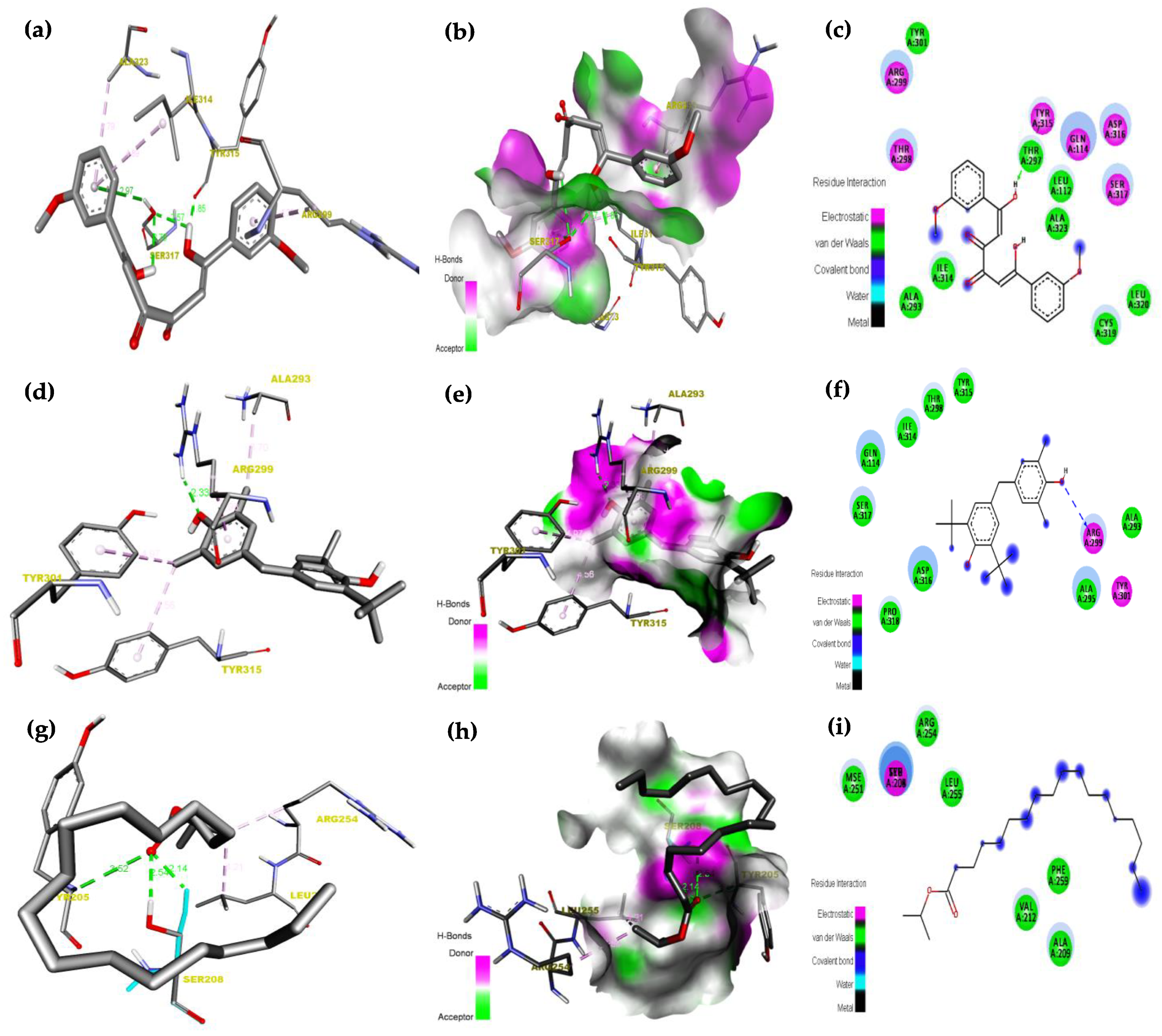
2.5. Molecular Docking Analysis

A molecular docking method was used against receptor protein 1N0W. Its purpose is to calculate the binding modalities of GC-MS resultant ligands, namely, (1) 3,4-Dihydroxy-1,6-bis-(3-methoxy-phenyl)-hexa-2,4-diene-1,6-dione, (2) Phenol,2,6-bis(1,1-dimethyl ethyl)-4-(4-hydroxy-3,5-dimethyl phenyl)methyl)-, (3) Isopropyl stearate is acting a vital role in the structure and function of biological molecules, the ligand–receptor interactions were examined based on hydrogen bonding. The findings imply that all three ligands might effectively bind to the active site of 1N0W, as demonstrated in Table 3. The compound 3,4-Dihydroxy-1,6-bis-(3-methoxy-phenyl)-hexa-2,4-diene-1,6-dione was exhibited as a high binding energy value of −9.1(Kcal/mol), is due to the presence of low band gap energy of hydrogen bond (1.8Å), π-Donor Hydrogen Bond, and π-alkyl Hydrophobic bonds interaction between this ligand and 1N0W protein. The ligand–receptor hydrogen bond interactions, receptor side hydrogen bond interactions, and their two-dimensional (2D) pictures are shown in Figure 7. Besides, molecular docking investigation depicted that all three ligands could strongly occupy the active site of receptor protein 1N0W. It has been determined that such chemicals may target this region, preventing its function.
The ethyl acetate extracts from vegetable seeds reveal promising antimicrobial and antioxidant properties. Extracts from these plants showed significant antibacterial activity against Gram-positive bacteria, which is crucial given the rise in antibiotic resistance [14,43,44,45,46,47,48]. These extracts may serve as natural sources of antioxidants based on their antioxidant activity [31,40,49,50,51,52,53,54,55]. It was revealed that several bioactive compounds exist in this extract, including methyl oleate, isopropyl stearate, and 3,4-dihydroxy-1,6-bis-(3-methoxy-phenyl)-hexa-2,4-diene-1,6-dione [54,56,57,58]. There is evidence that such compounds contribute to the observed antimicrobial and antioxidant properties [31,50,52,53,54,55]. Furthermore, human breast cancer cells (MCF-7) were found to be cytotoxic in a dose-dependent manner, while normal fibroblast cells (L929) were not affected. Apoptotic pathways may be involved in this cytotoxicity. Furthermore, molecular docking analysis confirmed their ability to bind to the 1N0W receptor protein, indicating their potential therapeutic utility. According to previous studies, plant seed extracts are important for preventing antibiotic resistance and developing natural antioxidants [35,59]. Research in this area could investigate the individual contributions of different compounds to these biological activities and how they might be used therapeutically in the future.

3. Materials and Methods

3.1. Seed Material Collection

Fresh, disease-free vegetable seeds (synergistic) were procured from the botanical park in Chennai, India. The complete seed types were combined, sorted, cleaned, and air-dried at room temperature for 8–10 days. The vegetable seed types were processed into fine powder. When powdered samples were needed for the extraction procedure, they were collected and kept in airtight containers shielded from heat and sunlight.

3.2. Extract Preparation

Vegetable seeds (300 g) Cucurbita maximas, Hibiscus cannabinus, Cyamopsis tetragonoloba, Phaseolus vulgaris, and Solanum melongena (synergistic) that had been ground up were mixed consecutively for 70–72 h in a rotator shaker operating at 130 rpm with (1:3 ratio) 900 mL of organic solvents, such as hexane, ethyl acetate, and methanol. Once dry and solvent-free, the filtrates were further concentrated at 40 °C in a rotating evaporator. To conduct additional in vitro studies, the resulting extracts were stored at 4 °C [60].

3.3. Soxhlet Extraction

In accordance with the methodology described elsewhere [61], the extract preparation was evaluated. Approximately 900 mL of solvents, including hexane, ethyl acetate, and methanol, were employed for the extract of powdered (synergistic) vegetable seed types weighing 300 g. The extract was filtered in a sterile environment using a Seitz filter and Whatman filter paper, and the extracts were then produced in powder form using a lyophilizer set at a temperature of −80 °C. The prepared extracts were subsequently stored at 4 °C for further analysis.

3.4. In Vitro Antibacterial Assessment

For the antimicrobial assay, Gram-positive bacteria of S. epidermis (MTTC 3615) and M. luteus (MTCC 106), as well as Gram-negative bacteria E. aerogens (MTCC 111) and P. vulgaris (MTCC 1771) were used in this study. The Gram-positive and Gram-negative bacterial cultures were grown separately on Mueller-Hinton broth (pH 7.0). The cultures were incubated for 24 h at 37 °C on a rotating shaker. After incubation, the bacterial cultures were adjusted to a concentration of 1.5 × 10⁸ CFU/mL and were swabbed onto sterile Mueller–Hinton agar plates. Vegetable seed extracts in three different solvents (20 mL each), hexane, ethyl acetate, and methanol, were applied to sterile discs (6 mm) and left to soak for 10 to 15 min. The discs were aseptically transferred to the plates seeded with the appropriate pathogens and incubated at 37 °C for 24 h using sterile forceps. Following the 24 h incubation period, the various solvent extracts of vegetable seeds were assessed for their ability to generate a zone of inhibition (measured in mm) against the indicator pathogenic bacteria. Streptomycin and the respective solvent-soaked discs were used as positive and negative controls, respectively. The tests were performed in triplicate to ensure the reliability and accuracy of the results.

3.5. DPPH Radical Scavenging Assay

The Sayah et al. [55] technique was used to assess the DPPH radical scavenging capacity of the ethyl acetate seed extract. At 517 nm, the absorbance was measured in comparison to a blank. The equation below was used to estimate the DPPH scavenging activity, and the IC50 value was calculated. DPPH scavenging capacity (%) = [(A sample − A blank)/A control] × 100.

3.6. Hydrogen Peroxide (H2O2) Scavenging Activity

According to the methods of Ruch et al. [62], hydrogen peroxide scavenging characteristics of ethyl acetate extracts of seed extract at various concentrations (200–1000 µg/mL) used the following equation to calculate: Hydrogen peroxide scavenging (%) = [(A0 − A1)/A0] × 100. where A0 denotes the absorbance of the control, and A1 denotes the absorbance of the sample.

3.7. GC–MS Analysis

Following the methodology outlined by Venkatadri [63,64] using GC-MS (SHIMADZU QP2010, China) instrument, spectrum analysis was conducted to identify the phytochemical constituents present in the mid-polar solvent of ethyl acetate seed extract.

3.8. Anticancer Activities

The human breast cancer cell line (MCF-7) with passage number 15 and normal fibroblast cell line (L929) with passage number 9 were occupied from the National Centre for Cell Science in Pune, India.

3.9. Cytotoxicity

The monolayer cell culture was trypsinized using a medium containing 10% FBS, and the cell density was increased to 1.0 × 10⁵ cells/mL. MTT assay was performed to analyze the inhibitory concentration (IC50) of ethyl acetate seed extract. Both the cell lines were cultured in a microtiter plate for about 2 days to obtain 75% confluence. The medium was discarded, MTT was added to the culture, and incubated for 4 h at 37 °C. Then, the supernatant was removed, and about 50 µL of DMSO was loaded with the sample and incubated for 10 min. The absorbance was read at 570 nm using a microtiter plate reader, and the percentage viability was calculated [64]. The following formula was used to calculate the degree of inhibition, and the values for the test drug concentrations required to reduce cell growth by 50% (CTC50) for each line were obtained from the dose-response curves.
Percentage inhibition (%) [(ODcontrol − ODsample/ODcontrol)] × 100

3.10. Molecular Docking Studies

To shed light on the examined ligands’ target-binding modes, molecular docking experiments were carried out (PDB code: 1N0W) [https://www.rcsb.org/structure/1N0W accessed between 12–20 March 2023 [39]. This protein controls the activity of RAD51, a recombinase enzyme, in the pathways for DNA repair by homologous recombination. It has been linked to a higher risk of developing breast cancer. Given the remarkable conservation of the RAD51 oligomerization motif, RecA-like recombinases have a common evolutionary origin for producing nucleoprotein filaments, reflected in the BRC repeat. Cancer-associated mutations disrupt the expected connection between the BRC repeat and RAD51, providing structural information about the processes underlying cancer susceptibility. We chose this 1N0W breast cancer-targeted protein as a result of docking investigations.
The molecular docking program Auto dock vina 4.2 was used to select the potential binding mode between the GC-MS resultant ligands, namely, (1) 3,4-Dihydroxy-1,6-bis-(3-methoxy-phenyl)-hexa-2,4-diene-1,6-dione, (2) Phenol,2,6-bis(1,1-dimethyl ethyl)-4-(4-hydroxy-3,5-dimethyl phenyl)methyl)-, (3) Isopropyl stearate and 1N0W receptor protein. The molecular docking program Auto dock vina4.2 was used to select the potential binding mode between the GC-MS resultant ligands, namely, (1) 3,4-Dihydroxy-1,6-bis-(3-methoxy-phenyl)-hexa-2,4-diene-1,6-dione, (2) Phenol,2,6-bis(1,1-dimethyl ethyl)-4-(4-hydroxy-3,5-dimethyl phenyl)methyl)-, (3) Isopropyl stearate and 1N0W receptor protein. ChemDraw 12.0 software was used to create the ligand structures. These 2D chemical structures undergo energy minimization before being translated to Protein Data Bank (PDB) format, which Auto Dock Vina4.2 then transforms into the PDBQT format structure. The Kollman unified atom charges and solvation parameters offered by Auto Dock Tools were used to add the appropriate hydrogen atoms to the 1N0W protein structure before docking. A-Chain, a protein from 1N0W, is chosen for docking investigations. To create ligand structures for docking, non-polar hydrogen (H) atoms, Gasteiger partial charges, and rotatable bonds were defined. A grid box with a maximum size of 92 × 94 × 100Å was created using Auto Grid. At the binding site for 1N0W, the grid box was allocated at the protein’s center using x, y, and z coordinates of 33.899, 26.022, and 1.196, respectively, with a grid spacing of 0.581 Å. Using Discovery studio visualizes, docked structures were shown. A two-dimensional (2D) graphic depicting the interaction between the protein and ligand was produced [42].

3.11. Statistical Analysis

Results from each experiment were given as Mean ± SD, with each experiment being performed in triplicate. The statistical study was conducted using Microsoft Excel 2007, and a straightforward linear regression curve was employed to determine the IC50 values. The p-value was only considered to be less than 0.05 for determining statistical significance.

4. Conclusions

The findings of this study highlight the significant antibacterial and antioxidant properties of the ethyl acetate extract derived from vegetable seeds. The extract exhibited cytotoxic effects against breast cancer cells (MCF-7) while demonstrating lower toxicity towards normal fibroblast cells (L-929). The synergistic extract of ethyl acetate seeds effectively inhibited cell growth and induced apoptosis in both MCF-7 and L-929 cells. These results indicate that vegetable seed extract possesses potential as a source of antibacterial, antioxidant, and anticancer agents. Further, in-silico molecular docking analysis 3,4-Dihydroxy-1,6-bis-(3-methoxy-phenyl)-hexa-2,4-diene-1,6-dione ligand, which has shown the best binding energy value of -9.1 kcal/mol to against target protein (PDB code: 1N0W) with Crystal Structure of Human and alkyl hydroperoxides by using reducing equivalents acquired from 3,4-Dihydroxy-1,6-bis-(3-methoxy-phenyl)-hexa-2,4-diene-1,6-dione. However, it is important to acknowledge certain limitations of this study. First, the study focused on a limited number of vegetable seed types, and further investigation involving a wider range of seed varieties would provide a more comprehensive understanding of their therapeutic potential. Additionally, the specific mechanisms underlying the observed antibacterial, antioxidant, and anticancer effects of the extract were not elucidated in this study. Future research should unravel the molecular pathways and signaling mechanisms involved. Despite these limitations, the results of this study pave the way for future research and development in the field of vegetable seed extracts. Further refinement and optimization of the extraction process are necessary to create a synergistic ethyl acetate extract from various vegetable seeds that can be effectively utilized as an active ingredient in pharmaceutical or nutraceutical formulations. Continued exploration of vegetable seed extracts holds promise for the development of novel therapeutic agents with enhanced efficacy and minimal adverse effects.

Author Contributions

Conceptualization, all authors.; methodology, V.B.; software, J.I.A. validation, S.A., I.A.R. and J.S.M.S., formal analysis, V.B. and S.A.; investigation, V.B.; resources, V.B.; data curation, I.A.R. and J.S.M.S.; writing—original draft preparation, all authors.; writing—review and editing, A.P., I.A.R., S.A. and J.S.M.S.; visualization, S.A. and I.A.R.; project administration, I.A.R. and J.S.M.S.; funding acquisition, I.A.R. and J.S.M.S. All authors have read and agreed to the published version of the manuscript.

Funding

This research work was funded by Institutional Fund Projects, under grant number (IFPIP:57-130-1443). The authors gratefully acknowledge technical and financial support provided by Ministry of Education and King Abdulaziz University, DSR, Jeddah, Saudi Arabia.

Data Availability Statement

The data created is/are cited in the manuscript.

Conflicts of Interest

The authors declare no conflict of interest.

References

  1. Doocey, C.M.; Finn, K.; Murphy, C.; Guinane, C.M. The Impact of the Human Microbiome in Tumorigenesis, Cancer Progression, and Biotherapeutic Development. BMC Microbiol. 2022, 22, 53. [Google Scholar] [CrossRef] [PubMed]
  2. Zheng, Y.; Chen, Y.; Yu, K.; Yang, Y.; Wang, X.; Yang, X.; Qian, J.; Liu, Z.X.; Wu, B. Fatal Infections Among Cancer Patients: A Population-Based Study in the United States. Infect. Dis. Ther. 2021, 10, 871–895. [Google Scholar] [CrossRef] [PubMed]
  3. Stahl, G.K.; Angwin, D.N.; Very, P.; Gomes, E.; Weber, Y.; Tarba, S.Y.; Noorderhaven, N.; Benyamini, H.; Bouckenooghe, D.; Chreim, S.; et al. Sociocultural Integration in Mergers and Acquisitions: Unresolved Paradoxes and Directions for Future Research. Thunderbird Int. Bus. Rev. 2013, 55, 333–356. [Google Scholar] [CrossRef]
  4. Pennathur, A.; Gibson, M.K.; Jobe, B.A.; Luketich, J.D. Oesophageal Carcinoma. Lancet 2013, 381, 400–412. [Google Scholar] [CrossRef] [PubMed]
  5. Enzinger, P.C.; Mayer, R.J. Esophageal Cancer. N. Engl. J. Med. 2004, 350, 1363–1364. [Google Scholar] [CrossRef]
  6. Akhtar, M.S.; Panwar, J.; Yun, Y.-S. Biogenic Synthesis of Metallic Nanoparticles by Plant Extracts. ACS Sustain. Chem. Eng. 2013, 1, 591–602. [Google Scholar] [CrossRef]
  7. Zhang, W.C.; Shyh-Chang, N.; Yang, H.; Rai, A.; Umashankar, S.; Ma, S.; Soh, B.S.; Sun, L.L.; Tai, B.C.; Nga, M.E.; et al. Glycine Decarboxylase Activity Drives Non-Small Cell Lung Cancer Tumor-Initiating Cells and Tumorigenesis. Cell 2012, 148, 259–272. [Google Scholar] [CrossRef]
  8. Sauer, A.G.; Siegel, R.L.; Jemal, A.; Fedewa, S.A. Current Prevalence of Major Cancer Risk Factors and Screening Test Use in the United States: Disparities by Education and Race/Ethnicity. Cancer Epidemiol. Biomark. Prev. 2019, 28, 629–642. [Google Scholar] [CrossRef]
  9. Man, S.M.; Jenkins, B.J. Context-Dependent Functions of Pattern Recognition Receptors in Cancer. Nat. Rev. Cancer 2022, 22, 397–413. [Google Scholar] [CrossRef]
  10. Saini, A.; Kumar, M.; Bhatt, S.; Saini, V.; Malik, A. Cancer causes and treatments. Int. J. Pharm. Sci. Res. 2020, 11, 3121–3134. [Google Scholar]
  11. Zhuang, X.; Kang, Y.; Zhao, L.; Guo, S. Design and Synthesis of Copper Nanoparticles for the Treatment of Human Esophageal Cancer: Introducing a Novel Chemotherapeutic Supplement. J. Exp. Nanosci. 2022, 17, 274–284. [Google Scholar] [CrossRef]
  12. Maynard, A.; McCoach, C.E.; Rotow, J.K.; Harris, L.; Haderk, F.; Kerr, D.L.; Yu, E.A.; Schenk, E.L.; Tan, W.; Zee, A.; et al. Therapy-Induced Evolution of Human Lung Cancer Revealed by Single-Cell RNA Sequencing. Cell 2020, 182, 1232–1251. [Google Scholar] [CrossRef] [PubMed]
  13. Gyamfi, J.; Lee, Y.H.; Eom, M.; Choi, J. Interleukin-6/STAT3 Signalling Regulates Adipocyte Induced Epithelial-Mesenchymal Transition in Breast Cancer Cells. Sci. Rep. 2018, 8, 8859. [Google Scholar] [CrossRef] [PubMed]
  14. Haque, M.; Sartelli, M.; McKimm, J.; Bakar, M.A. Health Care-Associated Infections—An Overview. Infect. Drug Resist. 2018, 11, 2321–2333. [Google Scholar] [CrossRef] [PubMed]
  15. Snow, R.W.; Guerra, C.A.; Noor, A.M.; Myint, H.Y.; Hay, S.I. The Global Distribution of Clinical Episodes of Plasmodium Falciparum Malaria. Nature 2005, 434, 214–217. [Google Scholar] [CrossRef]
  16. Fidock, D.A. Priming the Antimalarial Pipeline. Nature 2010, 465, 297–298. [Google Scholar] [CrossRef] [PubMed]
  17. Buchholz, K.; Burke, T.A.; Williamson, K.C.; Wiegand, R.C.; Wirth, D.F.; Marti, M. A High-Throughput Screen Targeting Malaria Transmission Stages Opens New Avenues for Drug Development. J. Infect. Dis. 2011, 203, 1445–1453. [Google Scholar] [CrossRef]
  18. Roxanis, I.; Colling, R.; Kartsonaki, C.; Green, A.R.; Rakha, E.A. The Significance of Tumour Microarchitectural Features in Breast Cancer Prognosis: A Digital Image Analysis. Breast Cancer Res. 2018, 20, 11. [Google Scholar] [CrossRef]
  19. Li, L.; Zhang, D.; Liu, B.; Lv, D.; Zhai, J.; Guan, X.; Yi, Z.; Ma, F.; Wang, N. Antibody-Drug Conjugates in HER2-Positive Breast Cancer. Chin. Med. J. Engl. 2022, 135, 261–267. [Google Scholar] [CrossRef]
  20. Eden, C.M.; Johnson, J.; Syrnioti, G.; Malik, M.; Ju, T. The Landmark Series: The Breast Cancer Burden of the Asian American Population and the Need for Disaggregated Data. Ann. Surg. Oncol. 2023, 30, 2121–2127. [Google Scholar] [CrossRef]
  21. Sasikala, M.; Mohan, S.; Swarnakumari, S.; Nagarajan, A. Isolation and in Vivo Evaluation of Anti-Breast Cancer Activity of Resin Glycoside Merremoside from Ipomoea Aquatica Forsskal in Overcoming Multi-Drug Resistance. Phytomed. Plus 2022, 2, 100359. [Google Scholar] [CrossRef]
  22. Ferlay, J.; Colombet, M.; Soerjomataram, I.; Parkin, D.M.; Piñeros, M.; Znaor, A.; Bray, F. Cancer Statistics for the Year 2020: An Overview. Int. J. Cancer 2021, 149, 778–789. [Google Scholar] [CrossRef] [PubMed]
  23. Chen, J.; Jin, L.; Chen, L.; Bian, Z.; Li, Z.; Cao, S.; Zhou, J.; Xu, L.; Zhao, W.; Wang, Q. Patients Achieved PCR during Neoadjuvant Chemotherapy Had Better Outcome than Adjuvant Chemotherapy Setting in Breast Cancer: A Comparative Study. Cancer Treat. Res. Commun. 2023, 36, 100719. [Google Scholar] [CrossRef] [PubMed]
  24. Akbari Nakhjavani, S.; Afsharan, H.; Khalilzadeh, B.; Ghahremani, M.H.; Carrara, S.; Omidi, Y. Gold and Silver Bio/Nano-Hybrids-Based Electrochemical Immunosensor for Ultrasensitive Detection of Carcinoembryonic Antigen. Biosens. Bioelectron. 2019, 141, 111439. [Google Scholar] [CrossRef]
  25. Zhong, L.; Li, Y.; Xiong, L.; Wang, W.; Wu, M.; Yuan, T.; Yang, W.; Tian, C.; Miao, Z.; Wang, T.; et al. Small Molecules in Targeted Cancer Therapy: Advances, Challenges, and Future Perspectives. Signal Transduct. Target. Ther. 2021, 6, 201. [Google Scholar] [CrossRef]
  26. Apu, A.S.; Liza, M.S.; Jamaluddin, A.T.M.; Howlader, M.A.; Saha, R.K.; Rizwan, F.; Nasrin, N. Phytochemical Screening and in Vitro Bioactivities of the Extracts of Aerial Part of Boerhavia Diffusa Linn. Asian Pac. J. Trop. Biomed. 2012, 2, 673–678. [Google Scholar] [CrossRef]
  27. Maya, S.; Sarmento, B.; Nair, A.; Rejinold, N.; Nair, S.; Jayakumar, R. Smart Stimuli Sensitive Nanogels in Cancer Drug Delivery and Imaging: A Review. Curr. Pharm. Des. 2013, 19, 7203–7218. [Google Scholar] [CrossRef]
  28. Heinrich, M.; Hesketh, A. 25 Years after the `Rio Convention’––Lessons Learned in the Context of Sustainable Development and Protecting Indigenous and Local Knowledge. Phytomedicine 2019, 53, 332–343. [Google Scholar] [CrossRef]
  29. Badri, D.V.; Quintana, N.; El Kassis, E.G.; Kim, H.K.; Choi, Y.H.; Sugiyama, A.; Verpoorte, R.; Martinoia, E.; Manter, D.K.; Vivanco, J.M. An ABC Transporter Mutation Alters Root Exudation of Phytochemicals That Provoke an Overhaul of Natural Soil Microbiota. Plant Physiol. 2009, 151, 2006–2017. [Google Scholar] [CrossRef]
  30. Anusha Siddiqu, S.; Dini, S.; Esmaeili, Y.; Roshanak, S.; Ali Redha, A.; Ahmad Wani, S. Uses of Carotenoid-Rich Ingredients to Design Functional Foods: A Review. J. Food Bioact. 2023, 21, 3–20. [Google Scholar] [CrossRef]
  31. Tundis, R.; Iacopetta, D.; Sinicropi, M.S.; Bonesi, M.; Leporini, M.; Passalacqua, N.G.; Ceramella, J.; Menichini, F.; Loizzo, M.R. Assessment of Antioxidant, Antitumor and pro-Apoptotic Effects of Salvia Fruticosa Mill. Subsp. Thomasii (Lacaita) Brullo, Guglielmo, Pavone & Terrasi (Lamiaceae). Food Chem. Toxicol. 2017, 106, 155–164. [Google Scholar] [CrossRef] [PubMed]
  32. Kwan, T.W.; Wong, S.S.; Hong, Y.; Kanaya, A.M.; Khan, S.S.; Hayman, L.L.; Shah, S.H.; Welty, F.K.; Deedwania, P.C.; Khaliq, A.; et al. Epidemiology of Diabetes and Atherosclerotic Cardiovascular Disease Among Asian American Adults: Implications, Management, and Future Directions: A Scientific Statement From the American Heart Association. Circulation 2023, 148, 74–94. [Google Scholar] [CrossRef] [PubMed]
  33. Efferth, T.; Li, P.C.H.; Konkimalla, V.S.B.; Kaina, B. From Traditional Chinese Medicine to Rational Cancer Therapy. Trends Mol. Med. 2007, 13, 353–361. [Google Scholar] [CrossRef]
  34. Mahmud, S.; Hasan, M.R.; Biswas, S.; Paul, G.K.; Afrose, S.; Mita, M.A.; Sultana Shimu, M.S.; Promi, M.M.; Hani, U.; Rahamathulla, M.; et al. Screening of Potent Phytochemical Inhibitors Against SARS-CoV-2 Main Protease: An Integrative Computational Approach. Front. Bioinform. 2021, 1. [Google Scholar] [CrossRef] [PubMed]
  35. Najmi, A.; Javed, S.A.; Al Bratty, M.; Alhazmi, H.A. Modern Approaches in the Discovery and Development of Plant-Based Natural Products and Their Analogues as Potential Therapeutic Agents. Molecules 2022, 27, 349. [Google Scholar] [CrossRef]
  36. Singh, S.; Tyagi, C.; Rather, I.A.; Sabir, J.S.M.; Hassan, M.I.; Singh, A.; Singh, I.K. Molecular Modeling of Chemosensory Protein 3 from Spodoptera Litura and Its Binding Property with Plant Defensive Metabolites. Int. J. Mol. Sci. 2020, 21, 4073. [Google Scholar] [CrossRef] [PubMed]
  37. Halgren, T.A.; Murphy, R.B.; Friesner, R.A.; Beard, H.S.; Frye, L.L.; Pollard, W.T.; Banks, J.L. Glide: A New Approach for Rapid, Accurate Docking and Scoring. 2. Enrichment Factors in Database Screening. J. Med. Chem. 2004, 47, 1750–1759. [Google Scholar] [CrossRef]
  38. Salim, B.; Noureddine, M. Identification of Compounds from Nigella Sativa as New Potential Inhibitors of 2019 Novel Coronasvirus (Covid-19): Molecular Docking Study. ChemRxiv 2020, 19. [Google Scholar]
  39. Pellegrini, M. Computational Methods for Protein Function Analysis. Curr. Opin. Chem. Biol. 2001, 5, 46–50. [Google Scholar] [CrossRef]
  40. Ma, Z.; Zhang, H. Phytochemical Constituents, Health Benefits, and Industrial Applications of Grape Seeds: A Mini-Review. Antioxidants 2017, 6, 71. [Google Scholar] [CrossRef]
  41. Morris, G.M.; Huey, R.; Lindstrom, W.; Sanner, M.F.; Belew, R.K.; Goodsell, D.S.; Olson, A.J. AutoDock4 and AutoDockTools4: Automated Docking with Selective Receptor Flexibility. J. Comput. Chem. 2009, 30, 2785–2791. [Google Scholar] [CrossRef] [PubMed]
  42. Spassov, V.Z.; Yan, L. pH-selective Mutagenesis of Protein–Protein Interfaces: In Silico Design of Therapeutic Antibodies with Prolonged Half-life. Proteins Struct. Funct. Bioinform. 2013, 81, 704–714. [Google Scholar] [CrossRef] [PubMed]
  43. Ventola, C.L. The Antibiotic Resistance Crisis: Causes and Threats. Pharm. Ther. 2015, 40, 277. [Google Scholar]
  44. Rossolini, G.M.; Arena, F.; Pecile, P.; Pollini, S. Update on the Antibiotic Resistance Crisis. Curr. Opin. Pharmacol. 2014, 18, 56–60. [Google Scholar] [CrossRef] [PubMed]
  45. Devanshi, S.; Lakshmi, D.B. The Antibiotic Resistance Crisis—An Indian Perspective. Int. J. Bus. Manag. Res. 2020, 8, 112–116. [Google Scholar] [CrossRef]
  46. Martens, E.; Demain, A.L. The Antibiotic Resistance Crisis, with a Focus on the United States. J. Antibiot. 2017, 70, 520–526. [Google Scholar] [CrossRef]
  47. Rather, I.A.; Kim, B.-C.; Bajpai, V.K.; Park, Y.-H. Self-Medication and Antibiotic Resistance: Crisis, Current Challenges, and Prevention. Saudi J. Biol. Sci. 2017, 24, 808–812. [Google Scholar] [CrossRef]
  48. Aslam, B.; Wang, W.; Arshad, M.I.; Khurshid, M.; Muzammil, S.; Rasool, M.H.; Nisar, M.A.; Alvi, R.F.; Aslam, M.A.; Qamar, M.U.; et al. Antibiotic Resistance: A Rundown of a Global Crisis. Infect. Drug Resist. 2018, 11, 1645–1658. [Google Scholar] [CrossRef]
  49. Sudhasupriya, P.; Shakina Begam, A.; Rajeshkumar, S. Screening for Antioxidant and Antimicrobial Activity of Seed Extracts of Avocado Pear. Res. J. Pharm. Technol. 2017, 10, 1991–1996. [Google Scholar] [CrossRef]
  50. Theansungnoen, T.; Nitthikan, N.; Wilai, M.; Chaiwut, P.; Kiattisin, K.; Intharuksa, A. Phytochemical Analysis and Antioxidant, Antimicrobial, and Antiaging Activities of Ethanolic Seed Extracts of Four Mucuna Species. Cosmetics 2022, 9, 14. [Google Scholar] [CrossRef]
  51. Qadir, A.; Khan, N.; Arif, M.; Warsi, M.H.; Ullah, S.N.M.N.; Yusuf, M. GC–MS Analysis of Phytoconstituents Present in Trigonella foenumgraecum L. Seeds Extract and Its Antioxidant Activity. J. Indian Chem. Soc. 2022, 99, 100503. [Google Scholar] [CrossRef]
  52. Mohammadi, N.; Ostovar, N. Essential Oil Composition of Polylophium Involucratum and Evaluation of Antioxidant Capacity of Seeds Ethanolic Extracts by DSC. Food Chem. Adv. 2022, 1, 100066. [Google Scholar] [CrossRef]
  53. Phuyal, N.; Jha, P.K.; Raturi, P.P.; Rajbhandary, S. Total Phenolic, Flavonoid Contents, and Antioxidant Activities of Fruit, Seed, and Bark Extracts of Zanthoxylum Armatum DC. Sci. World J. 2020, 2020, 8780704. [Google Scholar] [CrossRef] [PubMed]
  54. Santos, L.C.D.; Azevedo, L.S.; de Siqueira, E.P.; Castro, A.H.F.; Lima, L.A.R.D.S. Chemical Characterization, Antioxidant Activity, and Cytotoxicity of Fatty Acids Methyl Esters from Handroanthus Impetiginosus (Mart. Ex DC.) Mattos (Bignoniaceae) Seeds. Nat. Prod. Res. 2023, 1–5. [Google Scholar] [CrossRef]
  55. Sayah, K.; Marmouzi, I.; Naceiri Mrabti, H.; Cherrah, Y.; Faouzi, M.E.A. Antioxidant Activity and Inhibitory Potential of Cistus salviifolius (L.) and Cistus monspeliensis (L.) Aerial Parts Extracts against Key Enzymes Linked to Hyperglycemia. Biomed. Res. Int. 2017, 2017, 2789482. [Google Scholar] [CrossRef]
  56. Momodu, I.B.; Okungbowa, E.S.; Agoreyo, B.O.; Maliki, M.M. Gas Chromatography—Mass Spectrometry Identification of Bioactive Compounds in Methanol and Aqueous Seed Extracts of Azanza Garckeana Fruits. Niger. J. Biotechnol. 2022, 38. [Google Scholar] [CrossRef]
  57. Akalazu, J.N.; Uchegbu, R.I. Biochemical Composition and Antimicrobial Activities of Seed Extracts of Avocado (Persea americana). FASEB J. 2020, 34, 1. [Google Scholar] [CrossRef]
  58. Idris, S.; Ndukwe, G.; Gimba, C. Preliminary Phytochemical Screening and Antimicrobial Activity of Seed Extracts of Persea americana (Avocado Pear). Bayero J. Pure Appl. Sci. 2010, 2. [Google Scholar] [CrossRef]
  59. Alsheikh, H.M.A.; Sultan, I.; Kumar, V.; Rather, I.A.; Al-sheikh, H.; Jan, A.T.; Haq, Q.M.R. Plant-based Phytochemicals as Possible Alternative to Antibiotics in Combating Bacterial Drug Resistance. Antibiotics 2020, 9, 480. [Google Scholar] [CrossRef]
  60. Ahmed, H. Ethnomedicinal, Phytochemical and Pharmacological Investigations of Perilla frutescens (L.) Britt. Molecules 2018, 24, 102. [Google Scholar] [CrossRef]
  61. Ahmad, W.; Jantan, I.; Bukhari, S.N.A. Tinospora crispa (L.) Hook. f. & Thomson: A Review of Its Ethnobotanical, Phytochemical, and Pharmacological Aspects. Front. Pharmacol. 2016, 7, 59. [Google Scholar] [CrossRef] [PubMed]
  62. Ruch, R.J.; Crist, K.A.; Klaunig, J.E. Effects of Culture Duration on Hydrogen Peroxide-Induced Hepatocyte Toxicity. Toxicol. Appl. Pharmacol. 1989, 100, 451–464. [Google Scholar] [CrossRef] [PubMed]
  63. Venkatadri, B.; Khusro, A.; Aarti, C.; Rameshkumar, M.R.; Agastian, P. In Vitro Assessment on Medicinal Properties and Chemical Composition of Michelia Nilagirica Bark. Asian Pac. J. Trop. Biomed. 2017, 7, 782–790. [Google Scholar] [CrossRef]
  64. Venkatadri, B.; Arunagirinathan, N.; Rameshkumar, M.; Ramesh, L.; Dhanasezhian, A.; Agastian, P. In Vitro Antibacterial Activity of Aqueous and Ethanol Extracts of Aristolochia Indica and Toddalia Asiatica against Multidrug-Resistant Bacteria. Indian J. Pharm. Sci. 2015, 77, 788. [Google Scholar] [CrossRef] [PubMed]
Figure 1. Antioxidant activity of vegetable seed extracts of hexane, ethyl acetate, methanol extracts and ascorbic acid. (a) DPPH scavenging effect of different concentrations (200–1000 µg/mL). (b) H2O2 scavenging effect of different concentrations (200–1000 µg/mL) of vegetable seed extracts of hexane, ethyl acetate, methanol extracts, and ascorbic acid. Each value represents the mean ± SEM of triplicate experiments.
Plants 12 03352 g001
Figure 2. Gas chromatography–mass spectrometry chromatograms of the ethyl acetate extract of vegetable seed (Synergistic).
Plants 12 03352 g002
Figure 3. Mass spectrum for the major constituent methyl oleate from the ethyl acetate extract of vegetable seed (Synergistic).
Plants 12 03352 g003
Figure 4. Anticancer activity of L-929 (Fibroblast cells) ethyl acetate extracts (a) control, (b) 500 µg/ mL, (c) 250 µg/mL, (d) 100 µg/mL, (e) 50 µg/mL, and (f) 25 µg/mL.
Plants 12 03352 g004
Figure 5. Anticancer activity of ethyl acetate extracts on MCF-7 (a) control, (b) 25 µg/mL, (c) 50 µg/mL, (d) 100 µg/mL, (e) 250 µg/mL, and (f) 500 µg/ mL.
Plants 12 03352 g005
Figure 6. Anticancer activities of ethyl acetate extract from vegetable seed against MCF-7 and L929 cell line.
Plants 12 03352 g006
Figure 7. (a) Ligand–receptor hydrogen bond interactions of 3,4-Dihydroxy-1,6-bis-(3-methoxy-phenyl)-hexa-2,4-diene-1,6-dione. (b) Receptor side hydrogen bond interactions of3,4-Dihydroxy-1,6-bis-(3-methoxy-phenyl)-hexa-2,4-diene-1,6-dione. (c) 2D-ligand–receptor hydrogen bond interaction of 3,4-Dihydroxy-1,6-bis-(3-methoxy-phenyl)-hexa-2,4-diene-1,6-dione. (d) Ligand–receptor hydrogen bond interactions of Phenol,2,6-bis(1,1-dimethylethyl)-4-(4-hydroxy-3,5-dimethylphenyl)methyl). (e) Receptor side hydrogen bond interactions of Phenol,2,6-bis(1,1-dimethylethyl)-4-(4-hydroxy-3,5-dimethylphenyl)methyl). (f) 2D-ligand–receptor hydrogen bond interaction of Phenol,2,6-bis(1,1-dimethylethyl)-4-(4-hydroxy-3,5-dimethylphenyl)methyl). (g) Ligand–receptor hydrogen bond interactions of Isopropyl stearate. (h) Receptor side hydrogen bond interactions of Isopropyl stearate. (i) 2D-ligand–receptor hydrogen bond interaction of Isopropyl stearate.
Plants 12 03352 g007
Table 1. Antibacterial activity of vegetable seed extracts against Gram-positive and Gram-negative bacteria.
Tested MicroorganismsStreptomycin (mm)Zone of Inhibition (mm) at Concentration in (μg/mL)
Hexane
(mm)
Ethyl Acetate
(mm)
Methanol
(mm)
Staphylococcus epidermis16.6 ± 0.58.6 ± 0.512.6 ± 0.58.3 ± 0.5
Micrococcus luteus15.3 ± 0.57.6 ± 0.510.3 ± 0.58.6 ± 0.5
Enterobacter aerogens15.3 ± 0.58.3 ± 0.511.5 ± 0.57.6 ±0.5
Proteus vulgaris14.3 ± 0.57.6 ± 0.59.3 ± 0.58.6 ± 0.5
Table 2. Phyto-compounds analysis of ethyl acetate vegetable seed extracts present by GC MS.
Peak#Molecular FormulaRetention
Time
Identification
1C9H12O13.43Phenol,2-Prophyl-
2C11H8O514.17Cumarin-3-carboxylic acid,7-methoxy
3C15H10O215.17Flavone
4C16H28O16.155-Cyclohexadecen-1-one
5C17H34O217.12Hexadecanoic acid, methyl ester
6C19H36O218.85Methyl oleate
7C21H42O220.98Isopropyl stearate
8C23H32O222.82Phenol,2,6-bis(1,1-dimethylethyl)-4-(4-hydroxy-3,5-dimethylphenyl)methyl)-
9C20H18O624.33,4-Dihydroxy-1,6-bis-(3-methoxy-phenyl)-hexa-2,4-diene-1,6-dione
Table 3. The ligand–receptor residue interaction between Biscumarin ligand with 2JER protein.
S.NOPub Chem.
Id
Ligand NameBinding
Energy
(kcal/mol)
Residue InteractionType of BondDistance(Å)
11N0W3,4-Dihydroxy-1,6-bis-(3-methoxy-phenyl)-hexa-2,4-diene-1,6-dione−9.2: UNK0: H-A: TYR315: OHydrogen1.8
: UNK0: H-A: SER317: OHydrogen2.5
: UNK0: H-A: SER317 OHydrogen2.7
A: SER317: H-:UNK0π-Donor Hydrogen Bond2.9
: UNK0-A: ARG299π-alkyl Hydrophobic4.3
: UNK0-A: ILE314π-alkyl Hydrophobic4.8
: UNK0-A: ALA323π-alkyl Hydrophobic4.7
21N0WPhenol,2,6-bis(1,1-dimethylethyl)-4-(4-hydroxy-3,5-dimethylphenyl)methyl)-−6.8A: ARG299: H-:UNK0: OHydrogen Bond2.3
A: ALA293-:UNK0: Calkyl Hydrophobic3.6
: UNK0-A: ARG299π-alkyl Hydrophobic3.9
A: TYR301-:UNK0: Cπ-alkyl Hydrophobic4.9
A: TYR315-:UNK0: Cπ-alkyl Hydrophobic4.5
3.1N0WIsopropyl stearate−4.0A: SER208: H-:UNK0: OHydrogen Bond2.1
A: SER208: H-:UNK0: OHydrogen Bond2.5
A: TYR205: C-:UNK0: OHydrogen Bond3.5
: UNK0:C-A: ARG254alkyl Hydrophobic3.6
: UNK0-C-A: LEU255alkyl Hydrophobic4.2
Disclaimer/Publisher’s Note: The statements, opinions and data contained in all publications are solely those of the individual author(s) and contributor(s) and not of MDPI and/or the editor(s). MDPI and/or the editor(s) disclaim responsibility for any injury to people or property resulting from any ideas, methods, instructions or products referred to in the content.

