Article

Pulicaria dysenterica (L.) Bernh.—Rightfully Earned Name? Identification and Biological Activity of New 3-Methoxycuminyl Esters from P. dysenterica Essential Oil

by
Niko S. Radulović
1,*,
Marko Z. Mladenović
1,
Dušan R. Vukićević
2,
Nikola M. Stojanović
3,
Pavle J. Randjelović
3,
Zorica Z. Stojanović-Radić
4 and
Fabio Boylan
5
1
Department of Chemistry, Faculty of Sciences and Mathematics, University of Nis, Višegradska 33, 18000 Nis, Serbia
2
Faculty of Medicine, University of Kragujevac, Svetozara Markovića 69, 34000 Kragujevac, Serbia
3
Department of Physiology, Faculty of Medicine, University of Nis, Zorana Ðinđica 81, 18000 Nis, Serbia
4
Department of Biology and Ecology, Faculty of Sciences and Mathematics, University of Nis, Visegradska 33, 18000 Nis, Serbia
5
School of Pharmacy and Pharmaceutical Sciences, Panoz Institute, Trinity College, Westland Row, 2 Dublin, Ireland
*
Author to whom correspondence should be addressed.
Plants 2022, 11(23), 3340; https://doi.org/10.3390/plants11233340
Submission received: 6 November 2022 / Revised: 21 November 2022 / Accepted: 22 November 2022 / Published: 1 December 2022
(This article belongs to the Special Issue Medicinal Plants: Advances in Phytochemistry and Ethnobotany)

Abstract

:
Motivated by the ethnopharmacological use of Pulicaria dysenterica, in the present study, the antimicrobial potential of the extracted essential oil was investigated against a panel of eighteen microorganism strains. Additionally, anti-acetylcholinesterase and antispasmodic (isolated rat distal colon) activities, general acute toxicity (Artemia salina model), and immunomodulatory properties (cytotoxicity on isolated mouse macrophages) were studied. Detailed analyses of the essential oil led to the identification of 3-methoxycuminyl 2-methylbutanoate (a new natural product) and 3-methoxycuminyl 3-methylbutanoate (a rare natural product). The obtained esters and intermediates in the synthesis of the starting alcohol (3-methoxycuminol) were subjected to a battery of 1D- and 2D-NMR experiments. The synthesized esters were additionally characterized by GC–MS, IR, and UV–Vis. The synthesized compounds (ten in total) were biologically tested in the same way as the extracted P. dysenterica essential oil. The obtained low acute toxicity and promising antimicrobial potential suggest that the P. dysenterica essential oil might partially explain the ethnopharmacological application of P. dysenterica plant material for the treatment of gastrointestinal infections.

1. Introduction

Medicinal plants as industrial crops represent a renewable source of pharmaceuticals, essential oils, biocides, etc. [1]. Besides protecting plant biodiversity, the cultivation of new, potentially interesting medicinal plants is a way to strengthen local agro-economics. The choice of new medicinal plant crops could be based on ethnopharmacological knowledge, such as in the successful example of Artemisia annua [2]. Most medicinal plant species enlisted in modern pharmacopoeias have found their way to cultivation fields; however, numerous ethnopharmacologically renowned taxa have their use ceased over time due to various reasons, such as erroneous attributions of beneficial effects, toxicity, or being simply forgotten. Pulicaria dysenterica (L.) Bernh. (syn. Inula dysenterica, eng. great fleabane; family Asteraceae (Compositae)), native to Europe and Western Asia [3], represents an excellent example of an underused and almost abandoned folk remedy.
The name of the species in Latin, dysenterica, refers to the supposed property of this plant taxon to cure dysentery, which was the motivation for Carl Linnaeus to include it in his Flora Svecica [4]. The bruised leaves emit a characteristic smell, and they were used in medieval times to repel fleas and other insects. Additionally, the leaves were burned and the smoke was used as a domestic pesticide, hence the common name fleabane [5]. The use of P. dysenterica was mentioned many times in the works of later authors, such as the famous English herbalist Nicholas Culpeper. Thus, P. dysenterica could simultaneously provide access to specialty materials such as insecticides or an application in treating (infectious) gastrointestinal disorders. A possible common link between the two diametrical applications could be the anti-acetylcholinesterase activity of the constituents of P. dysenterica [6].
Up to now, the volatile secondary metabolites of P. dysenterica have only been the subject of a few studies. The essential oil was only investigated on two previous occasions [3,7], the latest of which was published 10 years ago [3]. This prompted us to re-analyze the composition of the essential oil from the aerial parts with the aim of finding the compounds responsible for the mentioned activities. The analysis required the synthesis of certain major and minor constituents, which enabled the testing of pure compounds and the essential oil for biological/pharmacological properties relevant to ethnopharmacological use. These included antimicrobial, antispasmodic (isolated rat distal colon), and anti-acetylcholinesterase activities, in addition to assessing the general acute toxicity (Artemia salina model) and cytotoxicity on isolated rat macrophages. Hence, in this work, we put forward various chemical and biological/pharmacological data needed to assess P. dysenterica as a potential plant crop.

2. Results

2.1. Composition of P. dysenterica Essential Oils and Their Variability

The aerial parts of P. dysenterica yielded yellowish essential oils (0.12–0.13%, w/w) with a pleasantly sweet odor. GC–MS, UV, IR, and NMR analyses, chromatographic separation, and synthetic work allowed for the identification of 296 constituents of the essential oils from dry P. dysenterica aerial parts (Table 1). The identified constituents represented 94.7–96.6% of the total essential oils, with oxygenated mono- and sesquiterpenoids (55.2–68.0% and 12.7–19.5%, respectively) as the most abundant compound classes. Among them, neryl isobutyrate and 3-methoxycuminyl isobutyrate represented major essential-oil constituents (16.4–22.1% and 25.5–31.1%, respectively). Only quantitative differences were noted between the essential oils collected from different P. dysenterica populations. Contrary to this slight quantitative compositional dissimilarity between the analyzed samples, the herein presented composition (Table 1) was very different from those that Basta et al. [7] and Mumivand et al. [3] published.

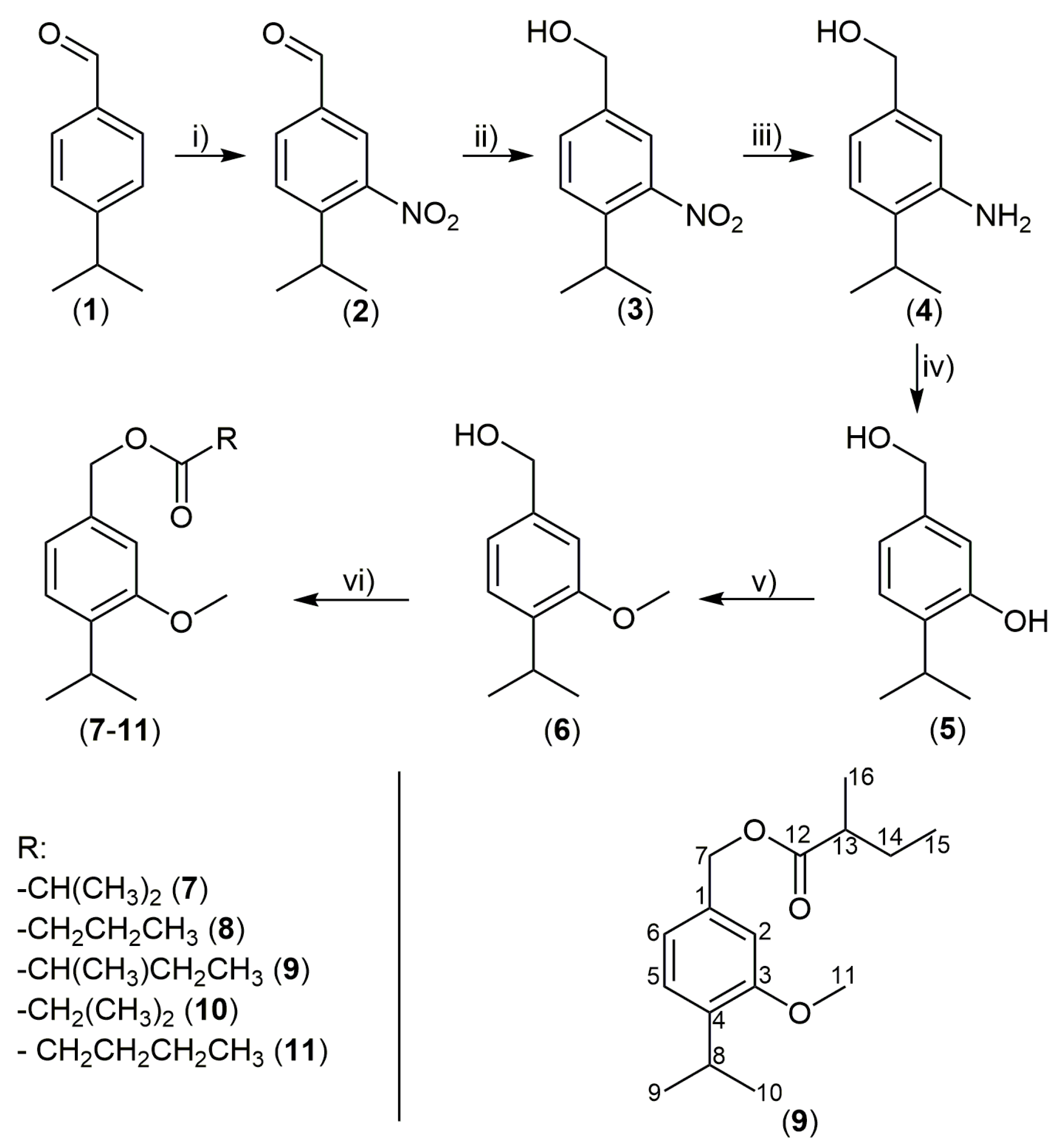
2.2. Identification, Synthesis, and NMR Spectral Characterization of the (New) 3-Methoxycuminyl Esters from P. dysenterica Essential Oil

One of the major essential-oil constituents (3-methoxycuminyl isobutyrate) was tentatively easily identified based solely on the matching of the corresponding retention indices and mass spectra with literature data [10]. Additionally, partial ion current (PIC, m/z 137, 163, and 180 ions) chromatograms of the essential-oil samples indicated the presence of additional constituents related to 3-methoxycuminyl isobutyrate, i.e., most probably other esters of 3-methoxycuminol. After a detailed consideration of the mass spectra and the GC retention data of these essential-oil constituents, we could tentatively identify them as 3-methoxycuminyl esters of 2-methylbutanoic and 3-methylbutanoic (isovaleric) acids. The specific 3-methoxycuminol, needed to prepare the synthetic samples of esters for a direct comparison, was commercially unavailable. For that reason, we followed an approach that included two parts: the synthesis 3-methoxycuminol and the preparation of a small synthetic library of five esters (3-methoxycuminyl 2-methylpropanoate, butanoate, 2-methylbutanoate, 3-methylbutanoate, and pentanoate) starting from 3-methoxycuminol and the appropriate acids via the Steglich procedure (Figure 1).
Co-injection experiments confirmed the mentioned tentative identifications, i.e., the essential oil contained the following esters of 3-methoxycuminol: 2-methylpropanoate (isobutyrate), 2-methylbutanoate, and 3-methylbutanoate (isovalerate). One of the synthesized esters (2-methylbutanoate), according to a detailed literature search, is a new natural product previously undescribed or mentioned in the literature so far. In contrast, the identified 3-methylbutanoate is a rare natural product that was only identified as a constituent of the Inula viscosa essential oil [11]. Additionally, the synthesized 3-methoxycuminyl butanoate and pentanoate are new compounds. A literature search showed that 3-methoxycuminyl esters are rare secondary metabolites in the plant kingdom. According to a SciFinder search of the Chemical Abstracts Service (CAS) database, at the time of the investigation, only 16 reports have dealt with 3-methoxycuminyl esters (2 with 3-methoxycuminyl acetate, 13 with the isobutyrate, and only 1 with the isovalerate). The mentioned literature search showed that their occurrence in nature is restricted to Asteraceae and seems typical for the tribes of Inuleae (genera Inula and Pulicaria) and Senecioneae (genus Doronicum).
The obtained esters and intermediates in the synthesis of the starting alcohol (3-methoxycuminol) were subjected to a battery of 1D- (1H and 13C, including 1H spectra with homonuclear and 13C spectra without heteronuclear decoupling, as well as DEPT90 and DEPT135) and 2D- (gradient NOESY, HSQC, and HMBC) NMR experiments, as well as MS, IR, and UV–Vis measurements. The spectral data and assignments are summarized in Table 2, the experimental section, and (Supplementary Materials Figures S1–S33); a numbering scheme of C atoms is given in Figure 1. Additionally, in the case of 3-methoxycuminol (6), a pivotal point in the structural elucidation was the complete spin analyses, i.e., 1H NMR simulation which was conducted as recently published by Radulović et al. [12]. Combining data from these spectra allowed for the assignation of all 1H and 13C NMR signals. The assignment of signals is later discussed in detail for the new natural product—3-methoxycuminyl 2-methylbutanoate (9). In the case of all other compounds, the assignment was analogous.
The 1H and 13C NMR spectra of compound 9 (Supplementary Materials Figures S14 and S15) contained the expected number of signals. A doublet at 1.20 ppm (J = 6.9 Hz, 6 H) was assigned to the two methyl groups from the isopropyl fragment (C-9 and C-10 protons). These protons were coupled with a one-proton septuplet at 3.30 ppm. The HSQC spectrum (Supplementary Materials Figure S20) enabled the assignation of 13C NMR signals of the carbon atoms from the same structural fragment (C-8–26.6 ppm, and C-9 and C-10–22.6 ppm). The HMBC spectrum (Supplementary Materials Figure S21) showed a correlation between C-8 proton from the isopropyl moiety and four 13C NMR signals. According to DEPT90 and DEPT135 (Supplementary Materials Figures S17 and S18), these were: two non-protonated carbon atoms at 137.0 and 156.8 ppm, two methyl carbon atoms at 22.6 ppm, and one methine carbon atom at 126.1 ppm, which were assigned to C-4, C-3, C-9, C-10, and C-5, respectively. Additionally, besides signals for C-3, C-4, and C-8 carbon atoms, the C-5 proton at 7.19 ppm (d, J = 7.7 Hz, 1H) displayed long-range coupling to carbon atoms at 134.7, 110.1, 120.3, and 66.1 ppm that were assigned to C-1, C-2, C-6, and C-7, respectively. In the case of the methoxy group, the protons appeared as a singlet at 3.83 ppm that was directly connected (according to the HSQC spectrum) to the carbon atom at 55.3 ppm. As in our previous assignations of 2-methylbutyrates [13], a methyl group carbon atom signal at 11.6 ppm was linked to protons at 0.91 (t, J = 7.4 Hz), and the carbon atom from another methyl group at 16.6 ppm was linked to protons at 1.17 (d, J = 7.0 Hz). Based on the HMBC correlations of the protons of these two methyl groups (C-15 and C-16), as well as data from HSQC, DEPT90, and DEPT135, the resonance at 2.43 ppm was assigned to C-13 protons and the resonances at 1.71 and 1.49 ppm were assigned to the two diastereotopic C-14 protons.

2.3. Biological Activity

The primary goal of this study was to provide data on the possible biological activity (AChE (acetylcholinesterase) inhibitory, antimicrobial, antispasmodic, and cytotoxicity activities) of the P. dysenterica essential oil (EO) and the main and new EO constituents, as well as to assess the safety of the EO and selected synthesized compounds by screening for acute toxicity in the model of Artemia salina. Alongside the isomeric 3-methoxycuminyl butanoates and pentanoates from the library, 3-nitrocuminaldehyde (2), 3-nitrocuminol (3), 3-aminocuminol (4), 3-hydroxycuminol (5), and 3-methoxycuminol (6) were also assayed in the mentioned biological tests (we were motivated to include these compounds in the assays because the presence of the phenolic hydroxyl, amino, or nitro group might significantly alter the activity of the natural compounds). These compounds (2–5; Figure 1) were intermediary products of the reaction sequences in synthesizing the starting alcohol (6).

2.3.1. AChE Inhibitory Activity

Recent studies showed that volatile natural products from various essential oils could be used as alternatives to synthetic insecticides against stored-product pests and insects in general [14]. The potential AChE inhibitory activity of the herein studied essential oil or some of the synthesized compounds (easily, rapidly, and cheaply available even on a large scale) could have enormous industrial value in the constant quest for safe insecticides. P. dysenterica essential oil, cuminal (1), and a spectrum of the multi-functionalized synthesized compounds (2–11) allowed for the systematic evaluation of their AChE inhibitory activity. The results of the AChE inhibition assays are summarized in Table 3. Due to solubility issues, the highest tested concentration providing reliable results was 125 mg/L for the EO or 500 µmol/L for compounds 1–11 (the final concentration in the wells).
As expected, among the tested compounds, the esters had the lowest inhibitory activity, which was lower than 5%. A low inhibitory activity was also noted for cuminal and the EO (12.8 and 14.9%, respectively). Interestingly, the presence of a nitro group was found to be necessary for this type of activity. The synthesized 3-nitrocuminaldehyde (2) and 3-nitrocuminol (3) displayed much greater AChE inhibitory activity compared with cuminal (1), whereas the reduction of the nitro group to the amino one drastically reduced the inhibitory effect (Table 3). Inhibitors of acetylcholinesterase are occasionally applied to treat some digestive problems [15]. As mentioned before, infusions of P. dysenterica are used for a similar purpose [4], but it appears that such activity does not come from the plant’s essential oil.

2.3.2. Brine Shrimp Lethality

The acute toxicity of the EO and the selected synthesized compounds was tested with an A. salina acute toxicity assay, as described previously by Radulović and coworkers [16]. The following compounds were chosen to be tested: compounds 7, 8, and 10 (constituents of the EO); 3-hydroxycuminol (5); and 3-methoxycuminol (6). Compounds 5 and 6, intermediary products of the reaction sequences depicted in Figure 1, could be potential essential-oil or plant constituents (e.g., compound 5 was already found as the constituent of the extracts of Eupatorium fortune [17]).
When applied at 3.9–125 mg/L, the tested samples showed a low to moderate toxicity compared with the positive control (the obtained LC50 values for SDS were comparable to literature values [18]). The synthesized alcohols (5 and 6) showed a low toxicity in the A. salina acute toxicity assay. Mortality for the highest tested concentrations of compound 5 after 24 h was only 20%, whereas the LC50 after 48 h was 125 mg/L (0.75 mM). In the case of compound 6, LC50 values were 92.2 mg/L (0.51 mM) and 65.6 mg/L (0.36 mM) after 24 and 48 h, respectively. It seems that the oxygenation in position 3 of the aromatic ring (i.e., the presence of a hydroxy or methoxy group in compounds 5 and 6, respectively) is important for toxicity. It is interesting to note that compound 6 showed a higher toxicity than 5, i.e., the methylation of the phenol group raised toxicity against A. salina, probably due to the changes in the polarity of the mentioned compounds. In the case of the tested esters (7, 9, and 10), the mortality of the nauplii of compounds 7–11 was up to 40% after 24 h (for this reason, we could not calculate LC50 with an acceptable degree of confidence). After 48 h, it was possible to calculate LC50 values of 0.66, 0.28, and 0.35 mM for compounds 7, 9, and 10, respectively. Interestingly, the EO turned out to be non-toxic to Artemia salina (the mortality for the highest tested concentrations of the EO was less than 5%, as in the case of the negative control [18]).

2.3.3. Antimicrobial Activity

The antimicrobial testing of the synthesized compounds showed prominent activity against all tested groups of microorganisms; the active concentrations ranged from 0.01 to 4.00 mg/mL (0.06-15.15 µmol/mL; see Table 4). The only exceptions where activity was not observed in the tested concentration range were compounds 10 (against S. aureus) and 11 (against S. epidermidis). It is notable that together with the EO, intermediate compounds 2–6 showed significantly higher antimicrobial potential than esters 7–11, which constituted the EO (7, 9, and 10), and their homologs (8 and 11). Considering the overall activity, the highest potency was observed for compounds 3 and 6, with average MIC values of 425 and 343 mg/L, respectively. In addition, interesting findings were observed regarding selectivity, where the EO, the intermediate 3-nitrocuminaldehyde (2), and alcohols (4 and 5) exhibited significantly higher potency against Gram-positive strains, which was not the case with compounds 3 and 6, where higher activity was observed against fungal strains. The same higher antifungal potency was noted in the case of all esters (7–11). This pattern of activity was prominent in the case of compound 10; a four times lower concentration inhibited fungal growth in comparison with those needed for bacterial growth inhibition. Among the bacterial strains, K. rhizophila and A. baumanii were the most sensitive ones, while P. aeruginosa and E. coli showed the highest resistance. As expected, the yeast showed the higher sensitivity to the two tested fungal strains.
Salmonella isolates were sensitive to the tested samples at concentrations in the range of 0.12–4.00 g/L (0.72–15.15 mmol/L; see Table 5), which was similar to that obtained for the reference strain of the same bacterial species (0.50–4.00 g/L). Notably, some of the isolates showed a slightly higher sensitivity than the ATCC strain, but regarding the testing of the activity of the tested compounds showed a very similar level of antimicrobial potency as against ATCC strains. Once again, a higher antimicrobial power was exhibited by the EO and 2–6, among which compound 4 showed the least antimicrobial effect, which is the same pattern as the one noted for the tested bacterial and fungal (ATCC) species. The most active compound in general, 5, also showed the highest activity against Salmonella isolates. In the case of compounds 711, which once again exhibited a significantly lower antimicrobial activity, compound 8 showed the highest anti-salmonella effect. According to these results, the application of the P. dysenterica EO might contribute to the curing of gastrointestinal infectious diseases owing to its antimicrobial action. However, it should be used with caution due to relatively high active concentrations and the observed activity against all tested microorganisms, which might influence the existence and/or recovery of commensal intestinal microbial flora.
Previous studies on P. dysenterica antimicrobial activity are scarce and only investigated aerial part extracts [19,20]. These studies showed the antimicrobial effect of an aqueous extract against Bacillus cereus and Vibrio cholerae and a methanol extract against S. aureus, V. cholerae, and B. cereus, and a chloroformic extract was found to be active against S. aureus and V. cholerae. However, the mentioned extracts were not chemically characterized in these two studies, so the herein tested essential oil activity cannot be compared to these results, especially considering the additional differences in the methods for the determination of antimicrobial activity (disc diffusion vs. microdilution). In another study, a high inhibitory potential of a fraction rich in 3-methoxycuminyl isobutyrate (40%) was observed, as a microbicidal effect at 0.025 mL/L against Helicobacter pylory [21] was demonstrated. Herein, the same compound in its pure state showed a weaker antimicrobial potential against the tested Gram-negative strains. These observed differences in the activities are probably related to the variability in the sensitivity of the bacterial species, as well as to the combined effect of 3-methoxycuminyl isobutyrate with other compounds present in the fraction tested in the mentioned study. Notably, the EO in the present study was found to possess a higher antimicrobial potential than the activities observed for the pure major compounds. This confirms that some other compounds, present at a lower percentages, significantly contributed to the observed effect of the EO. Some of them, such as nerol, (E)-caryophyllene, neryl isobutyrate, neryl isovalerate, and caryophyllene oxide, presented in a relatively high percentage (1.4–22.1%) in the herein studied EO, and others are antimicrobial agents, as confirmed by many studies [22,23,24,25,26,27].

2.3.4. Antispasmodic Activity

Different concentrations of the pooled EO sample, alongside papaverine as the positive control, were assayed for their effect on spontaneous contractions of the isolated rat distal colon. The negative control (diluted DMSO, 0.5%, v/v) did not affect spontaneous distal colon contractions. In contrast, the positive control, papaverine, exhibited gastrointestinal smooth muscle relaxation, with an EC50 value of 3.7 µM; it did not affect the frequency of contractions in the tested concentration range. The tested concentrations of the EO ranged from 0.025 mg/L to 0.25 g/L (the final concentration in the 20 mL tissue bath containing Tyrode’s solution). Higher concentrations of the EO were not tested due to the low solubility of the EO in Tyrode’s solution. Unexpectedly, monitoring distal colon contraction showed that the EO did not affect them. Even in the highest tested concentration, 0.25 g/L, the amplitude of distal colon contractions or the number of contractions per minute remained similar to those from the negative control. The antispasmodic potential of the EO would be an important aspect of this essential oil since the ethnopharmacologically suggested application involves alleviating symptoms from the hyperfunction of the colon, i.e., diarrhea [28]. The obtained results indicate that the EO did not exert any significant action on the isolated rat distal colon contractions, which is why we did not pursue the potential action of the synthetized compounds. It is worth mentioning that this is the first study to evaluate the antispasmodic action of the essential oil arriving from plants belonging to the Pulicaria genus. Different Pulicaria species, e.g., P. glutinosa, have been traditionally used by the United Arab Emirates population for treating different gastrointestinal disorders, including colitis and helminthiasis [29]. Leaf water extracts of P. glutinosa were found to modulate the spontaneous contractions of isolated rabbit jejunum, where an initial stimulation of contractions was seen in lower doses and higher doses caused an inhibition of contractions, reaching an IC50 of 2.3 mg/mL [29].

2.3.5. Cytotoxicity of EO and Pure Compounds

The essential oil of P. vulgaris was evaluated for its cytotoxicity toward breast and liver cancer cells, and it was shown to exert IC50 values ranging from 5 to 7 mg/L [30]. In contrast, for the oils of P. crispa, P. undulata, and P. incisa, a slightly less cytotoxic potential towards the same cancer cell lines was previously demonstrated [30]. The EO used in our experiments showed much lower cytotoxic potential, and a concentration of 100 mg/L reduced the viability of peritoneal macrophages by more than 50% (Table 6). In the following dilution (10 μg/mL), the toxicity was significantly reduced and the viability of the cells was comparable to that of RPMI-treated cells. This activity could have potentially arisen from a different composition of the EO sample at hand, as well as the higher resistance of normal cells isolated from healthy animals or the selectivity of this oil towards cancerous cells. The mentioned activity of the P. vulgaris essential oil was suggested to be arriving from carvotanacetone, thymol, and thymyl isobutyrate, which the oil possesses in abundance [31]; in comparison, the herein tested sample of EO possesses neryl isobutyrate and 3-methoxycuminyl isobutyrate as its major essential-oil constituents. On previous occasions, a plant extract of P. undulata and pure flavonoids isolated from it showed promising cytotoxic potential toward breast and liver cancer cells [32]. Similar results were found for P. orientalis ethanolic extracts, which showed significant cytotoxic potential in a culture of human amniotic epithelial cells with an IC50 value of 18 mg/L [33]. Some specific mechanisms of action of axillarin, isolated from P. crispa extract, suggest that it may serve as a potential agent in fighting cancers [34].
Besides the EO, the highest cytotoxic activity towards rat peritoneal macrophages in this study was exerted by compounds 2 and 5 in their highest concentrations (Table 6), while 6, 9, and 10 exerted moderate cytotoxic potential at the same concentrations. All other tested concentrations of the EO and the mentioned compounds did not show any cytotoxic potential, nor did 3, 4, and 7 in any of the applied concentrations (Table 6). Interestingly, the mutual presence of nitro and aldehyde groups in compound 2 was important for this activity. The synthesized 3-nitrocuminaldehyde (2) displayed a much greater activity than 3-nitrocuminol (3), and the change of the nitro group to the phenol group drastically magnified cytotoxicity (Table 6). It seems that the presence of a hydroxy group or a methoxy group in position 3 in compounds 5 and 6, respectively, is of importance for cytotoxicity, whereas the esterification of the phenol group ultimately reduces the mentioned activity (Table 6). The difference in the cytotoxic activity of the EO and synthesized natural products (7, 9, and 10) suggested that other identified essential-oil constituents, or potential synergistic effects of present plant metabolites, were responsible for the obtained cytotoxic activity towards rat peritoneal macrophages.
The observed relationship between the cytotoxic potential of the EO and synthesized compounds, as well as their correspondent MICs, can be rationalized/systematized in several possible ways. Firstly, the EO is an at least 10-fold more potent antimicrobial agent (Table 4 and Table 5) than it is a cytotoxic agent (Table 6), indicating that it might be adequate for application in the treatment of infectious diseases since there is a possible pharmacological window that does not overlap with its toxicity profile. This is especially true for the EO concentrations that exerted no notable toxicity towards macrophages at near-MIC values (Table 6). Secondly, compounds exerting the highest cytotoxic potential (2 and 5) at concentrations of 10−4 M (Table 6) exhibited antimicrobial potential in a close concentration range (MIC 0.01–3 μM) towards the majority of the tested microorganisms, with only a few outliers where the MIC was 100x higher (P. aeruginosa and A. brasiliensis; Table 4). These results indicate that, when applied, these compounds might act not only as antimicrobials but also as cytotoxic agents against immune system cells. Compounds with a moderate toxicity could include compounds 6, 9, and 10 that, at the highest tested concentration, decreased cell viability from around 20 to 30% (Table 6). These compounds also exerted modest antimicrobial activity, with MIC values of between 3 and 20 μM (Table 4 and Table 5). Finally, compounds with no notable cytotoxic potential towards macrophages at the highest tested concentration (compounds 3, 4, and 7) exhibited a weak cytotoxic potential, except for compound 3 (Table 4 and Table 5). These data suggest that the antimicrobial activity of the EO might not be directly associated with the activity of a single compound, but rather a synergistic action of compounds within. This issue remains to be clarified in future studies.

3. Materials and Methods

3.1. General

All used solvents (HPLC grade) and chemicals were purchased from Sigma-Aldrich (St. Louis, MO, USA), Merck (Darmstadt, Germany), or Carl Roth (Karlsruhe, Germany). All chemicals used in the bioassays were of the highest available grade (Sigma-Aldrich, Merck, TCI Co, Tokyo, Japan; Acros Organics, Morris Plains, NJ, USA; AppliChem, Darmstadt, Germany; Santa Cruz Biotechnology, Dallas, TX, USA; and Teva, Belgrade, Serbia). Silica gel 60, particle size distribution of 40–63 mm (Acros Organics, Geel, Belgium), was used for dry-flash chromatography, whereas precoated Al silica gel plates (Kieselgel 60 F254, 0.2 mm, Merck, Darmstadt, Germany) were used for analytical TLC analyses. The spots on TLC were initially visualized with UV light (254 nm), followed by spraying with 50% (v/v) aq. H2SO4 followed by heating. ATR-IR measurements (attenuated total reflectance) were carried out using a Thermo Nicolet model 6700 FTIR instrument (Waltham, MA, USA). UV spectra (in acetonitrile) were measured using a UV-1800 PC Shimadzu spectrophotometer (Tokyo, Japan). 1H, 13C NMR, and two-dimensional spectra were recorded on a Bruker Avance III 400 MHz NMR spectrometer (1H at 400 MHz and 13C at 100.6 MHz) using the built-in Bruker pulse sequences (Fällanden, Switzerland). All NMR spectra were measured at 25 °C in deuterated chloroform with tetramethylsilane as the internal standard. Chemical shifts are reported in ppm (δ) and referenced to tetramethylsilane (δH 0) in 1H NMR spectra or residual CHCl3H 7.26) and 13CDCl3C 77.16) in heteronuclear 2D spectra. The following abbreviations were used to designate multiplicities: br, broad signal; s, singlet; d, doublet; t, triplet; q, quartet; sext, sextet; sept, septet; dd, doublet of doublets; dquint, doublet of quintets; dtd, doublet of triplets of doublets; ddtd, doublet of doublets of triplets of doublets; dqd, doublet of quartets of doublets; septddd, septet of doublets of doublets of doublets; and tsept, triplet of septets. In the case of complex signals (overlapped or higher order), δH and J values were manually adjusted to fit the experimentally available values and further optimized using MestreNova software (tools/spin simulation) [12]. Elemental analysis (microanalysis of carbon, hydrogen, and oxygen) was carried out with a Carlo Erba Elemental Analyzer model 1106 (Carlo Erba Strumentazione, Milan, Italy). The GC–MS analyses (three repetitions) were carried out using a Hewlett-Packard 6890N gas chromatograph equipped with a fused silica capillary column DB-5MS (5% diphenylpolysiloxane, 95% dimethylpolysiloxane, 30 m × 0.25 mm, film thickness of 0.25 µm, Agilent Technologies, Lexington, USA) and coupled with a 5975B mass selective detector from the same company. The injector and interface were operated at 250 °C and 320 °C, respectively. The oven temperature was raised from 70 to 300 °C at a heating rate of 5 °C/min; the heating program ended with an isothermal period of 10 min. As a carrier gas, helium at 1.0 mL/min was used. The samples were injected in a split mode (injection volume was 1 µL; split ratio was 40:1). MS conditions were as follows: ionization voltage of 70 eV, acquisition mass range of 35–650, and scan time of 0.32 s. Essential-oil constituents were identified by comparisons of their GC retention indices (relative to C7–C31 n-alkanes on the DB-5MS column [35]) with literature values [8] and their mass spectra with those of authentic standards and values from Wiley 11, NIST17 [9], MassFinder 2.3, and a homemade MS library with the spectra corresponding to pure substances. Wherever possible, constituents were also identified by co-injection with an authentic sample. The GC–FID analyses (three repetitions of each sample) were carried out using an Agilent 7890A GC system equipped with a single injector, one flame ionization detector (FID), and a fused silica capillary column HP-5MS (5% phenylmethylsiloxane, 30 m × 0.32 mm, film thickness of 0.25 μm, Agilent Technologies, Palo Alto, CA, USA). The oven temperature was programmed from 70 °C to 300 °C at 15 °C/min and then isothermally held at 300 °C for 5 min; the carrier gas was nitrogen at 3.0 mL/min; the injector temperature was held at 250 °C. The samples, comprising 1.0 μL of corresponding solutions, were injected in a splitless mode. The parameters of the FID detector were as follows: heater temperature of 300 °C, H2 flow of 30 mL/min, air flow of 400 mL/min, makeup flow of 23.5 mL/min, and data collection with an Agilent GC Chemstation with a digitization rate of 20 Hz. The GC–FID quantification of 3-methoxycuminyl isobutyrate, 2-methylbutanoate, and isovalerate was carried out by constructing calibration curves, compound concentration versus peak area (C = f (A)), for twelve dilutions (12.8, 6.4, 3.2, 1.6, 0.8, 0.4, 0.2, 0.1, 0.05, 0.025, 0.0125, and 0.00625 mg/mL) of the standards dissolved in ethyl acetate. Each sample was analyzed for three consecutive runs. The quantification of other identified essential-oil components was carried out using peak-area normalization with response factors from the literature [36,37,38,39]. Experimentally obtained values of response factors for representatives of all groups of essential-oil constituents were in good agreement with those reported in previous reports [36,37,38,39]. Nonane was used as the internal standard for these analyses.

3.2. Plant Material

Flowering aerial parts of Pulicaria dysenterica were collected from two wild-growing populations: one from the village Skrapež (near Leskovac, Serbia, 450 m above sea level, 42°99′34″ N and 22°09′67″ E; sample (A) and another from urban settings of the city of Niš (43°32′06” N and 21°94′28” E, at an altitude of 195 m; sample (B) in August 2012 and 2010, respectively. Voucher specimens were deposited in the Herbarium of the Faculty of Sciences and Mathematics, University of Niš, Serbia, under the acquisition numbers MM0902 and MM0893, respectively. The identity of the plant material was confirmed by a trained botanist, the custodian of the mentioned herbarium.

3.3. Hydrodistillation

The dry aerial parts (two times three batches, ca. 200 g each) were submitted to hydrodistillation with 2.0 L of distilled water for 2.5 h, and a Clevenger-type apparatus was used to produce yellowish essential oils. The obtained essential oils were separated by extraction with diethyl ether and dried with anhydrous magnesium sulphate; the solvent was evaporated under a gentle stream of nitrogen at room temperature, and the essential oils were then immediately analyzed by GC–MS.

3.4. Synthesis of 3-Methoxycuminol

3.4.1. Nitration of Cuminaldehyde

Nitration was accomplished following a method by Atkinson and Simpson [40]. A mixture of concentrated nitric (46 mL) and sulfuric acids (52 mL) was cooled to 0 °C and stirred. Then, cuminaldehyde (1; 10 g, 67.57 mmol) was dropwise added to this solution (temperature control in the interval of 0–5 °C). The mixture was stirred for 30 min. Then the cooling bath was removed and the mixture was stirred for another 30 min. The reaction mixture was quenched with excess ice-water, and the product was taken up by diethyl ether (4 × 150 mL). The organic layers were combined, dried with anhydrous MgSO4, and concentrated under reduced pressure. Crude 4-isopropyl-3-nitrobenzaldehyde (2; 3-nitrocuminaldehyde) was purified by dry-flash column chromatography on silica gel using n-hexane/Et2O mixtures of increasing polarity as the eluents. The purity of 3-nitrocuminaldehyde (2) was checked by TLC, GC–MS, and NMR. The yield of 3-nitrocuminaldehyde (2; 12.26 g (63.52 mmol)) was 94%. The spectral data of 2 are given below:
4-Isopropyl-3-nitrobenzaldehyde (2; 3-nitrocuminaldehyde): retention index (RI) = 1533 (DB-5MS column); UV (CH3CN) λmax(log ε) 241 (4.16), 199 (4.13) nm; FTIR (neat; cm−1) 2971, 2873, 1700, 1613, 1568, 1527, 1461, 1388, 1354, 1297, 1214, 1192, 1131, 1053, 1008, 948, 924, 903, 838, 819, 767, 739, 704, 671, 624; MS (EI), m/z (%) 193(1) [M+], 192(2), 179(3), 178(23), 177(5), 176(48), 162(4), 160(8), 158(13), 151(10), 150(4), 149(25), 148(68), 147(15), 146(9), 145(8), 136(4), 135(39), 134(14), 133(39), 132(32), 131(15), 130(24), 121(8), 120(14), 119(11), 118(12), 117(19), 116(19), 115(77), 108(4), 107(36), 106(19), 105(21), 104(17), 103(45), 102(19), 101(4), 95(7), 94(11), 93(10), 92(14), 91(100), 90(8), 89(15), 87(4), 79(13), 78(21), 77(92), 76(14), 75(18), 74(14), 65(26), 64(5), 63(22), 62(8), 59(7), 53(6), 52(7), 51(31), 50(14), 43(48), 41(16), 39(20); analyzed C 62.20, H 5.73, N 7.23, O 24.84%, calculated for C10H11NO3, C 62.17, H 5.74, N 7.25, O 24.84%; 1H NMR (CDCl3) δ 1.35 (d, J = 6.8 Hz, 6H, CH3-9 and CH3-10), 3.47 (sept, J = 6.8 Hz, 1H, CH-8), 7.70 (d, J = 8.1 Hz, 1H, CH-5), 8.07 (dd, J = 8.1, 1.5 Hz, 1H, CH-6), 8.20 (d, J = 1.5 Hz, 1H, CH-2), 10.04 (s, 1H, CH-7); 13C NMR (CDCl3) δ 23.4 (C-9, and C-10), 29.1 (C-8), 125.1 (C-2), 128.7 (C-5), 132.4 (C-6), 134.9 (C-1), 148.9 (C-4), 150.1 (C-3), 189.6 (C-7).

3.4.2. Synthesis of 3-Nitrocuminol

A mixture of 3-nitrocuminaldehyde (2; 12 g, 62.18 mmol) and sodium borohydride (4.73 g, 125 mmol) in an anhydrous methanol/tetrahydrofuran mixture (75 mL; 1:9, v/v) was stirred at 0 °C for 30 min and additional 2 h at room temperature. A saturated solution of NaHCO3 (100 mL) was added, and the mixture was stirred for 10 min. The reaction mixture was extracted with diethyl ether (4 × 100 mL), followed by a usual work-up (drying with MgSO4 and solvent evaporation), and it yielded 11.88 g (60.93 mmol) of the pure 3-nitrocuminol (3; the purity of the product was checked by TLC, GC–MS, and NMR). The yield of 3-nitrocuminol (3) was 98%. The spectral data of 3 are given below:
(4-Isopropyl-3-nitrophenyl)methanol (3; 3-nitrocuminol): retention index (RI) = 1685 (DB-5MS column); UV (CH3CN) λmax(log ε) 292 (3.53), 242 (3.99), 206 (4.47) nm; FTIR (neat; cm−1) 3338, 2968, 2872, 1622, 1567, 1523, 1463, 1386, 1352, 1202, 1135, 1104, 1054, 887, 833, 808, 765, 675; MS (EI), m/z (%) 195(2) [M+], 194(1), 180(10), 179(5), 178(65), 160(21), 153(3), 152(5), 150(26), 149(8), 148(43), 144(4), 137(17), 136(9), 135(13), 134(28), 133(15), 132(7), 130(22), 128(9), 121(8), 120(12), 118(12), 117(35), 116(15), 115(56), 109(4), 108(6), 107(43), 106(27), 105(27), 104(4), 103(28), 102(10), 94(11), 93(14), 92(14), 91(98), 90(11), 89(25), 87(4), 79(56), 78(25), 77(100), 76(6), 75(4), 74(5), 65(20), 64(5), 63(19), 62(6), 59(4), 57(7), 55(9), 53(13), 52(9), 51(28), 50(9), 44(5), 43(52), 41(22), 39(24); analyzed C 61.55, H 6.70, N 7.20, O 24.55%, calculated for C10H13NO3, C 61.53, H 6.71, N 7.18, O 24.58%; 1H NMR (CDCl3) δ 1.29 (d, J = 6.8 Hz, 6H, CH3-9 and CH3-10), 3.39 (sept, J = 6.8 Hz, 1H, CH-8), 4.33 (br s, 1H, OH), 4.73 (br s, 2H, CH2-7), 7.46 (d, J = 8.1 Hz, 1H, CH-5), 7.53 (dd, J = 8.1, 1.5 Hz, 1H, CH-6), 7.70 (d, J = 1.5 Hz, 1H, CH-2); 13C NMR (CDCl3) δ 23.6 (C-9, and C-10), 28.4 (C-8), 63.8 (C-7), 121.9 (C-2), 127.8 (C-5), 130.7 (C-6), 139.8 (C-1), 141.6 (C-4), 149.7 (C-3).

3.4.3. Reduction of 3-Nitrocuminol

A solution of 3-nitrocuminol (3; 11.5 g, 58.97 mmol) and 5% Pd/C (1 g) in anhydrous ethyl acetate (50 mL) was stirred under hydrogen (atmospheric pressure) at room temperature for a duration of 6 h. After the completion of the reaction (monitored by TLC and GC–MS), the mixture was filtered and concentrated under reduced pressure. Crude 3-aminocuminol (4) was purified by dry-flash chromatography on silica gel using n-hexane/Et2O mixtures of increasing polarity as the eluents. The yield of 3-aminocuminol (4; 9.44 g (57.21 mmol)) was 97%. The spectral data of 4 are given below:
(3-Amino-4-isopropylphenyl)methanol (4; 3-aminocuminol): retention index (RI) = 1587 (DB-5MS column); UV (CH3CN) λmax(log ε) 292 (3.46), 241 (3.91), 207 (4.51) nm; FTIR (neat; cm−1) 3378, 3185, 2959, 2928, 2867, 2838, 1621, 1577, 1508, 1447, 1425, 1381, 1367, 1311, 1284, 1253, 1227, 1160, 1060, 1045, 988, 955, 921, 889, 853, 798, 730; MS (EI), m/z (%) 166(4), 165(36) [M+], 151(10), 150(100), 134(3), 133(3), 132(8), 130(3), 122(3), 121(3), 120(14), 118(5), 117(5), 115(5), 106(9), 105(8), 104(3), 103(6), 94(9), 93(6), 91(8), 79(5), 78(3), 77(13), 65(5), 59(4), 51(3), 41(3), 39(4); analyzed C 72.71, H 9.12, N 8.49, O 9.68%, calculated for C10H15NO, C 72.69, H 9.15, N 8.48, O 9.68%; 1H NMR (CDCl3) δ 1.24 (d, J = 6.8 Hz, 6H, CH3-9 and CH3-10), 2.87 (sept, J = 6.8 Hz, 1H, CH-8), 3.14 (br s, 3H, OH and NH2), 4.54 (br s, 2H, CH2-7), 6.66 (d, J = 1.6 Hz, 1H, CH-2), 6.75 (dd, J = 7.8, 1.6 Hz, 1H, CH-6), 7.11 (d, J = 7.8 Hz, 1H, CH-5); 13C NMR (CDCl3) δ 22.3 (C-9, and C-10), 27.5 (C-8), 65.1 (C-7), 114.5 (C-2), 117.7 (C-6), 125.6 (C-5), 132.2 (C-4), 139.4 (C-1), 143.4 (C-3).

3.4.4. Synthesis of 3-Hydroxycuminol

Nine grams (54.55 mmol) of 3-aminocuminol (4) were dissolved in a solution of concentrated sulfuric acid (12 mL) in 30 mL of water at 0 °C with efficient stirring. After 15 min, an aqueous solution of sodium nitrite (3.76 g (54.55 mmol) of NaNO2 dissolved in 10 mL of water) was dropwise added to this mixture (temperature was controlled in an interval of 0 to 5 °C). The solution was stirred for 2 h at room temperature and extracted three times with Et2O. The organic layers were combined, dried over anhydrous MgSO4, and concentrated under reduced pressure. Crude 3-hydroxycuminalcohol (5) was purified by dry-flash chromatography on silica gel using an n-hexane/Et2O mixture. The yield of 3-hydroxycuminol (5; 6.07 g (36.57 mmol)) was 67%. The spectral data of 5 are given below:
5-(Hydroxymethyl)-2-isopropylphenol (5; 3-hydroxycuminol): retention index (RI) = 1563 (DB-5MS column); UV (CH3CN) λmax(log ε) 282 (3.50), 276 (3.49), 218 (3.94), 202 (4.22) nm; FTIR (neat; cm−1) 3271, 2960, 2870, 1616, 1585, 1504, 1424, 1382, 1362, 1288, 1236, 1193, 1152, 1112, 1087, 1060, 1002, 939, 863, 817, 755, 739, 710; MS (EI), m/z (%) 167(4), 166(37) [M+], 152(10), 151(100), 135(4), 133(10), 123(4), 121(22), 115(6), 107(7), 105(10), 103(10), 95(11), 93(4), 91(12), 79(8), 78(3), 77(17), 65(5), 53(3), 51(4), 41(3), 39(4); analyzed C 72.31, H 8.45, O 19.24%, calculated for C10H14O2, C 72.26, H 8.49, O 19.25%; 1H NMR (CDCl3) δ 1.21 (d, J = 6.9 Hz, 6H, CH3-9 and CH3-10), 3.22 (sept, J = 6.9 Hz, 1H, CH-8), 4.27 (br s, 2H, C10-OH and C3-OH), 4.54 (br s, 2H, CH2-7), 6.78–6.81 (overlapping peaks, 2H, CH-2, CH-6), 7.14 (d, J = 8.2 Hz, 1H, CH-5); 13C NMR (CDCl3) δ 22.6 (C-9, and C-10), 26.8 (C-8), 65.0 (C-7), 114.2 (C-6), 119.3 (C-2), 126.5 (C-5), 134.5 (C-1), 138.9 (C-4), 153.3 (C-3).

3.4.5. Synthesis of 3-Methoxycuminol

3-Hydroxycuminol (5; 5 g, 30.12 mmol) was added to a suspension of anhydrous potassium carbonate (16.58 g, 120 mmol) in acetone (50 mL). Then, methyl iodide (8.5 g, 60 mmol) was added, and the solution was heated for 4 h under reflux. After that, another portion of methyl iodide (4.3 g, 30.30 mmol) was added, and the solution was stirred at room temperature for another 24 h and concentrated in vacuo. The residue was dissolved in water (50 mL) and extracted three times with Et2O. The combined organic extracts were dried with anhydrous MgSO4 and concentrated under reduced pressure. Crude 3-methoxycuminol (6) was purified by dry-flash chromatography on silica gel using n-hexane/Et2O mixtures of increasing polarity as the eluents. The yield of 3-methoxycuminol (6; 5.2 g (28.89 mmol)) was 96%. The spectral data of 6 are given below:
(4-Isopropyl-3-methoxyphenyl)methanol (6; 3-methoxycuminol): retention index (RI) = 1492 (DB-5MS column); UV (CH3CN) λmax(log ε) 318 (3.12), 281 (3.63), 275 (3.62), 223 (4.13), 203 (4.46) nm; FTIR (neat; cm−1) 3310, 2958, 2869, 1612, 1579, 1505, 1462, 1416, 1382, 1360, 1287, 1254, 1191, 1160, 1093, 1061, 1040, 921, 854, 818, 733; MS (EI), m/z (%) 181(3), 180(28) [M+], 166(11), 165(100), 149(4), 147(3), 135(9), 121(5), 117(7), 115(7), 109(4), 107(4), 105(21), 103(7), 91(17), 79(11), 78(4), 77(15), 65(5), 53(3), 51(4), 41(4), 39(4); analyzed C 73.28, H 8.96, O 17.76%, calculated for C11H16O2, C 73.30, H 8.95, O 17.75%; 1H NMR (CDCl3) δ 1.20 (d, J = 6.9 Hz, 6H, CH3-9 and CH3-10), 2.00 (br s, 1H, OH), 3.30 * (septddd, J = 6.9, 0.4, 0.3, 0.25 Hz, 1H, CH-8), 3.83 (s, 3H, CH3-11), 4.63 * (dd, J = 0.6, 0.5 Hz, 2H, CH2-7), 6.8745 * (dtd, J = 1.6, 0.5, 0.25 Hz, 1H, CH-2), 6.8877 * (ddtd, J = 7.34, 1.6, 0.6, 0.3 Hz, 1H, CH-6), 7.1787 * (dd, J = 7.34, 0.4 Hz, 1H, CH-5); 13C NMR (CDCl3) δ 22.7 (C-9, and C-10), 26.5 (C-8), 55.4 (C-11), 65.4 (C-7), 109.1 (C-2), 119.0 (C-6), 126.1 (C-5), 136.5 (C-1), 139.4 (C-4), 156.9 (C-3). * The values of chemical shift and coupling constants were determined by a simulation of the 1H NMR spectrum (manual iterative full spin analysis (Radulović et al., 2019).

3.5. Synthesis of 3-Methoxycuminyl Esters

Esters of 3-methoxycuminol (6) with isobutanoic (7), butanoic (8), 2-methylbutanoic (9), 3-methylbutanoic (10), and pentanoic (11) acids were prepared according to the general Steglich approach (N,N’-dicyclohexylcarbodiimide (DCC)/4-(dimethylamino)pyridine (DMAP)). A solution of 3-methoxycuminol (400 mg, 2.2 mmol), the appropriate carboxylic acid (2.3 mmol), DMAP (80 mg, 0.7 mmol), and DCC (470 mg, 2.3 mmol) in 30 mL of dry CH2Cl2 was stirred overnight at room temperature. Then, the precipitated urea was filtered off and the filtrate was concentrated in vacuo. The resulting residue was purified by dry-flash chromatography on silica gel using an n-hexane/Et2O mixture (19:1, v/v) as the eluent. The spectral data (except NMR spectral data for 7, 9, and 10 that are given in Table 2) of the synthesized esters 711 are given below and in the Supplementary Materials:
4-Isopropyl-3-methoxybenzyl isobutanoate (7; 3-methoxycuminyl isobutanoate): colorless liquid; retention index (RI) = 1725 (DB-5MS column); UV (CH3CN) λmax(log ε) 281 (3.71), 275 (3.72), 224 (4.25), 204 (4.57) nm; FTIR (neat; cm−1) 2961, 2872, 1732, 1613, 1581, 1507, 1463, 1418, 1386, 1362, 1342, 1289, 1256, 1188, 1148, 1110, 1094, 1062, 1041, 965, 926, 852, 817, 758, 735; MS (EI), m/z (%) 251(8), 250(54) [M+], 236(14), 235(100), 181(8), 180(76), 179(6), 165(5), 164(5), 163(37), 162(3), 149(3), 148(13), 147(16), 137(25), 135(9), 133(7), 132(3), 131(7), 121(15), 119(5), 118(3), 117(16), 116(5), 115(16), 109(12), 105(9), 103(6), 91(15), 79(4), 78(3), 77(8), 71(11), 65(3), 55(3), 43(21), 41(8), 39(3); analyzed C 71.95, H 8.85, O 19.20%, calculated for C15H22O3, C 71.97, H 8.86, O 19.17%.
4-Isopropyl-3-methoxybenzyl butanoate (8; 3-methoxycuminyl butanoate): colorless liquid; retention index (RI) = 1776 (DB-5MS column); UV (CH3CN) λmax(log ε) 281 (3.29), 275 (3.30), 224 (3.83), 201 (4.52) nm; FTIR (neat; cm−1) 2961, 2873, 1733, 1613, 1581, 1507, 1461, 1418, 1382, 1349, 1288, 1256, 1165, 1094, 1062, 1040, 972, 921, 851, 817, 734; MS (EI), m/z (%) 251(7), 250(49) [M+], 236(14), 235(100), 181(8), 180(77), 179(3), 165(6), 164(3), 163(19), 162(3), 149(3), 148(9), 147(15), 137(23), 135(5), 133(6), 132(3), 131(5), 121(10), 119(5), 118(3), 117(13), 116(4), 115(13), 109(8), 105(7), 103(5), 91(12), 79(3), 78(3), 77(7), 71(12), 65(3), 43(13), 41(6), 39(3); analyzed C 71.96, H 8.84, O 19.20%, calculated for C15H22O3, C 71.97, H 8.86, O 19.17%; 1H NMR (CDCl3) δ 0.95 (t, J = 7.4 Hz, 3H, CH3-15), 1.20 (d, J = 6.9 Hz, 6H, CH3-9 and CH3-10), 1.68 (sext, J = 7.4 Hz, 1H, CH2-14), 2.34 (t, J = 7.4 Hz, 3H, CH2-13), 3.30 (sept, J = 6.9 Hz, 1H, CH-8), 3.83 (s, 3H, CH3-11), 5.08 (s, 2H, CH2-7), 6.83 (d, J = 1.5 Hz, 1H, CH-2), 6.92 (dd, J = 7.7, 1.5 Hz, 1H, CH-6), 7.19 (d, J = 7.7 Hz, 1H, CH-5); 13C NMR (CDCl3) δ 13.7 (C-15), 18.5 (C-14), 22.6 (C-9, and C-10), 26.6 (C-8), 36.3 (C-13), 55.4 (C-11), 66.2 (C-7), 110.3 (C-2), 120.5 (C-6), 126.1 (C-5), 134.5 (C-1), 137.1 (C-4), 156.9 (C-3), 173.6 (C-12).
4-Isopropyl-3-methoxybenzyl 2-methylbutanoate (9; 3-methoxycuminyl 2-methylbutanoate): colorless liquid; retention index (RI) = 1808 (DB-5MS column); UV (CH3CN) λmax(log ε) 281 (3.40), 275 (3.42), 224 (3.95), 200 (4.72) nm; FTIR (neat; cm−1) 2962, 2936, 2874, 1731, 1613, 1581, 1507, 1461, 1418, 1382, 1350, 1289, 1257, 1178, 1144, 1118, 1094, 1062, 1041, 1013, 957, 850, 817, 757, 735; MS (EI), m/z (%) 265(8), 264(47) [M+], 249(77), 181(11), 180(100), 179(6), 178(4), 165(5), 164(6), 163(42), 162(3), 149(3), 148(11), 147(13), 137(24), 135(7), 133(6), 131(6), 121(15), 119(4), 118(3), 117(14), 116(5), 115(14), 109(11), 105(8), 103(5), 91(13), 85(6), 79(4), 78(3), 77(7), 57(23), 55(4), 41(8), 39(3); analyzed C 71.93, H 8.88, O 19.19%, calculated for C15H22O3, C 71.97, H 8.86, O 19.17%.
4-Isopropyl-3-methoxybenzyl 3-methylbutanoate (10; 3-methoxycuminyl 3-methylbutanoate): colorless liquid; retention index (RI) = 1817 (DB-5MS column); UV (CH3CN) λmax(log ε) 281 (3.00), 275 (3.02), 224 (3.54), 200 (4.32) nm; FTIR (neat; cm−1) 2958, 2871, 1732, 1613, 1581, 1507, 1463, 1418, 1371, 1350, 1291, 1255, 1182, 1164, 1117, 1093, 1062, 1041, 986, 926, 851, 816, 736; MS (EI), m/z (%) 265(7), 264(42) [M+], 250(12), 249(79), 181(11), 180(100), 179(3), 165(6), 164(5), 163(29), 162(3), 149(3), 148(9), 147(13), 137(26), 135(5), 133(6), 131(5), 121(11), 119(4), 118(3), 117(12), 116(4), 115(12), 109(8), 105(7), 103(5), 91(11), 85(9), 79(3), 78(3), 77(6), 57(12), 55(3), 43(5), 41(7), 39(3); analyzed C 71.97, H 8.82, O 19.21%, calculated for C15H22O3, C 71.97, H 8.86, O 19.17%.
4-Isopropyl-3-methoxybenzyl pentanoate (11; 3-methoxycuminyl pentanoate): colorless liquid; retention index (RI) = 1863 (DB-5MS column); UV (CH3CN) λmax(log ε) 281 (3.25), 275 (3.26), 224 (3.79), 200 (4.56) nm; FTIR (neat; cm−1) 2958, 2872, 1736, 1613, 1581, 1507, 1463, 1418, 1380, 1349, 1288, 1259, 1166, 1094, 1062, 1020, 851, 804; MS (EI), m/z (%) 265(7), 264(40) [M+], 250(13), 249(80), 181(12), 180(100), 179(3), 165(6), 164(5), 163(26), 162(4), 149(4), 148(11), 147(14), 137(28), 135(6), 133(7), 131(6), 121(13), 119(5), 118(3), 117(16), 116(5), 115(16), 109(9), 105(9), 103(6), 91(15), 85(13), 79(4), 78(3), 77(8), 57(16), 55(6), 43(3), 41(9), 39(3); analyzed C 71.95, H 8.86, O 19.19%, calculated for C15H22O3, C 71.97, H 8.86, O 19.17%; 1H NMR (CDCl3) δ 0.91 (t, J = 7.4 Hz, 3H, CH3-16), 1.20 (d, J = 6.9 Hz, 6H, CH3-9 and CH3-10), 1.35 (sext, J = 7.4 Hz, 1H, CH2-15), 1.63 (qui, J = 7.4 Hz, 1H, CH2-14), 2.36 (t, J = 7.4 Hz, 3H, CH2-13), 3.30 (sept, J = 6.9 Hz, 1H, CH-8), 3.83 (s, 3H, CH3-11), 5.08 (s, 2H, CH2-7), 6.83 (d, J = 1.5 Hz, 1H, CH-2), 6.92 (dd, J = 7.7, 1.5 Hz, 1H, CH-6), 7.19 (d, J = 7.7 Hz, 1H, CH-5); 13C NMR (CDCl3) δ 13.7 (C-16), 22.3 (C-15), 22.6 (C-9, and C-10), 26.6 (C-8), 27.1 (C-14), 34.1 (C-13), 55.4 (C-11), 66.3 (C-7), 110.3 (C-2), 120.5 (C-6), 126.1 (C-5), 134.5 (C-1), 137.1 (C-4), 156.9 (C-3), 173.8 (C-12).

3.6. Biological Activity

3.6.1. Animals and Housing

Disease-free male Wistar rats (300–350 g) were obtained from the Vivarium of the Scientific Research Center for Biomedicine, Faculty of Medicine, University of Niš, Serbia. The animals were maintained under standard husbandry conditions with a temperature of 23 ± 2 °C, relative humidity of 55 ± 10%, and 12/12 h light/dark cycle. All animals were fed with commercially available standard laboratory food pellets, and water was provided ad libitum. The experiments were performed following the declaration of Helsinki and European Community guidelines for the ethical handling of laboratory animals (EU Directive of 2010; 2010/63/EU), and the experimental protocols were commenced after being approved by the institutional animal ethics committee (No. 323-07-06862/2016-05/2).

3.6.2. Preparation of Distal Colon Strips

After the animals were sacrificed, their abdomens were opened and the distal colon, a few centimeters from the anus, was dissected and placed in a Petri dish filled with Tyrode’s solution of the following composition: 136.75 mM NaCl, 2.68 mM KCl, 1.05 mM MgCl2, 1.80 mM CaCl2, 0.42 mM NaH2PO4, 11.90 mM NaHCO3, and 5.55 mM glucose, pH 7.4. The luminal contents were flushed out using the same solution, and the distal colon strips (approximately 1.0–1.5 cm in length) were longitudinally mounted in a 20 mL tissue bath containing Tyrode’s solution bubbled with a mixture containing 5% CO2 (v/v) in oxygen and maintained at 37 °C. One edge of the distal colon was anchored with a silk suture to the bottom of the organ bath, and the other edge was connected using a cotton thread to the isometric force transducer (Elunit, Belgrade, Serbia). The data were recorded and analyzed with PC Biodata-F software (Elunit, Belgrade, Serbia).

3.6.3. Exposition of the Distal Colon to P. dysenterica Essential-Oil Sample

After a stabilization period of 45 min, the distal colon tissue was exposed to increasing concentrations of the essential-oil sample (EO) from 0.025 µg/mL to 0.25 mg/mL. The two samples of essential oil were of very similar composition, so they were pooled and used in the biological assays. Due to the poor solubility of the essential oil in Tyrode’s solution, higher concentrations (0.25 mg/mL) were not tested. The distal colon strip was exposed to each EO concentration for 5 min, after which the tissue segments were washed with fresh Tyrode’s solution and left to stabilize for 10 min before being exposed to the corresponding EO concentration. Different EO concentrations were tested in parallel using two segments of the distal colon, and the experiments were repeated four times on distal colon segments obtained from different animals.

3.6.4. Measurement of Changes in the Contraction Pattern

For each tested concentration of the essential-oil sample (EO), the maximal and minimal amplitudes were measured during 5 min of exposure to the EO sample. The change in the amplitude of distal colon contractions, relative to the one measured in the period before the addition of the test compounds, was expressed as a percentage and used to calculate EC values. The number of contractions was counted before the addition of the EO samples or papaverine (positive control). For each of the tested concentrations of the EO, the number of contractions was counted during each minute of a 5 min exposure period, and the obtained data were used to calculate the percentage of the increase or decrease in the number of distal colon contractions.

3.6.5. AChE (Acetylcholinesterase) Inhibitory Activity

The AChE inhibitory activities of the EO sample, commercially available cuminal (1), and synthesized compounds 2–11 were measured by a quantitative colorimetric assay based on Ellman’s method [41]. Briefly, mixtures of 25 µL of AChE (0.22 U/mL in buffer A), 50 µL of buffer A (50 mM Tris–HCl, pH 7.9, containing 0.1% bovine serum albumin), and 25 µL of the test solutions (3.9–1250 µg of EO per mL or 0.0095–5 mmol/L of compounds 1–11 in absolute methanol; ten different concentrations) were incubated for 20 min at 37 °C. After that, Ellman’s reagent (125 µL of 3 mM 5,5′-dithiobis(2-nitrobenzoic acid) in buffer B (50 mM Tris–HCl, pH 7.9, containing 0.1 M NaCl and 0.02 M MgCl2 × 6H2O)) and 25 µL of 15 mM acetylthiocholine iodide were added, and the absorbance at 405 nm was recorded every 15 s over 15 min. Absolute methanol was used as the negative control (10%, v/v, in the plate well). For validation, different concentrations of rivastigmine served as a positive control. Each experiment was carried out in triplicate and repeated three times.

3.6.6. Test Microorganisms

The essential oil of P. dysenterica and the synthesized compounds were tested against a panel of microbial strains belonging to the American Type Culture Collection reference strains; Gram-positive bacteria (Staphylococcus aureus (ATCC 6538), S. epidermidis (ATCC 12228), Bacillus cereus (ATCC 11778), and Kocuria rhizophila (formerly Sarcina lutea under the same ATCC number of ATCC 9341)), Gram-negative bacteria (Pseudomonas aeruginosa (ATCC 9027), Escherichia coli (ATCC 8739), Salmonella enterica subsp. enterica serovar Enteritidis (ATCC 13076) and Acinetobacter baumanii (ATCC 19606)), yeast Candida albicans (ATCC 10231) and mold Aspergillus brasiliensis (ATCC 16404). The testing was also performed against eight isolates of Salmonella spp. obtained from human stool samples. Bacterial strains were maintained on Nutrient Agar (NA) at 37 °C and fungal strains were maintained on Sabouraud Dextrose Agar (SDA) at 30 °C at the Microbiology Laboratory (Department of Biology and Ecology, Faculty of Sciences and Mathematics, University of Niš).

3.6.7. Screening of Antimicrobial Activity (Microdilution Method)

Antimicrobial activity was evaluated using a broth microdilution method in microtiter plates, as described earlier [42]. Briefly, cell suspensions standardized to McFarland standard No. 0.5 (DEN-1, Biosan) were made using the test microorganisms’ overnight cultures (18 h). Stock solutions of the synthesized compounds were made in pure DMSO and further diluted with an appropriate sterile broth (Sabouraud Dextrose or Mueller Hinton broth); the lowest dilution of the solvent (10%, v/v) did not affect bacterial or fungal growth. These solutions were further serially diluted (the diluting factor 2) in a concentration range of 0.01–4.00 g/L. After making the dilutions of the test substances, the inoculum was added to all wells and the plates were incubated at 37 °C for 24 h in the case of bacteria or at 30 °C for 48 h in the case of fungi. Streptomycin, chloramphenicol, and nystatin served as positive controls, and one non-inoculated well, free of any antimicrobial agent, was also included to ensure medium sterility. The bacterial growth was determined by adding 20 μL of a 0.5% triphenyltetrazolium chloride (TTC) aqueous solution. MIC was defined as the lowest concentration of the test compound that inhibited visible growth (red-colored pellet on the bottom of the wells after the addition of TTC). All experiments were performed in triplicate.

3.6.8. Evaluation of Acute Toxicity in the Model of Artemia salina

The method for Artemia salina (brine shrimp) cyst hatching used here was previously described by Radulović et al. [42]. The final concentrations of the tested samples (EO and synthesized compounds 5, 6, 7, 9, and 10) were as follows: 3.9, 7.8, 15.6, 31.3, 62.5, and 125 µg/mL. The final concentration of DMSO was much less than 1% (v/v). The tested samples were not aerated, and the test dishes were left at room temperature under constant illumination; brine shrimps were not fed during the test. Dead nauplii were counted after 24 and 48 h. Statistical analysis determined a concentration lethal to 50% of nauplii (LC50). Sodium dodecyl sulphate (SDS) was used as a positive control. DMSO was inactive under the stated conditions, as demonstrated by a negative control. All the tests were performed in triplicate and repeated twice.

3.6.9. Preparation and Culture of Rat Macrophages

Animals were sacrificed and opened under sterile conditions. To obtain a single-cell suspension, the peritoneal cavity was washed with PBS. Suspensions of the rat peritoneal macrophages obtained after centrifugation at 1200 rpm for 10 min (at 4 °C) were re-suspended in an RPMI medium, cell density was adjusted to 2.5 × 10⁶ cells/mL, and their viability was confirmed using trypan blue staining (>95% of viable cells). These cells were further cultured in 96-well cell culture plates (Greiner Bio-One, Frickenhausen, Germany); each well contained 100 µL of the suspension containing the RPMI medium. Control cells were cultured with 100 µL of RPMI per well. Dexamethasone (a steroid drug with anti-inflammatory and immunosuppressant effects) was used as the positive control at a final concentration of 1 × 10⁻⁴ M in the wells. The EO sample was assayed in five different concentrations from 100 to 0.001 µg/mL. The compounds were tested in doses from 10−4 to 10−8 mol/L. The plates were incubated at 37 °C for 24 h under an atmosphere of 95% air and 5% CO2 (v/v). All experiments were performed in quadruplicate and repeated three times.

3.6.10. Determination of Cell Viability by MTT Assay

The mitochondrial-dependent reduction of MTT to formazan crystals was used to determine cell viability in cultures. The assay was performed 24 h after the incubation of macrophages with different concentrations of the oil or appropriate control. After the removal of the cell medium, 100 µL of a fresh RPMI medium and an MTT solution (5 mg/mL) were added, and the plates were incubated for an additional 4 h. Acidified isopropanol was added to all wells, and the plates were shaken to dissolve the dark blue crystals of the formazan. A few minutes after the dissolution of crystals, the absorbance was read at 550 nm [16] using an automated microplate reader (Multiscan Ascent, Labsystems, Helsinki, Finland).

3.6.11. Statistical Treatment of the Results of In Vitro Animal Assays

The results are expressed as the mean ± SD. Statistically significant differences between the treatments in in vitro assays conducted on isolated rat distal colon tissue and peritoneal macrophages were determined by a One-Way Analysis of Variance (ANOVA) followed by Tukey’s post hoc test for multiple comparisons (GraphPad Prism version 5.03, San Diego, CA, USA). Probability values (p) ≤ 0.05 were considered to be statistically significant.

4. Conclusions

A sum of the organic synthesis and GC–MS, UV–Vis, FTIR, and 1D and 2D NMR analyses provide unequivocal proof that Pulicaria dysenterica produces 3-methoxycuminyl esters: isobutanoate (major essential oil constituent), 2-methylbutanoate (a new natural product), and 3-methylbutanoate (a rare natural product that was only identified as a constituent of Inula viscosa essential oil [11]). The herein presented results regarding the acute toxicity, antimicrobial activity, AChE inhibitory activity, antispasmodic activity, and cytotoxic properties of the essential oil and 3-methoxycuminyl esters further corroborate the fact that the P. dysenterica essential oil could be responsible for the ethnopharmacological use of this taxon for the treatment of some digestive problems. Surprisingly, although the essential oil moderately inhibited acetylcholinesterase (at the concentration of 0.125 μg/mL, it caused a 14.9% reduction in acetylcholinesterase activity), it did not affect spontaneous distal colon contractions. Additionally, the oil and its constituents only exerted a high cytotoxic potential when cells were exposed to the highest tested concentrations; in the subsequently tested dilutions, the toxicity almost wholly disappeared.
Based on the present results, the essential oil of P. dysenterica can be considered a natural agent that can be further explored as a crop for treating digestive problems caused by some microorganisms. However, although we have provided new data regarding the phytochemistry and bioactivity of P. dysentericas essential oil and oil constituents, this is just a tiny piece of the whole picture. We focused our attention on the essential oil and several volatile metabolites. To confirm P. dysenterica as medicinal taxa and potential industrial crops, we need to provide answers about the non-volatile metabolites and their bioactivity/toxicity.

Supplementary Materials

The following supporting information can be downloaded at: www.mdpi.com/article/10.3390/plants11233340/s1, Figures S1–S34.

Author Contributions

Conceptualization, N.S.R.; Methodology, N.S.R. and M.Z.M.; Software, N.M.S. and M.Z.M.; Formal Analysis, D.R.V., N.M.S., P.J.R. and M.Z.M.; Investigation, D.R.V., N.M.S., Z.Z.S.-R., P.J.R. and M.Z.M.; Data Curation, D.R.V., N.M.S., Z.Z.S.-R., P.J.R. and M.Z.M.; Writing—Original Draft Preparation, D.R.V., N.M.S., Z.Z.S.-R. and M.Z.M.; Writing—Review and Editing, N.S.R. and F.B.; Supervision, N.S.R.; Funding Acquisition, N.S.R. All authors have read and agreed to the published version of the manuscript.

Funding

This work was supported by the Ministry of Education, Science and Technological Development of Serbia (grant No. 451-03-68/2022-14/200124 and 451-03-68/2022-14/200113).

Data Availability Statement

Not applicable.

Conflicts of Interest

All authors declare that there are no conflicts of interest regarding this submission.

References

  1. Lubbe, A.; Verpoorte, R. Cultivation of Medicinal and Aromatic Plants for Specialty Industrial Materials. Ind. Crop. Prod. 2011, 34, 785–801. [Google Scholar] [CrossRef]
  2. Ferreira, J.; Laughlin, J.; Delabays, N.; De Magalhães, P. Cultivation and genetics of Artemisia annua L. for increased production of the antimalarial artemisinin. Plant Genet. Resour. 2005, 3, 206–229. [Google Scholar] [CrossRef] [Green Version]
  3. Mumivand, H.; Rustaii, A.-R.; Jahanbin, K.; Dastan, D. Essential Oil Composition of Pulicaria dysenterica (L.) Bernh from Iran. J. Essent. Oil Bear. Plants 2010, 13, 717–720. [Google Scholar] [CrossRef]
  4. Grieve, M. A Modern Herbal: The Medicinal, Culinary, Cosmetic and Economic Properties, Cultivation and Folk-Lore of Herbs, Grasses, Fungi, Shrubs & Trees with Their Modern Scientific Uses; Dover Publications: Mineola, New York, USA, 1971; Volume 1. [Google Scholar]
  5. Williams, C.A.; Harborne, J.B.; Greenham, J. Geographical variation in the surface flavonoids of Pulicaria dysenterica. Biochem. Syst. Ecol. 2000, 28, 679–687. [Google Scholar] [CrossRef] [PubMed]
  6. Dou, D.; Park, J.G.; Rana, S.; Madden, B.J.; Jiang, H.; Pang, Y.-P. Novel Selective and Irreversible Mosquito Acetylcholinesterase Inhibitors for Controlling Malaria and Other Mosquito-Borne Diseases. Sci. Rep. 2013, 3, 1068. [Google Scholar] [CrossRef] [PubMed] [Green Version]
  7. Basta, A.; Tzakou, O.; Couladis, M.; Pavlović, M. Chemical Composition of Pulicaria dysenterica (L.) Bernh. from Greece. J. Essent. Oil Res. 2007, 19, 333–335. [Google Scholar] [CrossRef]
  8. Adams, R.P. Identification of Essential Oil Components by Gas Chromatography/mass Spectrometry, 4th ed.; Allured Publishing Corporation: Carol Stream, IL, USA, 2007. [Google Scholar]
  9. NIST 17; Mass Spectral Library (NIST/EPA/NIH). National Institute of Standards and Technology: Gaithersburg, MD, USA, 2017.
  10. Radulović, N.; Blagojević, P.; Palić, R.; Zlatković, B. Volatiles of Telekia speciosa (Schreb.) Baumg. (Asteraceae) from Serbia. J. Essent. Oil Res. 2010, 22, 250–254. [Google Scholar] [CrossRef]
  11. Shtacher, G.; Kashman, Y. Chemical investigation of volatile constituents of Inula viscosa. Tetrahedron 1971, 27, 1343–1349. [Google Scholar] [CrossRef]
  12. Radulović, N.S.; Mladenović, M.Z.; Stojanović, N.M.; Randjelović, P.; Blagojević, P.D. Structural Elucidation of Presilphiperfolane-7α,8α-diol, a Bioactive Sesquiterpenoid from Pulicaria vulgaris: A Combined Approach of Solvent-induced Chemical Shifts, GIAO Calculation of Chemical Shifts, and Full Spin Analysis. J. Nat. Prod. 2019, 82, 1874–1885. [Google Scholar] [CrossRef]
  13. Radulović, N.S.; Mladenović, M.Z.; Blagojević, P.D.; Stojanović-Radić, Z.Z.; Ilic-Tomic, T.; Senerovic, L.; Nikodinovic-Runic, J. Toxic essential oils. Part III: Identification and biological activity of new allylmethoxyphenyl esters from a Chamomile species (Anthemis segetalis Ten.). Food Chem. Toxicol. 2013, 62, 554–565. [Google Scholar] [CrossRef]
  14. López, M.D.; Pascual-Villalobos, M.J. Mode of inhibition of acetylcholinesterase by monoterpenoids and implications for pest control. Ind. Crop. Prod. 2010, 31, 284–288. [Google Scholar] [CrossRef]
  15. Law, N.M.; Bharucha, A.E.; Undale, A.S.; Zinsmeister, A.R. Cholinergic stimulation enhances colonic motor activity, transit, and sensation in humans. Am. J. Physiol. Gastrointest. Liver Physiol. 2001, 281, G1228–G1237. [Google Scholar] [CrossRef]
  16. Radulović, N.S.; Filipović, S.I.; Zlatković, D.B.; Đorđević, M.R.; Stojanović, N.M.; Randjelović, P.J.; Mitić, K.V.; Jevtović-Stoimenov, T.M.; Ranđelović, V.N. Immunomodulatory pinguisane-type sesquiterpenes from the liverwort Porella cordaeana (Porellaceae): The “new old” furanopinguisanol and its oxidation product exert mutually different effects on rat splenocytes. RSC Adv. 2016, 6, 41847–41860. [Google Scholar] [CrossRef]
  17. Jiang, H.-X.; Li, Y.; Pan, J.; Gao, K. Terpenoids from Eupatorium fortunei Turcz. Helv. Chim. Acta 2006, 89, 558–566. [Google Scholar] [CrossRef]
  18. Libralato, G.; Prato, E.; Migliore, L.; Cicero, A.M.; Manfra, L. A review of toxicity testing protocols and endpoints with Artemia spp. Ecol. Indic. 2016, 69, 35–49. [Google Scholar] [CrossRef] [Green Version]
  19. Nickavar, B.; Amin, G.; Ghavamian, P. Antimicrobial Activity of Pulicaria dysenterica L. Iran. J. Pharm. Res. 2002, 5, 31–32. [Google Scholar] [CrossRef]
  20. Nickavar, B.; Mojab, F. Antibacterial activity of Pulicaria dysenterica extracts. Fitoterapia 2003, 74, 390–393. [Google Scholar] [CrossRef]
  21. Miguel, G.; Faleiro, L.; Caveliro, C.; Salgueiro, L.; Casanova, J. Susceptibility of Helicobacter pylory to essential oil of Dittrichia viscosa subsp. revoluta. Phytother. Res. 2008, 22, 259–263. [Google Scholar] [CrossRef] [Green Version]
  22. Ali, N.; Chhetri, B.; Dosoky, N.; Shari, K.; Al-Fahad, A.; Wessjohann, L.; Setzer, W. Antimicrobial, antioxidant, and cytotoxic activities of Ocimum forskolei and Teucrium yemense (Lamiaceae) essential oils. Medicines 2017, 4, 17. [Google Scholar] [CrossRef] [Green Version]
  23. Coté, H.; Boucher, M.-A.; Pichette, A.; Legault, J. Anti-Inflammatory, antioxidant, antibiotic, and cytotoxic activities of Tanacetum vulgare L. essential oil and its constituents. Medicines 2017, 4, 34. [Google Scholar] [CrossRef]
  24. Jirovetz, L.; Buchbauer, G.; Schmidt, E.; Stoyanova, A.S.; Denkova, Z.; Nikolova, R.; Geissler, M. Purity, antimicrobial activities and olfactoric evaluations of geraniol/nerol and various of their derivatives. J. Essent. Oil Res. 2007, 19, 288–291. [Google Scholar] [CrossRef]
  25. Kotan, R.; Kordali, S.; Cakir, A. Screening of antibacterial activities of twenty-one oxygenated monoterpenes. Z. Naturforsch. C 2007, 62, 507–513. [Google Scholar] [CrossRef] [PubMed]
  26. Schmidt, J.M.; Noletto, J.A.; Vogler, B.; Setzer, W.N. Abaco bush medicine: Chemical composition of the essential oils of four aromatic medicinal plants from Abaco Island, Bahamas. J. Herbs Spices Med. Plants 2006, 12, 43–65. [Google Scholar] [CrossRef]
  27. Tian, J.; Zeng, X.; Zeng, H.; Feng, Z.; Miao, X.; Peng, X. Investigations on the antifungal effect of nerol against Aspergillus flavus causing food spoilage. Sci. World J. 2013, 2013, 230795. [Google Scholar] [CrossRef] [Green Version]
  28. Schulz, V.; Hänsel, R.; Blumenthal, M.; Tyler, V.E. Digestive System. In Rational Phytotherapy; Schulz, V., Hänsel, R., Blumenthal, M., Tyler, V.E., Eds.; Springer: Berlin/Heidelberg, Germany, 2004; pp. 225–290. [Google Scholar] [CrossRef]
  29. Tanira, M.O.; Ali, B.H.; Bashir, A.K.; Wasfi, I.A.; Chandranath, I. Evaluation of the relaxant activity of some United Arab Emirates plants on intestinal smooth muscle. J. Pharm. Pharmacol. 1996, 48, 545–550. [Google Scholar] [CrossRef]
  30. Salleh, W.M.N.H.W.; Kassim, H.; Tawang, A. Volatile components and biological activities of Pulicaria essential oils. A review. Riv. Ital. Sostanze Grasse 2021, XCVIII, 49–58. [Google Scholar]
  31. Sharifi-Rad, J.; Miri, A.; Hoseini-Alfatemi, S.M.; Sharifi-Rad, M.; Setzer, W.N.; Hadjiakhoondi, A. Chemical composition and biological activity of Pulicaria vulgaris essential oil from Iran. Nat. Prod. Commun. 2014, 9, 1633–1636. [Google Scholar] [CrossRef] [PubMed] [Green Version]
  32. Hussien, T.A.; El-Toumy, S.A.; Hassan, H.M.; Hetta, M.H. Cytotoxic and antioxidant activities of secondary metabolites from Pulicaria undulata. Int. J. Pharm. Pharm. 2016, 8, 150–155. [Google Scholar] [CrossRef] [Green Version]
  33. Ali, N.A.; Jülich, W.D.; Kusnick, C.; Lindequist, U. Screening of Yemeni medicinal plants for antibacterial and cytotoxic activities. J. Ethnopharmacol. 2001, 74, 173–179. [Google Scholar] [CrossRef]
  34. al-Yahya, M.A.; el-Sayed, A.M.; Mossa, J.S.; Kozlowski, J.F.; Antoun, M.D.; Ferin, M.; Baird, W.M.; Cassady, J.M. Potential cancer chemopreventive and cytotoxic agents from Pulicaria crispa. J. Nat. Prod. 1988, 51, 621–624. [Google Scholar] [CrossRef]
  35. Van den Dool, H.; Kratz, P.D. A generalization of the retention index system including linear temperature programmed gas-liquid partition chromatography. J. Chromatogr. 1963, 11, 463–471. [Google Scholar] [CrossRef] [PubMed]
  36. Bicchi, C.; Liberto, E.; Matteodo, M.; Sgorbini, B.; Mondello, L.; d’Acampora Zellner, B.; Costa, R.; Rubiolo, P. Quantitative analysis of essential oils: A complex task. Flavour Fragr. J. 2008, 23, 382–391. [Google Scholar] [CrossRef]
  37. de Saint Laumer, J.-Y.; Leocata, S.; Tissot, E.; Baroux, L.; Kampf, D.M.; Merle, P.; Boschung, A.; Seyfried, M.; Chaintreau, A. Prediction of response factors for gas chromatography with flame ionization detection: Algorithm improvement, extension to silylated compounds, and application to the quantification of metabolites. J. Sep. Sci. 2015, 38, 3209–3217. [Google Scholar] [CrossRef] [PubMed]
  38. Radulović, N.S.; Mladenović, M.Z.; Blagojević, P.D. A ‘Low-Level’ Chemotaxonomic Analysis of the Plant Family Apiaceae: The Case of Scandix balansae Reut. ex Boiss. (Tribe Scandiceae). Chem. Biodivers. 2013, 10, 1202–1219. [Google Scholar] [CrossRef] [PubMed]
  39. Costa, R.; Zellner, B.; Crupi, M.L.; De Fina, M.R.; Valentino, M.R.; Dugo, P.; Dugo, G.; Mondello, L. GC-MS, GC-O and enantio-GC investigation on the essential oil of Tarchonanthus camphoratus L. Flavour Fragr. J. 2008, 23, 40–48. [Google Scholar] [CrossRef]
  40. Atkinson, C.M.; Simpson, J.C.E. 156. Cinnolines. Part XII. 4-Methylcinnolines. J. Chem. Soc. 1947, 808–812. [Google Scholar] [CrossRef]
  41. Ellman, G.L.; Courtney, K.D.; Andres, V.; Feather-Stone, R.M. A new and rapid colorimetric determination of acetylcholinesterase activity. Biochem. Pharmacol. 1961, 7, 88–95. [Google Scholar] [CrossRef]
  42. Radulović, N.; Genčić, M.; Stojanović, N.; Randjelović, P.; Stojanović-Radić, Z.; Stojiljković, N. Toxic essential oils. Part V: Behaviour modulating and toxic properties of thujones and thujone-containing essential oils of Salvia officinalis L., Artemisia absinthium L., Thuja occidentalis L. and Tanacetum vulgare L. Food Chem. Toxicol. 2017, 105, 355–369. [Google Scholar] [CrossRef]
Figure 1. Synthesis of 3-methoxycuminyl esters: (i) HNO3 and H2SO4; (ii) NaBH4 and MeOH/THF; (iii) H2, Pd/C, and EtOAc; (iv) H2SO4, H2O, and NaNO2; (v) MeI, K2CO3, and CH3COCH3; (vi) RCOOH, DCC/DMAP, and CH2Cl2.
Plants 11 03340 g001
Table 1. Chemical composition of the essential oil of Pulicaria dysenterica (L.) Bernh. from Serbia.
RI aConstituents bC cSamples dID e
ExpLitAB
%c%c
765765(Z)-2-Penten-1-olFADtr0.07--f, g
801801HexanalFADtr0.11--f, g, h
830828FurfuralFADtr0.11--f, g, h
845841(Z)-2-HexenalFADtr0.11--f, g
852846(E)-2-HexenalFAD0.31.14--f, g
8658631-HexanolFAD0.10.35tr0.10f, g, h
901901HeptanalFADtr0.11--f, g, h
910907(2E,4E)-2,4-HexadienalFADtr0.11tr0.11f, g
935932α-PineneMHtr0.08--f, g, h
9499554-Methyl-1-hexanolFAD--tr0.10f, g
950947(E)-2-HeptenalFADtr0.11tr0.11f, g
954952BenzaldehydeSMtr0.11tr0.11f, g, h
973969SabineneMHtr0.08--f, g
976974β-PineneMH--tr0.08f, g, h
9779741-Octen-3-olFADtr0.11tr0.10f, g, h
9849802,3-OctanedioneFADtr0.11tr0.10f, g
9859816-Methyl-5-hepten-2-oneFADtr0.11tr0.10f, g
992988MyrceneMHtr0.08--f, g
9939842-PentylfuranFAD--tr0.12f, g
993993Butyl butanoateFADtr0.13--f, g
994997(2E,4Z)-2,4-HeptadienalFADtr0.11--f, g
9969983-MethoxypyridineFAD--tr0.12f, g
999999Yomogi alcoholMO--tr0.10f, g, h
10001000DecaneFAD--tr0.07f, g, h
10021001(E)-2-(2-Pentenyl)furanFAD1.04.30tr0.12f, g
1002998OctanalFAD0.10.38tr0.11f, g
10121005(2E,4E)-2,4-HeptadienalFAD0.10.38tr0.11f, g
10141013α-TerpineneMHtr0.08--f, g
10281021p-CymeneMHtr0.08--f, g
10331024LimoneneMHtr0.08tr0.08f, g, h
103410261,8-CineoleMOtr0.13--f, g, h
10361026Benzyl alcoholSM--tr0.10f, g, h
10361025(Z)-β-OcimeneMHtr0.08--f, g
10491036PhenylacetaldehydeSMtr0.11tr0.11f, g
10511044(E)-β-OcimeneMHtr0.08--f, g
10581049(E)-2-OctenalFADtr0.11--f, g
10631054γ-TerpineneMH--tr0.08f, g
10641056Artemisia ketoneMO--tr0.10f, g
10691060(E)-2-Octen-1-olFAD--tr0.10f, g
107210631-OctanolFADtr0.11tr0.10f, g, h
10731064m-TolualdehydeSMtr0.11tr0.11f, g
10731071(3E,5E)-3,5-Octadien-2-oneFAD--tr0.10f, g
10751067cis-Linalool oxide (furanoid)MOtr0.13--f, g
10801080Artemisia alcoholMO--tr0.10f, g, h
108210774-MethylbenzaldehydeSM--tr0.11f, g
10931086TerpinoleneMHtr0.08tr0.08f, g
11001100UndecaneFAD--tr0.00f, g, h
11001095LinaloolMO0.10.350.10.32f, g, h
11051100NonanalFAD0.10.380.20.70f, g, h
11071107(3E)-6-Methyl-3,5-heptadien-2-oneFAD--tr0.10f, g
11161118cis-p-Menth-2-en-1-olMOtr0.11tr0.10f, g
11171119trans-p-Mentha-2,8-dien-1-olMO--tr0.10f, g
11281134cis-p-Mentha-2,8-dien-1-olMO--tr0.10f, g
11331136trans-p-Menth-2-en-1-olMO--tr0.10f, g
11341137(1R *,3S *,5R *)-Sabinol (syn. trans-sabinol)MO--tr0.10f, g, h
11391140trans-VerbenolMO--tr0.10f, g
11411141CamphorMO--0.10.33f, g, h
11431142(Z)-3-Hexenyl isobutanoateFADtr0.13--f, g
11441150(2E,6Z)-2,6-NonadienalFAD--tr0.11f, g
11561154Nerol oxideMO0.10.430.10.40f, g
11601157(E)-2-NonenalFADtr0.11tr0.11f, g
11611154AlbeneO--tr0.08f, g
116211653,4-DimethylphenolSM--tr0.10f, g
11621160PinocarvoneMO--0.10.33f, g
11681165LavandulolMO0.20.70--f, g, h
11701165BorneolMOtr0.11tr0.10f, g, h
11701166p-Mentha-1,5-dien-8-olMO--tr0.10f, g
11771172cis-PinocamphoneMO--tr0.10f, g
11801174Terpinen-4-olMO--tr0.10f, g
11861179p-Cymen-8-olMOtr0.11tr0.10f, g
11921186Butyl hexanoateFADtr0.13--f, g
11941191Hexyl butanoateFAD0.10.42--f, g
11951186α-TerpineolMO0.10.350.20.65f, g
11991190Methyl salicylateSMtr0.13--f, g
11991196SafranalCtr0.11tr0.11f, g
12001200DodecaneFAD--tr0.07f, g, h
12011190MyrtenalMO--tr0.11f, g
12061201DecanalFADtr0.11tr0.11f, g, h
12101207trans-PiperitolMO--tr0.10f, g
121912218,9-DehydrothymolMO0.10.350.10.32f, g
12261217β-CyclocitralMOtr0.11tr0.11f, g
12301227NerolMO2.07.041.96.17f, g, h
12371232Methyl thymyl etherMO0.10.43tr0.12f, g
12401235trans-Chrysanthenyl acetateMO--tr0.12f, g
12441235NeralMOtr0.11tr0.11f, g, h
12461240Carvacryl methyl etherMO--tr0.12f, g
12511249GeraniolMOtr0.11--f, g, h
12571250trans-Piperitone epoxideMO--tr0.12f, g
12621260(E)-2-DecenalFADtr0.11tr0.11f, g
12651267Nonanoic acidFADtr0.13tr0.12f, g, h
12721264GeranialMOtr0.11--f, g, h
127212661-DecanolFAD--0.10.32f, g, h
12901287Bornyl acetateMO--tr0.12f, g
12901288Lavandulyl acetateMO0.10.42--f, g
12921289ThymolMOtr0.11tr0.10f, g, h
12931298trans-Pinocarvyl acetateMO--tr0.12f, g
12941992Dihydroedulan IAC--tr0.07f, g
12951292(2E,4Z)-2,4-DecadienalFADtr0.11tr0.11f, g
12961290IndoleSM--tr0.10f, g
13001300TridecaneFAD--0.10.25f, g, h
13011298Theaspirane AC--tr0.08f, g
13081305UndecanalFADtr0.11tr0.11f, g, h
13171315Theaspirane BC----f, g
13181315(2E,4E)-2,4-DecadienalFAD0.10.38tr0.11f, g
13221319(Z)-3-Hexenyl tiglateFADtr0.13--f, g
13271324Myrtenyl acetateMOtr0.13--f, g
133113297H-α-Silphiperfol-5-eneSHtr0.08tr0.08f, g
13381334Presilphiperfol-7-eneSH--0.10.25f, g
13391344exo-2-Hydroxycineole acetateMO--tr0.12f, g
135013527H-β-Silphiperfol-5-eneSH0.10.270.30.76f, g
13531350α-LongipineneSH--tr0.08f, g
13591356EugenolSMtr0.11tr0.10f, g, h
13641364Decanoic acidFAD--tr0.12f, g, h
13651359Neryl acetateMOtr0.13tr0.12f, g
13741373Linalyl isobutyrateMOtr0.13tr0.12f, g
13771374α-CopaeneSH--tr0.08f, g
13811377Silphiperfol-6-eneSH--0.51.27f, g
13811383(E)-β-DamascenoneC--tr0.10f, g
13871382Modheph-2-eneSHtr0.110.10.25f, g
13901391Octyl butanoateFAD0.20.84--f, g
13901387β-BourboneneSH--tr0.08f, g
139413907-epi-SesquithujeneSHtr0.080.20.51f, g
13951387α-IsocomeneSH--tr-f, g
13961389β-ElemeneSHtr0.080.30.76f, g
13951392(Z)-JasmoneCtr0.11--f, g
14001400TetradecaneFAD--tr0.07f, g, h
14031398PetasiteneSH--tr0.08f, g
14061403Methyl eugenolSM0.10.43--f, g
14111405ItaliceneSH0.20.550.30.76f, g
14121407β-IsocomeneSH--0.10.25f, g
14121408(Z)-CaryophylleneSH--tr0.08f, g
14151411cis-α-BergamoteneSH--tr0.08f, g
14201422Bornyl isobutyrateMO0.10.420.62.34f, g
142114247,8-Dihydro-3,4-dehydro-β-iononeC--tr0.10f, g
142614242,5-Dimethoxy-p-cymeneMO--tr0.12f, g
14281417(E)-CaryophylleneSH5.615.358.220.75f, g, h
14361430β-CopaeneSH--tr0.08f, g
14361430Neryl acetoneCtr0.11tr0.10f, g
14431432trans-α-BergamoteneSHtr0.08tr0.08f, g
14471446Sesquisabinene BSH--tr0.08f, g
14471440(Z)-β-FarneseneSH0.41.101.02.53f, g
14541453Geranyl acetoneC0.10.360.10.33f, g
14611452α-HumuleneSH0.30.820.51.27f, g
146314672-MethyltetradecaneFAD--tr0.07f, g
14651458allo-AromadendreneSH--tr0.08f, g
14701464α-AcoradieneSHtr0.08tr0.08f, g
1473147410-epi-β-AcoradieneSHtr0.08tr0.08f, g
147414691-DodecanolFAD--0.20.65f, g
147514714,5-di-epi-AristolocheneSHtr0.08--f, g
14831481γ-CurcumeneSH0.82.191.23.04f, g
14861479ar-CurcumeneSH0.51.371.02.53f, g
14861484Germacrene DSHtr0.08--f, g, h
14881487(E)-β-IononeC--tr0.10f, g
14871480Thymyl isobutyrateMOtr0.13tr0.12f, g
14931490Neryl isobutyrateMO22.193.2516.463.87f, g
14941493trans-Muurola-4(14),5-dieneSHtr0.08--f, g
14961498EremophileneSHtr0.08--f, g
14961496ViridifloreneSHtr0.08--f, g
1499/6-Methoxythymyl acetate *MO--tr0.12f
15041498α-SelineneSHtr0.08--f, g
15061500α-MuuroleneSH--tr0.08f, g
15131515β-BisaboleneSH0.61.642.15.31f, g
151415077-epi-Eremophila-1(10),8,11-trieneSH----f, g
15161514β-CurcumeneSH0.41.100.20.51f, g
15191510Cameroonan-7α-olSO--tr0.10f, g
15201513γ-CadineneSH--tr0.08f, g
15231514CubebolSO--tr0.10f, g
1523151510-epi-Italicene etherSO--tr0.12f, g
152315113,4-Dimethyl-5-pentyl-2(5H)-furanoneFAD--tr0.10f, g
15241515SesquicineoleSOtr0.11--f, g
15281519Silphiperfolan-7β-olSO--0.10.32f, g
15281521Bornyl isovalerateMO--tr0.12f, g
15291522δ-CadineneSH0.20.550.30.76f, g
15301523cis-BovolideFAD--tr0.08f, g
15321531(Z)-NerolidolSO--tr0.10f, g
15331528cis-CalameneneSHtr0.08--f, g
15351529KessaneSO0.10.270.30.76f, g
15411536Italicene etherSO0.10.43tr0.12f, g
15411537α-CadineneSHtr0.08--f, g
15381534LiguloxideSOtr0.13--f, g
15481542cis-Sesquisabinene hydrateSO0.10.350.20.65f, g
15501544α-CalacoreneSHtr0.08--f, g
15521547Italicene epoxideSOtr0.13--f, g
1553 Unidentified constituentj 0.4-0.1-
155915517-epi-trans-Sesquisabinene hydrateSO--tr0.10f, g
15601555ElemicinSM0.10.35--f, g
15611564Isocaryophyllene oxideSO0.20.860.41.59f, g
15661561(E)-NerolidolSOtr0.11--f, g
15691567LongipinanolSO0.72.461.13.57f, g
15741565(Z)-3-Hexenyl benzoateFADtr0.13--f, g
15791582Neryl 2-methylbutanoateMO5.523.214.517.53f, g
15801577SpathulenolSO0.62.11--f, g
15811577trans-Sesquisabinene hydrateSO--1.75.52f, g
15861582Neryl isovalerateMO1.45.910.72.73f, g
15921582Caryophyllene oxideSO3.715.913.714.69f, g
15931585Presilphiperfolan-8-olSO--tr0.10f, g
15961590GlobulolSOtr0.11--f, g
16001596FokienolSO0.20.700.30.97f, g
160215994(14)-Salvialene-1-oneSOtr0.11--f, g
16061592ViridiflorolSOtr0.11--f, g
16151608Humulene epoxide IISO0.62.580.31.19f, g
16181611TetradecanalFADtr0.11--f, g
16181620Humulene epoxide IIISO0.30.82--f, g
16261613epi-MarsupellolSO0.41.410.61.95f, g
163316271-epi-CubenolSOtr0.11--f, g
16441639Caryophylla-3(15),7(14)-dien-6α-olSO0.31.060.41.30f, g
16461632Eudesm-3,11-dien-5-olSOtr0.11--f, g
16481638epi-α-CadinolSO--tr0.10f, g
16491639Caryophylla-3(15),7(14)-dien-6β-olSO1.03.521.34.22f, g
1655164313-TetradecanolideFAD--tr0.08f, g
16581668BicyclohumulenoneSO0.10.36--f, g
16621652α-CadinolSO0.31.060.41.30f, g
16641656(Z)-Caryophylla-3(15),6-dien-14-ol (syn. 14-hydroxy-(Z)-caryophyllene)SO0.41.411.23.90f, g
16671658neo-IntermedeolSOtr0.11--f, g
1667165811-Selinen-4α-olSOtr0.11tr0.10f, g
16721665IntermedeolSO--tr0.10f, g
16741675(E)-trans-α-Bergamota-2,10-dien-12-alSOtr0.11tr0.11f, g
16781670epi-β-BisabololSO--tr0.10f, g
16791674β-BisabololSO--tr0.10f, g
16791668(E)-2-epi-Caryophylla-3(15),6-dien-14-ol (syn. 14-hydroxy-9-epi-(E)-caryophyllene)SO0.82.821.44.55f, g
16801693β-SinensalSO0.20.76--f, g
16851685Germacra-4(15),5,10(14)-trien-1α-olSO0.20.70--f, g
168916586-Methoxythymyl isobutyrateMO0.20.840.31.17f, g
16901690(Z)-α-trans-BergamotolSO0.62.110.61.95f, g
16971688ShyobunolSO--tr0.10f, g
16981704Bicyclogermacren-14-alSO1.03.82--f, g
17001700HeptadecaneFADtr0.08--f, g, h
17011708Italicen-13-alSO--0.31.06f, g
1709/6-(Isobutyryloxy)thymyl methyl ether *MO0.31.290.31.19f
17091700Amorpha-4,9-dien-2-olSO--tr0.10f, g
17121715PentadecanalFAD--0.31.06f, g
17131712ar-Curcumen-15-alSO--0.10.35f, g
172517233-Methoxycuminyl isobutyrateMO31.1131.2225.599.32f, g, h
17321730(E,E)-FarnesalSO0.20.760.51.76f, g
17341733(E)-γ-Curcumen-12-olSO--tr0.10f, g
17341724(Z)-NuciferolSOtr0.11tr0.10f, g
17411732ZerumboneSO0.20.710.30.99f, g
17501740Mint sulfideOtr0.08--f, g
17511754(Z)-β-Curcumen-12-olSO0.10.350.20.65f, g
17621762β-AcoradienolSO0.10.350.30.97f, g
17641762Tetradecanoic acidFAD0.10.42tr0.12f, g, h
17661760(Z)-LanceolSO0.10.350.10.32f, g
17671759Benzyl benzoateFADtr0.13tr0.12f, g
17751768β-BisabolenalSO0.20.760.51.76f, g
1776176510-epi-Italicen-12-yl acetateSOtr0.130.20.78f, g
17861784PhenanthreneO--tr0.08f, g
17911789β-BisabolenolSO--0.51.62f, g
17941796Eudesma-3,11-dien-2-oneSO0.10.360.41.32f, g
17981780Italicen-12-yl acetateSO--tr0.12f, g
18001800OctadecaneFADtr0.08--f, g, h
1808-3-Methoxycuminyl 2-methylbutyrateMO1.35.491.76.62f, g, h
1817-3-Methoxycuminyl 3-methylbutyrateMO0.10.420.10.39f, g, h
18201818HexadecanalFADtr0.11--f, g
18331820(Z)-γ-Curcumen-12-yl acetateSO--0.72.73f, g
18361830(Z)-Nuciferyl acetateSO0.10.420.51.95f, g
1834183215-PentadecanolideFADtr0.08--f, g
18401845(2E,6E)-Farnesyl acetateSO--tr0.12f, g
18471845Hexahydrofarnesyl acetoneC0.41.430.41.32f, g
1859184815-HexadecanolideFAD0.20.550.10.25f, g
18621854(Z)-Lanceyl acetateSO--tr0.12f, g
18761864Benzyl salicylateSM--tr0.12f, g
18871889(5Z,9E)-Farnesyl acetoneC--tr0.10f, g
18971896(8Z,11Z,14Z)-8,11,14-HeptadecatrienalFAD--tr0.11f, g
19001900NonadecaneFAD--tr0.07f, g, h
19131913(5E,9E)-Farnesyl acetoneCtr0.11--f, g
191519243-(Isobutyryloxy)-4-isopropylbenzyl isobutyrateMO0.31.270.41.56f, g
19241920HeptadecanalFADtr0.11--f, g
19301921Methyl hexadecanoateFAD--tr0.12f, g
19511934(Z)-γ-Curcumen-12-yl isobutyrateSO0.10.420.10.39f, g
19551945(Z)-Nuciferyl isobutyrateSO0.41.690.41.56f, g
19591959Hexadecanoic acidFAD0.31.270.51.95f, g, h
20002000IcosaneFAD--tr0.07f, g, h
2015200913-epi-Manool oxideDO0.73.010.41.59f, g
2028203610-Isobutyryloxy-8,9-epoxythymyl isobutyrateMO2.29.281.55.84f, g, i
20392025(Z)-γ-Curcumen-12-yl isovalerateSOtr0.130.10.39f, g
20422025(Z)-Nuciferyl isovalerateSO--0.10.39f, g
208920831-OctadecanolFAD--tr0.10f, g, h
209120901-HeneicoseneFAD--tr0.08f, g
21002100HeneicosaneFADtr0.08tr0.07f, g, h
210621065-Dodecyldihydro-2(3H)-furanoneFAD--tr0.10f, g
21162122cis-PhytolDO0.10.350.51.62f, g
2117-10-(2-Methylbutyryloxy)-8,9-epoxythymyl isobutyrate *MO0.52.110.51.95f
2120212210-Isovaleryloxy-8,9-epoxythymyl isobutyrateMO0.10.420.10.39f, g, i
21462143(9Z,12Z,15Z)-9,12,15-Octadecatrienoic acidFAD--tr0.12f, g
22002200DocosaneFAD--tr0.07f, g, h
22272218cis-Phytyl acetateDO--tr0.12f, g
22322224EicosanalFAD--tr0.11f, g, h
229922961-EicosanolFAD--tr0.10f, g
23002300TricosaneFAD0.10.27tr0.07f, g, h
24002400TetracosaneFADtr0.08tr0.07f, g, h
25002500PentacosaneFAD1.12.96tr0.07f, g, h
26002600HexacosaneFAD0.10.27tr0.07f, g, h
27002700HeptacosaneFAD0.30.810.92.23f, g, h
28002800OctacosaneFADtr0.08tr0.07f, g, h
29002900NonacosaneFAD0.10.27tr0.07f, g, h
30003000TriacontaneFADtr0.08tr0.07f, g, h
31003100HentriacontaneFADtr0.08tr0.07f, g, h
Total identified [%] 96.6 94.7
Carotenoid derivativesC0.5 0.5
DiterpenoidsDO0.8 0.9
Fatty acid and fatty acid-related compoundsFAD4.4 2.4
Monoterpene hydrocarbonsMHtr tr
Oxygenated monoterpenesMO68.0 55.2
Oxygenated sesquiterpenesSO13.5 19.8
OthersOtr tr
Sesquiterpene hydrocarbonsSH9.1 16.4
Shikimate metabolitesSM0.2 tr
a RI = retention indices; Exp = determined relative to a homologous series of n-alkanes (C7–C31) on a DB-5MS column; Lit = literature retention index values taken from Adams [8] and NIST 17 [9];/There is no available literature RI data for the identified essential-oil constituent. b syn. = synonym.c C = Class; for compound class abbreviations, cf. last rows of this Table. d The essential oil of P. dysenterica aerial parts collected in the village Skrapež (2012; sample A) and urban settings of the city of Niš (2010; sample B); %: tr = trace amounts (<0.05%); - = not detected; c: mean value of concentration in mg per 100 g of dry plant material. e ID = identification method; f = constituent identified by mass-spectra comparison with those listed in Wiley 11, NIST17 [9], MassFinder 2.3, and a homemade mass spectral library; g = constituent identified by retention index matching with literature data; h = constituent identity confirmed by GC co-injection of an authentic sample; i = structure of the identified essential-oil constituent was confirmed by 1D and 2D NMR analysis. j Unidentified constituent: MS (EI), m/z (%) 220(17) [M+], 205(5), 177(2), 150(11), 133(100), 131(4), 118(15), 117(32), 115(13), 107(16), 105(21), 103(5), 91(19), 79(5), 77(7), 71(11), 65(3), 51(2), 43(20), 41(10). * Tentatively identified essential-oil constituent by analysis of mass fragmentation and the prediction of retention index.
Table 2. 1H and 13C NMR spectroscopic data (CDCl3; 400 and 100.6 MHz, respectively) for 3-methoxycuminyl isobutanoate (7), 3-methoxycuminyl 2-methylbutanoate (9), and 3-methoxycuminyl isovalerate (10).
PositionCompound
7910
1H13C1H13C1H13C
1-134.6-134.7-134.5
26.82 (d, J = 1.5 Hz, 1H)110.06.82 (d, J = 1.5 Hz, 1H)110.16.83 (d, J = 1.5 Hz, 1H)110.3
3-156.8-156.8-156.8
4-137.0-137.0-137.1
57.19 (d, J = 7.7 Hz, 1H)126.17.19 (d, J = 7.7 Hz, 1H)126.17.19 (d, J = 7.7 Hz, 1H)126.1
66.91 (dd, J = 7.7, 1.5 Hz, 1H)120.26.91 (dd, J = 7.7, 1.5 Hz, 1H)120.36.92 (dd, J = 7.7, 1.5 Hz, 1H)120.5
75.08 (s, 2H)66.25.09 (s, 2H)66.15.08 (s, 2H)66.2
83.30 (sept, J = 6.9 Hz, 1H)26.63.30 (sept, J = 6.9 Hz, 1H)26.63.30 (sept, J = 6.9 Hz, 1H)26.6
9 and 101.20 (d, J = 6.9 Hz, 6H)22.61.20 (d, J = 6.9 Hz, 6H)22.61.20 (d, J = 6.9 Hz, 6H)22.6
113.83 (s, 3H)55.33.83 (s, 3H)55.33.83 (s, 3H)55.3
12-177.1-176.6-173.0
132.60 (sept, J = 7.0 Hz, 1H)34.02.43 a (pseudo sext, J = 7.17, 7.0, 6.65 Hz, 1H)41.12.24 (d, J = 6.9 Hz, 2H)43.5
141.20 (d, J = 7.0 Hz, 6H)19.01.7119 a (dqd, J = −13.65, 7.40, 7.17 Hz, 1H); 1.4950 a (dqd, −13.65, 7.41, 6.65 Hz, 1H)26.82.14 (tsept, J = 6.9, 6.6 Hz, 1H)25.8
150.91 a (pseudo t, J = 7.41, 7.40 Hz, 3H)11.60.96 (d, J = 6.6 Hz, 6H)22.4
16 1.17 (d, J = 7.0 Hz, 3H)16.6
a The presented values of chemical shifts and coupling constants, including their sign (Supplementary Materials Figure S34), were determined from manual iterative total spin 1H NMR simulation [12].
Table 3. Acetylcholinesterase inhibitory activity of P. dysenterica essential oil, cuminal (1), and synthesized compounds (2–11).
CompoundCode% of AChE Inhibition a,b
Cuminaldehyde112.8
3-Nitrocuminaldehyde247.5
3-Nitrocuminol340.4
3-Aminocuminol411.7
3-Hydroxycuminol526.2
3-Methoxycuminol632.1
3-Methoxycuminyl isobutanoate7<5
3-Methoxycuminyl butanoate8<5
3-Methoxycuminyl 2-methylbutanoate9<5
3-Methoxycuminyl 3-methylbutanoate10<5
3-Methoxycuminyl pentanoate11<5
Pulicaria dysenterica essential oilEO14.9
a When applied in the highest tested concentration (0.5 mmol/L (1–11) or 125 mg/L in the case of the EO sample). b IC50 (µmol/L) was not determined as higher concentrations of the EO or the synthesized compounds were not accessible due to their low solubility in a 10% aqueous methanol solution.
Table 4. Antimicrobial activity of P. dysenterica essential oil and pure synthesized compounds against ATTC strains of bacteria and fungi.
SampleStrains
Gram-PositiveGram-NegativeFungi
S. aureusB. cereusK. rhizophilaS. epidermidisP. aeruginosaE. coliA. baumaniiS. entericaC. albicansA. brasiliensis
EOa0.120.120.120.124.002.001.001.000.501.00
2b0.310.310.310.6220.735.182.592.590.311.30
3b2.562.561.282.562.562.562.562.561.281.28
4b12.1212.126.0612.1224.2424.246.0612.1212.1212.12
5b0.180.180.060.0612.053.011.513.013.0112.05
6b2.780.670.332.781.392.782.782.781.391.39
7b16.0016.008.0016.0016.0016.008.0016.008.008.00
8b8.008.008.008.008.008.004.008.004.004.00
9b15.1515.157.5815.1515.1515.157.5815.157.587.58
10b>15.1515.1515.1515.1515.1515.157.5815.157.583.79
11b15.1515.157.58>15.1515.1515.157.5815.157.587.58
CHLc10.765.371.342.6921.512.6943.0321.51- d- d
STRc1.210.284.834.839.661.2138.709.66- d- d
NYSc- d- d- d- d- d- d- d- d2.530.32
a mg/mL; b mmol/L; c µmol/L; d/= not tested; CHL (chloramphenicol), STR (streptomycin), and NYS (nystatin) served as positive controls.
Table 5. Antimicrobial activity of P. dysenterica essential oil and pure synthesized compounds against human isolates of Salmonella spp.
SampleSalmonella spp. Isolates from Stool
S1S2S3S4S5S6S7S8ATCC
EOa0.500.502.000.500.500.500.500.121.00
2b2.592.592.591.302.590.621.302.592.59
3b1.282.562.562.562.562.562.562.562.56
4b12.1212.123.0312.1212.126.0612.1212.1212.12
5b0.723.011.511.511.510.723.011.513.01
6b1.392.785.562.782.782.782.782.782.78
7b16.0016.0016.008.008.0016.0016.0016.0016.00
8b8.008.008.002.002.008.008.008.008.00
9b15.1515.1515.1515.1515.1515.1515.157.5815.15
10b15.1515.1515.1515.1515.1515.1515.157.5815.15
11b15.157.587.587.587.583.7915.1515.1515.15
a mg/mL; b mmol/L.
Table 6. Macrophage viability estimated using an MTT assay following incubation with different concentrations of EO and selected pure compounds.
SampleConcentration (mol/L)
10−410−510−610−710−8
EOaMean40.1 *88.1 **95.898.699.1
SD0.76.510.95.82.5
2Mean46.7 *106.1105.6107.1108.6
SD3.313.48.714.50.4
3Mean99.4107.4108.2111.2106.4
SD1.18.014.07.39.1
4Mean91.4110.2104.595.0107.9
SD18.95.413.413.14.0
5Mean46.2 *114.9108.1109.4108.4
SD2.98.71.515.67.3
6Mean70.4 *96.1105.6108.4107.1
SD5.14.08.77.25.1
7Mean97.1111.5100.499.9104.8
SD4.714.98.45.812.4
9Mean79.6 *106.6115.4113.996.6
SD1.55.113.714.212.7
10Mean75.8 *106.696.1103.3112.6
SD9.15.110.22.28.7
CPMean 54.3 *
SD 8.2
RPMIMean100
SD5.3
** p < 0.05; * p < 0.001 vs. RPMI; a concentrations of the essential oil were 100, 10, 1, 0.1, and 0.01 mg/L.
Publisher’s Note: MDPI stays neutral with regard to jurisdictional claims in published maps and institutional affiliations.

