Article

Evidence from Phylogenomics and Morphology Provide Insights into the Phylogeny, Plastome Evolution, and Taxonomy of Kitagawia

1
Key Laboratory of Bio-Resources and Eco-Environment of Ministry of Education, College of Life Sciences, Sichuan University, Chengdu 610065, China
2
Sichuan Key Laboratory of Conservation Biology on Endangered Wildlife, College of Life Sciences, Sichuan University, Chengdu 610065, China
*
Authors to whom correspondence should be addressed.
Plants 2022, 11(23), 3275; https://doi.org/10.3390/plants11233275
Submission received: 8 November 2022 / Revised: 24 November 2022 / Accepted: 25 November 2022 / Published: 28 November 2022
(This article belongs to the Special Issue Plant Molecular Phylogenetics and Evolutionary Genomics III)

Abstract

:
Kitagawia Pimenov is one of the segregate genera of Peucedanum sensu lato within the Apiaceae. The phylogenetic position and morphological delimitation of Kitagawia have been controversial. In this study, we used plastid genome (plastome) and nuclear ribosomal DNA (nrDNA) sequences to reconstruct the phylogeny of Kitagawia, along with comparative plastome and morphological analyses between Kitagawia and related taxa. The phylogenetic results identified that all examined Kitagawia species were divided into Subclade I and Subclade II within the tribe Selineae, and they were all distant from the representative members of Peucedanum sensu stricto. The plastomes of Kitagawia and related taxa showed visible differences in the LSC/IRa junction (JLA) and several hypervariable regions, which separated Subclade I and Subclade II from other taxa. Fruit anatomical and micromorphological characteristics, as well as general morphological characteristics, distinguished the four Kitagawia species within Subclade I from Subclade II and other related genera. This study supported the separation of Kitagawia from Peucedanum sensu lato, confirmed that Kitagawia belongs to Selineae, and two species (K. praeruptora and K. formosana) within Subclade II should be placed in a new genus. We believe that the “core” Kitagawia should be limited to Subclade I, and this genus can be distinguished by the association of a series of morphological characteristics. Overall, our study provides new insights into the phylogeny, plastome evolution, and taxonomy of Kitagawia.

1. Introduction

Peucedanum sensu lato, with 100–120 species distributed in Eurasia and Africa, is taxonomically one of the most complex groups in the Apiaceae [1]. Peucedanum sensu lato has long been regarded as extremely heterogeneous and contains a great diversity of life-forms, leaf and fruit structures, and chemical constituents [2,3]. Based on morphological and molecular studies, the genus is now reduced to only a few species allied to the type species Peucedanum officinale L., called Peucedanum sensu stricto, and several segregates are recognized as distinct genera [4,5,6,7,8,9].
Kitagawia Pimenov is one of the segregate genera of Peucedanum sensu lato. This genus was first described by the Russian botanist M. G. Pimenov in 1986 [10]. By investigating the carpological, morphological, and biochemical characteristics of species of Peucedanum sensu lato from the Far East and Siberia, Pimenov identified five species and one subspecies in the new genus Kitagawia and designated Kitagawia terebinthacea (Fisch. ex Trevir.) Pimenov as the nomenclatural type [10]. According to the original description of the genus by Pimenov [10], Kitagawia possesses distinguishing characteristics, such as partial lignification of mesocarp parenchyma and the absence of several flavonoids common to Peucedanum sensu lato.
However, the taxonomy of Kitagawia has long been controversial, and the boundaries of Kitagawia are poorly determined. Since the establishment of Kitagawia, many authors still include species treated as Kitagawia in Peucedanum sensu lato, and Kitagawia has been treated as a synonym of Peucedanum sensu lato [11,12,13,14]. In addition, several recently discovered Korean endemics that morphologically resemble Kitagawia species were also included in Peucedanum sensu lato [15,16]. Only a few botanists have thought it necessary to separate Kitagawia from Peucedanum sensu lato. Of them, Pimenov, the authority on Kitagawia, stated in 1986 that the boundaries of Kitagawia may expand further (“some uncovered species of Peucedanum sensu lato and Angelica in China and Japan should probably be transferred to Kitagawia”) [10]. Pimenov and Ostroumova [17] revised the description of the genus and included nine species in Kitagawia. Meanwhile, four species of Peucedanum sensu lato endemic in China, Peucedanum formosanum Hayata, Peucedanum ampliatum K.T. Fu, Peucedanum harry-smithii Fedde ex H. Wolff, and Peucedanum songpanense R.H. Shan & F.T. Pu, were considered as possible candidates for inclusion in Kitagawia [17]. In 2017, Pimenov identified five new nomenclatural combinations for Kitagawia by consulting the type specimens of Chinese Apiaceae [18]. Currently, ten species have been identified in Kitagawia [4]. Eight of these species, including the type species K. terebinthacea, are found in China.
The morphological delimitation of Kitagawia is ambiguous. In traditional taxonomy, fruit characteristics are of crucial importance in the classification system of the Apiaceae, and this is true for the identification of Kitagawia [10,19,20,21,22]. However, three newly identified Kitagawia species, Kitagawia macilenta (Franch.) Pimenov, Kitagawia formosana (Hayata) Pimenov, and Kitagawia praeruptora (Dunn) Pimenov have more than one vallecular vitta and more than two commissural vittae, and the latter two species have hairy mericarps, all incongruent with the original description of Kitagawia [10,11,12]. In addition, similar characteristics such as mesocarp cells with pitted walls, vallecular vittae one, and commissural vittae two are shared by Kitagawia, Peucedanum sensu stricto, and other segregate genera of Peucedanum sensu lato [4], which blur the morphological delimitations among them. Recently, fruit micromorphological studies performed by Ostroumova [23,24,25] have revealed a new diagnostic characteristic for Kitagawia (fruits are either pubescent or have areas with rugulate cuticles). Nevertheless, this characteristic was not reported in a similar study by Lee et al. [26]. Therefore, whether fruit micromorphological characteristics can provide support for the distinction of Kitagawia needs further examination.
The molecular phylogeny of Kitagawia is complicated and unresolved. Kitagawia terebinthacea (Fisch. ex Trevir.) Pimenov, the type species of Kitagawia, has shown different phylogenetic placements in several main clades (i.e., the tribe Selineae, Pleurospermeae, and Acronema Clade) in various phylogenetic analyses [9,27,28,29]. The bewildering results undoubtedly imply that some accessions of Kitagawia were misidentifications, as confirmed by Downie et al. [30]. The result of Downie et al. [30] that Kitagawia was divided into the tribe Selineae and Acronema Clade may also have been misleading. Recent phylogenetic studies conducted by Pimenov et al. suggested that Kitagawia was not closely related to Peucedanum sensu stricto and other segregates of Peucedanum sensu lato but clustered with Saposhnikovia Schischk., which to some extent supported the separation of Kitagawia [5,9,31]. However, some Kitagawia species belonging to Selineae did not cluster together, according to Zhou et al.’s [32,33] phylogenetic analyses. Thus, more reliable identification and more robust phylogenetic reconstruction are required for Kitagawia.
It is notable that the aforementioned phylogenetic analyses relied on single DNA fragments [e.g., nuclear ribosomal DNA internal transcribed spacer (nrDNA ITS) and chloroplast DNA (cpDNA) rpl16 and rps16 intron], and they suffered from low branch support. Plastids are important organelles in plants. In most angiosperms, including Daucus L. and Foeniculum Mill. in the Apiaceae, plastid genomes (plastomes) are predominantly maternally inherited [34,35]. Plastomes lack recombination and have low rates of nucleotide substitutions [36,37]. Adequate phylogenetic informative characters are another important advantage of plastomes. These characteristics have led to their widespread use in analyses of phylogenetic relationships [38,39,40]. Plastome sequences can effectively improve the support and resolution of phylogenies at the generic level and beyond [41,42,43,44,45,46]. In particular, the plastome-based phylogeny performed by Liu et al. [47] constructed a robust phylogenetic framework for Peucedanum sensu lato and provided a valuable reference to our investigation of Kitagawia. In addition, comparative analyses of plastomes can also provide useful information for eliciting evolutionary and interspecific relationships [48,49,50], which should further improve our understanding of the taxonomic classification of Kitagawia.
The combination of molecular data and morphological characteristics has proven to yield solid evidence for the phylogeny and taxonomy of many Apiaceae [51,52,53,54,55]. In the present study, we performed comprehensive phylogenetic analyses based on plastomes and nrDNA sequences, complemented by detailed comparative plastome and morphological analyses of Kitagawia and related taxa. The objectives of this study were to: (1) reconstruct the phylogeny of Kitagawia; (2) investigate the plastome features of Kitagawia and related taxa; (3) verify the taxonomic value of fruit micromorphology for Kitagawia; (4) examine previous taxonomic treatments and proposals for Kitagawia.

2. Results

2.1. Phylogenetic Analyses

Single-copy coding sequences (CDS) and nrDNA (ITS + ETS) sequences were used to conduct the phylogenetic analyses. The phylogenetic trees based on the plastome CDS dataset and nrDNA dataset produced incongruent tree topologies, while they identified that all six Kitagawia species examined fell into Selineae but were not clustered in one branch (Figure 1). Two types of support values, maximum likelihood (ML) bootstrap values (BS) and Bayesian inference (BI) posterior probabilities (PP), are shown on the phylogenetic trees (Figure 1).
The analyses of ML and BI generated identical tree topologies for the plastome CDS dataset (Figure 1). In Selineae, Kitagawia baicalensis (Redow. ex Willd.) Pimenov, Kitagawia stepposa (Y.H. Huang) Pimenov, Kitagawia komarovii Pimenov, and K. terebinthacea clustered with Peucedanum hakuunense Nakai and Peucedanum chujaense K. Kim, S.H. Oh, Chan S. Kim & C.W. Park forming a robust branch, namely Subclade I (BS = 100%, PP = 1.00). Kitagawia praeruptora (Dunn) Pimenov, K. formosana, P. harry-smithii var. grande (K.T. Fu) R.H. Shan & M.G. Sheh, and P. ampliatum formed Subclade II with strong support (BS = 100%, PP = 1.00). Subclade I and Subclade II were sister to Carlesia sinensis Dunn and Saposhnikovia divaricata (Turcz.) Schischk. (BS = 100%, PP = 1.00), respectively. Within Subclade I, K. baicalensis was sister to K. stepposa (BS = 100%, PP = 1.00), then they were clustered with K. komarovii forming one branch of Subclade I (BS = 100%, PP = 1.00). Peucedanum hakuunense Nakai was more closely related to P. chujaense (BS = 100%, PP = 1.00), then clustered with K. terebinthacea forming another branch of Subclade I (BS = 100%, PP = 1.00). Within Subclade II, P. ampliatum first diverged from the remaining species (BS = 100%, PP = 1.00), followed by K. formosana (BS = 100%, PP = 1.00), and the branch K. praeruptora + P. harry-smithii var. grande (BS = 100%, PP = 1.00). The branch Subclade I + C. sinensis were sister to four Angelica species (BS = 100%, PP = 1.00); they were then clustered with the branch Subclade II + S. divaricata (BS = 72%, PP = 0.98). Peucedanum morisonii Besser ex Schult. was located at the base of Selineae (BS = 100%, PP = 1.00) and was distant from Subclade I and Subclade II.
The nrDNA (ITS + ETS) tree topologies generated by ML and BI analyses were also consistent (Figure 1). In Selineae, the branch Subclade I + C. sinensis were no longer sister to four Angelica species but were sister to the branch Subclade II + S. divaricata (BS = 100%, PP = 1.00). For Subclade I, K. stepposa was no longer sister to K. baicalensis, but formed a sister branch to K. komarovii (BS = 69%, PP = 0.94), then clustered with K. baicalensis (BS = 100%, PP = 1.00). Peucedanum hakuunense Nakai was sister to K. terebinthacea (BS = 85%, PP = 0.91), and they continued to cluster with K. stepposa, K. komarovii, and K. baicalensis, but with only moderate support (BS = 68%, PP = 0.87). Within Subclade II, K. formosana, K. praeruptora, P. ampliatum, and P. harry-smithii var. grande formed a polytomy with strong support (BS = 100%, PP = 1.00). Peucedanum officinale L. was sister to P. morisonii (BS = 100%, PP = 1.00), and they were clustered with four Angelica species (BS = 97%, PP = 0.99).
Three clades, Tordyliinae, Coriandreae, and Hymenidium Clade, had very different phylogenetic placements in the two phylogenetic trees (Figure 1). In the CDS tree, Tordyliinae was more closely related to Coriandreae (BS = 100%, PP = 1.00), and they were then clustered with Hymenidium Clade forming a branch with high support (BS = 97%, PP = 1.00). In the nrDNA (ITS + ETS) tree, the three clades were not clustered in one branch. Hymenidium Clade was first to diverge (BS = 76%, PP = 0.99), followed by Tordyliinae (BS = 97%, PP = 1.00), and a Coriandreae + Selineae branch (BS = 63%, PP = 0.77).

2.2. Comparative Analyses of Plastomes

2.2.1. Plastome Features of Kitagawia and Related Taxa

The complete plastome sequences range in size from 146,718 bp (K. formosana) to 148,327 bp (K. terebinthacea) for the 14 species of Kitagawia and related taxa (Table 1, Figure S1). All these plastomes comprised a pair of inverted repeat (IR) regions (17,987–19,043 bp) separated by the large single copy (LSC, 91,829–93,700 bp) and small single copy (SSC) regions (17,377–17,631 bp), exhibiting a typical quadripartite structure [43,47] (Table 1, Figure S1). The IR regions of three species (17,987 bp for P. morisonii, 18,058 bp for Angelica sylvestris L., and 18,095 bp for C. sinensis) were the shortest in length (Table 1). The overall GC content was between 37.4–37.6%, with the IR regions richer (44.3–44.8%) than the LSC (35.9–36.1%) or SSC (30.8–31.1%) regions (Table 1, Figure S1). All 14 plastomes encoded 113–114 unique genes, with 80 protein-coding genes (PCGs), 29–30 transfer RNA (tRNA) genes, and 4 ribosomal RNA (rRNA) genes (Table 1 and Table S1). In comparison to other samples in this study, the plastomes of K. praeruptora, K. formosana, and Peucedanum harry-smithii var. grande lacked the trnT-GGU gene (Figure S1, Table S1). Fifteen genes were duplicated in the IR regions, including five PCGs, six tRNA genes, and four rRNA genes (Table S1). Incomplete copy of gene ycf1 in the IRb region was regarded as a pseudogene (ψycf1) (Table S1). There were 16 intron-containing genes found, 13 of which were PCGs, and the remainder were tRNA genes (Table S1). Three genes (ycf3, clpP, and rps12) possessed two introns, whereas the others each contained only one (Table S1).

2.2.2. Simple Sequence Repeat Analysis

Among the 14 selected plastomes, the total number of simple sequence repeats (SSRs) ranged from 65 (K. formosana) to 78 (P. hakuunense) (Figure 2, Table S2). The number of mononucleotides (35–48) was the largest, followed by dinucleotides (14–23), tetranucleotides (8–11), and trinucleotides (1–4) (Figure 2, Table S2). Hexanucleotides were the least common, only found in K. terebinthacea and A. sylvestris (Figure 2, Table S2). In addition, bases A and T were the dominant components for all identified SSRs in the 14 plastomes (Figure 2, Table S2). In all plastomes, mononucleotides, dinucleotides, and trinucleotides were composed almost entirely of A and T, except for the C/G motif (0–11.11%) of mononucleotides (Figure 2, Table S2).

2.2.3. IR Border Analysis

The IR border regions and adjacent genes of the complete plastomes from 14 selected species of Kitagawia and related taxa were compared to analyze expansion and contraction in junction regions. Overall, Subclade I and Subclade II were similar at the four junctions, while both markedly differed from P. morisonii, C. sinensis, and A. sylvestris. The LSC/IRa junction (JLA) of the 14 plastomes occurred between genes trnL and trnH. There were 14–111 bp of non-coding sequence between JLA and the 3′ end of gene trnH in Subclade I and Subclade II, which were considerably less than 928 bp in P. morisonii, 1068 bp in C. sinensis, and 978 bp in A. sylvestris (Figure 3). Conversely, there were 1255–1848 bp of non-coding sequence in Subclade I and Subclade II between JLA and the 3′ end of gene trnL, which were much more than 746 bp in P. morisonii, 873 bp in C. sinensis, and 882 bp in A. sylvestris (Figure 3). The four junctions in Subclade II were relatively consistent, whereas, within Subclade I, they were more diverse. The LSC/IRb junction (JLB) was mostly located within gene ycf2, with 37–576 bp of gene ycf2 duplicated in the IRb region, except that the distance from JLB to the 3′ end of gene ycf2 was 35–53 bp in K. baicalensis, K. stepposa, and P. hakuunense in Subclade I (Figure 3). Similarly, the SSC/IRb junction (JSB) were mostly located between pseudogene ψycf1 and gene ndhF, and were 3–156 bp away from the 3′ end of gene ndhF, except for K. baicalensis, K. stepposa, and K. komarovii, where gene ndhF spanned JSB, and 33 bp of gene ndhF were duplicated in the IRb region (Figure 3). In the 14 plastomes, gene ycf1 spanned the SSC/IRa junction (JSA) (Figure 3). For K. baicalensis, K. stepposa, and K. komarovii in Subclade I, 1884 bp of gene ycf1 duplicated in the IRa region, while for the remaining plastomes, the duplication portions of gene ycf1 in the IRa region were longer (1953–2269 bp) (Figure 3).

2.2.4. DNA Rearrangement and Sequence Divergence Analyses

The DNA arrangement of the 14 plastomes examined was relatively conserved. No gene rearrangements were detected among them (Figure S2). With reference to K. terebinthacea, the overall sequence identity and divergent regions across the 14 selected plastomes were analyzed. Clearly, the LSC and SSC regions were more divergent than the two IR regions, and coding regions showed more sequence conservation than non-coding regions (Figure 4). In several highly variable regions (e.g., atpF-atpH, petN-psbM, accD-psaI, ycf2-trnL, rpl32-trnL, ycf1, and ycf2), Subclade I showed a high degree of similarity, which differed from Subclade II and P. morisonii (Figure 4).

2.3. Morphological Analyses

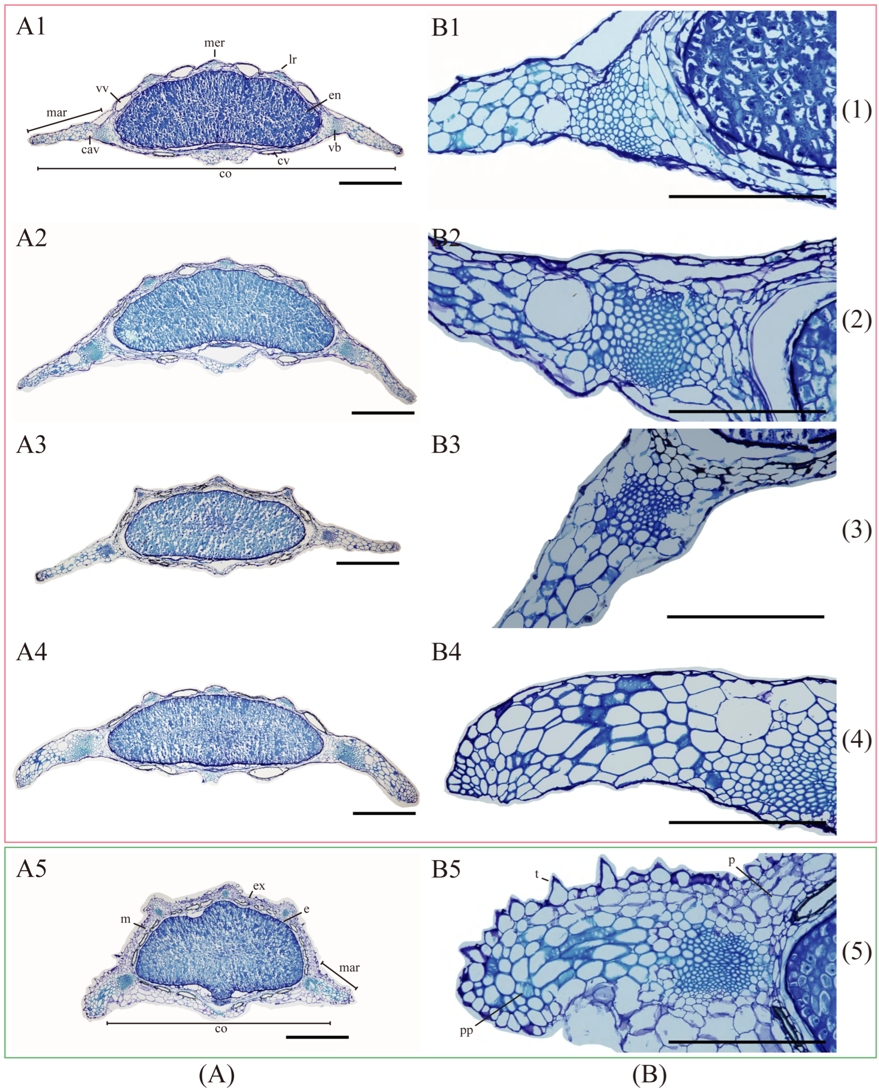
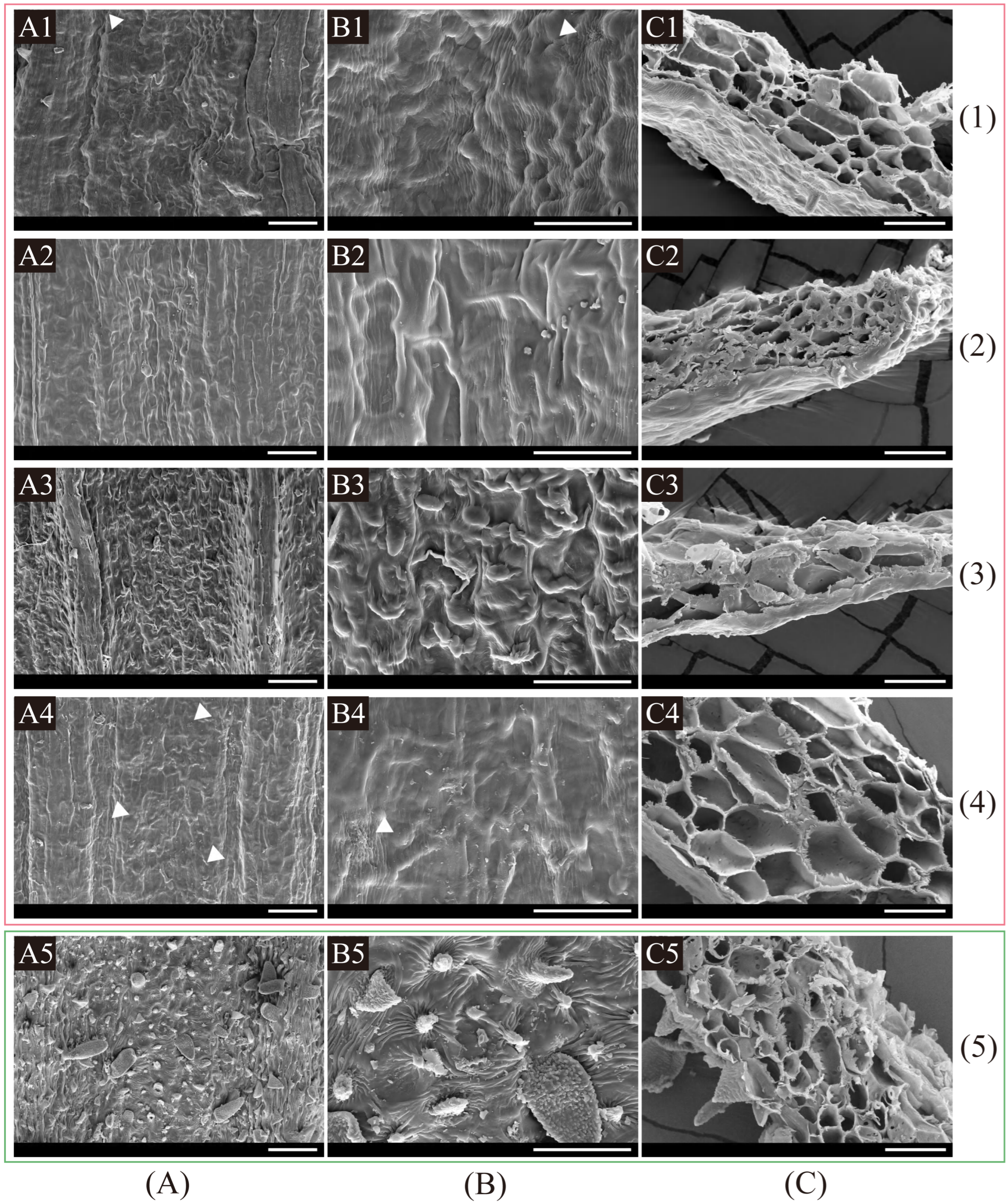
2.3.1. Fruit Anatomical and Micromorphological Examination

Fruit anatomical and micromorphological characteristics of the five Kitagawia species studied are shown in Table 2.
Fruit anatomical characteristics showed that the four Kitagawia species within Subclade I (i.e., K. baicalensis, K. komarovii, K. stepposa, and K. terebinthacea) all had strongly dorsally compressed mericarps, filiform and slightly prominent dorsal (one median and two lateral) ribs, winged marginal ribs, flat or slightly concave endosperm commissural face, solitary vitta in each furrow, 2 vittae on commissure, 1–2 layers of mesocarp cells closest to the epidermis, and lignified parenchyma with pitted walls in mesocarp (Table 2, Figure 5). Kitagawia praeruptora (Dunn) Pimenov within Subclade II had slightly dorsally compressed mericarps, filiform and prominent dorsal ribs, narrowly winged and thick marginal ribs, flat endosperm commissural face, 3–4 vittae in each furrow, 6 vittae on commissure, 3–5 layers of mesocarp cells closest to the epidermis, and lignified parenchyma with pitted walls in mesocarp (Table 2, Figure 5). The average ratios of the total widths of both marginal ribs to the entire fruit width were 42.3–50.3% in the four Kitagawia species within Subclade I (K. baicalensis, K. komarovii, K. stepposa, and K. terebinthacea), while the average ratios of the commissure width to the fruit width were 94.1–96.4% (Table 2, Figure 5). In K. praeruptora, these two average ratios were 39.8% and 85.2%, respectively (Table 2, Figure 5).
Fruit micromorphological studies revealed that the five Kitagawia species had inconspicuous cell borders of fruit surfaces, different fruit surfaces (undulate: K. baicalensis and K. terebinthacea; rugate: K. stepposa; tuberculate: K. komarovii; raised: K. praeruptora), various cuticular foldings (cuticle striate and rugulate: K. baicanlensis and K. terebinthacea; cuticle smooth and striate: K. stepposa and K. komarovii; dense, tiny prickles and short hairs with rugulate and tuberculate cuticle: K. praeruptora), without epicuticular wax, and parenchyma cells with pitted walls in marginal ribs (Table 2, Figure 6).

2.3.2. Comparison of General Morphological Characteristics

Considering general morphological characteristics, the four Kitagawia species within Subclade I shared glabrous stems, 2–3-pinnate leaf blades, lanceolate bracts and bracteoles, unequal and adaxial surface hispid rays, white petals, subulate or triangular calyx teeth, conical stylopodia, and elliptic, glabrous mericarps (Table S3). Kitagawia praeruptora (Dunn) Pimenov and K. formosana within Subclade II shared hairy stems and rays, linear or lanceolate bracts and bracteoles, white petals, obsolete or inconspicuous calyx teeth, conical stylopodia, and ovate-elliptic, sparsely pubescent or hispid mericarps (Table S3). More morphological characteristics for all 15 species studied are summarized in Table S3.

3. Discussion

3.1. Phylogenetic Position and Non-Monophyly of Kitagawia

Previous phylogenetic analyses did not specifically examine the genus Kitagawia and may have been misled by misidentifications of some Kitagawia accessions, failing to identify a clear phylogenetic position for Kitagawia [27,29,30]. Recent studies have revealed that Kitagawia is distantly related to Peucedanum sensu stricto and other segregates of Peucedanum sensu lato, but they relied on single DNA fragments and were limited by low support [5,9,31]. In this study, we combined complete plastome sequences with nrDNA sequences to produce a well-supported phylogeny for Kitagawia.
In our study, single-copy CDS plastome sequences produced a robust phylogeny for Kitagawia. In the CDS tree, all six Kitagawia species examined fell into Selineae and were divided into two branches (Subclade I and Subclade II) (Figure 1). They were all considerably distant from P. morisonii, the member of Peucedanum sensu stricto confirmed in previous studies [2,8,29] (Figure 1). Similar results were presented in the nrDNA (ITS + ETS) tree (Figure 1). Peucedanum officinale L., the type species of Peucedanum sensu stricto, formed a sister branch to P. morisonii, which was closely related to four Angelica species and more distantly related to the six Kitagawia species examined (Figure 1). In both phylogenetic trees, Subclade I and C. sinensis, and Subclade II and S. divaricata formed sister branches, respectively (Figure 1). Although there were differences in the topologies of plastome CDS and nrDNA (ITS + ETS) trees, they consistently indicated considerably distant phylogenetic relationships between Kitagawia and Peucedanum sensu stricto, thus providing strong support for the separation of Kitagawia. These trees also revealed that Kitagawia, as defined by Pimenov, was not monophyletic [4,10,17,18].
We believe that the “core” Kitagawia should be limited to Subclade I, while K. praeruptora and K. formosana should be removed from the genus. Furthermore, P. hakuunense was treated as a synonym of K. komarovii by Pimenov [18]; while our results showed that P. hakuunense should be identified as a distinct species, both P. hakuunense and P. chujaense may belong to Kitagawia. Our phylogenetic analyses confirmed that Kitagawia does not belong to the Acronema Clade but to Selineae. There was no doubt that some accessions of Kitagawia used in previous studies were misidentified [27,29,30]. Our results demonstrate the rationale behind the separation of Kitagawia and the necessity for its further taxonomic revision.

3.2. Plastome Structure and Evolution and Their Phylogenetic Implications

Our comparative analyses revealed that all 14 plastomes examined displayed a typical quadripartite structure [43,47] (Figure S1). The plastome structure, GC content, and gene order were similar to those of species in the Apiaceae tribe Selineae (Figures S1 and S2, Table 1), implying that plastome structure was highly conserved [43,47,55]. Among the 14 species examined, the plastomes of K. praeruptora, K. formosana, and P. harry-smithii var. grande lacked the trnT-GGU gene (Table S2), which may suggest that they have different evolutionary histories when compared to other plastomes [56,57].
SSRs have been widely used as molecular markers in plant population genetics and evolutionary studies [58,59,60]. In the 14 studied plastomes of Kitagawia and related taxa, mononucleotides were the most abundant SSRs, followed by dinucleotides, tetranucleotides, trinucleotides, pentanucleotides, and hexanucleotides. Such findings are widespread in Apiaceae, Liliaceae, and Allium L. [47,61,62]. All identified SSRs were mainly composed of bases A and T, causing an AT richness in the overall plastome [43,63]. IR expansion and contraction have been recognized as evolutionary markers for illustrating relationships among taxa [43,55,64]. In this study, the JLA of species within Subclade I and Subclade II visibly differed from other species examined (Figure 3). Distances between gene trnL and JLA of species within Subclade I and Subclade II were considerably longer than P. morisonii, C. sinensis, and A. sylvestris (Figure 3). In contrast, distances from gene trnH to JLA of species within Subclade I and Subclade II were shorter than for the three above-mentioned species (Figure 3). Furthermore, the lengths of the IR regions of P. morisonii, C. sinensis, and A. sylvestris were shorter than for other species examined (18,457–19,043 bp) (Figure 3, Table 1). The similar features of JLA and the IR length of P. morisonii, C. sinensis, and A. sylvestris may imply a common but different evolutionary history that differentiated them from the others (Subclade I and II and S. divaricata). In addition, sequence divergences noted in several regions (e.g., ycf1, ycf2, and accD-psaI) separated Subclade I from Subclade II and P. morisonii (Figure 4). These differences in plastomes may reflect distinct phylogenetic affinities between the species of Subclade I and the others [55]. Overall, the results of comparative plastome analyses revealed different evolutionary relationships of Subclade I and Subclade II to other taxa examined and added some support to the distinct phylogenetic position and non-monophyly of Kitagawia.

3.3. Morphological Delimitations between Kitagawia and Related Taxa

In this study, we examined fruit anatomical and micromorphological characteristics of five Kitagawia species (i.e., K. baicalensis, K. komarovii, K. praeruptora, K. stepposa, and K. terebinthacea). The results showed that all species shared filiform dorsal ribs, winged marginal ribs, flat or slightly concave endosperm commissural face, partly lignified parenchyma cells with pitted walls in marginal ribs, inconspicuous cell borders of fruit surfaces, without epicuticular wax (Figure 5 and Figure 6, Table 2). These findings support the recognition of Kitagawia at a generic level [10]. However, compared with the four “core” Kitagawia species within Subclade I (K. baicalensis, K. komarovii, K. stepposa, and K. terebinthacea), K. praeruptora within Subclade II had more vallecular vittae (3–4 vs. solitary) and commissural vittae (6 vs. 2), more layers of mesocarp cells closest to the epidermis (3–5 vs. 1–2), and narrower marginal ribs and commissure (Figure 5, Table 2). These differences run counter to the descriptions of Kitagawia [4,10,17], implying that K. praeruptora should be removed from Kitagawia. Furthermore, the five Kitagawia species examined had different fruit surfaces and various cuticular foldings (Figure 6, Table 2). These characteristics partly overlapped with the fruit micromorphological characteristics of Peucedanum sensu stricto [23,25,65] and were incongruent with the results of Ostroumova [23,24,25]. Thus, micromorphological characteristics of fruit surfaces might vary among populations, which would make them poor diagnostic characteristics for Kitagawia.
Furthermore, the four “core” Kitagawia species within Subclade I differed from C. sinensis in bracts and bracteoles (linear or lanceolate vs. linear), mericarp shape (elliptic vs. oblong-ovate), mericarp surfaces (glabrous vs. densely hirtellous), and marginal rib shape of mericarps (winged vs. filiform) [66] (Table S3). Similarly, they were distinct from A. sylvestris, which has linear bracts and bracteoles, pubescent rays, obsolete calyx teeth, broadly ovate mericarps, and narrowly winged and obtuse dorsal ribs [17,67] (Table S3). It is notable that the four “core” Kitagawia species within Subclade I were slightly different from the representative members of Peucedanum sensu stricto (P. morisonii and P. officinale) in leaf blades (2–3-pinnate vs. 3–6-ternate), bracts (lanceolate vs. subulate to linear), bracteoles (lanceolate vs. linear), rays (adaxial surface hispid vs. glabrous), and petals (white vs. yellow) [11,12,68] (Table S3). These partially overlapping traits reflect the difficulties in dividing Peucedanum sensu lato and indicate that it is ill-advised to distinguish Kitagawia from Peucedanum sensu stricto by individual morphological characteristics.
The association of a series of morphological characteristics is regarded as a more powerful tool to distinguish taxonomically difficult taxa compared to a single morphological characteristic [55]. Through our analyses, the morphological delimitation of Kitagawia (the four “core” Kitagawia species within Subclade I) should be “stems glabrous, leaf blades 2–3-pinnate, ultimate leaf segments linear to lanceolate, bracts and bracteoles lanceolate, rays adaxial surface hispid, petals white, calyx teeth subulate or triangular, stylopodia conical, mericarps strongly compressed dorsally, elliptic and glabrous, dorsal ribs filiform and slightly prominent, marginal ribs winged, commissure broad, solitary vitta in each furrow, two vittae on commissure, partly lignified parenchyma cells with pitted walls in marginal ribs, 1–2 layers of mesocarp cells closest to the epidermis, inconspicuous cell borders of fruit surfaces, without epicuticular wax”. Considering that P. hakuunense and P. chujaense probably belong to Kitagawia, the morphological delimitation of Kitagawia may need further revision.

3.4. Taxonomic Suggestions for Six Species from Kitagawia and Peucedanum Sensu Lato

Pimenov stated that the generic boundaries of Kitagawia might need to be expanded and highlighted several Peucedanum sensu lato species that may belong to Kitagawia [10,17]. One example was discussed in the taxonomic treatment of K. formosana [18].
In this study, two Peucedanum sensu lato species considered as candidates for Kitagawia, P. ampliatum and P. harry-smithii var. grande, were included to investigate the generic boundaries of Kitagawia. In our phylogenetic trees, P. ampliatum and P. harry-smithii var. grande clustered with K. praeruptora and K. formosana, forming Subclade II (Figure 1). The phylogenetic positions of the four species within Subclade II were distant from Subclade I (“core” Kitagawia) and Peucedanum sensu stricto (Figure 1). Instead, they are closely related to S. divaricata, the type species of a monotypic genus Saposhnikovia (Figure 1). The rather distant relationship of Subclade II to Subclade I and Peucedanum sensu stricto suggest that the four species within Subclade II (including K. praeruptora and K. formosana) should neither be considered members of Kitagawia nor included in Peucedanum sensu lato, which is consistent with previous phylogenetic results [32,33,47,69].
In our morphological analyses, the four species within Subclade II, K. praeruptora, K. formosana, P. ampliatum, and P. harry-smithii var. grande, shared characteristics “stems pubescent or tomentose, bracts absent or few, linear or lanceolate, bracteoles numerous, linear or lanceolate, rays pubescent or tomentose on adaxial surface or throughout, petals white, mericarps pubescent or hispid, dorsal ribs filiform and prominent, marginal ribs narrowly winged, 3–5 vittae in each furrow, 6–8 vittae on commissure” [11,12,70], which differed from the four Kitagawia species within Subclade I, supporting the distant phylogenetic relationship of K. praeruptora and K. formosana to “core” Kitagawia (Figure 1, Table S3). These characteristics shared by the four species within Subclade II could also distinguish them from Peucedanum sensu stricto [4] and Saposhnikovia (the latter have glabrous stems, rays, and mericarp, few bracteoles, solitary vitta in each furrow, two vittae on commissure, and one pseudovitta under each primary rib) [71] (Table S3). As far as the present study is concerned, the members of Subclade II possessed unique fruit anatomical and micromorphological characteristics, as well as general morphological characteristics, that could not be identified in other related genera (Tables S2 and S3, Figure 5 and Figure 6). This may suggest that their current taxonomic placements are unreasonable. The phylogenetic and morphological results consistently suggested that K. praeruptora and K. formosana should be placed in a new genus and the two possible candidate taxa (P. ampliatum and P. harry-smithii var. grande) should not be included in Kitagawia. For the possible new genus, more extensive population sampling and study of additional, potentially related taxa are required to determine generic boundaries and morphological delimitations.
Two Peucedanum sensu lato species found in Korea, P. hakuunense and P. chujaense, fell into Subclade I. Of these, P. hakuunense was treated as a synonym of K. komarovii [18]. Peucedanum chujaense K. Kim, S.H. Oh, Chan S. Kim & C.W. Park was thought to be morphologically most similar to Kitagawia species [15]. Our phylogenetic analyses revealed that P. hakuunense and P. chujaense nested into Subclade I, suggesting they may need to be included in Kitagawia (Figure 1). However, specimens of P. hakuunense and P. chujaense were not available and morphological evaluations could not be undertaken. In our morphological analyses, therefore, the general morphological characteristics of P. hakuunense and P. chujaense were directly referenced from previous literature [15,72,73] to document the morphological similarity of these two species with the four Kitagawia species within Subclade I (Table S3). Phylogenetic and morphological evidence suggests that P. hakuunense and P. chujaense probably belong to Kitagawia. More evidence is needed for a complete taxonomic treatment of P. hakuunense and P. chujaense and for the clarification of interspecific relationships within Subclade I. To that end, international cooperation will be needed to produce a more detailed revision of Kitagawia.

3.5. Topologic Incongruence between Phylogenies based on Plastome and NrDNA Sequences

Previous molecular phylogenetic studies have identified a common phylogenetic incongruence between plastid and nuclear gene trees [43,74]. Several evolutionary processes, such as hybridization, introgression, and incomplete lineage sorting (ILS), are regarded as plausible explanations for the discordance of plastid and nuclear DNA phylogenies [75,76].
In our phylogenetic analyses, the phylogenetic positions of three main clades (i.e., Tordyliinae, Coriandreae, and Hymenidium Clade) differed between the two phylogenetic trees (Figure 2). These differences were also reported by Wen et al. [46]. We agree with Wen et al. [46] that the conflicting positions of the three main clades (Tordyliinae, Coriandreae, and Hymenidium Clade) between the plastome CDS and nrDNA (ITS + ETS) trees were mainly caused by chloroplast capture. In Selineae, some branches (e.g., the branch Subclade I + C. sinensis and the branch Subclade II + S. divaricata) and even some species within the same branch differed in their phylogenetic placements in the phylogenies based on plastome CDS and nrDNA (ITS + ETS) sequences (Figure 1). Similar to the three main clades (Tordyliinae, Coriandreae, and Hymenidium Clade), the incongruent positions of the four branches (i.e., “Peucedanum sensu stricto group”, “Angelica group”, Subclade I + Carlesia, and Subclade II + Saposhnikovia) between the plastome CDS and nrDNA (ITS + ETS) trees may also have been generated by early chloroplast capture events due to hybridization. Our phylogenetic results supported a chloroplast capture hypothesis that the early lineage of the branch Subclade I + Carlesia may have successively captured the chloroplast genome of ancestors of the “Angelica group”. The strongly supported polytomy of Subclade II in the nrDNA (ITS + ETS) tree suggested the insufficiency of phylogenetic informative characters in the nrDNA dataset, which is also thought to contribute to the incongruent topologies between plastid and nuclear gene trees [77,78]. The lack of informative characters is usually derived from rapid radiation and hybridization [79,80]. In our study, hybridization is not a reasonable explanation for the polytomy of Subclade II because the four species within Subclade II included two stenochoric species (K. formosana and P. ampliatum) with non-overlapping distributions [12,70]. Rapid radiation may be the most likely mechanism for the polytomy of Subclade II in the nrDNA (ITS + ETS) tree. The hairy stems and fruits and numerous mericarp vittae may be synapomorphies of the four species within Subclade II (Table S4), and they may have undergone rapid morphological differentiation by a founder effect [81], which can coincide with rapid radiation.

4. Materials and Methods

4.1. Taxon Sampling

Fresh green leaves from adult plants of seven taxa, including five Kitagawia species (K. baicalensis, K. komarovii, K. praeruptora, K. stepposa, and K. terebinthacea), C. sinensis, and S. divaricata were collected from the wild, and then immediately dried with silica gel to preserve them for DNA extraction. Special permission was not required to collect these materials because they are not key-protected plants. In addition, we requested and obtained the genomic DNA of K. formosana from the Herbarium of the Institute of Botany (PE), Chinese Academy of Sciences. The formal identification of all samples was undertaken by Professor Xingjin He (Sichuan University). The voucher information of the eight samples is summarized in Table S4.

4.2. DNA Extraction, Sequencing, Assembly, and Annotation

The total genomic DNA of the eight taxa was extracted from silica gel-dried leaves or herbarium specimens with a modified CTAB protocol [82]. The primers ITS4 (5′-TCC TCC GCT TAT TGA TAT GC-3′) and ITS5 (5′-GGA AGT AAA AGT CGT AAC AAG G-3′) were used in the polymerase chain reaction (PCR) amplification of the complete ITS region [83]. The ETS sequences were amplified with primers 18S-ETS (5′-ACT TAC ACA TGC ATG GCT TAA TCT-3′) and Umb-ETS (5′-GCG CAT GAG TGG TGA WTK GTA-3′) [84,85]. The 30-µL PCR reactions contained 2 µL extracted total genomic DNA, 10 µL ddH2O, 1.5 µL each of 10 pmol µL−1 forward and reverse primers, and 15 µL Taq MasterMix (CWBio, Beijing, China). The PCR program of ITS and ETS regions started with an initial denaturation at 94 °C for 4 min, followed by 30 cycles of denaturation at 94 °C for 45 s, annealing at 54 °C for 45 s, and extension at 72 °C for 1 min, with a final extension at 72 °C for 10 min. All PCR products were separated on a 1.5% (w v−1) agarose TAE gel and sent to Sangon (Shanghai, China) for sequencing. The newly sequenced ITS and ETS sequences of the eight taxa were examined and edited with Geneious v9.0.2 [86], and consensus sequences were obtained separately.
Plastomes of the eight taxa, namely K. baicalensis, K. formosana, K. komarovii, K. praeruptora, K. stepposa, K. terebinthacea, C. sinensis, and S. divaricata, were sequenced. We provided 20 µL of total genomic DNA per species for the sequencing process, and total genomic DNA was sequenced on an Illumina HiSeq X Ten platform (paired-end, 150 bp) by Novogene (Beijing, China). At least 5 GB of raw data per species were generated. Quality control of the raw reads was performed using fastP v0.15.0 (-n 10 and -q 15) [87], yielding high-quality reads for each species. The program GetOrganelle v1.7.5.3 [88] was used to assemble the complete plastomes with the custom parameters (-F embplant_pt; -R 15; -k 21, 45, 65, 85, 105, 121). Assembled plastomes of the eight taxa were annotated with PGA [89]. Geneious v9.0.2 [86] was then used to manually adjust the annotation for uncertain start and stop codons based on comparisons with homologous genes from other annotated plastomes. The circular plastome maps were generated with OGDRAW v1.3.1 [90].
The newly obtained complete plastomes, ITS, and ETS sequences of the eight taxa studied were submitted to GenBank under accession numbers OP379697-OP379704, OP377020-OP377027, and OP379688-OP379695, respectively (Table S5).

4.3. Phylogenetic Analyses

After preliminary analyses, 28 species from Selineae, Tordyliinae, Coriandreae, Hymenidium Clade, Apieae, Careae, Pyramidoptereae, and Acronema Clade were selected to conduct the phylogenetic reconstruction. The type species of Peucedanum sensu stricto and Angelica, P. officinale and A. sylvestris, were included due to the blurred boundaries and potential expansion of Kitagawia [10]. Similarly, P. ampliatum and P. harry-smithii var. grande were added to examine the possible candidates for Kitagawia according to Pimenov and Ostroumova [17]. Pternopetalum davidii Franch. and Pternopetalum vulgare (Dunn) Hand.-Mazz., which belong to Acronema Clade, were chosen as the outgroup to root the phylogenetic tree, based on the results of Downie et al. [30]. The names of the main clades refer to the contributions of Downie et al. [30] and Gou et al. [51].
Two datasets were constructed for the phylogenetic analyses. The CDS dataset consisted of single-copy CDS sequences from complete plastomes of 27 taxa (6 species from Kitagawia and 21 species from other genera in the above-mentioned clades). To provide greater branch support [85], the nrDNA dataset concatenated the complete ITS and ETS regions from 27 taxa of the same genera used in the CDS dataset. Peucedanum officinale L. and P. chujaense could not be added to the CDS dataset and nrDNA dataset, respectively, due to a lack of online data. All sequences used in phylogenetic analyses are available in GenBank (Table S5).
The 79 single-copy CDS sequences from 27 complete plastomes were extracted and connected using PhyloSuite v1.2.2 [91]. The nrDNA (ITS + ETS) sequences of 27 taxa were also concatenated with PhyloSuite v1.2.2 [91]. Sequences of the two datasets were aligned with MAFFT v7.221 [92] and then manually corrected with MEGA7 [93]. Maximum likelihood (ML) and Bayesian inference (BI) methods were adopted to infer phylogenetic relationships. RAxML v8.2.10 [94] was used to perform the ML analyses for the two datasets based on the GTRGAMMA model and 1000 rapid bootstrap replicates. MrBayes v3.2.7 [95] was used to perform the BI analyses with the best substitution model as determined by MrModeltest v2.4 [96]. The selected models for the CDS dataset and nrDNA dataset in BI analyses were GTR + I + G and GTR + G, respectively. Four independent Markov chains were run for 10,000,000 generations with random initial trees, sampling every 1000 generations. The first 25% of trees were discarded as burn-in, and the remaining trees were used to build a 50% majority-rule consensus tree. The phylogenetic trees were visualized and edited with FigTree v1.4.4 [97] and MEGA7 [93].

4.4. Comparative Analyses of Plastomes

To better understand the genomics and evolution of Kitagawia plastomes, 14 plastomes representing Kitagawia and related taxa were selected for comparative analyses of plastomes based on our phylogenetic results. They consisted of six Kitagawia plastomes (K. baicalensis, K. formosana, K. komarovii, K. praeruptora, K. stepposa, and K. terebinthacea), five Peucedanum sensu lato plastomes (P. ampliatum, P. chujaense, P. hakuunense, P. harry-smithii var. grande, and P. morisonii), A. sylvestris, C. sinensis, and S. divaricata.
Simple sequence repeats (SSRs) for each plastome were detected with MISA (http://pgrc.ipk-gatersleben.de/misa/ (accessed on 21 May 2022)) [98]. The minimum numbers of the SSRs were set as 10, 5, 4, 3, 3, and 3 for mono-, di-, tri-, tetra-, penta-, and hexa-nucleotides, respectively.
The expansion or contraction between IR border regions of the 14 plastomes of Kitagawia and related taxa were drawn by IRscope [99] and adjusted manually.
The DNA rearrangements among the 14 selected plastomes were detected using Mauve Aligner [100] implemented in Geneious v9.0.2 [86]. Sequence divergence of the 14 complete plastomes was visualized with mVISTA [101] under Shuffle-LAGAN alignment mode [102] with K. terebinthacea as the reference.

4.5. Morphological Analyses

4.5.1. Fruit Anatomical and Micromorphological Characteristics

Mature fruits of five Kitagawia species (K. baicalensis, K. komarovii, K. praeruptora, K. stepposa, and K. terebinthacea) were collected from the wild and preserved in formaldehyde–acetic acid–alcohol (FAA) for morphological examination. Fruit anatomy was evaluated by paraffin section. For each of the five species, the fruits of at least three individuals from the same population were examined. All fruits were treated following the method of Feder and O’Brien [103] for embedding in glycol methacrylate (GMA). A Leica RM2016 microtome was used to prepare transverse sections through the center of the mericarp, about 3–5 mm in thickness, and they were stained with toluidine blue. Sections were observed and photographed under an Olympus BX43 microscope. Morphological terminology followed Kljuykov et al. [104].
Fruit micromorphology was examined by scanning electron microscope (SEM). After dehydration using graded ethanol, all mature fruits were directly mounted on clean aluminum stubs with conducting carbon adhesive tabs, coated, and then scanned with a JSM-7500F scanning electron microscope. The micromorphological characteristics studied and micromorphological terminology follow the contributions of Ostroumova [23,24].

4.5.2. General Morphological Characteristics

We examined the general morphological characteristics of the 14 species used in the above comparative plastome analyses together with P. officinale, the type species of Peucedanum sensu stricto. The characteristics examined included features of the stem, leaf, bract, bracteole, ray, flower, and fruit. Data from the five Kitagawia species (K. baicalensis, K. komarovii, K. praeruptora, K. stepposa, and K. terebinthacea) were obtained mainly from our field-based observations and anatomy research. For some taxa and characteristics, data were obtained directly from previous literature [11,12,15,17,66,67,68,71,72,73].

5. Conclusions

In this study, we newly sequenced complete plastomes of eight species from Kitagawia, Carlesia, and Saposhnikovia. The phylogeny reconstruction of Kitagawia was performed based on plastome CDS and nrDNA (ITS + ETS) sequences. Our results revealed that Kitagawia is a member of the tribe Selineae but is divided into two branches (Subclade I and Subclade II). The distant phylogenetic relationships between Kitagawia and Peucedanum sensu stricto verified the rationale behind the separation of Kitagawia. The visible differences in JLA and several highly variable regions added some support to the distinct phylogenetic position and non-monophyly of Kitagawia. Similarly, fruit anatomical and micromorphological characteristics, as well as general morphological characteristics, distinguished Subclade I from Subclade II and other related taxa, further supporting the results of phylogenetic analyses. These findings demonstrated that P. ampliatum and P. harry-smithii var. grande should not be included in Kitagawia and suggested that K. praeruptora and K. formosana should be placed in a new genus and that P. hakuunense and P. chujaense probably belong to Kitagawia. We believe that the “core” Kitagawia should be limited to Subclade I, and this genus can be distinguished by the association of a series of morphological characteristics. In short, our study confirmed the distinct generic status of Kitagawia and clarified the morphological delimitations between Kitagawia and related taxa, providing a foundation for further taxonomic and evolutionary research on Kitagawia.

Supplementary Materials

The following supporting information can be downloaded at: https://www.mdpi.com/article/10.3390/plants11233275/s1, Figure S1: Gene maps of the eight plastomes of Kitagawia, Carlesia, and Saposhnikovia; Table S1: List of genes identified in the 14 plastomes of Kitagawia and related taxa; Table S2: Number of all identified SSRs in the 14 plastomes; Figure S2: Mauve alignment of the 14 plastomes of Kitagawia and related taxa; Table S3: Synopsis of general morphological characteristics from the 15 species involved in this study; Table S4: Collection locality and voucher information are provided for the eight sequenced plastomes; Table S5: GenBank accession numbers of DNA sequences used in this study.

Author Contributions

Conceptualization, J.-Q.L., S.-D.Z. and X.-J.H.; methodology, J.-Q.L. and C.-K.L.; formal analysis, J.-Q.L. and J.C.; investigation, J.-Q.L., C.-K.L. and J.C.; data curation, J.-Q.L.; writing—original draft preparation, J.-Q.L.; writing—review and editing, J.-Q.L., M.P., S.-D.Z. and X.-J.H.; supervision, S.-D.Z. and X.-J.H.; funding acquisition, S.-D.Z. and X.-J.H. All authors have read and agreed to the published version of the manuscript.

Funding

This work was supported by the National Natural Science Foundation of China, grant numbers 32070221 and 32170209.

Data Availability Statement

The annotated plastomes, ITS, and ETS sequences of the eight taxa studied have been submitted to NCBI (https://www.ncbi.nlm.nih.gov (accessed on 7 September 2022)) with accession numbers that can be found in Table S5.

Acknowledgments

We are grateful to Ting Ren, Rui-Yu Cheng, Xian-Lin Guo, Bo-Ni Song, An-Qi Zhao, and Li-Jia Liu for their help in sample collection. We also thank Chao Xu, Xin-Tang Ma, Zhi-Rong Yang, and other staff of the Herbarium of the Institute of Botany (PE), Chinese Academy of Sciences, for providing genomic DNA.

Conflicts of Interest

The authors declare no conflict of interest.

References

  1. Pimenov, M.G.; Leonov, M.V.; Constance, L. The Genera of the Umbelliferae: A Nomenclator; Royal Botanic Gardens: Kew, UK, 1993; ISBN 978-0-947643-58-4. [Google Scholar]
  2. Shneyer, V.S.; Kutyavina, N.G.; Pimenov, M.G. Systematic Relationships within and between Peucedanum and Angelica (Umbelliferae–Peucedaneae) Inferred from Immunological Studies of Seed Proteins. Plant Syst. Evol. 2003, 236, 175–194. [Google Scholar] [CrossRef]
  3. Zhou, J.; Wang, W.C.; Gong, X.; Liu, Z.W. Leaf Epidermal Morphology in Peucedanum L. (Umbelliferae) from China. Acta Bot. Gallica 2014, 161, 21–31. [Google Scholar] [CrossRef]
  4. Plunkett, G.M.; Pimenov, M.G.; Reduron, J.-P.; Kljuykov, E.V.; van Wyk, B.-E.; Ostroumova, T.A.; Henwood, M.J.; Tilney, P.M.; Spalik, K.; Watson, M.F.; et al. Apiaceae. In Flowering Plants. Eudicots: Apiales, Gentianales (Except Rubiaceae); Kadereit, J.W., Bittrich, V., Eds.; The Families and Genera of Vascular Plants; Springer International Publishing: Cham, Switzerland, 2018; Volume 15, pp. 9–206. ISBN 978-3-319-93605-5. [Google Scholar]
  5. Pimenov, M.G.; Ostroumova, T.A.; Degtjareva, G.V.; Samigullin, T.H. Sillaphyton, a New Genus of the Umbelliferae, Endemic to the Korean Peninsula. Bot. Pac. 2016, 5, 31–41. [Google Scholar] [CrossRef] [Green Version]
  6. Brullo, C.; Brullo, S.; Downie, S.R.; Danderson, C.A.; del Galdo, G.G. Siculosciadium, a New Monotypic Genus of Apiaceae from Sicily. Ann. Mo. Bot. Gard. 2013, 99, 1–18. [Google Scholar] [CrossRef] [PubMed]
  7. Winter, P.J.D.; Magee, A.R.; Phephu, N.; Tilney, P.M.; Downie, S.R.; van Wyk, B.E. A New Generic Classification for African Peucedanoid Species (Apiaceae). Taxon 2008, 57, 347–364. [Google Scholar] [CrossRef]
  8. Spalik, K.; Reduron, J.-P.; Downie, S.R. The Phylogenetic Position of Peucedanum Sensu Lato and Allied Genera and Their Placement in Tribe Selineae (Apiaceae, Subfamily Apioideae). Plant Syst. Evol. 2004, 243, 189–210. [Google Scholar] [CrossRef]
  9. Ostroumova, T.A.; Pimenov, M.G.; Degtjareva, G.V.; Samigullin, T.H. Taeniopetalum Vis. (Apiaceae), a Neglected Segregate of Peucedanum, L., Supported as a Remarkable Genus by Morphological and Molecular Data. Skvortsovia 2016, 3, 20–44. [Google Scholar]
  10. Pimenov, M.G. Kitagawia—A New Asiatic Genus of the Family Umbelliferae. Bot. Zhurnal 1986, 71, 942–949. [Google Scholar]
  11. Sheh, M.L. Peucedanum. In Flora Reipublicae Popularis Sinicae; Science Press: Beijing, China, 1992; Volume 55, pp. 127–158. [Google Scholar]
  12. Sheh, M.L.; Watson, M.F. Peucedanum Linnaeus. In Flora of China; Science Press & Missouri Botanical Garden Press: Beijing, China; St. Louis, MO, USA, 2005; Volume 14, pp. 182–192. [Google Scholar]
  13. Yamazaki, T. Umbelliferae in Japan II. J. Jpn. Bot. 2001, 76, 283–285. [Google Scholar]
  14. Chang, C.S.; Kim, H.; Chang, K.S. Provisional Checklist of Vascular Plants for the Korea Peninsula Flora (KPF); Designpost: Pajo, Philippines, 2014; ISBN 978-89-968648-2-0. [Google Scholar]
  15. Kim, K.; Kim, C.S.; Oh, S.H.; Park, C.W. A New Species of Peucedanum (Apiaceae) from Korea. Phytotaxa 2019, 393, 75. [Google Scholar] [CrossRef]
  16. Kim, K.; Suh, H.-J.; Song, J.-H. Two New Endemic Species, Peucedanum miroense and P. tongkangense (Apiaceae), from Korea. PhytoKeys 2022, 210, 35–52. [Google Scholar] [CrossRef]
  17. Pimenov, M.G.; Ostroumova, T.A. Umbelliferae of Russia; Association of Scientific Publications: Moscow, Russia, 2012; ISBN 978-5-87317-813-1. [Google Scholar]
  18. Pimenov, M.G. Updated Checklist of Chinese Umbelliferae: Nomenclature, Synonymy, Typification, Distribution. Turczaninowia 2017, 20, 106–239. [Google Scholar] [CrossRef] [Green Version]
  19. Drude, O. Umbelliferae. In Die Natürlichen Pflanzenfamilien; Wilhelm Engelman: Leipzig, Germany, 1898; Volume 3, pp. 63–150, 271. [Google Scholar]
  20. Kljuykov, E.V.; Zakharova, E.A.; Ostroumova, T.A.; Tilney, P.M. Most Important Carpological Anatomical Characters in the Taxonomy of Apiaceae. Bot. J. Linn. Soc. 2021, 195, 532–544. [Google Scholar] [CrossRef]
  21. Liu, M.; van Wyk, B.-E.; Tilney, P.M. The Taxonomic Value of Fruit Structure in the Subfamily Saniculoideae and Related African Genera (Apiaceae). Taxon 2003, 52, 261–270. [Google Scholar] [CrossRef]
  22. Morison, R. Plantarum Umbelliferarum Distributio Nova; Theatro Sheldoniano: Oxford, UK, 1672. [Google Scholar]
  23. Ostroumova, T.A. Fruit Micromorphology in the Umbelliferae of the Russian Far East. Bot. Pac. J. Plant Sci. Conserv. 2018, 7, 41–49. [Google Scholar] [CrossRef] [Green Version]
  24. Ostroumova, T.A. Fruit Micromorphology in the Umbelliferae in Siberia and Patterns of Morphological Diversity in the Family. Probl. Bot. South Sib. Mong. 2020, 19, 44–48. [Google Scholar] [CrossRef]
  25. Ostroumova, T.A. Fruit Micromorphology of Siberian Apiaceae and Its Value for Taxonomy of the Family. Turczaninowia 2021, 24, 120–143. [Google Scholar] [CrossRef]
  26. Lee, C.; Kim, J.; Darshetkar, A.M.; Choudhary, R.K.; Park, S.H.; Lee, J.; Choi, S. Mericarp Morphology of the Tribe Selineae (Apiaceae, Apioideae) and Its Taxonomic Implications in Korea. Bangladesh J. Plant Taxon. 2018, 25, 175–186. [Google Scholar] [CrossRef]
  27. Downie, S.R.; Watson, M.F.; Spalik, K.; Katz-Downie, D.S. Molecular Systematics of Old World Apioideae (Apiaceae): Relationships among Some Members of Tribe Peucedaneae Sensu Lato, the Placement of Several Island-Endemic Species, and Resolution within the Apioid Superclade. Can. J. Bot. 2000, 78, 506–528. [Google Scholar] [CrossRef]
  28. Downie, S.R.; Plunkett, G.M.; Watson, M.F.; Spalik, K.; Katz-Downie, D.S.; Valiejo-Roman, C.M.; Terentieva, E.I.; Troitsky, A.V.; Lee, B.-Y.; Lahham, J.; et al. Tribes and Clades within Apiaceae Subfamily Apioideae: The Contribution of Molecular Data. Edinb. J. Bot. 2001, 58, 301–330. [Google Scholar] [CrossRef]
  29. Feng, T.; Downie, S.R.; Yu, Y.; Zhang, X.M.; Chen, W.W.; He, X.J.; Liu, S. Molecular Systematics of Angelica and Allied Genera (Apiaceae) from the Hengduan Mountains of China Based on NrDNA ITS Sequences: Phylogenetic Affinities and Biogeographic Implications. J. Plant Res. 2009, 122, 403–414. [Google Scholar] [CrossRef] [PubMed]
  30. Downie, S.R.; Spalik, K.; Katz-Downie, D.S.; Reduron, J.P. Major Clades within Apiaceae Subfamily Apioideae as Inferred by Phylogenetic Analysis of NrDNA ITS Sequences. Plant Divers. Evol. 2010, 128, 111. [Google Scholar] [CrossRef] [Green Version]
  31. Pimenov, M.; Degtjareva, G.; Ostroumova, T.; Samigullin, T.; Averyanov, L.V. Xyloselinum laoticum (Umbelliferae), a New Species from Laos, and Taxonomic Placement of the Genus in the Light of NrDNA ITS Sequence Analysis. Phytotaxa 2016, 244, 248. [Google Scholar] [CrossRef]
  32. Zhou, J.; Gong, X.; Downie, S.R.; Peng, H. Towards a More Robust Molecular Phylogeny of Chinese Apiaceae Subfamily Apioideae: Additional Evidence from NrDNA ITS and CpDNA Intron (Rpl16 and Rps16) Sequences. Mol. Phylogenet. Evol. 2009, 53, 56–68. [Google Scholar] [CrossRef]
  33. Zhou, J.; Gao, Y.Z.; Wei, J.; Liu, Z.W.; Downie, S.R. Molecular Phylogenetics of Ligusticum (Apiaceae) Based on NrDNA ITS Sequences: Rampant Polyphyly, Placement of the Chinese Endemic Species, and a Much-Reduced Circumscription of the Genus. Int. J. Plant Sci. 2020, 181, 306–323. [Google Scholar] [CrossRef]
  34. Corriveau, J.L.; Coleman, A.W. Rapid Screening Method to Detect Potential Biparental Inheritance of Plastid DNA and Results for over 200 Angiosperm Species. Am. J. Bot. 1988, 75, 1443–1458. [Google Scholar] [CrossRef]
  35. Mandel, J.R.; Ramsey, A.J.; Holley, J.M.; Scott, V.A.; Mody, D.; Abbot, P. Disentangling Complex Inheritance Patterns of Plant Organellar Genomes: An Example from Carrot. J. Hered. 2020, 111, 531–538. [Google Scholar] [CrossRef] [PubMed]
  36. Erixon, P.; Oxelman, B. Whole-Gene Positive Selection, Elevated Synonymous Substitution Rates, Duplication, and Indel Evolution of the Chloroplast ClpP1 Gene. PLoS ONE 2008, 3, e1386. [Google Scholar] [CrossRef] [Green Version]
  37. Korpelainen, H. The Evolutionary Processes of Mitochondrial and Chloroplast Genomes Differ from Those of Nuclear Genomes. Sci. Nat. 2004, 91, 505–518. [Google Scholar] [CrossRef]
  38. Gou, W.; Jia, S.B.; Price, M.; Guo, X.L.; Zhou, S.D.; He, X.J. Complete Plastid Genome Sequencing of Eight Species from Hansenia, Haplosphaera and Sinodielsia (Apiaceae): Comparative Analyses and Phylogenetic Implications. Plants 2020, 9, 1523. [Google Scholar] [CrossRef]
  39. Guo, X.L.; Zheng, H.Y.; Price, M.; Zhou, S.D.; He, X.J. Phylogeny and Comparative Analysis of Chinese Chamaesium Species Revealed by the Complete Plastid Genome. Plants 2020, 9, 965. [Google Scholar] [CrossRef] [PubMed]
  40. Huang, J.; Yu, Y.; Liu, Y.M.; Xie, D.F.; He, X.J.; Zhou, S.D. Comparative Chloroplast Genomics of Fritillaria (Liliaceae), Inferences for Phylogenetic Relationships between Fritillaria and Lilium and Plastome Evolution. Plants 2020, 9, 133. [Google Scholar] [CrossRef] [PubMed] [Green Version]
  41. Li, J.; Cai, J.; Qin, H.H.; Price, M.; Zhang, Z.; Yu, Y.; Xie, D.F.; He, X.J.; Zhou, S.D.; Gao, X.F. Phylogeny, Age, and Evolution of Tribe Lilieae (Liliaceae) Based on Whole Plastid Genomes. Front. Plant Sci. 2021, 12, 699226. [Google Scholar] [CrossRef]
  42. Parks, M.; Cronn, R.; Liston, A. Increasing Phylogenetic Resolution at Low Taxonomic Levels Using Massively Parallel Sequencing of Chloroplast Genomes. BMC Biol. 2009, 7, 84. [Google Scholar] [CrossRef] [Green Version]
  43. Ren, T.; Li, Z.X.; Xie, D.F.; Gui, L.J.; Peng, C.; Wen, J.; He, X.J. Plastomes of Eight Ligusticum Species: Characterization, Genome Evolution, and Phylogenetic Relationships. BMC Plant Biol. 2020, 20, 519. [Google Scholar] [CrossRef] [PubMed]
  44. Wang, Y.H.; Wicke, S.; Wang, H.; Jin, J.J.; Chen, S.Y.; Zhang, S.D.; Li, D.Z.; Yi, T.S. Plastid Genome Evolution in the Early-Diverging Legume Subfamily Cercidoideae (Fabaceae). Front. Plant Sci. 2018, 9, 138. [Google Scholar] [CrossRef] [Green Version]
  45. Yao, G.; Jin, J.J.; Li, H.T.; Yang, J.B.; Mandala, V.S.; Croley, M.; Mostow, R.; Douglas, N.A.; Chase, M.W.; Christenhusz, M.J.M. Plastid Phylogenomic Insights into the Evolution of Caryophyllales. Mol. Phylogenet. Evol. 2019, 134, 74–86. [Google Scholar] [CrossRef]
  46. Wen, J.; Xie, D.F.; Price, M.; Ren, T.; Deng, Y.Q.; Gui, L.J.; Guo, X.L.; He, X.J. Backbone Phylogeny and Evolution of Apioideae (Apiaceae): New Insights from Phylogenomic Analyses of Plastome Data. Mol. Phylogenet. Evol. 2021, 161, 107183. [Google Scholar] [CrossRef]
  47. Liu, C.K.; Lei, J.Q.; Jiang, Q.P.; Zhou, S.D.; He, X.J. The Complete Plastomes of Seven Peucedanum Plants: Comparative and Phylogenetic Analyses for the Peucedanum Genus. BMC Plant Biol. 2022, 22, 101. [Google Scholar] [CrossRef] [PubMed]
  48. Xie, D.F.; Yu, H.X.; Price, M.; Xie, C.; Deng, Y.Q.; Chen, J.P.; Yu, Y.; Zhou, S.D.; He, X.J. Phylogeny of Chinese Allium Species in Section Daghestanica and Adaptive Evolution of Allium (Amaryllidaceae, Allioideae) Species Revealed by the Chloroplast Complete Genome. Front. Plant Sci. 2019, 10, 460. [Google Scholar] [CrossRef] [PubMed] [Green Version]
  49. Zhang, F.; Wang, N.; Cheng, G.; Shu, X.; Wang, T.; Zhuang, W.; Lu, R.; Wang, Z. Comparative Chloroplast Genomes of Four Lycoris Species (Amaryllidaceae) Provides New Insight into Interspecific Relationship and Phylogeny. Biology 2021, 10, 715. [Google Scholar] [CrossRef] [PubMed]
  50. Ren, T.; Xie, D.; Peng, C.; Gui, L.; Price, M.; Zhou, S.; He, X. Molecular Evolution and Phylogenetic Relationships of Ligusticum (Apiaceae) Inferred from the Whole Plastome Sequences. BMC Ecol. Evol. 2022, 22, 55. [Google Scholar] [CrossRef] [PubMed]
  51. Gou, W.; Guo, X.L.; Zhou, S.D.; He, X.J. Phylogeny and Taxonomy of Meeboldia, Sinodielsia and Their Relatives (Apiaceae: Apioideae) Inferred from NrDNA ITS, Plastid DNA Intron (Rpl16 and Rps16) Sequences and Morphological Characters. Phytotaxa 2021, 482, 121–142. [Google Scholar] [CrossRef]
  52. Lyskov, D.F.; Samigullin, T.H.; Pimenov, M.G. Molecular and Morphological Data Support the Transfer of the Monotypic Iranian Genus Alococarpum to Prangos (Apiaceae). Phytotaxa 2017, 299, 223–233. [Google Scholar] [CrossRef]
  53. Pimenov, M.; Degtjareva, G.; Ostroumova, T.; Samigullin, T.; Zakharova, E. Polyphyletic Trachyspermum (Umbelliferae) Revisited: A Contribution of Molecular and Carpological Data. Plant Biosyst. 2022, 156, 722–742. [Google Scholar] [CrossRef]
  54. Xiao, Y.P.; Guo, X.L.; Price, M.; Gou, W.; Zhou, S.D.; He, X.J. New Insights into the Phylogeny of Sinocarum (Apiaceae, Apioideae) Based on Morphological and Molecular Data. Phytokeys 2021, 175, 13. [Google Scholar] [CrossRef] [PubMed]
  55. Li, Z.X.; Guo, X.L.; Price, M.; Zhou, S.D.; He, X.J. Phylogenetic Position of Ligusticopsis (Apiaceae, Apioideae): Evidence from Molecular Data and Carpological Characters. AoB Plants 2022, 14, plac008. [Google Scholar] [CrossRef] [PubMed]
  56. Mohanta, T.K.; Mishra, A.K.; Khan, A.; Hashem, A.; Abd_Allah, E.F.; Al-Harrasi, A. Gene Loss and Evolution of the Plastome. Genes 2020, 11, 1133. [Google Scholar] [CrossRef] [PubMed]
  57. Abdullah; Mehmood, F.; Heidari, P.; Rahim, A.; Ahmed, I.; Poczai, P. Pseudogenization of the Chloroplast Threonine (TrnT-GGU) Gene in the Sunflower Family (Asteraceae). Sci. Rep. 2021, 11, 21122. [Google Scholar] [CrossRef] [PubMed]
  58. Chung, S.-M.; Staub, J.E.; Chen, J.-F. Molecular Phylogeny of Cucumis Species as Revealed by Consensus Chloroplast SSR Marker Length and Sequence Variation. Genome 2006, 49, 219–229. [Google Scholar] [CrossRef] [Green Version]
  59. Powell, W.; Morgante, M.; Andre, C.; McNicol, J.W.; Machray, G.C.; Doyle, J.J.; Tingey, S.V.; Rafalski, J.A. Hypervariable Microsatellites Provide a General Source of Polymorphic DNA Markers for the Chloroplast Genome. Curr. Biol. 1995, 5, 1023–1029. [Google Scholar] [CrossRef] [Green Version]
  60. Kaur, S.; Panesar, P.S.; Bera, M.B.; Kaur, V. Simple Sequence Repeat Markers in Genetic Divergence and Marker-Assisted Selection of Rice Cultivars: A Review. Crit. Rev. Food Sci. Nutr. 2015, 55, 41–49. [Google Scholar] [CrossRef] [PubMed]
  61. Chen, J.; Xie, D.; He, X.; Yang, Y.; Li, X. Comparative Analysis of the Complete Chloroplast Genomes in Allium Section Bromatorrhiza Species (Amaryllidaceae): Phylogenetic Relationship and Adaptive Evolution. Genes 2022, 13, 1279. [Google Scholar] [CrossRef] [PubMed]
  62. Li, J.; Price, M.; Su, D.M.; Zhang, Z.; Yu, Y.; Xie, D.F.; Zhou, S.D.; He, X.J.; Gao, X.F. Phylogeny and Comparative Analysis for the Plastid Genomes of Five Tulipa (Liliaceae). BioMed Res. Int. 2021, 2021, 6648429. [Google Scholar] [CrossRef] [PubMed]
  63. Qian, J.; Song, J.; Gao, H.; Zhu, Y.; Xu, J.; Pang, X.; Yao, H.; Sun, C.; Li, X.; Li, C.; et al. The Complete Chloroplast Genome Sequence of the Medicinal Plant Salvia miltiorrhiza. PLoS ONE 2013, 8, e57607. [Google Scholar] [CrossRef] [PubMed]
  64. Wang, R.J.; Cheng, C.L.; Chang, C.C.; Wu, C.L.; Su, T.M.; Chaw, S.M. Dynamics and Evolution of the Inverted Repeat-Large Single Copy Junctions in the Chloroplast Genomes of Monocots. BMC Evol. Biol. 2008, 8, 36. [Google Scholar] [CrossRef] [PubMed] [Green Version]
  65. Yildirim, H.; Duman, H. Peucedanum guvenianum (Apiaceae), a New Species from West Anatolia, Turkey. Doga Turk. J. Bot. 2017, 41, 600–608. [Google Scholar] [CrossRef]
  66. Dunn, S.T. Carlesia Dunn. In Hooker’s Icones Plantarum; Ser. 4; Icones Plantarum; London, UK, 1902; Volume 28, p. 2739. [Google Scholar]
  67. Dihoru, G.; Comanescu, M.; Nicoara, R. Analysis of the Characters on Some Angelica Taxa. Rom. J. Biol.-Plant Biol. 2011, 56, 79–89. [Google Scholar]
  68. Randall, R.E.; Thornton, G. Peucedanum officinale L. J. Ecol. 1996, 84, 475–485. [Google Scholar] [CrossRef]
  69. Zhou, J.; Wang, W.; Liu, M.; Liu, Z. Molecular Authentication of the Traditional Medicinal Plant Peucedanum praeruptorum and Its Substitutes and Adulterants by DNA-Barcoding Technique. Pharmacogn. Mag. 2014, 10, 385–390. [Google Scholar] [CrossRef] [PubMed] [Green Version]
  70. Kao, M.T. Peucedanum L. In Flora of Taiwan, 2nd ed.; Huang, T.C., Ed.; Department of Botany, National Taiwan University: Taipei, Taiwan, 1993; Volume 3, pp. 1034–1037. [Google Scholar]
  71. Liou, T.N. Saposhnikovia Schischk. In Flora Plantarum Herbacearum Chinae Boreali-Orientalis; Science Press: Beijing, China, 1977; Volume 6, pp. 213–216. [Google Scholar]
  72. Nakai, T. Notulae ad Plantas Asiae Orientalis (XI). J. Jpn. Bot. 1939, 15, 740–741. [Google Scholar]
  73. Park, C.; Lee, B.; Song, J.-H.; Kim, K. Peucedanum L. In Flora of Korea Volume 5c Rosidae: Rhamnaceae to Apiaceae; National Institute of Biological Resources: Incheon, Republic of Korea, 2017; ISBN 978-89-6811-276-8. [Google Scholar]
  74. Stull, G.W.; Soltis, P.S.; Soltis, D.E.; Gitzendanner, M.A.; Smith, S.A. Nuclear Phylogenomic Analyses of Asterids Conflict with Plastome Trees and Support Novel Relationships among Major Lineages. Am. J. Bot. 2020, 107, 790–805. [Google Scholar] [CrossRef] [PubMed]
  75. Doyle, J.J. Gene Trees and Species Trees: Molecular Systematics as One-Character Taxonomy. Syst. Bot. 1992, 17, 144–163. [Google Scholar] [CrossRef]
  76. Smith, S.A.; Moore, M.J.; Brown, J.W.; Yang, Y. Analysis of Phylogenomic Datasets Reveals Conflict, Concordance, and Gene Duplications with Examples from Animals and Plants. BMC Evol. Biol. 2015, 15, 150. [Google Scholar] [CrossRef] [PubMed] [Green Version]
  77. Pelser, P.B.; Kennedy, A.H.; Tepe, E.J.; Shidler, J.B.; Nordenstam, B.; Kadereit, J.W.; Watson, L.E. Patterns and Causes of Incongruence between Plastid and Nuclear Senecioneae (Asteraceae) Phylogenies. Am. J. Bot. 2010, 97, 856–873. [Google Scholar] [CrossRef] [PubMed]
  78. Zhang, Y.X.; Zeng, C.X.; Li, D.Z. Complex Evolution in Arundinarieae (Poaceae: Bambusoideae): Incongruence between Plastid and Nuclear GBSSI Gene Phylogenies. Mol. Phylogenet. Evol. 2012, 63, 777–797. [Google Scholar] [CrossRef]
  79. Lavretsky, P.; Hernández-Baños, B.E.; Peters, J.L. Rapid Radiation and Hybridization Contribute to Weak Differentiation and Hinder Phylogenetic Inferences in the New World Mallard Complex (Anas spp.). Auk 2014, 131, 524–538. [Google Scholar] [CrossRef] [Green Version]
  80. Fernández, M.; Ezcurra, C.; Calviño, C.I. Chloroplast and ITS Phylogenies to Understand the Evolutionary History of Southern South American Azorella, Laretia and Mulinum (Azorelloideae, Apiaceae). Mol. Phylogenet. Evol. 2017, 108, 1–21. [Google Scholar] [CrossRef]
  81. Milne, R.I.; Abbott, R.J. The Origin and Evolution of Tertiary Relict Floras. In Advances in Botanical Research; Academic Press: Cambridge, MA, USA, 2002; Volume 38, pp. 281–314. ISBN 0065-2296. [Google Scholar]
  82. Doyle, J.J.; Doyle, J.L. A Rapid DNA Isolation Procedure for Small Quantities of Fresh Leaf Tissue. Phytochem. Bull. 1987, 19, 11–15. [Google Scholar]
  83. White, T.J.; Bruns, T.; Lee, S.; Taylor, J. Amplification and Direct Sequencing of Fungal Ribosomal RNA Genes for Phylogenetics. In PCR Protocols, A Guide to Methods and Applications; Innis, M.A., Gelfand, D.H., Sninsky, J.J., White, T.J., Eds.; Academic Press: San Diego, CA, USA, 1990; pp. 315–322. ISBN 978-0-12-372180-8. [Google Scholar]
  84. Baldwin, B.G.; Markos, S. Phylogenetic Utility of the External Transcribed Spacer (ETS) of 18S–26S RDNA: Congruence of ETS and ITS Trees of Calycadenia (Compositae). Mol. Phylogenet. Evol. 1998, 10, 449–463. [Google Scholar] [CrossRef] [PubMed]
  85. Logacheva, M.D.; Valiejo-Roman, C.M.; Degtjareva, G.V.; Stratton, J.M.; Downie, S.R.; Samigullin, T.H.; Pimenov, M.G. A Comparison of NrDNA ITS and ETS Loci for Phylogenetic Inference in the Umbelliferae: An Example from Tribe Tordylieae. Mol. Phylogenet. Evol. 2010, 57, 471–476. [Google Scholar] [CrossRef]
  86. Kearse, M.; Moir, R.; Wilson, A.; Stones-Havas, S.; Cheung, M.; Sturrock, S.; Buxton, S.; Cooper, A.; Markowitz, S.; Duran, C. Geneious Basic: An Integrated and Extendable Desktop Software Platform for the Organization and Analysis of Sequence Data. Bioinformatics 2012, 28, 1647–1649. [Google Scholar] [CrossRef] [Green Version]
  87. Chen, S.; Zhou, Y.; Chen, Y.; Gu, J. Fastp: An Ultra-Fast All-in-One FASTQ Preprocessor. Bioinformatics 2018, 34, i884–i890. [Google Scholar] [CrossRef] [PubMed]
  88. Jin, J.J.; Yu, W.B.; Yang, J.B.; Song, Y.; DePamphilis, C.W.; Yi, T.S.; Li, D.Z. GetOrganelle: A Fast and Versatile Toolkit for Accurate de Novo Assembly of Organelle Genomes. Genome Biol. 2020, 21, 241. [Google Scholar] [CrossRef] [PubMed]
  89. Qu, X.J.; Moore, M.J.; Li, D.Z.; Yi, T.S. PGA: A Software Package for Rapid, Accurate, and Flexible Batch Annotation of Plastomes. Plant Methods 2019, 15, 50. [Google Scholar] [CrossRef] [PubMed] [Green Version]
  90. Greiner, S.; Lehwark, P.; Bock, R. OrganellarGenomeDRAW (OGDRAW) Version 1.3.1: Expanded Toolkit for the Graphical Visualization of Organellar Genomes. Nucleic Acids Res. 2019, 47, W59–W64. [Google Scholar] [CrossRef] [PubMed]
  91. Zhang, D.; Gao, F.; Jakovlić, I.; Zou, H.; Zhang, J.; Li, W.X.; Wang, G.T. PhyloSuite: An Integrated and Scalable Desktop Platform for Streamlined Molecular Sequence Data Management and Evolutionary Phylogenetics Studies. Mol. Ecol. Resour. 2020, 20, 348–355. [Google Scholar] [CrossRef]
  92. Katoh, K.; Standley, D.M. MAFFT Multiple Sequence Alignment Software Version 7: Improvements in Performance and Usability. Mol. Biol. Evol. 2013, 30, 772–780. [Google Scholar] [CrossRef] [PubMed] [Green Version]
  93. Kumar, S.; Stecher, G.; Tamura, K. MEGA7: Molecular Evolutionary Genetics Analysis Version 7.0 for Bigger Datasets. Mol. Biol. Evol. 2016, 33, 1870–1874. [Google Scholar] [CrossRef] [Green Version]
  94. Stamatakis, A. RAxML Version 8: A Tool for Phylogenetic Analysis and Post-Analysis of Large Phylogenies. Bioinformatics 2014, 30, 1312–1313. [Google Scholar] [CrossRef] [Green Version]
  95. Ronquist, F.; Teslenko, M.; van der Mark, P.; Ayres, D.L.; Darling, A.; Höhna, S.; Larget, B.; Liu, L.; Suchard, M.A.; Huelsenbeck, J.P. MrBayes 3.2: Efficient Bayesian Phylogenetic Inference and Model Choice across a Large Model Space. Syst. Biol. 2012, 61, 539–542. [Google Scholar] [CrossRef] [PubMed] [Green Version]
  96. Nylander, J. MrModeltest v2. Program Distributed by the Author. Evolutionary Biology Centre, Uppsala University. Available online: https://github.com/nylander/MrModeltest2 (accessed on 8 October 2021).
  97. Rambaut, A.; Drummond, A. FigTree v1.4.4. Available online: http://tree.bio.ed.ac.uk/software/figtree/ (accessed on 10 November 2021).
  98. Beier, S.; Thiel, T.; Münch, T.; Scholz, U.; Mascher, M. MISA-Web: A Web Server for Microsatellite Prediction. Bioinformatics 2017, 33, 2583–2585. [Google Scholar] [CrossRef] [PubMed] [Green Version]
  99. Amiryousefi, A.; Hyvönen, J.; Poczai, P. IRscope: An Online Program to Visualize the Junction Sites of Chloroplast Genomes. Bioinformatics 2018, 34, 3030–3031. [Google Scholar] [CrossRef] [PubMed]
  100. Darling, A.C.E.; Mau, B.; Blattner, F.R.; Perna, N.T. Mauve: Multiple Alignment of Conserved Genomic Sequence with Rearrangements. Genome Res. 2004, 14, 1394–1403. [Google Scholar] [CrossRef] [PubMed] [Green Version]
  101. Frazer, K.A.; Pachter, L.; Poliakov, A.; Rubin, E.M.; Dubchak, I. VISTA: Computational Tools for Comparative Genomics. Nucleic Acids Res. 2004, 32, W273–W279. [Google Scholar] [CrossRef] [PubMed] [Green Version]
  102. Brudno, M.; Malde, S.; Poliakov, A.; Do, C.B.; Couronne, O.; Dubchak, I.; Batzoglou, S. Glocal Alignment: Finding Rearrangements during Alignment. Bioinformatics 2003, 19, i54–i62. [Google Scholar] [CrossRef] [PubMed]
  103. Feder, N.; O’Brien, T.P. Plant Microtechnique: Some Principles and New Methods. Am. J. Bot. 1968, 55, 123–142. [Google Scholar] [CrossRef]
  104. Kljuykov, E.V.; Liu, M.; Ostroumova, T.A.; Pimenov, M.G.; Tilney, P.M.; van Wyk, B.-E.; van Staden, J. Towards a Standardised Terminology for Taxonomically Important Morphological Characters in the Umbelliferae. S. Afr. J. Bot. 2004, 70, 488–496. [Google Scholar] [CrossRef]
Figure 1. Phylogenetic trees constructed by maximum likelihood (ML) and Bayesian inference (BI). The bootstrap values (BS) of ML and posterior probabilities (PP) of BI are listed at each node. (*) represents maximum support in both analyses. Colored blocks indicate the main clades or branches with inconsistent phylogenetic placements in both phylogenetic trees. (Left): CDS tree; (Right): nrDNA (ITS + ETS) tree.
Plants 11 03275 g001
Figure 2. Comparison of simple sequence repeats (SSRs) in the 14 plastomes of Kitagawia and related taxa. (A) number of six types of SSRs; (B) number of all repeat motifs of identified SSRs. Boxes in pink and green are for Subclade I and Subclade II, respectively.
Plants 11 03275 g002
Figure 3. Comparison of the border regions of the 14 plastomes of Kitagawia and related taxa. Different boxes for genes represent the gene position.
Plants 11 03275 g003
Figure 4. Sequence identity plot of the 14 plastomes with K. terebinthacea as the reference. The vertical scale represents the percentage of identity ranging from 50 to 100%. Coding and non-coding regions are marked in purple and pink, respectively.
Plants 11 03275 g004
Figure 5. Fruit anatomical characteristics of five Kitagawia species. (A) transverse sections; (B) mesocarp parenchyma in marginal ribs. (1) K. baicalensis; (2) K. stepposa; (3) K. komarovii; (4) K. terebinthacea; (5) K. praeruptora. Abbreviations: cav = cavity; co = commissure; cv = commissural vittae; e = endocarp; en = endosperm; ex = exocarp; lr = lateral ribs; m = mesocarp; mar = marginal ribs; mer = median rib; p = parenchyma without pits; pp = lignified parenchyma with pitted walls; t = trichomes; vb = vallecular bundles; vv = vallecular vittae. Boxes in pink and green are for Subclade I and Subclade II, respectively. Scale bar = 0.5 mm in (A); 0.25 mm in (B).
Plants 11 03275 g005
Figure 6. Fruit micromorphological characteristics of five Kitagawia species. (A) surface patterns of fruits; (B) cuticular foldings of fruit surfaces; (C) inner structure in marginal ribs of fruits. (1) K. baicalensis; (2) K. stepposa; (3) K. komarovii; (4) K. terebinthacea; (5) K. praeruptora. Rugulate cuticles are marked with white triangles. Boxes in pink and green are for Subclade I and Subclade II, respectively. Scale bar = 100 µm in (A); 50 µm in (B,C).
Plants 11 03275 g006
Table 1. Features of the 14 plastid genomes of Kitagawia and related taxa.
TaxaLength (bp)Number of Unique GenesGC Content (%)
GenomeLSCSSCIRTotalPCGtRNArRNATotalLSCSSCIR
K. baicalensis147,98593,39617,61918,4851148030437.535.931.044.6
K. formosana146,71891,82917,58118,6541138029437.636.131.044.5
K. komarovii147,79093,22517,63118,4671148030437.535.931.144.6
K. praeruptora147,03592,07217,53518,7141138029437.636.131.144.5
K. stepposa147,96593,37617,61918,4851148030437.536.031.044.6
K. terebinthacea148,32793,07817,60518,8221148030437.535.930.944.3
P. ampliatum147,40392,52617,51918,6791148030437.636.031.044.5
P. chujaense147,83993,33517,59018,4571148030437.435.930.844.6
P. hakuunense147,42691,91517,42519,0431148030437.535.930.944.4
P. harry-smithii var. grande147,04692,13517,52718,6921138029437.636.031.144.5
P. morisonii147,10593,59417,53717,9871148030437.636.131.144.8
A. sylvestris147,15893,47917,56318,0581148030437.535.931.044.8
C. sinensis147,43693,70017,54618,0951148030437.535.931.044.7
S. divaricata147,93893,23317,37718,6641148030437.535.930.844.6
Table 2. Fruit anatomical and micromorphological characteristics of five Kitagawia species.
TaxaK. baicalensisK. stepposaK. komaroviiK. terebinthaceaK. praeruptora
MericarpsStrongly compressed dorsallyStrongly compressed dorsallyStrongly compressed dorsallyStrongly compressed dorsallySlightly compressed dorsally
Dorsal rib shapeFiliform, slightly prominentFiliform, slightly prominentFiliform, slightly prominentFiliform, slightly prominentFiliform, prominent
Marginal rib shapeWingedWingedWingedWingedNarrowly winged
Vallecular vittae11113–4
Commissural vittae22226
EndospermSlightly concaveSlightly concaveFlatFlatFlat
Layers of mesocarp cells1–21–21–21–23–5
Mesocarp parenchymaLignified parenchyma with pitted wallsLignified parenchyma with pitted wallsLignified parenchyma with pitted wallsLignified parenchyma with pitted wallsLignified parenchyma with pitted walls
Average of marw/fw (%)42.348.844.650.339.8
Average of cow/fw (%)96.494.095.494.185.2
Cell borders (outer surface of fruits)InconspicuousInconspicuousInconspicuousInconspicuousInconspicuous
Fruit surfacesUndulateRugateTuberculateUndulateRaised
Cuticular foldingsStriate and rugulateSmooth and striateSmooth and striateStriate and rugulateRugulate and tuberculate
Epicuticular waxAbsentAbsentAbsentAbsentAbsent
Inner fruit structureParenchyma cells with pitted wallsParenchyma cells with pitted wallsParenchyma cells with pitted wallsParenchyma cells with pitted wallsParenchyma cells with pitted walls
Note: marw/fw: the ratio of the total widths of both marginal ribs to the fruit width; cow/fw: the ratio of the commissure width to the fruit width.
Publisher’s Note: MDPI stays neutral with regard to jurisdictional claims in published maps and institutional affiliations.

