
Oxypeucedanin: Chemotaxonomy, Isolation, and Bioactivities


Center for Molecular Biosciences (CMBI), Institute of Pharmacy/Pharmacognosy, University of Innsbruck, Innrain 80-82, 6020 Innsbruck, Austria
Plants 2021, 10(8), 1577; https://doi.org/10.3390/plants10081577
Submission received: 19 June 2021
/
Revised: 19 July 2021
/
Accepted: 28 July 2021
/
Published: 30 July 2021
(This article belongs to the Special Issue Trends in Plants Phytochemistry and Bioactivity Analysis)
Abstract
:The present review comprehensively gathered phytochemical, bioactivity, and pharmacokinetic reports on a linear furanocoumarin, namely oxypeucedanin. Oxypeucedanin (OP), which structurally contains an epoxide ring, has been majorly isolated from ethyl acetate-soluble partitions of several genera, particularly Angelica, Ferulago, and Prangos of the Apiaceae family; and Citrus, belonging to the Rutaceae family. The methanolic extract of Angelica dahurica roots has been analytically characterized as the richest natural OP source. This naturally occurring secondary metabolite has been described to possess potent antiproliferative, cytotoxic, anti-influenza, and antiallergic activities, as assessed in preclinical studies. In order to explore potential drug candidates, oxypeucedanin, its derivatives, and semi-synthetically optimized analogues can be considered for the complementary assessments of biological assays.
1. Introduction
Coumarins, as a broad class of secondary metabolites, are divided into diverse derivatives according to their structural categories, such as simple coumarins, 4-phenylcoumarins, pyranocoumarins, benzocoumarins, and furanocoumarins (syn. furocoumarins) [1]. The fundamental furanocoumarin structure is based on fusing a furan ring to umbelliferone (7-hydroxycoumarin). According to the furan ring position, linear and angular derivatives are generated through two biosynthesis pathways, including phenylpropanoid and mevalonic acid [1].
Aside from several documented biological properties, furanocoumarins are mostly distinguished for photosensitizing potencies. Some of the linear furanocoumarins, specifically psoralen, bergapten, and xanthotoxin, are phototoxic [2]. Phototoxicity of these naturally occurring compounds can be employed beneficially to treat some dermatitis disorders. Psoralen, the simplest linear furanocoumarin, is used to remedy inflammatory skin diseases such as psoriasis. This constituent induces inflammatory cell apoptosis via a UVA interaction, suppressing DNA synthesis, and cell proliferation [3,4]. Notwithstanding this activity, furanocoumarins are rarely allergenic; among them, isobergapten (linear) and sphondin (angular) are verified as the major allergenic furanocoumarins [2].
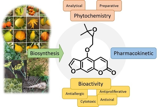
Most of the furanocoumarins have been chemo-taxonomically characterized from the Apiaceae, Rutaceae, and Fabaceae families [5,6]. Oxypeucedanin (OP, C16H14O5, molecular weight: 286.28 g/mol, 4-[(3,3-dimethyloxiran-2-yl)methoxy]furo[3,2-g]chromen-7-one) is a linear furanocoumarin containing an epoxide ring in which, at position 5, 7H-furo[3,2-g][1]benzopyran-7-one is substituted by a [(2S)-3,3-dimethyloxiran-2-yl]methoxy group (Figure 1) [7].
Considering the substantial furanocoumarin contents comprising OP in plant foods [1,8,9,10], and the diverse biological effects on human health, investigations of phytochemicals and their bioactivities are crucial approaches. The present context aims at overviewing all the phytochemical (preparative and analytical) and pharmacological (including pharmacokinetic) studies of OP that have been carried out. “Oxypeucedanin” was applied as a keyword to seek the correlated data through the English-language papers indexed in the Web of Science, SciFinder, and PubMed scientific databases, in which all the plants’ parts were considered (accessed date: 25 May 2021).
2. Phytochemical Studies of Oxypeucedanin
Several experiments have previously been performed to isolate, purify, and identify OP from various plant species entirely belonging to the Apiaceae and Rutaceae families.
Some of the plants possessing OP are being consumed as foods and spices; for instance, the leaves of dill (Anethum graveolens), Ferulago spp., Angelica archangelica, and Prangos spp. (Apiaceae), along with the renowned Citrus fruits (Rutaceae). Traditional applications of the major plants containing OP will be mentioned in the following sections, but for example, the consumption of Angelica dahurica roots as a toothache reliever and for the treatment of the common cold in China, and the use of Prangos leaves as tonic, carminative, and anthelmintic agents in Iran can be mentioned.
High-performance liquid chromatography coupled with both ultraviolet (HPLC-UV) and diode array detectors (HPLC-DAD) have been utilized for analytical assessment of OP from plants, during which the qualification and quantification analysis were mostly accomplished on Angelica dahurica roots (Apiaceae). The following sections present the phytochemical aspects of OP, with detailed information documented in Table 1 and Table 2.
2.1. Preparative Analysis of Oxypeucedanin
2.1.1. Isolation and Purification of Oxypeucedanin from the Apiaceae Family
The Apiaceae (syn. Umbelliferae) family has been identified as one of the major natural sources of coumarin derivatives [11]. So far, OP has been isolated from 23 species belonging to 11 different genera by utilizing diverse separation techniques. Among them, the genera of Angelica, Ferulago, and Prangos have been investigated in six, five, and three phytochemical studies, respectively, leading to isolation of OP. Overall, 50% of the identified OP has been isolated from root parts of the plants, while the best soluble partitions were ethyl acetate (EtOAc, 12 cases), chloroform (CHCl3, 7 cases), and dichloromethane (CH2Cl2, 6 cases). Table 1 documents all the preparative information on OP purified from different plant species.
Anethum graveolens
Oxypeucedanin has been separated from whole part of A. graveolens and its n-hexane (NHEX)-soluble fraction by applying vacuum liquid chromatography (VLC) and reverse-phase (RP) preparative thin-layer chromatography (PTLC) [12].
Angelica spp.
Medium-pressure liquid chromatography (MPLC) on silica gel has previously been exploited to isolate OP from the root CHCl3-soluble fraction of A. archangelica [13]. In traditional Chinese medicine, the roots of A. dahurica are renowned as “Hangbaizhi” or “Baizhi”, and famed for their therapeutic properties, specifically treatment of the common cold and toothaches, and as a food spice [14]. The root part of this species has been described to contain several furanocoumarins; however, among them, the presence of OP was proven by many studies compared to the other natural sources, thus it can be introduced as the major OP origin [15].
Column chromatography on silica gel (CC) has been used for the isolation of OP from the root methanolic fraction of A. dahurica [16,17,18]. This compound has been isolated from ethanolic and n-butanol (BuOH)-soluble partitions of this species by exploiting CC [19] and recrystallization [20], respectively; as well as from the CHCl3 and CH2Cl2 soluble fractions using CC [21] and recrystallization methods [22], and CC on silica gel [23,24], respectively.
Sephadex® LH-20 (SLH) has played a key role in isolation and purification of diverse classes of natural products [25]. This method has further been used for the isolation and separation of coumarins, including OP. Ethyl acetate-soluble extract of A. dahurica root can be considered as the richest OP source. The eluting solvent system comprising NHEX–EtOAc (3:2) through CC on silica gel [26,27], as well as SLH [28] and recrystallization [29], have been applied as the final separation procedures.
High-speed counter-current chromatography (HSCCC) is a liquid stationary-phase chromatographic technique in which the mixtures are separated according to their polarity into two immiscible solvent systems [30]. It also has been utilized as an OP purification instrument, while a two-phase solvent system of NHEX–EtOAc–MeOH–H2O was optimized and employed [31,32,33]. OP also was isolated from nonpolar soluble partitions of A. dahurica using semipreparative HPLC [34] and octadecyl silica gel column chromatography (ODS-CC) [35].
Numerous chromatographic steps were applied to isolate OP from EtOAc extract of A. furcijuga flower, whilst HPLC was chosen as the final stage [36]. OP has been isolated from the root EtOAc fraction of A. koreana [37], as well as from the methanolic and CH2Cl2 soluble partitions of A. pancicii by utilizing flash chromatography (FC) and HPLC [38]. The fruit part of A. purpurascens has been extracted via various organic solvents, and the NHEX fraction was subsequently subjected to CC on silica gel, leading to OP purification [39].
Diplolophium buchanani
Dichloromethane partition of D. buchanani leaf has previously been chromatographed via a final recrystallization method; however, in this study, centrifugal partition chromatography was primarily used [40].
Ducrosia anethifolia
In a phytochemical study, OP was isolated and characterized from the CHCl3-soluble fraction of the aerial part of D. anethifolia by means of CC on silica gel, MPLC, and centrifugal preparative thin-layer chromatography (CPTLC) [41].
Levisticum officinale
The root parts of L. officinale have recently been extracted using different solvents. After solvent–solvent partitioning, the EtOAc fraction was subjected to CC via NHEX–EtOAc (100:0 to 0:100) and EtOAc–MeOH (100:0 to 80:20) as mobile phase systems, and OP was subsequently isolated [42].
Ferulago spp.
Various nonvolatile and volatile components of F. angulata (syn. F. trifida) have been identified as its significant and predominant constituents [43]. The root part was fractionated, the NHEX-soluble partition was chromatographed, and OP was accordingly isolated by preparative thin-layer chromatography (PTLC) [44]. The CHCl3-partition of the root was also separated by CC on silica gel, and OP was purified [45,46]. Oxypeucedanin has been isolated and identified from the NHEX aerial part of F. bernardii, whilst CC on silica gel (mobile phase: PET–EtOAc–MeOH), and lastly recrystallization (from H2O–EtOH), were exploited [47].
The aerial and root parts of F. capillaris have been further subjected to separation of their constituents. Oxypeucedanin was isolated from the NHEX-soluble partition, considering that CC on silica gel with a gradient solvent system including NHEX–EtOAc was applied [48]. OP has further been separated from the PET soluble fraction of the F. humulis rhizome. In this recent study, CC (mobile phase: NHEX‒EtOAc; EtOAc‒MeOH) and PTLC (elution system: cyclohexane‒EtOAc 2:1) were utilized as chromatographic tools [49]. The separation procedure, including CC on silica gel through eluting solvent systems of NHEX–CHCl3 and CHCl3–EtOAc, and recrystallization, led to the isolation of OP from the root EtOAc fraction of F. subvelutina [50].
Harbouria trachypleura
Oxypeucedanin has been isolated from the methanolic fraction of the aerial part of H. trachypleura by applying the following separation techniques: vacuum liquid chromatography (VLC, mobile phase: NHEX–EtOAc), FC (mobile phase: CH2Cl2–Me2CO 97:3), CC (mobile phase: CH2Cl2–Me2CO 97:3 to 4:1), RP-VLC (mobile phase: H2O–MeOH 50:50 to 100:0), and PTLC (mobile phase: CH2Cl2–Me2CO 97:3) [51].
Ostericum koreanum
The dried root of O. koreanum is known as “Osterici radix” in China. It has traditionally been consumed as a natural analgesic agent in oriental medicine [52]. The root parts of O. koreanum has been subjected for the preparative phytochemical investigations. From the CHCl3 and EtOAc-soluble fractions, OP was isolated and identified by using CC on Silica gel, eluting with benzene (Bz)–EtOAc (9:1) [52] and NHEX–EtOAc [53,54], respectively.
Petroselinurn crispurn
Four studies described isolation of OP from the flake and leaf parts of P. crispurn. The diethyl ether (DEE)-soluble partition of this species has been separated via HPLC [55,56], in addition the implementation of CC on Silica gel, SLH, and PTLC on the leaves’ CH2Cl2 [57] and EtOAc-partitions was led to purify OP [58].
Peucedanum spp.
The plants of the Peucedanum genus have been recognized for using in folk medicine to treat joint pain, sore throat, epilepsy, gastrointestinal and respiratory disorders [59]. Counter-current chromatography (CCC) has previously been used to isolate OP from the CH2Cl2-partition of P. cervaria fruit [59]. A gradient solvent system comprising H2O – MeOH was applied for the isolation of OP through semipreparative-HPLC from the root EtOAc-partition of P. ostruthium [60].
Prangos spp.
In addition to various phytochemical reports on P. ferulacea [11,61], Shokoohinia et al. [62] also isolated OP from the root’s Me2CO-extract by utilizing VLC and MPLC. Operation of CC was led to the OP isolation from the CHCl3-soluble partition of P. pabularia root [63] and the P. uloptera NHEX leaf fraction [64].
2.1.2. Isolation and Purification of Oxypeucedanin from the Rutaceae Family
Oxypeucedanin has been isolated and characterized from six species belonging to Rutaceae family; among them, the peel’s essential oils of various Citrus species have been documented as the major source of this furanocoumarin (Table 1).
Citrus spp.
Previously, oxypeucedanin was isolated and identified from the EtOAc extract of C. hystrix fruit by applying first ODS-CC, then PTLC [65]. The essential oils extracted from the C. limon peel has been stated to contain OP. In a study performed by Arimoto et al. [66], using of CC on Silica gel and HPLC, and in the research of Dugo et al. [67] CC on Silica gel and recrystallization were led to isolation and purification of this furanocoumarin; additionally, HSCCC method has been exploited to separate OP from the Key’s (C. aurantifolia) and Persian lemon’s (C. latifolia) volatile oils [68].
Skimmia japonica
Preparative-TLC, and CC on Silica gel (mobile phase: NHEX – CHCl3 – EtOAc 8:1:1, 6:3:1) have previously been applied in the isolation of OP from the leaf’s EtOAc fraction of S. japonica [69].
Zanthoxylum flavum
In another study, an isocratic solvent system using H2O–MeOH (6:4) was employed through an RP-solid-phase extraction (SPE) method, in which OP was isolated and characterized as a pure compound from the root methanolic extract of Z. flavum [70].

Table 1.
Isolated oxypeucedanin from different plant species.
| Plant Family | Plant Species | Subjected Plant Part/Soluble Extract | Method of Isolation/Purification | Reference |
|---|---|---|---|---|
| Apiaceae | Anethum graveolens | WP/NHEX | VLC [NHEX‒EtOAc 100:0 to 90:10], VLC [NHEX–EtOAc 50:50], RP-PTLC [NHEX–EtOAc 60:40] | [12] |
| Angelica archangelica | R/CHCl3 | MPLC [butanone‒CHCl3‒DEE‒NHEX 6.9:1:1.4;90.7% to 48:6.8:10:35.2%], MPLC [THF–Propanol–MeCN–H2O 7.5:1.3:1.7:89.5 to 45.1:7.5:10:37.4] | [13] | |
| Angelica dahurica | R/MeOH | CC [NHEX–EtOH–MeOH], CC [NHEX–EtOH], CC[NHEX–EtOH– MeOH 5:1:1], CC [NHEX–EtOH 1:1] | [16] | |
| R/MeOH | CC [NHEX–EtOAc–MeOH], CC [NHEX–EtOAc–MeOH 5:1:1], CC [NHEX–EtOAc 1:1] | [17] | ||
| R/MeOH | CC [NHEX–EtOAc 5:1, 1:1, 0:1], CC [NHEX–EtOAc 5:1] | [18] | ||
| R/EtOH | CC [NHEX–Me2CO 20:1, 15:1, 10:1, 7:1, 4:1, 2:1, 1:1; CHCl3–MeOH 10:1, 8:1, 6:1, 4:1, 2:1, 1:1], CC [NHEX–Me2CO 20:1, 15:1, 10:1, 8:1, 6:1, 4:1, 2:1], CC [NHEX–Me2CO 14:1, 4:1] | [19] | ||
| R/n-BuOH | CC [NHEX‒Me2CO 20:1 to 2:1], RP-CC [MeOH‒H2O 75:25], RP-HPLC [MeCN‒H2O 40:60], recrys. | [20] | ||
| R/CHCl3 | CC | [21] | ||
| R/CHCl3 | CC [CHCl3‒Me2CO 1:0, 0:1], CC [NHEX-EtOAc], recrys. [DEE‒CH2Cl2] | [22] | ||
| R/CH2Cl2 | CC [NHEX–EtOAc 5:1 to 0:1] | [23] | ||
| R/CH2Cl2 | CC [NHEX–EtOAc], CC [NHEX–EtOAc 3:1] | [24] | ||
| R/EtOAc | CC [NHEX–EtOAc 3:2] | [26,27] | ||
| R/EtOAc | CC [CH2Cl2‒MeOH 100:0 to 0:100], ODS-CC [MeOH‒H2O 50:50 to 95:5], SLH [MeOH] | [28] | ||
| R/EtOAc | HSCCC [NHEX–EtOAc–MeOH–H2O 5:5:4:6] | [31] | ||
| R/EtOAc | HSCCC [NHEX–EtOAc–MeOH–H2O 1:1:1:1, 5:5:4.5:5.5] | [32] | ||
| R/EtOAc | HSCCC [NHEX–EtOAc–MeOH–H2O 1:1:1:1, 5:5:4:6] | [33] | ||
| R/EtOAc | CC [NHEX–EtOAc 20:80 to 0:100], CC [EtOAc–MeOH 100:0 to 0:100], recrys. | [29] | ||
| R/PET | CCC [NHEX–MeOAc–MeCN–H2O 4:3:4:4], HPLC | [34] | ||
| R/NHEX | CC [NHEX–EtOAc], ODS-CC [MeOH–H2O 80:20] | [35] | ||
| R/nd | nd | [15] | ||
| Angelica furcijuga | Fl/EtOAc | CC [NHEX–EtOAc 10:1 to 5:1 to 1:1, to MeOH 100%], RP-CC [MeOH–H2O 70:30 to 80:20 to 90:10 to MeOH 100%], RP-CC [MeOH–H2O 60:40 to 75:25, 0:100], HPLC [MeOH–H2O 75:25] | [36] | |
| Angelica koreana | R/EtOAc | HPLC [MeCN–H2O 30:70] | [37] | |
| Angelica pancicii | R/MeOH R/CH2Cl2 | FC [petrol; DEE], HPLC [MeCN–H2O (HCO2H 2%) 50:50, 65:35] HPLC [MeCN–H2O (HCO2H 2%) 50:50, 65:35] | [38] | |
| Angelica purpurascens | Fr/NHEX | CC [NHEX–EtOAc 100:0 to 0:100], CC [NHEX–EtOAc 80:20] | [39] | |
| Diplolophium buchanani | L/CH2Cl2 | CPC [NHEX–EtOAc–MeOH–H2O 10:5:5:1], recrys. [NHEX–EtOAc] | [40] | |
| Ducrosia anethifolia | AP/CHCl3 | CC [CHCl3–MeOH 100:0 to 20:80], MPLC [NHEX–CH2Cl2 50:50 to 0:100, CH2Cl2–MeOH 100:0 to 100:0], MPLC [NHEX–EtOAc 95:5 to 0:100], CPTLC [NHEX–EtOAc 95:5 to 0:100] | [41] | |
| Ferulago angulate (syn. F. trifida) | R/NHEX | PTLC [CHCl3–Me2CO 95:5] | [44] | |
| R/CHCl3 | CC [CHCl3‒EtOAc 10:0, 5:5], CC [CHCl3‒EtOAc 9:1] | [45,46] | ||
| Ferulogo bernardii | AP/NHEX | CC [PET‒EtOAc‒MeOH 60:40], recrys. [EtOH‒H2O] | [47] | |
| Ferulago capillaris | AP, R/NHEX | CC [NHEX–EtOAc] | [48] | |
| Ferulago humulis | Rhizome/PET | CC [NHEX‒EtOAc; EtOAc‒MeOH], PTLC [cyclohexane‒EtOAc 2:1] | [42] | |
| Ferulago subvelutina | R/EtOAc | CC [NHEX‒CHCl3, CHCl3, CHCl3‒EtOAc], CC [CHCl3‒EtOAc 9:1, EtOAc], recrys. | [49] | |
| Harbouria trachypleura | AP/MeOH | VLC [NHEX‒EtOAc], FC [CH2Cl2–Me2CO 97:3], CC [CH2Cl2–Me2CO 97:3 to 4:1], RP-VLC [MeOH‒H2O 50:50 to 100:0], PTLC [CH2Cl2–Me2CO 97:3] | [50] | |
| Levisticum officinale | R/EtOAc | CC [NHEX–EtOAc 100:0 to 0:100; EtOAc–MeOH 100:0 to 80:20] | [42] | |
| Ostericum koreanum | R/CHCl3 | CC [Bz‒EtOAc 9:1] | [51] | |
| R/EtOAc | CC [NHEX–EtOAc] | [52,53] | ||
| Petroselinurn crispurn | flake/DEE | RP-CC [MeCN–H2O 3:2], HPLC [MeCN–H2O 37:63], HPLC [CHCl3‒MeOH 99.9:0.1] | [54] | |
| L/DEE | CC [NHEX–DEE 1:1], HPLC [MeCN–H2O 35:65] | [55] | ||
| L/EtOAc | nd | [57] | ||
| L/CH2Cl2 | CC [PET–EtOAc 1:0, 9:1, 8:2], CC [CH2Cl2–EtOAc 9:1], CC [CH2Cl2–MeOH 9:1, 8:2, 0:1], SLH [PE–CH2Cl2–MeOH 3:2:1], SLH [PET–CH2Cl2–MeOH 4:2:1], PTLC [PET–EtOAc 7:3] | [56] | ||
| Peucedanum cervaria | Fr/CH2Cl2 | CCC [heptane–EtOAc–MeOH–H2O 3:2:3:2] | [58] | |
| Peucedanum ostruthium | R/EtOAc | HPLC [MeOH–H2O (HOAc 0.1%) 0:100 to 100:0] | [59] | |
| Prangos ferulacea | R/Me2CO | VLC [heptane–EtOAc 10:0 to 0:10], MPLC [heptane–EtOAc 7:3 to 5:5] | [61] | |
| Prangos pabularia | R/CHCl3 | CC [NHEX–EtOAc 20:1; 10:1 to 0:1; EtOAc–MeOH 15:1 to 2:1], CC [NHEX–EtOAc 15:1] | [62] | |
| Prangos uloptera | L/NHEX | CC [NHEX–EtOAc 100:0, 1:99, 5:95, 10:99, 20:80, 40:60, 60:40, 80:20, 100:0, MeOH 100], CC [NHEX–EtOAc 20:80, 0:100, MeOH 100%], PTLC [Me2CO–CHCl3 5:95] | [63] | |
| Rutaceae | Citrus hystrix | Fr/EtOAc | ODS-CC [MeOH–H2O 50:50], PTLC [EtOAc–NHEX 4:1] | [64] |
| Citrus limon | peel/EO | PTLC [EtOAc–Me2CO 95:5], HPLC | [65] | |
| peel/EO | CC [PET–EtOAc 80:20], recrys. | [66] | ||
| Citrus aurantifolia & C. latifolia | nd/EO | HSCCC [NHEX–EtOAc–MeOH–H2O 6:4:5:5] | [67] | |
| Skimmia japonica | L/EtOAc | PTLC [CHCl3–EtOAc 8:2], PTLC [NHEX–CHCl3–EtOAc 7:2:1], CC [NHEX–CHCl3–EtOAc 8:1:1, 7:2:1, 6:3:1] | [68] | |
| Zanthoxylum flavum | R/MeOH | RP-SPE [MeOH–H2O 6:4] | [69] |
AP: aerial part, Bz: benzene, CC: column chromatography on silica gel, CCC: counter-current chromatography, CH2Cl2: dichloromethane, CHCl3: chloroform, CPC: centrifugal partition chromatography, CPTLC: centrifugal preparative thin layer chromatography, EO: essential oil, EtOAc: ethyl acetate, EtOH: ethanol, DEE: diethyl ether, Fl: flower, Fr: fruit, H2O: water, HCO2H: formic acid, HPLC: high-performance liquid chromatography, HSCCC: high-speed counter-current chromatography, L: leaf, Me2CO: Me2CO, MeCN: acetonitrile, MeOAc: methyl acetate, MeOH: methanol, MPLC: medium pressure liquid chromatography, n-BuOH: n-butanol, NHEX: n-hexane; nd: not determined, ODS: octadecyl silica gel, PET: petroleum ether, PTLC: preparative thin layer chromatography, R: root, recrys.: recrystallization, RP: reverse phase, SLH: Sephadex® LH-20, SPE: solid-phase extraction, THF: tetrahydrofuran, VLC: vacuum liquid chromatography, WP: whole part.
2.2. Structural Identification of Oxypeucedanin
Spectroscopic techniques, mainly nuclear magnetic resonance (NMR) comprising carbon-13 (13C), along with one-dimensional (1D) or 2D proton (1H) such as COSY (¹H‒¹H correlation spectroscopy), NOESY (nuclear Overhauser effect spectroscopy), HMBC (heteronuclear multiple bond correlation), and HSQC (heteronuclear single quantum coherence); mass spectrometry (MS); spectrophotometric ultra-violet (UV); and infrared (IR) have been applied in the structure elucidation of OP. Assured determination of physical properties such as melting point plays a role. Furthermore, in some studies, circular dichroism (CD) and optical rotatory power ([α]D) have been utilized to ascertain the absolute stereochemistry.
In accordance with the characteristic signals of furanocoumarins in 1H-NMR, which consists of a pair signals of cis olefinic protons linked α and β to a carbonyl (δ H 6.30 d, J = 10.0 Hz, H-3 and δ H 8.19 dd, J = 10.0, 0.5 Hz, H-4) and signals for furan olefinic protons (δ H 7.60 d, J = 2.0 Hz, H-2′ and δ H 6.94 dd, J = 2.5, 1.0 Hz, H-3′). In the 13C-NMR spectrum, 16 carbons can be detected, whilst regarding its being structurally prenylated, its chemical structure can be interpreted [12].
2.3. Analytical Investigations of Oxypeucedanin
2.3.1. Identification of Oxypeucedanin in the Apiaceae Family
In general, among 24 studies identifying OP from eight species belonging to five genera of Apiaceae, 45% were carried out on Angelica dahurica roots. Moreover, HPLC coupled with UV and DAD detectors have been applied to analyze various soluble fractions of the plant samples specifically the methanolic extracts (12 cases). Table 2 shows the used plant species’ names, instrument, method of identification, and the OP quantities.
Angelica spp.
Hydro-ethanolic (96%) extracts of three Icelandic A. archangelica cultivars have been subjected to fingerprint HPLC-UV analysis of the major components; Accordingly, OP was qualified and quantified (0–6.45 mg/g) [71]. Moreover, the root and leaf methanolic extract was quantitatively analyzed by applying HPLC tandem MS (mass spectrometry) [72].
Considering that the root (syn. radix) part of A. dahurica has been described as having the richest OP content, in order to explore the optimized identification method, several analytical investigations were previously carried out to qualification and quantitation assessment of this compound. Overall, OP has mostly been identified from the methanolic extract, whereas the hydro-alcoholic fractions were also asserted. In a study reported by Fan et al. [14], reverse-phase rapid-resolution liquid chromatography (RRLC) led to OP qualification in the A. dahurica radix methanolic extract, possessing quantities of 0.066 to 1.45 mg/g.
In order to analyze the predominant secondary metabolites in the radix methanolic fraction of A. dahurica, exploiting HPLC-UV led to qualifying and quantifying OP, with quantities 0.81 µg/mL [73] and 22.30 µg/g [74]. Oxypeucedanin was quantified from the root methanolic fractions with the highest content among the studied furanocoumarins (xanthotoxin, bergapten, imperatorin, phellopterin, and isoimperatorin) at different plant growth stages, in amounts 1.5–3.0 mg/g [75]. Rising the MeOH ratio in H2O (60 to 100%) as the eluent system via HPLC-DAD led to measurement of the highest and lowest OP content in the root methanolic specimens dried by freezing and at 70 °C, respectively [76].
Supercritical fluid chromatography (SFC) has been utilized to analyze the methanolic partitions of the five root samples of A. dahurica, and OP was subsequently quantified at 1.54–2.93 g/100 g [77]. In a recent study performed by Yang et al. [78], quantitative 1H-NMR was exploited to examine 11 Chinese plant samples, and the OP amount ranged from 0.17 to 0.35%.
The radix hydro-ethanolic (70%) extract of A. dahurica has been subjected to HPLC-DAD analysis, and the OP content was recorded at 1.24 to 4.98 mg/g among 19 plant batches [79]. The same plant fraction was characterized by HPLC-UV, and OP quantities were assessed as 0.063 and 0.024% in the harvested samples from Korea and China, respectively [80]. In a rapid identification study by developing LC-NMR-MS, OP was qualified in the root ethanolic extract of A. dahurica [81].
Yu et al. [82] studied the bitter compounds (six coumarins) of A. dahurica root by using HPLC-DAD-ESI (electrospray ionization)-MS. The plant materials were prepared via boiling in H2O and frying in soybean oil, and OP contents were found to be 15 and 8 µg/mL, respectively.
Ostericum koreanum
Two samples of O. koreanum roots have previously been collected from different locations in South Korea, extracted with MeOH, and subjected to HPLC-UV [83]. In another experiment, a hydro-ethanolic (70%) fraction of O. koreanum was subjected to an HPLC-UV fingerprint measurement. The OP quantities were recorded 0.70 to 21.11% and 1.02 to 12.60% in nine and five samples harvested from Korea and China, respectively [53].
Petroselinum crispum
In a study carried out by Caboni et al. [84], OP was quantified at 46.04 mg/kg from the aerial part methanolic extract of parsley (P. crispum) via HPLC-QTOF (quadrupole and time-of-flight analyzer)-MS. HPLC-UV also has been applied in qualification and quantification assessment of the curled leaf, root, and flake parts of parsley EtOAc extracts. Consequently, OP content was recorded with a remarkable amount in the leaf of up to 102.87 µg/g [55].
Peucedanum spp.
In a preliminary computer-assisted qualification study accomplished by using ChromSword® to construct QSRR (quantitative structure–retention relationships), the PET-soluble partition of the P. alsaticum fruit part has been chromatographed by means of UPLC (ultra-performance liquid chromatography), and OP was qualitatively characterized [85].
Among seven P. ostruthium rhizome samples collected from Austria and Germany, OP content ranged from 1.58 to 25.05 mg/g in the CH2Cl2 extracts after utilizing HPLC-DAD-MS [86]. Furthermore, OP was identified in the EtOAc root fraction by applying HPLC-DAD and HPLC-UV-ESI-MS [59].
Yrjönen et al. [87] experimented with 132 root, stem, leaf, and umbel specimens of P. palustre at flowering stages harvested from 43 Finland locations by means of HPLC-DAD-ESI-HR (high-resolution)-MS. Subsequently, OP was analyzed as the dominant compound of the root samples (24.3 mg/g), compared to the umbel (22.8 mg/g), stem (2.62 mg/g), and leaf (2.25 mg/g) parts. Oxypeucedanin also has been qualified in the root and umbel methanolic fractions of this species by applying HPLC-MS eluting with three solvent systems consisting of H2O and MeOH (gradient and isocratic) as mobile phase [72]. Nonpolar soluble fraction of the P. palustre root was furtherly characterized to possess OP, indicating quantities of 0.16 to 0.44 mg/100 g via HPLC-DAD on six Finnish samples [88].
Prangos ferulacea
The impact of three extraction methods, including Soxhlet, ultrasonic-assisted extraction (UAE), and maceration on three coumarin contents of P. ferulacea root was elaborated comparably. The HPLC-UV results revealed that the highest OP quantity was determined in the samples extracted via UAE, at 79.27 mg/g [89].
Table 2.
Qualification and quantification fingerprint analysis of oxypeucedanin in different plant species.
Table 2.
Qualification and quantification fingerprint analysis of oxypeucedanin in different plant species.
| Plant Family/Herbal Product | Plant Species | Subjected Plant Part/Soluble Extract | Analytical Instrument | Eluent System for Chromatography | Quantity | Reference |
|---|---|---|---|---|---|---|
| Apiaceae | Angelica archangelica | Fr/hydro-ethanolic (96%) | HPLC-UV | H2O‒MeOH [40:60 to 5:95] | 0‒6.45 mg/g | [71] |
| R, L/MeOH | HPLC-MS | H2O (1% HCO2H)‒MeOH [40:60]; H2O (0.1% HCO2H)‒MeOH [100:0 to 70:30, 40:60, 0:100, 100:0] | na | [72] | ||
| Angelica dahurica | Radix/MeOH | RRLC | H2O‒MeOH [55:45, 50:50, 42:58, 36:64, 30:70, 20:80] | 0.066‒1.45 mg/g | [14] | |
| Radix/MeOH | HPLC-UV | H2O (0.2% H3PO4)‒MeOH [52:48, 40:60, 55:45, 48:52, 25:75] | 0.816 µg/mL (by IL-DLLME) | [73] | ||
| Radix/MeOH | HPLC-UV | H2O‒MeCN [70:30, 40:60, 30:70, 40:60, 70:30] | 22.30 µg/g | [74] | ||
| R/MeOH | HPLC-DAD | H2O‒MeOH [40:60, 20:80, 10:90, 0:100] | 1.5–3.0 mg/g | [75] | ||
| R/MeOH | HPLC-DAD | H2O‒MeOH [40:60, 20:80, 10:90, 0:100] | 36.95 ± 1.45 (freeze dried) 24.96 ± 0.75 (shade dried) 24.22 ± 1.75 (40 °C) 32.13 ± 1.42 (70 °C) | [76] | ||
| R/MeOH | SFC | CO2‒MeOH (0.1% DEA) [100:0, 97:3, 90:10] | 1.54–2.93 g/100 g (0.16‒0.77%) | [77] | ||
| nd/MeOH | 1H-qNMR | solvent: DMSO-d6 | 6.38–6.39 ppm (0.17–0.35%) | [78] | ||
| Radix/hydro-ethanolic (70%) | HPLC-DAD | H2O (0.1% HCO2H)‒MeOH [95:5, 35:65, 5:95] (for qualification) H2O (0.1% HCO2H)‒MeOH [70:30, 40:60, 40, 5:95] (for quantification) | 1.24–4.98 mg/g | [79] | ||
| Radix/hydro-ethanolic (70%) | HPLC-UV | H2O‒MeCN [30:70] | 0.063 ± 0.01 % (collected from Korea) 0.024 ± 0.02 % (collected from China) | [80] | ||
| R/EtOH | LC-NMR-MS | H2O‒MeCN [40:60] | na | [81] | ||
| R/H2O (for boiled sample) and soybean oil, MeOH (for fried sample) | HPLC-DAD-ESI-MS | H2O (0.1% HCO2H)‒MeOH [for boiled sample: 95:5, 75:25, 26:64, 95:5] [for fried sample: 95:5, 85:15, 77:23, 47:53, 20:80, 95:5] | 15 µg/mL (for boiled sample) 8 µg/mL (for fried sample) | [82] | ||
| Ostericum koreanum | R/MeOH | HPLC-UV | H2O‒MeCN [65:35 to 25:75] | 0.57 ± 0.26 0.99 ± 0.89 | [83] | |
| nd/hydro-ethanolic (70%) | HPLC-UV | H2O‒MeCN [40:60] | 0.70 ± 0.02–21.11 ± 0.07% (Korean samples) 1.02 ± 0.01–12.60 ± 0.10% (Chinese samples) | [53] | ||
| Petroselinum crispum | AP/MeOH | HPLC-QTOF-MS | H2O (0.1% HCO2H)‒MeCN [90:10, 60:40, 20:80, 10:90, 0:100, 90:10] | 46.04 ± 5.50 mg/kg | [84] | |
| L, R, flake/EtOAc | HPLC-UV | cyclohexane‒isopropyl ether‒n-amyl alcohol [15:4:0.5] | 102.87 ± 14.08 µg/g (curled L) 88.68 ± 6.04 µg/g (curled R) 88.60 ± 17.90 µg/g (flake) | [55] | ||
| Peucedanum alsaticum | Fr/PET | UPLC | H2O‒MeCN [74:26 to 55:45] | na | [85] | |
| Peucedanum ostruthium | Rh/CH2Cl2 | HPLC-DAD-MS | H2O (0.01% HOAc)‒MeCN (0.01% HOAc) [75:25 to 63:37 to 55:45 to 35:65 to 5:95] | 1.58 ± 0.03–25.05 ± 0.11 mg/g | [86] | |
| R/EtOAc | HPLC-DAD (RP-C30) HPLC-UV-ESI-MS | H2O (0.1% HOAc)‒MeOH [100:0 to 0:100] | na | [59] | ||
| Peucedanum palustre | R, St, L, umbel/MeOH | HPLC-DAD-ESI-HR-MS | H2O (0.01 M HCO2H)‒MeCN [100:0 to 40:60, 10:90] | 24.3 ± 14.0 mg/g (in R) 2.62 ± 1.56 mg/g (in St) 2.25 ± 1.28 mg/g (in L) 22.8 ± 30.9 mg/g (in umbel) | [87] | |
| R, umbel/MeOH | HPLC-MS | H2O (1% HCO2H)‒MeOH [40:60] H2O‒MeOH (1% HCO2H) [71:29, 0:100], H2O‒MeOH (1% HCO2H) [100:0 to 70:30, 40:60, 0:100, 100:0] | nd | [72] | ||
| R/NHEX | HPLC-DAD | THF‒MeCN‒MeOH‒H2O [3.1:35:5.4:56.5] | 0.16–0.44 mg/100 g | [88] | ||
| Prangos ferulacea | R/NHEX, hydro-ethanolic (95%), MeOH | HPLC-UV | H2O‒MeOH [30:70] | 59.38 ± 0.007 mg/g (extraction with Soxhlet, NHEX) 79.27 ± 0.22 mg/g (extraction with UAE, hydro-ethanolic 95%) 55.29 ± 0.01 mg/g (extraction with maceration, MeOH) | [89] | |
| Rutaceae | Atalantia ceylanica | Se/MeOH | HPLC-UV | nd | na | [90] |
| Citrus spp. | EO | HPLC-UV | NHEX‒isopropanol [98:2] | 0.7–1.65 g/L (Lemon EO from Sicily) 0.95 g/L (Lemon EO from Spain) 2.02 g/L (Lemon EO from Argentina) 0.49 g/L (Lime EO from Mexican 1) 0.96 g/L (Lime EO from Mexican 2) 0.68 g/L (Lime EO from Iran) | [91] | |
| EO | HPTLC | CH2Cl2‒DEE [100:3], CHCl3‒heptane [95:5] | na | [92] | ||
| EO | HPLC-UV | NHEX + EtOAc (92:8)‒NHEX + EtOH (90:10) | 1.55 ± 0.38 g/kg (extracted EO by Sfumatrice technology) 2.2 ± 0.41 g/kg (extracted EO by Pelatrice technology) 1.9 ± 0.45 g/kg (extracted EO by FMC technology) 0.86 ± 0.26 g/kg (extracted EO by Torchi technology) | [93] | ||
| Citrus aurantifolia & C. latifolia | EO | HPLC-DAD | H2O + MeOH + THF (85:10:5)‒MeOH + THF (95:5) [100:0, 60:40, 10:90, 100:0] | 4.09–10.54 g/L (C. aurantifolia) 0.27–10.72 g/L (C. latifolia) | [94] | |
| EO | HPLC-UV | NHEX + EtOAc (93:7)‒NHEX + EtOH (90:10) [100:0 to 5:95, 100:0] | 144 mg/100 g (C. aurantifolia) 210–328 mg/100 g (C. latifolia) | [95] | ||
| Citrus aurantifolia & C. latifolia & C. paradisi | EO | HPLC-DAD | H2O‒MeCN [70:30 to 60:40] | na | [67] | |
| Citrus limon | Wax/EO | UHPLC-DAD | H2O (0.1% HCO2H)‒MeCN [25:75, 100:0, 50:50, 30:70] | 62 ± 0.8 mg/kg | [96] | |
| EO | HPLC-DAD | NHEX + EtOAc (92:8)‒NHEX + EtOH (90:10) [100:0 to 0:100] | 89–157 mg/100 g | [66] | ||
| Citrus medica | Fr/EO | HPLC-DAD | H2O‒MeCN [70:30, 40:60, 0:100, 70:30] | 2.03–21.30 g/100 g | [97] | |
| Eureka limon | peel/EO | GC-MS | - | na | [98] | |
| Yuanhu zhitong (Chinese herbal drug) | Hydro-methanolic (75%) | UPLC-Q-TOF-MS | H2O (0.2% HCO2H)‒MeCN | na | [99] | |
| Hydro-methanolic (75%) | RRLC-QQQ | H2O (0.3% HCO2H)‒MeCN [80:20, 60:40, 20:80] | 0.12–4.01 µg/g | [100] |
1H-qNMR: quantitative 1H NMR, AAc: ammonium acetate, AF: ammonium format, AP: aerial part, CH2Cl2: CH2Cl2, CHCl3: chloroform, CO2: carbon dioxide, Cmax: maximum concentration, DAD: diode array detector, DEA: diethyl amine, DEE: diethyl ether, DMSO: dimethyl sulfoxide, EO: essential oil, ELSD: evaporative light-scattering detector, ESI: electrospray ionization, EtOAc: ethyl acetate, EtOH: ethanol, Fr: fruit, GC: gas chromatography, H3PO4: phosphoric acid, H2O: water, HCO2H: formic acid, HOAc: acetic acid, HR: high resolution, HPLC: high-performance liquid chromatography, IL-DLLME: ionic liquid dispersive liquid–liquid microextraction, L: leaf, LC: liquid chromatography, MeCN: acetonitrile, MeOH: methanol, MS: mass spectrometry, NHEX: n-hexane, na: not analyzed, NMR: nuclear magnetic resonance, PET: petroleum ether, QTOF: quadrupole and a time-of-flight analyzer, R: root, RP: reverse phase, Rh: rhizome, RRLC: rapid-resolution liquid chromatography, RRLC-QQQ: rapid-resolution liquid chromatography-triple quadrupole mass spectrometry, Se: seed, SFC: supercritical fluid chromatography, St: stem, THF: tetrahydrofuran, TNBS: trinitrobenzenesulfonic acid, UAE: ultrasound assisted extraction, UPLC: ultra-performance liquid chromatography, UV: ultraviolet-Visible.
2.3.2. Characterization of Oxypeucedanin in the Rutaceae Family
As depicted in Table 2, oxypeucedanin has predominantly been qualified from the essential oils of Citrus spp.; however its peel, wax, and fruit parts were also rich in OP. An HPLC instrument coupled with DAD and UV as detectors was utilized as the main analytical tool to characterize its content. The OP contents were richest in the Citrus limon and C. latifolia essential oils.
Atalantia ceylanica
Oxypeucedanin, along with the linear furanocoumarins bergapten, xanthotoxin, heraclenin, and imperatorin, as well as two novel oximes, have previously been identified in the seed methanolic extract of A. ceylanica by means of HPLC-UV; however, no more information of the chromatographic conditions and OP content has been reported [90].
Citrus spp.
Essential oils extracted from plants of the genus Citrus can be considered as the richest samples among the Rutaceae family in terms of the OP occurrence. Dugo et al. [91] quantitatively and qualitatively analyzed OP content by applying HPLC-UV through an isocratic solvent mixture of NHEX–isopropanol (98:2) as the mobile phase. The highest and lowest OP concentrations were determined in lemon oil harvested from Argentina and lime oil from Mexico, at 2.02 and 0.49 g/L, respectively. HPTLC (high-performance thin-layer chromatography) also was used to analytically qualify OP in Citrus spp. oil, while the mobile phases were CH2Cl2–DEE (100:3) and CHCl3–heptane (95:5) [92].
In a similar study performed by Verzera et al. [93], four Citrus spp. essential oils were extracted through diverse technologies and subjected to an analytical HPLC-UV eluting with the combined solvents of NHEX–EtOAc (92:8) and NHEX–EtOH (90:10) as the mobile phases. Overall, the samples extracted with Pelatrice technology indicated the highest OP content (2.2 g/kg).
Volatile oils of Citrus latifolia (Persian lemon) and C. aurantifolia (Key lime) have further been characterized for their coumarin contents, including OP. In an analytical experiment, HPLC-DAD was employed by using a rising ratio of MeOH + THF (95:5) from 0 to 90% in H2O + MeOH + THF (85:10:5) to detect OP at quantities of 4.09–10.54 g/L and 0.27–10.72 g/L in the selected Key and Persian lime samples, respectively [94]. The oils of one Key and seven Persian lime samples have previously been extracted to analytically test their OP content. HPLC-UV eluting via the mixture of solvents NHEX + EtOAc (93:7) and NHEX + EtOH (90:10) from 100:0 to 5:95 led to quantifying OP contents of 144 mg/100 g and 210–328 mg/100 g in the Key and Persian lime specimens, respectively [95].
In another investigation, OP was qualified in the essential oils extracted from Key and Persian limes, and grapefruit (C. paradisi), and H2O in MeCN (70:30 to 60:40) was applied as a gradient mobile phase through an HPLC-DAD instrument [67]. Ultra-HPLC (UHPLC) coupled with DAD has previously been exploited to analyze the extracted fruit oils of C. limon harvested from Sicily, Italy—locally named as “Diamante citron”; whilst by increasing concentration of MeCN in H2O (0.1% HCO2H) as the mobile phase, OP was qualified and quantified (62 mg/kg) in this plant material [96]. A similar study was accomplished by hiring a gradient binary solvent system of NHEX + EtOAc (92:8) in NHEX + EtOH (90:10) via HPLC-DAD, leading to the identification of bergamottin as the major coumarin compound (160 mg/100 g) among 37 industrial cold-pressed Italian lemon oils; however, OP was also quantified with remarkable amounts of 89–157 mg/g [66].
The essential oils extracted from nine green Italian C. medica fruit samples also have been studied by exploiting HPLC-DAD through mobile phase H2O–MeCN (70:30 to 0:100) as the eluent system, and OP concentration was quantified at 2.03–21.3 g/100 g [97]. Barth et al. [98] further experimentally analyzed the Eureka limon peel’s essential oil in samples collected from Ivory Coast in France by application of GC-MS. Oxypeucedanin could be detected in the residue part, which was gained from the remaining materials after washing the SFC column with EtOH.
2.3.3. Identification of Oxypeucedanin from Other Natural Sources
The Chinese herbal drug “Yuanhu zhitong” is combined of several medicinal herbs, mainly Angelica dahurica radix (baizhi) and Corydalis rhizome (yanhusuo). Its healing applications in the treatment of costalgia, gastralgia, dysmenorrheal, and headache have been documented in China [99].
In two experiments, its hydro-ethanolic (75%) extract was prepared and subjected to an assessment of its major components. In a study by Xu et al. [99], OP was characterized by applying UPLC-Q-TOF-MS in 15 drug samples, whilst H2O (0.2% HCO2H) and MeCN were employed in the mobile phase system. Moreover, rapid-resolution liquid chromatography-tandem triple quadrupole mass spectrometry (RRLC-QQQ) was employed to qualify and quantify the assessment of 17 compounds in 15 drug samples. Enhancing the ratio of MeCN from 20 to 80% in H2O (0.3% HCO2H) was chosen as the optimized eluent system, leading to quantify OP varying from 0.12 to 4.01 µg/g [100].
3. Biological Activities of Oxypeucedanin
So far, numerous preclinical biological properties of OP have been determined in vitro and in vivo. Most of these studies were focused on in vitro assessment of cytotoxic (n: 9); anti-inflammatory, antimicrobial, and antioxidant (n: 5); and antiproliferative and acetylcholinesterase inhibitory (AChE) (n: 4) activities, taking into account that due to the probable toxicity of this compound, its efficacy had not been elaborated in clinical trials yet.
In brief, OP proved good antifeedant activities against Spodoptera littoralis and S. litura larvae in the tested dosages. Moreover, it indicated potent antiallergic inflammation (histamine H1 receptor antagonist), antibiofilm activity against Pseudomonas aeruginosa PAO1, considerable antiproliferative effects on HTC15 (colon cancer), and UVA-irradiated B16F10 (melanoma) cell lines, in addition to high anti-influenza potencies against influenza A-H1N1 and influenza A-H9N2 (higher than ribavirin as the positive control), significant cytotoxic activity against LNCaP (androgen-sensitive human prostate adenocarcinoma) compared to finasteride, and remarkable synergistic activity with docetaxel (anticancer drug). Table S1 illustrates the comprehensive biological information of OP, including the activities, experimented media, applied assays, positive controls, and cell lines/strains.
3.1. Antiallergic Activity
In an in vivo study, OP showed a weak inhibitory effect on compound 48/80 (condensation products of N-methyl-p-methoxy phenylethylamine with formaldehyde)-induced histamine in mouse peritoneal, and no antiallergic activity in the analyzed dosages was observed. Nevertheless, the histamine contents at doses of 10 and 20 mg/kg were assessed 12.6 ± 1.75 and 12.2 ± 1.00 µg/100 mL of cavity fluids, respectively [26].
3.2. Antiarrhythmic Activity
Application of an hKv1.5 channel inhibitory assay indicated that OP prolonged the cardiac action duration of rat atrial and ventricular muscles in a dose-dependent manner. The IC50 determined was 76.12 ± 8.07 nM, while it had no effect on human Eag-related gene (HERG) current [16].
3.3. Anticonvulsant Activity
No anticonvulsant effect was recorded for OP at a concentration of 300 mg/kg, performed using maximal electroshock-induced seizure in mice model (in vivo) [101]. In another experiment on 18 coumarin derivatives, the modulation of GABA-induced chloride currents (IGABA) on recombinant α1β2γ2S GABAA receptors expressed in Xenopus laevis oocytes were analyzed. Accordingly, an EC50 value of 26 ± 8 μM and IGABA of 550 ± 71% (at 100 μM) were measured for OP [102].
3.4. Antifeedant Activity
The leaf-disk bioassay has previously been utilized for evaluation antifeedant activity of OP. It revealed inhibitory effects against Spodoptera littoralis larvae, with ED50 of 41.92 ± 18.74 mg/L [103]. In a bioassay-guided isolation of antifeedant compounds from Skimmia japonica, the antifeedant index was calculated at 19.83 ± 6.91% at a concentration of 1 mg/mL of OP against Spodoptera litura larvae, in which the control group demonstrated feeding-deterrent activity, with an index of 23.62 ± 9.64% [68].
3.5. Antigenotoxic Activity
Marumoto et al. [104] investigated antigenotoxic impacts of several natural coumarins by exploiting Umu Chromotest™ in vitro. No genotoxic activity was detected for OP against carcinogens induced by AF-2 (furylfuramide) and MNNG (N-methyl-N’-nitro-N-nitrosoguanidine). The OP antigenotoxicity also was determined against procarcinogen of PBTA-4 (2-[2-(acetylamino)24-amino-5-methoxyphenyl]25-amino-7-bromo-4-chloro-2H-benzotriazole, at 26 µM in the presence of rat S9 mix with IC50 of 1.69 ± 0.28 µM; whereas on the genotoxic activation of PBTA-4 and MeIQ (2-amino-3,4-di-methylimidazo[4,5-f]quinoline) catalyzed by rat and human CYP1A1 enzymes, IC50 were 0.24 ± 0.08 and 30.14 ± 0.08 µM, respectively. Moreover, OP was efficient against MeIQ (0.016 µM) on the above-mentioned enzymes.
3.6. Anti-Inflammatory Activity
In an in vitro experiment, using a NO production assay, no anti-inflammatory activity was identified for OP in the culture medium of LPS (10 µg/mL)/IFN-γ (10 units/mL)-stimulated RAW 264.7 cell line (murine macrophage) [81]. In a study carried out by Li and Wu [28], OP exhibited a potent histamine H1 receptor antagonist via virtual screening by a docking program. This compound demonstrated antiallergic inflammation by reducing histamine release. Moreover, OP inhibited the secretion of tumor necrosis factor-α (TNF-α), interleukin (IL)-1β, and IL-4 in RBL-2H3 cells (rat basophilic leukemia), possessing secretion concentrations of 100, 120, and 39 pg/mL, respectively; whereas DNP-HAS (dinitrophenyl-human serum albumin) was applied as a positive control, with secretion of 160, 240, and 55 pg/mL, respectively [28].
L-NG-Nitro arginine methyl ester (L-NAME, 100 mM) has been applied as positive control in a NO production assay against lipopolysaccharide (LPS)-activated RAW 264.7 macrophage cells; OP demonstrated a moderate anti-inflammatory activity, with IC50 of 16.8 µg/mL [15]. Using the same method, OP showed anti-inflammatory potency (IC50: 57 µM) in LPS-activated mouse peritoneal macrophage cell, while the activity was lower than L-NMMA (NG-monomethyl-L-arginine) with IC50 28 µM [36]. Moreover, Murakami et al. [64] illustrated a weak NO accumulation inhibitory activity of OP against RAW 264.7 macrophage cells, possessing IC50 of 310 µM, compared to N-(iminoethyl)-L-ornithine with IC50 value 7.9 µM, applied as the positive control.
3.7. Antimalarial Activity
3.8. Antimicrobial Activity
Mileski et al. [38] elaborated antibacterial potency of OP against several Gram-negative and Gram-positive bacteria by using a microbroth dilution method. The highest effect was detected against Bacillus cereus, with MIC and MBC values of 2.00 ± 0.03 and 4.00 ± 0.06 mg/mL, respectively; whilst the antibacterial drug streptomycin exhibited 0.09 ± 0.00 and 0.37 ± 0.02 mg/mL, respectively. In the same study, antiquorum sensing and antibiofilm activities of different extracts of Angelica pancicii and its isolated compounds were used against Pseudomonas aeruginosa PAO1. The maximum diameter reduction was recorded in the presence of OP among all samples. This furanocoumarin presented a lower colony diameter of 8.66 ± 4.04 mm, compared to streptomycin and ampicillin, with 11.00 ± 1.00 and 13.33 ± 5.03 mm, respectively [38].
Oxypeucedanin revealed no antifungal activity against three plant pathogens comprising the bacteria Xanthomonas compestris Erwinia cartovorum, and a fungus Sclerotinia sclerotorium by applying a disk diffusion assay [63]. A microbroth dilution assay was used to assess antimycobacterial activity of OP isolated from dill (Anethum graveolens) against five microorganisms. The highest potencies were measured against Mycobacterium smegmatis and M. aurum, each with the same MIC value of 32 μg/mL, in comparison with ethambutol (positive control), possessing an MIC of 0.5 μg/mL [12]. Moreover, Tavakoli et al. [46] reported an antibacterial effect of OP (concentration: 50 μg/disk) against Klebsiella pneumoniae and Shigella dysenteriae, indicating inhibition zones of 13 mm evaluated by disk diffusion assay.
OP indicated moderate effects among the compounds isolated from Levisticum officinale against several bacteria pathogens. When a microbroth dilution method was applied, it revealed potencies, with MIC values of 64 and 256 against multidrug-resistant Mycobacterium tuberculosis and Staphylococcus aureus, respectively, although it exhibited weaker effects compared to the applied positive controls [70].
3.9. Antioxidant Activity
Oxypeucedanin isolated from five plant species has been evaluated for its ability to scavenge free radicals. A DPPH (2,2-diphenyl-1-picrylhydrazyl) assay displayed antiradical activity of OP identified in Angelica dahurica, with an IC50 value higher than 200 μg/mL [23]. This bioassay also has been utilized to evaluate the antiradical potency of OP extracted from Ferulago subvelutina. It exhibited a weak activity (IC50: 217 μg/mL) compared to BHT (butylated hydroxytoluene, IC50: 27 μg/mL) as the reference standard [54]. Furthermore, by applying the FRAP (ferric-reducing antioxidant power) method, OP isolated from F. trifida showed an effect of 9.15 ± 1.7 mm FSE (ferrous sulphate equivalents)/100 g; however, BHT indicated a greater effect of 267.2 ± 1.7 mm FSE/100 g [46].
Razavi et al. [63] reported an antiradical activity of OP with an RC50 value of 51.25 mg/mL, determined with DPPH assay. OP also indicated remarkable antioxidant activity among all 14 isolated compounds from Zanthoxylum flavum, possessing an IC50 of 8.3 μg/mL, compared to ascorbic acid (IC50: 1.4 μg/mL), when the cell-based DCFH-DA (dichloro-dihydro-fluorescein diacetate) method was employed [69].
3.10. Antiproliferative Activity
In a study by Kim et al. [35], the SRB (sulforhodamine B) assay was exploited to investigate in vitro cell-proliferation activity of OP against five selected cell lines. In a dose-dependent manner, the highest potency was detected on HTC15 (colon cancer cell line) with an ED50 of 3.4 ± 0.3 μg/mL, whilst cisplatin showed an ED50 of 2.2 ± 0.4 μg/mL as the positive control.
Psoralen (IC50 of 0.11 μM) has previously been used as positive control for assessing the in vitro antiproliferative activity against the UVA-irradiated B16F10 melanoma cell line, and OP demonstrated an effect, with an IC50 0.22 μM. These promising findings can be considered for further OP application in the treatment of photo-dermatosis disorders. Moreover, in this study, OP’s impact on the aforementioned cell line was evaluated in vivo in a mouse model as well. Interestingly, OP reduced tumor volume from 2200 (in control group) to 500 mm3 after 20 days of treatment (0.5 mg/kg), along with decreasing the tumor weight from 2000 mg to 250 mg (at 0.5 mg/kg of administration) [27].
Five cancer cell lines comprising MDA-MB-231 (breast cancer cell), T47D (breast cancer cell), SNU638 (stomach cancer cell), SK-Hep-1 (human hepatoma cell), and A549 (lung cancer cell) were subjected to assess proliferation activity of OP by utilization of an SRB assay. Among them, OP showed the highest potency against SK-Hep-1 cells, with an IC50 of 32.4 μM; however, it was lower than etoposide as the control (IC50: 0.3 μM). The authors asserted that this activity may be correlated with the induction of G2/M phase cell-cycle arrest and upregulation of the p53/MDM2/p21 axis in SK-HEP-1 hepatoma cells by OP after 72 h of treatment [19].
In our previous study, doxorubicin, a chemotherapy medication, has been applied as the positive control to study the antiproliferative effect of the isolated furanocoumarins from Ducrosia anethifolia via an MTT assay. Moderate effects of OP were observed against PAR (L5178Y mouse T-cell lymphoma) and MDR (ABCB1-expressing L5178Y) cell lines, possessing IC50s of 25.98 and 28.89 µM, respectively; while doxorubicin showed stronger potencies, with IC50s of 0.05 and 0.46 µM, respectively. Oxypeucedanin also exhibited a slight synergistic effect with doxorubicin when using a checkerboard combination assay against the MDR cell line with CI (combination index), with an ED50 of 0.85 ± 0.07 at 1:50 ratio [41].
3.11. Antiviral Activity
Oxypeucedanin isolated from Angelica dahurica notably exhibited higher anti-influenza activity compared to the control drug. In this study, administration of OP (20 μM, after 2 h) considerably decreased the levels of viral proteins NP (nucleoprotein) and NA (neuraminidase) in the treated cells to less than 20% via inhibition of their synthesis. The EC50 values were measured at 5.98 ± 0.71 and 4.52 ± 0.39 μM against influenza A-H1N1 and influenza A-H9N2, respectively; while ribavirin possessed 6.29 ± 0.89 and 6.13 ± 0.19 μM, respectively [20]. Nonetheless, in accordance with Shokoohinia et al. [61], no anti-HSV (antiherpes) activity was detected for OP against the Vero cell line (African monkey kidney).
3.12. Calcium Antagonistic Activity
Calcium antagonistic activity of 15 compounds isolated from A. dahurica has previously been evaluated by measuring the depolarization-induced 45Ca2+ uptake in clonal GH4C1 rat pituitary cells, and OP showed an inhibitory effect of 43 ± 4.3% at a concentration of 20 μg/mL [13].
3.13. Cytotoxic Activity
An MTT assay has been used to evaluate cytotoxic activities of certain coumarins on tacrine-induced cytotoxicity in Hep-G2 cells. OP indicated no cytotoxicity, while the other studied coumarins (imperatorin, byakangelicin, and byakangelicol) were active. The EC50 of OP was found to be 286.7 ± 6.36 µM, a lower effect than silybin (EC50: 69.0 ± 3.4 µM) as the control [18]. Moderate cytotoxic activities of OP were determined by means of an MTT assay (in vitro) against four tested cancer cell lines: L1210 (murine leukemia), HL-60 (human leukemia), K562 (human leukemia), and B16F10 (murine melanoma). Although two of OP’s hydrated derivatives, namely pangelin and oxypeucedanin hydrate acetonide, indicated the highest effects, OP with an IC50 of 27.5 µg/mL showed weaker cytotoxicity than Adriamycin (syn. doxorubicin, IC50: 2.8 µg/mL) against the HL-60 cell line [24].
In a further study, OP isolated from A. koreanum displayed an equal cytotoxic activity with finasteride (control drug), both possessing IC50 values >20 µg/mL against the LNCaP cell line (androgen-sensitive human prostate adenocarcinoma) [37]. Nevertheless, doxorubicin as the chemotherapy drug showed higher in vitro cytotoxic effects on three experimented cancer cells, and OP was the most potent furanocoumarin derivative against parent (PAR, IC50: 40.33 ± 0.63 µM)and multidrug-resistant (MDR, IC50: 66.68 ± 0.00 µM) cells, and had slight toxicity on normal murine fibroblasts (NIH/3T3, IC50: 57.18 ± 3.91 µM) [41]. The highest OP cytotoxicity was recorded against the A-549 cell line (lung carcinoma), with an IC50 of 0.80 mM among four tested cells, compared to the positive control (tamoxifen, IC50: 0.017 mM) [46].
The human prostate carcinoma cell (DU145) has been used to assess OP’s cytotoxicity. Results demonstrated that this compound significantly inhibited cell growth, with a maximum of 93.3% (at 100 µM after 72 h treatment) by G2-M cell cycle arrest, and induced apoptotic cell death up to 50% (at 100 µM after 72 h treatment) [52]. By applying an MTT assay, Choi et al. [51] exhibited that OP was not able to reduce cell viability up to 100 μM for 24 h against mice neuroblastoma neuro-2A cells. A higher CC50 (50% cytotoxic concentration) for OP (272.6 ± 6.9 μM) asserted lower effectiveness of this naturally occurring compound in comparison with the anticancer drug etoposide (CC50: 46.9 ± 10.5 μM).
Using the same assay, cytotoxicity of OP was determined on the HeLa cell line, possessing an IC50 value of 314 μg/mL [63]. Cytotoxicity of OP isolated from Zanthoxylum flavum also has been investigated against the HL-60 (IC50: 8.9 µg/mL) and Vero cell lines (no activity) [69]. According to Dong et al. [106], multidrug resistance of the chemotherapeutic drugs comprising vincristine sulphate (VCR) and docetaxel (DTX) were mainly caused by P-glycoprotein. An MTT assay was applied to evaluate permeability (apical to basolateral side) of OP to Mardin Darby canine kidney (MDCK) cells. As a result, OP drastically increased the apparent permeability (Papp) of VCR (264.46 µmol/L) from 0.37 ± 0.04 × 10⁻⁶ to a maximum of 0.52 ± 0.04 × 10⁻⁶ cm/s at an OP concentration of 76.30 µmol/L.
3.14. Enzyme Inhibitory Activity
The reversible inhibitors of AChE (acetylcholinesterase enzyme) are being utilized generally in the treatment of neurodegenerative disorders, including Alzheimer’s disease (AD). Inhibition of brain AChE is considered as the major therapeutic strategy for treating this ailment [107]. So far, AChE inhibitory activity of many natural products has been investigated to develop novel drug candidates [108,109,110,111]. Oxypeucedanin demonstrated a dose-dependent AChE inhibitory activity in the study of Kim et al. [17]. In this study, an Ellman assay was applied to analyze its slight effect (IC50: 89.1 μM) in comparison with berberine (IC50: 2.9 μM) as the control drug. In another study, OP indicated a weak anti-AChE activity, with an IC50 of 69.3 ± 1.1 µg/mL, while eserine had an IC50 of 0.51 ± 0.03 µg/mL as the positive control [19]. Moreover, by utilizing the Ellman method, OP revealed the strongest AChE (19.36 ± 1.87%) and BChE (butyrylcholinesterase enzyme, 36.89 ± 1.23%) inhibitory effects among the compounds isolated from Angelica purpurascens, including stigmasterol, β-sitosterol, and bergapten [39].
Furthermore, inhibitory activity of OP against BACE1 (β-secretase) as an effective target in AD treatment has been carried out in vitro. OP was determined as the weakest compound among the other four isolated coumarins (isoimperatorin, imperatorin, (+)-byakangelicol, and (+)-byakangelicin), with an IC50 value of 359.2 ± 1.23 µM, whereas the control (Lys-Thr-Glu-Glu-Ile-Ser-Glu-Val-Asn-(statine)-Val-Ala-Glu-Phe-OH) possessed an IC50 of 0.2 ± 0.01 µM [22].
Oxypeucedanin isolated from A. koreana exhibited a weak inhibitory activity against 5α-reductase type I, an enzyme responsible for changing androgen testosterone into its activated form, dihydrotestosterone (DHT) [112]. This compound, with an IC50 higher than 20 µg/mL, demonstrated inactivity against LNCaP (androgen-sensitive human prostate adenocarcinoma) cells, although an IC50 of 19.8 µg/mL was recorded for finasteride as the control drug [37].
Recently, Karakaya et al. [39] studied the antilipid peroxidation activity of OP in vitro, and a moderate effect was identified, with an IC50 of 91.27 µg/mL. Oxypeucedanin also was evaluated for its abilities in molecular docking. The results exhibited dock scores of −7.52 and −4.23 kcal/mol against AChE (1EVE) and BChE (1P01), respectively. Moreover, OP suppressed the cytosolic isoenzyme of human carbonic anhydrase (hCA) I and II, with IC50s of 6.72 ± 0.98 and 5.29 ± 0.98 µM, respectively. In another investigation, OP demonstrated a good affinity to InhA (2-trans-enoyl-ACP reductase) enzyme, possessing a docking score of −7.764 kcal/mol, whilst the utilized control isoniazid indicated a docking score of −6.013 kcal/mol [70].
3.15. Insecticidal Activity
Oxypeucedanin possessed a moderate larvicidal effect against Anopheles stephensi, one of the main malaria vectors, with lethal concentrations causing 50% (LC50) and 90% (LC90) of mortality by 116.54 and 346.41 ppm, respectively [45].
3.16. Phytotoxic Activity
A lettuce assay has been applied to assess OP phytotoxicity in two studies. Sbai et al. [56] reported that OP isolated from Petroselinum crispum indicated a good effect, in particular on the inhibition of the Lactuca sativa (lettuce, 0.06 g/L) shoot length, at −24.37 ± 7.63% of control. However, this secondary metabolite showed effective impacts on the total seed germination (93.33 ± 7.63%) and root length (26.41 ± 15.88%). In a similar study, using the abovementioned method, OP showed remarkable phytotoxic activity in inhibition of seed germination (IC50: 0.21 mg/mL), and growth inhibitions of the shoot (IC50: 0.59 mg/mL) and root (IC50: 0.62 mg/mL) of lettuce at concentrations over 0.1 mg/mL [63].
4. Pharmacokinetic Analysis of Oxypeucedanin
As shown in Table 3, so far, seven studies have reported pharmacokinetic aspects of OP in living organisms. In an analysis performed by Chen et al. [113], the tissue distribution of OP was assessed in rat’s lung and liver after oral administration (0.46 g/kg). UHPLC coupled with an evaporative light-scattering detector (UHPLC-ELSD) was analytically applied via an enhancing ratio of MeCN (0.1% formic acid) from 5 to 95% in ammonium format. The OP concentrations were subsequently recorded at 0.05 and 0.013 µg/g in the lung and liver tissues, respectively.
An isocratic eluting solvent system consisting of H2O (0.1% HCO2H) in MeOH (28:72) was developed in HPLC-ESI-MS to determine the OP content in rat’s bile and urine by oral administration of the Angelica dahurica radix extract. OP was excreted in the rat’s bile and urine at 0.17 and 0.082%, respectively [114].
Hwang et al. [115] analytically quantified the OP content in A. dahurica (6.67%) by exploiting UHPLC-MS/MS eluting with MeCN (35 to 60%) in H2O. Moreover, maximum concentrations of OP were elaborated after administration of the plant radix to normal and colitis-induced rats. In the case of normal rats, the OP content was increased from 38.5 to 101.2 ng/mL by raising of the plant extract doses from 0.5 to 1.0 g/kg, respectively. Notably, OP concentration also was increased in TNBS (2,4,6-trinitrobenzene sulfonic acid)-treated rats from 29.0 to 61.2 ng/mL by administration of the plant extract at 0.5 and 1.0 g/kg, respectively.
A sensitive gas chromatography-mass spectrometry (GC-MS) method was previously developed for pharmacokinetic assessment of eight coumarins of A. dahurica. After OP oral administration (10 mg/kg) to rats, its maximum concentration reached 0.46 ± 0.01 µg/mL within 0.51 h [116]. In another experiment, an eluting gradient solvent system comprising H2O−MeOH (60:40 to 10:90) was established using an HPLC-ESI-MS/MS instrument for the pharmacokinetic study of OP. The findings revealed that Glehnia littoralis contained 19.0 µg/mL of OP, whereas after 72 h of oral administration, 13 and 18 ng were recorded in the rat’s urine and bile, respectively [117].
After single oral administration of 4.5 g/kg of A. dahurica extract containing 5.2 mg/kg OP, its maximum concentration was determined at 111 ± 25 ng/mL in the rat’s plasma after 12 h. In this study, LC-MS/MS eluting with MeCN (30 to 70%) in H2O (0.1% HCO2H) was employed [118]. In another pharmacokinetic study, an LC-MS/MS method (mobile phase: H2O−MeCN 65:35 to 15:85) was established to evaluate OP’s impact on docetaxel (a cancer medication) absorption in the rat plasma model. The results demonstrated that OP remarkably increased its absorption; the maximum concentrations of docetaxel in the presence and absence of OP were determined to be 118.40 ± 10.93 and 178.80 ± 8.81 µg/L after 163.80 and 104.60 min of administration, respectively [106].
5. Conclusions and Perspectives
Oxypeucedanin has been abundantly identified in several species belonging to the Apiaceae and Rutaceae families. Notably, the root part of Angelica dahurica has been characterized as the richest natural source of this compound. Along with various reported preclinical bioactivity assessments, OP represented significant antifeedant, antiallergic, antibiofilm, antiproliferative, anti-influenza, and cytotoxic characteristics. However, no clinical study on OP has been carried out; toxicological studies could support its safety evaluation.
Regarding the phototoxic activities of some linear furanocoumarins, majorly psoralen, bergapten, and xanthotoxin [119], more investigations focusing on the effect and toxicity of OP could be of interest. Until now, several studies have reported the adverse effects of some furanocoumarins that are caused by undesirable interactions with certain drugs, particularly furanocoumarins rich in grapefruit [9], but no similar study has been performed to analyze OP, and indeed this can also be considered a futuristic research aim.
In accordance with the literature, further bioassay experiments on OP and its optimized semisynthesized derivatives may lead to developing promising prospective drug candidates.
Supplementary Materials
The following are available online at https://www.mdpi.com/article/10.3390/plants10081577/s1, Table S1: Biological activities of oxypeucedanin.
Funding
This research received no external funding.
Institutional Review Board Statement
Not applicable.
Informed Consent Statement
Not applicable.
Conflicts of Interest
The authors declare no conflict of interest.
References
- Bruni, R.; Barreca, D.; Protti, M.; Brighenti, V.; Righetti, L.; Anceschi, L.; Mercolini, L.; Benvenuti, S.; Gattuso, G.; Pellati, F. Botanical sources, chemistry, analysis, and biological activity of furanocoumarins of pharmaceutical interest. Molecules 2019, 24, 2163. [Google Scholar] [CrossRef] [PubMed] [Green Version]
- Christensen, L.P. Polyphenols and polyphenol-derived compounds and contact dermatitis. In Polyphenols in Human Health and Disease; Elsevier: Amsterdam, The Netherlands, 2013; Volume 1, pp. 793–818. ISBN 9780123984562. [Google Scholar]
- Richard, E.G. The science and (Lost) art of psoralen plus UVA phototherapy. Dermatol. Clin. 2020, 38, 11–23. [Google Scholar] [CrossRef] [PubMed]
- Berakha, G.J.; Lefkovits, G. Psoralen phototherapy and phototoxicity. Ann. Plast. Surg. 1985, 14, 458–461. [Google Scholar] [CrossRef] [PubMed]
- Melough, M.M.; Cho, E.; Chun, O.K. Furocoumarins: A review of biochemical activities, dietary sources and intake, and potential health risks. Food Chem. Toxicol. 2018, 113, 99–107. [Google Scholar] [CrossRef] [PubMed]
- Bourgaud, F.; Olry, A.; Hehn, A. Recent advances in molecular genetics of furanocoumarin synthesis in higher plants. In Recent Advances in Redox Active Plant and Microbial Products: From Basic Chemistry to Widespread Applications in Medicine and Agriculture; Springer: Dordrecht, The Netherlands, 2014; pp. 363–375. ISBN 9789401789530. [Google Scholar]
- National Center for Biotechnology Information PubChem Compound Summary for CID 5359227. Available online: https://pubchem.ncbi.nlm.nih.gov/compound/Pentyl-acetate#section=Names-and-Identifiers%0Ahttps://pubchem.ncbi.nlm.nih.gov/compound/alpha-Mangostin%0Ahttps://pubchem.ncbi.nlm.nih.gov/compound/5359227 (accessed on 31 December 2020).
- Schulzová, V.; Hajšlová, J.; Botek, P.; Peroutka, R. Furanocoumarins in vegetables: Influence of farming system and other factors on levels of toxicants. J. Sci. Food Agric. 2007, 87, 2763–2767. [Google Scholar] [CrossRef]
- Hung, W.L.; Suh, J.H.; Wang, Y. Chemistry and health effects of furanocoumarins in grapefruit. J. Food Drug Anal. 2017, 25, 71–83. [Google Scholar] [CrossRef] [Green Version]
- Dugrand-Judek, A.; Olry, A.; Hehn, A.; Costantino, G.; Ollitrault, P.; Froelicher, Y.; Bourgaud, F. The distribution of coumarins and furanocoumarins in Citrus species closely matches Citrus phylogeny and reflects the organization of biosynthetic pathways. PLoS ONE 2015, 10, e0142757. [Google Scholar] [CrossRef] [PubMed] [Green Version]
- Mottaghipisheh, J.; Kiss, T.; Tóth, B.; Csupor, D. The Prangos genus: A comprehensive review on traditional use, phytochemistry, and pharmacological activities. Phytochem. Rev. 2020, 19, 1449–1470. [Google Scholar] [CrossRef]
- Stavri, M.; Gibbons, S. The antimycobacterial constituents of dill (Anethum graveolens). Phyther. Res. 2005, 19, 938–941. [Google Scholar] [CrossRef]
- Härmälä, P.; Vuorela, H.; Hiltunen, R.; Nyiredy, S.; Sticher, O.; Törnquist, K.; Kaltia, S. Strategy for the isolation and identification of coumarins with calcium antagonistic properties from the roots of Angelica archangelica. Phytochem. Anal. 1992, 3, 42–48. [Google Scholar] [CrossRef]
- Fan, G.; Deng, R.; Zhou, L.; Meng, X.; Kuang, T.; Lai, X.; Zhang, J.; Zhang, Y. Development of a rapid resolution liquid chromatographic method combined with chemometrics for quality control of Angelicae dahuricae radix. Phytochem. Anal. 2012, 23, 299–307. [Google Scholar] [CrossRef]
- Wang, C.C.; Lai, J.E.; Chen, L.G.; Yen, K.Y.; Yang, L.L. Inducible nitric oxide synthase inhibitors of Chinese herbs. Part 2: Naturally occurring furanocoumarins. Bioorganic Med. Chem. 2000, 8, 2701–2707. [Google Scholar] [CrossRef]
- Eun, J.S.; Park, J.A.; Choi, B.H.; Cho, S.K.; Kim, D.K.; Kwak, Y.G. Effects of oxypeucedanin on hKv1.5 and action potential duration. Biol. Pharm. Bull. 2005, 28, 657–660. [Google Scholar] [CrossRef] [PubMed] [Green Version]
- Dae, K.K.; Jong, P.L.; Jae, H.Y.; Dong, O.E.; Jae, S.E.; Kang, H.L. Acetylcholinesterase inhibitors from the roots of Angelica dahurica. Arch. Pharm. Res. 2002, 25, 856–859. [Google Scholar] [CrossRef]
- Oh, H.; Lee, H.S.; Kim, T.; Chai, K.Y.; Chung, H.T.; Kwon, T.O.; Jun, J.Y.; Jeong, O.S.; Kim, Y.C.; Yun, Y.G. Furocoumarins from Angelica dahurica with hepatoprotective activity on tacrine-induced cytotoxicity in Hep G2 cells. Planta Med. 2002, 68, 463–464. [Google Scholar] [CrossRef] [PubMed]
- Seo, W.D.; Kim, J.Y.; Ryu, H.W.; Kim, J.H.; Han, S.I.; Ra, J.E.; Seo, K.H.; Jang, K.C.; Lee, J.H. Identification and characterisation of coumarins from the roots of Angelica dahurica and their inhibitory effects against cholinesterase. J. Funct. Foods 2013, 5, 1421–1431. [Google Scholar] [CrossRef]
- Lee, B.W.; Ha, T.K.Q.; Cho, H.M.; An, J.P.; Kim, S.K.; Kim, C.S.; Kim, E.; Oh, W.K. Antiviral activity of furanocoumarins isolated from Angelica dahurica against influenza a viruses H1N1 and H9N2. J. Ethnopharmacol. 2020, 259, 112945. [Google Scholar] [CrossRef]
- Park, S.H.; Hong, J.Y.; Park, H.J.; Lee, S.K. The antiproliferative activity of oxypeucedanin via induction of G2/M phase cell cycle arrest and p53-dependent MDM2/p21 expression in human hepatoma cells. Molecules 2020, 25, 501. [Google Scholar] [CrossRef] [Green Version]
- Marumoto, S.; Miyazawa, M. β-secretase inhibitory effects of furanocoumarins from the root of Angelica dahurica. Phyther. Res. 2010, 24, 510–513. [Google Scholar] [CrossRef]
- Piao, X.L.; Park, I.H.; Baek, S.H.; Kim, H.Y.; Park, M.K.; Park, J.H. Antioxidative activity of furanocoumarins isolated from Angelicae dahuricae. J. Ethnopharmacol. 2004, 93, 243–246. [Google Scholar] [CrossRef]
- Thanh, P.N.; Jin, W.Y.; Song, G.Y.; Bae, K.H.; Kang, S.S. Cytotoxic coumarins from the root of Angelica dahurica. Arch. Pharm. Res. 2004, 27, 1211–1215. [Google Scholar] [CrossRef] [PubMed]
- Mottaghipisheh, J.; Iriti, M. Sephadex® LH-20, Isolation, and purification of flavonoids from plant species: A comprehensive review. Molecules 2020, 25, 4146. [Google Scholar] [CrossRef] [PubMed]
- Kimura, Y.; Okuda, H.; Baba, K. Histamine-release effectors from Angelica dahurica var. dahurica root. J. Nat. Prod. 1997, 60, 249–251. [Google Scholar] [CrossRef] [PubMed]
- Kimura, Y.; Sumiyoshi, M.; Sakanaka, M.; Taniguchi, M.; Baba, K. In vitro and in vivo antiproliferative effect of a combination of ultraviolet-A and alkoxy furocoumarins isolated from umbelliferae medicinal plants, in melanoma cells. Photochem. Photobiol. 2013, 89, 1216–1225. [Google Scholar] [CrossRef] [PubMed]
- Li, D.; Wu, L. Coumarins from the roots of Angelica dahurica cause anti-allergic inflammation. Exp. Ther. Med. 2017, 14, 874–880. [Google Scholar] [CrossRef] [Green Version]
- Song, D.K.; Kim, J.Y.; Li, G.; Lee, K.S.; Seo, C.S.; Yan, J.J.; Jung, J.S.; Kim, H.J.; Chang, H.W.; Son, J.K. Agents protecting against sepsis from the roots of Angelica dahurica. Biol. Pharm. Bull. 2005, 28, 380–382. [Google Scholar] [CrossRef] [PubMed] [Green Version]
- Khan, B.M.; Liu, Y. High speed counter current chromatography: Overview of solvent-system and elution-mode. J. Liq. Chromatogr. Relat. Technol. 2018, 41, 629–636. [Google Scholar] [CrossRef]
- Wei, Y.; Ito, Y. Isolation of imperatorin, oxypeucedanin, and isoimperatorin from Angelica dahurica (Fisch. ex Hoffm) Benth. et Hook by stepwise flow rate high-speed countercurrent chromatography. J. Liq. Chromatogr. Relat. Technol. 2006, 29, 1609–1618. [Google Scholar] [CrossRef]
- Wei, Y.; Ito, Y. Preparative isolation of imperatorin, oxypeucedanin and isoimperatorin from traditional Chinese herb “bai zhi” Angelica dahurica (Fisch. ex Hoffm) Benth. et Hook using multidimensional high-speed counter-current chromatography. J. Chromatogr. A 2006, 1115, 112–117. [Google Scholar] [CrossRef] [Green Version]
- Wei, Y.; Xie, Q.; Fisher, D.; Sutherland, I.A. Preparative isolation of imperatorin, oxypeucedanin and isoimperatorin from a traditional Chinese herb using a HSCCC strategy based on optimization of rapid flow rate. Chromatographia 2009, 70, 1185–1189. [Google Scholar] [CrossRef]
- Wu, X.; Chao, Z.; Wang, C.; Yu, L. Separation of chemical constituents from three plant medicines by counter-current chromatography using a three-phase solvent system at a novel ratio. J. Chromatogr. A 2015, 1384, 107–114. [Google Scholar] [CrossRef]
- Kim, Y.K.; Young, S.K.; Shi, Y.R. Antiproliferative effect of furanocoumarins from the root of Angelica dahurica on cultured human tumor cell lines. Phyther. Res. 2007, 21, 288–290. [Google Scholar] [CrossRef] [PubMed]
- Matsuda, H.; Morikawa, T.; Ohgushi, T.; Ishiwada, T.; Nishida, N.; Yoshikawa, M. Inhibitors of nitric oxide production from the flowers of Angelica furcijuga: Structures of hyuganosides IV and V. Chem. Pharm. Bull. 2005, 53, 387–392. [Google Scholar] [CrossRef] [Green Version]
- Seo, E.K.; Kyeong, H.K.; Min, K.K.; Cho, M.H.; Choi, E.W.; Kim, K.N.; Mar, W. Inhibitors of 5α-reductase type I in LNCaP cells from the roots of Angelica koreana. Planta Med. 2002, 68, 162–163. [Google Scholar] [CrossRef] [PubMed]
- Mileski, K.S.; Trifunović, S.S.; Ćirić, A.D.; Šakić, Ž.M.; Ristić, M.S.; Todorović, N.M.; Matevski, V.S.; Marin, P.D.; Tešević, V.V.; Džamić, A.M. Research on chemical composition and biological properties including antiquorum sensing activity of Angelica pancicii Vandas aerial parts and roots. J. Agric. Food Chem. 2017, 65, 10933–10949. [Google Scholar] [CrossRef] [Green Version]
- Karakaya, S.; Bingol, Z.; Koca, M.; Dagoglu, S.; Pınar, N.M.; Demirci, B.; Gulcin, İ.; Brestic, M.; Sytar, O. Identification of non-alkaloid natural compounds of Angelica purpurascens (Avé-Lall.) Gilli. (Apiaceae) with cholinesterase and carbonic anhydrase inhibition potential. Saudi Pharm. J. 2020, 28, 1–14. [Google Scholar] [CrossRef] [PubMed]
- Marston, A.; Hostettmann, K.; Msonthi, J.D. Isolation of antifungal and larvicidal constituents of Diplolophium buchanani by centrifugal partition chromatography. J. Nat. Prod. 1995, 58, 128–130. [Google Scholar] [CrossRef] [PubMed]
- Mottaghipisheh, J.; Nové, M.; Spengler, G.; Kúsz, N.; Hohmann, J.; Csupora, D. Antiproliferative and cytotoxic activities of furocoumarins of Ducrosia anethifolia. Pharm. Biol. 2018, 56, 658–664. [Google Scholar] [CrossRef] [Green Version]
- Monsefesfahani, H.; Farimani, M.M.; Ebrahimi, S.N.; Jung, J.H.; Aliahmadi, A.; Abbas-Mohammadi, M.; Skropeta, D.; Kazemian, H.; Feizabadi, M.; Miran, M. Antibacterial components of Levisticum officinale koch against multidrug-resistant Mycobacterium tuberculosis. Pharm. Sci. 2020, 26, 441–447. [Google Scholar] [CrossRef]
- Bagherifar, S.; Sourestani, M.M.; Zolfaghari, M.; Mottaghipisheh, J.; Zomborszki, Z.P.; Csupor, D. Variation of chemical constituents and antiradical capacity of nine Ferulago angulata populations from Iran. Chem. Biodivers. 2019, 16, 21–25. [Google Scholar] [CrossRef] [Green Version]
- Razavi, S.M.; Ravansalar, A.; Mirinejad, S. The investigation on phytochemicals from Ferulago angulata (Schlecht) Boiss, indigenous to central parts of Iran. Nat. Prod. Res. 2015, 29, 2037–2040. [Google Scholar] [CrossRef]
- Goodarzi, S.; Tavakoli, S.; Abai, M.R.; Amini, Z.; Vatandoost, H.; Yassa, N.; Hadjiakhoondi, A.; Tofighi, Z. Strong insecticidal potential of methanol extract of Ferulago trifida fruits against Anopheles stephensi as malaria vector. Environ. Sci. Pollut. Res. 2019, 26, 7711–7717. [Google Scholar] [CrossRef] [PubMed]
- Tavakoli, S.; Delnavazi, M.R.; Hadjiaghaee, R.; Jafari-Nodooshan, S.; Khalighi-Sigaroodi, F.; Akhbari, M.; Hadjiakhoondi, A.; Yassa, N. Bioactive coumarins from the roots and fruits of Ferulago trifida Boiss., an endemic species to Iran. Nat. Prod. Res. 2018, 32, 2724–2728. [Google Scholar] [CrossRef] [PubMed]
- Khalighi-Sigaroodi, F.; Hadjiakhoondi, A.; Shafiee, A.; Mozaffarian, V.A.; Shahverdi, A.R.; Alavi, S.H.R. Phytochemical analysis of Ferulogo bernardii Tomk & M.Pimen. Daru 2006, 14, 214–221. [Google Scholar]
- Jiménez, B.; Grande, M.C.; Anaya, J.; Torres, P.; Grande, M. Coumarins from Ferulago capillaris and F. brachyloba. Phytochemistry 2000, 53, 1025–1031. [Google Scholar] [CrossRef]
- Süzgeç-Selçuk, S.; Anıl, S.; Rayaman, E.; Gürer, S.; Özsoy, N.; Uruşak, E.A. The biological activities and phytochemical content of Ferulago humulis boiss. Indian J. Tradit. Knowl. 2020, 19, 728–735. [Google Scholar]
- Gohari, A.R.; Naseri, M.; Monsef-Esfehani, H.R.; Saeidnia, S.; Dastan, D. Antioxidative coumarins from the roots of Ferulago subvelutina. Asian J. Chem. 2013, 25, 1875–1878. [Google Scholar] [CrossRef]
- Guz, N.R.; Lorenz, P.; Stermitz, F.R. New coumarins from Harbouria trachypleura: Isolation and synthesis. Tetrahedron Lett. 2001, 42, 6491–6494. [Google Scholar] [CrossRef]
- Choi, J.S.; Shin, H.Y.; Kwon, K.S.; Shin, S.; Choung, S.Y.; Kwon, Y.S.; Lee, J.W.; Choi, B.H.; Lee, C.K. Effects of oxypeucedanin on global gene expression and MAPK signaling pathway in mouse neuroblastoma neuro-2A cells. Planta Med. 2011, 77, 1512–1518. [Google Scholar] [CrossRef] [Green Version]
- Kang, T.J.; Lee, S.Y.; Singh, R.P.; Agarwal, R.; Yim, D.S. Anti-tumor activity of oxypeucedanin from Ostericum koreanum against human prostate carcinoma DU145 cells. Acta Oncol. 2009, 48, 895–900. [Google Scholar] [CrossRef] [PubMed] [Green Version]
- Lee, M.K.; Ling, J.H.; Chun, M.H.; Jeong, J.H.; Na, Y.C.; Lee, K.W.; Jung, J.H.; Hong, J. Simultaneous determination of biological marker compounds in Ostericum koreanum by HPLC method and discrimination by principal component analysis. Bull. Korean Chem. Soc. 2008, 29, 2465–2470. [Google Scholar] [CrossRef] [Green Version]
- Beier, R.C.; Ivie, G.W.; Oertli, E.H. Linear furanocoumarins and graveolone from the common herb parsley. Phytochemistry 1994, 36, 869–872. [Google Scholar] [CrossRef]
- Chaudhary, S.K.; Ceska, O.; Tetu, C. Oxypeucedanin, a major furocoumarin in parsley, Petroselinum crispum. Planta Med. 1986, No. 6, 462–464. [Google Scholar] [CrossRef]
- Sbai, H.; Saad, I.; Ghezal, N.; Della Greca, M.; Haouala, R. Bioactive compounds isolated from Petroselinum crispum L. leaves using bioguided fractionation. Ind. Crops Prod. 2016, 89, 207–214. [Google Scholar] [CrossRef]
- Manderfeld, M.M.; Schafer, H.W.; Davidson, P.M.; Zottola, E.A. Isolation and identification of antimicrobial furocoumarins from parsley. J. Food Prot. 1997, 60, 72–77. [Google Scholar] [CrossRef] [PubMed]
- Skalicka-Woźniak, K.; Mroczek, T.; Kozioł, E. High-performance countercurrent chromatography separation of Peucedanum cervaria fruit extract for the isolation of rare coumarin derivatives. J. Sep. Sci. 2015, 38, 179–186. [Google Scholar] [CrossRef] [PubMed]
- Gökay, O.; Kühner, D.; Los, M.; Götz, F.; Bertsche, U.; Albert, K. An efficient approach for the isolation, identification and evaluation of antimicrobial plant components on an analytical scale, demonstrated by the example of Radix imperatoriae. Anal. Bioanal. Chem. 2010, 398, 2039–2047. [Google Scholar] [CrossRef] [PubMed] [Green Version]
- Bagherifar, S.; Sourestani, M.M.; Zolfaghari, M.; Mottaghipisheh, J.; Zomborszki, Z.P.; Csupor, D. Chemodiversity of volatile oil contents of various parts of 10 Iranian Prangos ferulacea accessions, with analysis of antiradical potential. Nat. Prod. Commun. 2019, 14. [Google Scholar] [CrossRef] [Green Version]
- Shokoohinia, Y.; Sajjadi, S.E.; Gholamzadeh, S.; Fattahi, A.; Behbahani, M. Antiviral and cytotoxic evaluation of coumarins from Prangos ferulacea. Pharm. Biol. 2014, 52, 1543–1549. [Google Scholar] [CrossRef] [Green Version]
- Numonov, S.; Bobakulov, K.; Numonova, M.; Sharopov, F.; Setzer, W.N.; Khalilov, Q.; Begmatov, N.; Habasi, M.; Aisa, H.A. New coumarin from the roots of Prangos pabularia. Nat. Prod. Res. 2018, 32, 2325–2332. [Google Scholar] [CrossRef] [PubMed]
- Razavi, S.M.; Zahri, S.; Motamed, Z.; Ghasemi, G. Bioscreening of oxypeucedanin, a known furanocoumarin. Iran. J. Basic Med. Sci. 2010, 13, 133–138. [Google Scholar] [CrossRef]
- Murakami, A.; Gao, G.; Kim, O.K.; Omura, M.; Yano, M.; Ito, C.; Furukawa, H.; Jiwajinda, S.; Koshimizu, K.; Ohigashi, H. Identification of coumarins from the fruit of Citrus hystrix DC as inhibitors of nitric oxide generation in mouse macrophage raw 264.7 cells. J. Agric. Food Chem. 1999, 47, 333–339. [Google Scholar] [CrossRef]
- Arimoto, Y.; Sugawara, F.; Yoshida, S.; Yamaguchi, I. Prangolarin is a chemical facilitator for the enhanced development of the infection process in the epicarp of Citrus limon by Penicillium digitatum. J. Agric. Food Chem. 1995, 43, 2283–2285. [Google Scholar] [CrossRef]
- Dugo, P.; Mondello, L.; Cogliandro, E.; Cavazza, A.; Dugo, G. On the genuineness of citrus essential oils. Part LIII. Determination of the composition of the oxygen heterocyclic fraction of lemon essential oils (Citrus limon (L.) Burm. f.) by normal-phase high performance liquid chromatography. Flavour Fragr. J. 1998, 13, 329–334. [Google Scholar] [CrossRef]
- Feger, W.; Brandauer, H.; Gabris, P.; Ziegler, H. Nonvolatiles of commercial lime and grapefruit oils separated by high-speed countercurrent chromatography. J. Agric. Food Chem. 2006, 54, 2242–2252. [Google Scholar] [CrossRef]
- Escoubas, P.; Fukushi, Y.; Lajide, L.; Mizutani, J. A new method for fast isolation of insect antifeedant compounds from complex mixtures. J. Chem. Ecol. 1992, 18, 1819–1832. [Google Scholar] [CrossRef]
- Ross, S.A.; Krishnaven, K.; Radwan, M.M.; Takamatsu, S.; Burandt, C.L. Constituents of Zanthoxylum flavum and their antioxidant and antimalarial activities. Nat. Prod. Commun. 2008, 3, 791–794. [Google Scholar] [CrossRef] [Green Version]
- Sigurdsson, S.; Jonsdottir, S.; Gudbjarnason, S. Geographical variation of the furanocoumarin composition of the fruits of icelandic Angelica archangelica. Zeitschrift fur Naturforsch. Sect. C J. Biosci. 2012, 67, 1–7. [Google Scholar] [CrossRef] [PubMed]
- Eeva, M.; Rauha, J.P.; Vuorela, P.; Vuorela, H. Computer-assisted, high-performance liquid chromatography with mass spectrometric detection for the analysis of coumarins in Peucedanum palustre and Angelica archangelica. Phytochem. Anal. 2004, 15, 167–174. [Google Scholar] [CrossRef] [PubMed]
- Li, L.H.; Zhang, H.F.; Hu, S.; Bai, X.H.; Li, S. Dispersive liquid-liquid microextraction coupled with high-performance liquid chromatography for determination of coumarin compounds in Radix Angelicae dahuricae. Chromatographia 2012, 75, 131–137. [Google Scholar] [CrossRef]
- Li, F.; Song, Y.; Wu, J.; Chen, X.; Hu, S.; Zhao, H.; Bai, X. Hollow fibre cell fishing and hollow fibre liquid phase microextraction research on the anticancer coumarins of Radix Angelicae dahuricae in vitro and in vivo. J. Liq. Chromatogr. Relat. Technol. 2019, 42, 79–88. [Google Scholar] [CrossRef]
- Liang, W.H.; Chang, T.W.; Charng, Y.C. Influence of harvest stage on the pharmacological effect of Angelica dahurica. Bot. Stud. 2018, 59, 14. [Google Scholar] [CrossRef]
- Liang, W.H.; Chang, T.W.; Charng, Y.C. Effects of drying methods on contents of bioactive compounds and antioxidant activities of Angelica dahurica. Food Sci. Biotechnol. 2018, 27, 1085–1092. [Google Scholar] [CrossRef]
- Pfeifer, I.; Murauer, A.; Ganzera, M. Determination of coumarins in the roots of Angelica dahurica by supercritical fluid chromatography. J. Pharm. Biomed. Anal. 2016, 129, 246–251. [Google Scholar] [CrossRef]
- Yang, L.; Li, Q.; Feng, Y.; Qiu, D. Simultaneous determination of three coumarins in Angelica dahurica by 1H-qNMR method: A fast and validated method for crude drug quality control. J. Anal. Methods Chem. 2020, 2020, 1–7. [Google Scholar] [CrossRef] [PubMed] [Green Version]
- Liu, X.; Liu, J.; Cai, H.; Li, S.; Ma, X.; Lou, Y.; Qin, K.; Guan, H.; Cai, B. Novel characterization of Radix Angelicae dahuricae before and after the sulfur-fumigation process by combining high performance liquid chromatographic fingerprint and multi-ingredients determination. Pharmacogn. Mag. 2014, 10, 338–345. [Google Scholar] [CrossRef] [PubMed] [Green Version]
- Park, A.Y.; Park, S.Y.; Lee, J.; Jung, M.; Kim, J.; Kang, S.S.; Youm, J.R.; Han, S.B. Simultaneous determination of five coumarins in Angelicae dahuricae Radix by HPLC/UV and LC-ESI-MS/MS. Biomed. Chromatogr. 2009, 23, 1034–1043. [Google Scholar] [CrossRef] [PubMed]
- Kang, S.W.; Kim, C.Y.; Song, D.G.; Pan, C.H.; Cha, K.H.; Lee, D.U.; Um, B.H. Rapid identification of furanocoumarins in Angelica dahurica using the online LC-MMR-MS and their nitric oxide inhibitory activity in RAW 264.7 cells. Phytochem. Anal. 2010, 21, 322–327. [Google Scholar] [CrossRef]
- Yu, M.; Li, T.; Raza, A.; Wang, L.; Song, H.; Zhang, Y.; Li, L.; Hua, Y. Sensory-guided identification of bitter compounds in Hangbaizhi (Angelica dahurica). Food Res. Int. 2020, 129, 108880. [Google Scholar] [CrossRef]
- Kim, S.; Kim, K.Y.; Han, C.S.; Ki, K.S.; Min, K.J.; Zhang, X.; Whang, W.K. Simultaneous analysis of six major compounds in osterici radix and notopterygii rhizoma et radix by HPLC and discrimination of their origins from chemical fingerprint analysis. Arch. Pharm. Res. 2012, 35, 691–699. [Google Scholar] [CrossRef]
- Caboni, P.; Saba, M.; Oplos, C.; Aissani, N.; Maxia, A.; Menkissoglu-Spiroudi, U.; Casu, L.; Ntalli, N. Nematicidal activity of furanocoumarins from parsley against Meloidogyne spp. Pest Manag. Sci. 2015, 71, 1099–1105. [Google Scholar] [CrossRef]
- Skalicka-Woźniak, K.; Markowski, W.; Świeboda, R.; Głowniak, K. Computer-assisted searching for coumarins in Peucedanum alsaticum L. and Peucedanum cervaria (L.) Lap. Acta Chromatogr. 2009, 21, 531–546. [Google Scholar] [CrossRef]
- Vogl, S.; Zehl, M.; Picker, P.; Urban, E.; Wawrosch, C.; Reznicek, G.; Saukel, J.; Kopp, B. Identification and quantification of coumarins in Peucedanum ostruthium (L.) Koch by HPLC-DAD and HPLC-DAD-MS. J. Agric. Food Chem. 2011, 59, 4371–4377. [Google Scholar] [CrossRef]
- Yrjönen, T.; Eeva, M.; Kauppila, T.J.; Martiskainen, O.; Summanen, J.; Vuorela, P.; Vuorela, H. Profiling of coumarins in Peucedanum palustre (L.) Moench populations growing in Finland. Chem. Biodivers. 2016, 13, 700–709. [Google Scholar] [CrossRef] [PubMed]
- Vuorela, H.; Dallenbach-Tölke, K.; Nyiredy, S.; Hiltunen, R.; Sticher, O. HPLC analysis of the main furanocoumarins from Peucedanum palustre. Planta Med. 1989, 55, 181–184. [Google Scholar] [CrossRef] [PubMed]
- Gholivand, M.B.; Yamini, Y.; Dayeni, M.; Shokoohinia, Y. The influence of the extraction mode on three coumarin compounds yield from Prangos ferulacea (L.) Lindl roots. J. Iran. Chem. Soc. 2015, 12, 707–714. [Google Scholar] [CrossRef]
- Bacher, M.; Brader, G.; Hofer, O.; Greger, H. Oximes from seeds of Atalantia ceylanica. Phytochemistry 1999, 50, 991–994. [Google Scholar] [CrossRef]
- Dugo, P.; Russo, M.; Sarò, M.; Carnovale, C.; Bonaccorsi, I.; Mondello, L. Multidimensional liquid chromatography for the determination of chiral coumarins and furocoumarins in Citrus essential oils. J. Sep. Sci. 2012, 35, 1828–1836. [Google Scholar] [CrossRef]
- Le Borgne, E.; Cicchetti, E.; Bertrand, T. HPTLC methods for qualitative and quantitative analysis of selected furocoumarins in essential oils. Flavour Fragr. J. 2017, 32, 330–339. [Google Scholar] [CrossRef]
- Verzera, A.; Dugo, P.; Mondello, L.; Trozzi, A.; Cotroneo, A. Extraction technology and lemon oil composition. Ital. J. Food Sci. 1999, 11, 361–370. [Google Scholar]
- Bonaccorsi, I.; Dugo, P.; Mondello, L.; Sciarrone, D.; Dugo, G.; Haro-Guzman, L. Analytical characterization of industrial essential oils from fruits and leaves of C. Aurantifolia Tan. and C. latifolia Swing. J. Essent. Oil Res. 2011, 23, 68–79. [Google Scholar] [CrossRef]
- Dugo, P.; Mondello, L.; Lamonica, G.; Dugo, G. Characterization of cold-pressed Key and Persian lime oils by gas chromatography, gas chromatography/mass spectroscopy, high-performance liquid chromatography, and physicochemical indices. J. Agric. Food Chem. 1997, 45, 3608–3616. [Google Scholar] [CrossRef]
- Costa, R.; Albergamo, A.; Arrigo, S.; Gentile, F.; Dugo, G. Solid-phase microextraction-gas chromatography and ultra-high performance liquid chromatography applied to the characterization of lemon wax, a waste product from citrus industry. J. Chromatogr. A 2019, 1603, 262–268. [Google Scholar] [CrossRef]
- Gabriele, B.; Fazio, A.; Dugo, P.; Costa, R.; Mondello, L. Essential oil composition of Citrus medica L. Cv diamante (diamante citron) determined after using different extraction methods. J. Sep. Sci. 2009, 32, 99–108. [Google Scholar] [CrossRef] [PubMed]
- Barth, D.; Chouchi, D.; Della Porta, G.; Reverchon, E.; Perrut, M. Desorption of lemon peel oil by supercritical carbon dioxide: Deterpenation and psoralens elimination. J. Supercrit. Fluids 1994, 7, 177–183. [Google Scholar] [CrossRef]
- Xu, H.; Zhang, Y.; Tao, Y.; Huang, B.; Shen, D.; Li, G.; Yang, H. Study of chemical fingerprint for yuanhu zhitong tablet by UPLC/Q-TOF-MS. J. Liq. Chromatogr. Relat. Technol. 2013, 36, 807–820. [Google Scholar] [CrossRef]
- Zhang, Y.; Xu, H.; Chen, X.; Chen, C.; Wang, H.; Meng, F.; Yang, H.; Huang, L. Simultaneous quantification of 17 constituents from Yuanhu Zhitong tablet using rapid resolution liquid chromatography coupled with a triple quadrupole electrospray tandem mass spectrometry. J. Pharm. Biomed. Anal. 2011, 56, 497–504. [Google Scholar] [CrossRef]
- Łuszczki, J.J.; Andres-Mach, M.; Gleńsk, M.; Skalicka-Woźniak, K. Anticonvulsant effects of four linear furanocoumarins, bergapten, imperatorin, oxypeucedanin, and xanthotoxin, in the mouse maximal electroshock-induced seizure model: A comparative study. Pharmacol. Rep. 2010, 62, 1231–1236. [Google Scholar] [CrossRef]
- Singhuber, J.; Baburin, I.; Ecker, G.F.; Kopp, B.; Hering, S. Insights into structure-activity relationship of GABA A receptor modulating coumarins and furanocoumarins. Eur. J. Pharmacol. 2011, 668, 57–64. [Google Scholar] [CrossRef] [Green Version]
- Ballesta-Acosta, M.C.; Pascual-Villalobos, M.J.; Rodríguez, B. Short communication. The antifeedant activity of natural plant products towards the larvae of Spodoptera littoralis. Spanish J. Agric. Res. 2008, 6, 85–91. [Google Scholar] [CrossRef]
- Marumoto, S.; Oda, Y.; Miyazawa, M. Antigenotoxic activity of naturally occurring furanocoumarins. Environ. Mol. Mutagen. 2011, 52, 646–657. [Google Scholar] [CrossRef]
- Buffet, P.A.; Safeukui, I.; Deplaine, G.; Brousse, V.; Prendki, V.; Thellier, M.; Turner, G.D.; Mercereau-Puijalon, O. The pathogenesis of Plasmodium falciparum malaria in humans: Insights from splenic physiology. Blood 2011, 117, 381–392. [Google Scholar] [CrossRef] [PubMed] [Green Version]
- Dong, W.; Liao, Z.G.; Zhao, G.W.; Guan, X.J.; Zhang, J.; Liang, X.L.; Yang, M. Reversal effect of oxypeucedanin on p-glycoprotein-mediated drug transport. Molecules 2018, 23, 1841. [Google Scholar] [CrossRef] [PubMed] [Green Version]
- Colovic, M.B.; Krstic, D.Z.; Lazarevic-Pasti, T.D.; Bondzic, A.M.; Vasic, V.M. Acetylcholinesterase inhibitors: Pharmacology and toxicology. Curr. Neuropharmacol. 2013, 11, 315–335. [Google Scholar] [CrossRef] [PubMed] [Green Version]
- Murray, A.; Faraoni, M.; Castro, M.; Alza, N.; Cavallaro, V. Natural AChE inhibitors from plants and their contribution to Alzheimer’s disease therapy. Curr. Neuropharmacol. 2013, 11, 388–413. [Google Scholar] [CrossRef] [PubMed] [Green Version]
- Barbosa Filho, J.M.; Medeiros, K.C.P.; de Diniz, M.F.F.M.; Batista, L.M.; Athayde-Filho, P.F.; Silva, M.S.; da Cunha, E.V.L.; Almeida, J.R.G.S.; Quintans-Júnior, L.J. Natural products inhibitors of the enzyme acetylcholinesterase. Rev. Bras. Farmacogn. 2006, 16, 258–285. [Google Scholar] [CrossRef] [Green Version]
- Moodie, L.W.K.; Sepcic, K.; Turk, T.; Frangez, R.; Svenson, J. Natural cholinesterase inhibitors from marine organisms. Nat. Prod. Rep. 2019, 36, 1053–1092. [Google Scholar] [CrossRef]
- Dos Santos, T.C.; Gomes, T.M.; Pinto, B.A.S.; Camara, A.L.; De Andrade Paes, A.M. Naturally occurring acetylcholinesterase inhibitors and their potential use for Alzheimer’s disease therapy. Front. Pharmacol. 2018, 9. [Google Scholar] [CrossRef] [Green Version]
- Kumar, T.; Chaiyasut, C.; Rungseevijitprapa, W.; Suttajit, M. Screening of steroid 5α-reductase inhibitory activity and total phenolic content of Thai plants. J. Med. Plants Res. 2011, 5, 1265–1271. [Google Scholar]
- Chen, L.; Yang, H.; Yu, C.; Yuan, M.; Li, H. High hepatic exposure of furanocoumarins in Radix Angelica dahuricae is associated with transporter mediated active uptake. J. Ethnopharmacol. 2018, 212, 74–85. [Google Scholar] [CrossRef]
- Huo, H.; Yu, S.; Liu, X.; Meng, Y.; Ren, Y.; Zhang, L. Simultaneous and sensitive determination of eight coumarins in rat bile and urine after oral administration of Radix Angelicae dahuricae extract by liquid chromatography-electrospray ionization-mass spectrometry. Acta Chromatogr. 2013, 25, 201–219. [Google Scholar] [CrossRef]
- Hwang, Y.H.; Yang, H.J.; Ma, J.Y. Simultaneous determination of three furanocoumarins by UPLC/MS/MS: Application to pharmacokinetic study of Angelica dahurica radix after oral administration to normal and experimental colitis-induced rats. Molecules 2017, 22, 416. [Google Scholar] [CrossRef] [PubMed]
- Zhao, G.; Peng, C.; Du, W.; Wang, S. Pharmacokinetic study of eight coumarins of Radix Angelicae dahuricae in rats by gas chromatography-mass spectrometry. Fitoterapia 2013, 89, 250–256. [Google Scholar] [CrossRef] [PubMed]
- Liu, M.; Shi, X.; Yang, W.; Liu, S.; Wang, N.; Shi, R.; Qiao, S.; Wang, Q.; Wang, Y. Quantitative analysis of nine coumarins in rat urine and bile after oral administration of Radix Glehniae extract by high-performance liquid chromatography-electrospray ionization tandem mass spectrometry. Biomed. Chromatogr. 2011, 25, 783–793. [Google Scholar] [CrossRef] [PubMed]
- Chen, T.L.; Zhang, Y.B.; Xu, W.; Kang, T.G.; Yang, X.W. Biotransformation of isoimperatorin by rat liver microsomes and its quantification by LC-MS/MS method. Fitoterapia 2014, 93, 88–97. [Google Scholar] [CrossRef] [PubMed]
- Thakur, A.; Sharma, R.; Jaswal, V.S.; Nepovimova, E.; Chaudhary, A.; Kuca, K. Psoralen: A Biologically important coumarin with emerging applications. Mini. Rev. Med. Chem. 2020, 20, 1838–1845. [Google Scholar] [CrossRef]
Figure 1.
The chemical structure of oxypeucedanin.

Table 3.
Pharmacokinetic studies of oxypeucedanin.
| Plant Species/Purchased | Analytical Instrument | Method of Assessment | Concentration in Living Organisms | Reference |
|---|---|---|---|---|
| Angelica dahurica | UHPLC‒ELSD | AF (0.1% HCO2H)‒MeCN (0.1% HCO2H) [95:5, 86:14, 78:22, 70:30, 66:34, 62:38, 56:44, 16:84, 5:95, 95:5] | C: 0.05 µg/g in lung C: 0.013 µg/g in liver | [113] |
| HPLC–ESI–MS | H2O (0.1% HCO2H)‒MeOH [28:72] | 0.177% (in bile) ‒ 0.082% (in rat urine) | [114] | |
| UHPLC-MS/MS | H2O (0.1% HCO2H)‒MeCN [65:35, 40:60] | 6.67% Cmax: 38.5 ± 1.6 ng/mL at 0.5 g/kg (in normal rats) Cmax: 101.2 ± 21.2 ng/mL at 1.0 g/kg (in normal rats) Cmax: 29.0 ± 4.0 ng/mL at 0.5 g/kg (in TNBS-treated rats) Cmax: 61.2 ± 11.9 ng/mL at 1.0 g/kg (in TNBS-treated rats) | [115] | |
| GC-MS | - | Cmax: 0.46 ± 0.01 µg/mL Tmax: 0.51 h | [116] | |
| LC-MS/MS | H2O (0.1% HCO2H)‒MeCN [65:35, 53:47, 15:85, 65:35] | Cmax: 111 ± 25 ng/mL at 4.5 g/kg extract/5.2 mg/kg OP (in rat plasma) Tmax: 12 ± 0 h | [118] | |
| Glehnia littoralis | HPLC-ESI-MS/MS | H2O (1 mmol AAc)‒MeOH [60:40, 10:90, 60:40] | 19.0 µg/mL (in plant sample) 13 ng (in rat urine after 72 h oral administration) 18 ng (in rat bile after 72 h oral administration) | [117] |
| Purchased | LC-MS/MS | H2O (0.1% HCO2H)‒MeCN [70:30, 52:48, 30:70, 70:30] | Cmax: 118.40 ± 10.93 µg/L (docetaxel in rat plasma) Cmax: 178.80 ± 8.81 µg/L (OP + docetaxel in rat plasma) Tmax: 163.80 ± 11.82 min (docetaxel) Tmax: 104.60 ± 11.68 min (OP + docetaxel) | [106] |
AAc: Ammonium acetate, AF: ammonium format, C: concentration, Cmax: maximum concentration, ELSD: evaporative light-scattering detector, ESI: electrospray ionization, GC-MS: gas chromatography-mass spectrometry, H2O: water, HCO2H: formic acid, HPLC: high-performance liquid chromatography, LC-MS: liquid chromatography-mass spectrometry, MeCN: acetonitrile, MeOH: methanol, MS: mass spectrometry, OP: oxypeucedanin, Tmax: time to reach the maximum concentration, TNBS: 2,4,6-trinitrobenzene sulfonic acid, UHPLC: ultra-high-performance liquid chromatography.
Publisher’s Note: MDPI stays neutral with regard to jurisdictional claims in published maps and institutional affiliations. |
© 2021 by the author. Licensee MDPI, Basel, Switzerland. This article is an open access article distributed under the terms and conditions of the Creative Commons Attribution (CC BY) license (https://creativecommons.org/licenses/by/4.0/).
Share and Cite
MDPI and ACS Style
Mottaghipisheh, J. Oxypeucedanin: Chemotaxonomy, Isolation, and Bioactivities. Plants 2021, 10, 1577. https://doi.org/10.3390/plants10081577
AMA Style
Mottaghipisheh J. Oxypeucedanin: Chemotaxonomy, Isolation, and Bioactivities. Plants. 2021; 10(8):1577. https://doi.org/10.3390/plants10081577
Chicago/Turabian StyleMottaghipisheh, Javad. 2021. "Oxypeucedanin: Chemotaxonomy, Isolation, and Bioactivities" Plants 10, no. 8: 1577. https://doi.org/10.3390/plants10081577
Note that from the first issue of 2016, this journal uses article numbers instead of page numbers. See further details here.