
Antibacterial and Antifungal Activity of the Extracts of Different Parts of Avicennia marina (Forssk.) Vierh

 and
and 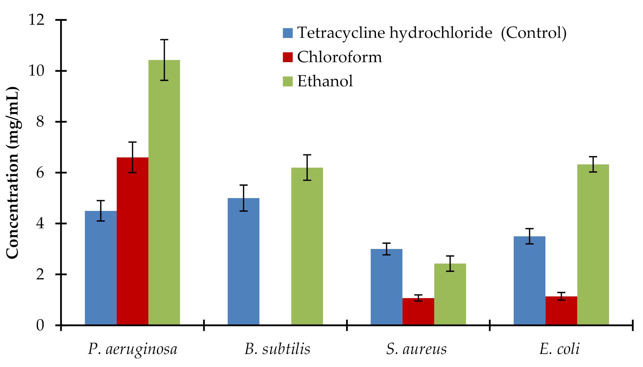
Abstract
:1. Introduction
2. Results
2.1. Antibacterial Activity of Extract of the Roots of Avicennia marina
2.2. Antibacterial Activity of the Leaf Extract of Avicennia marina
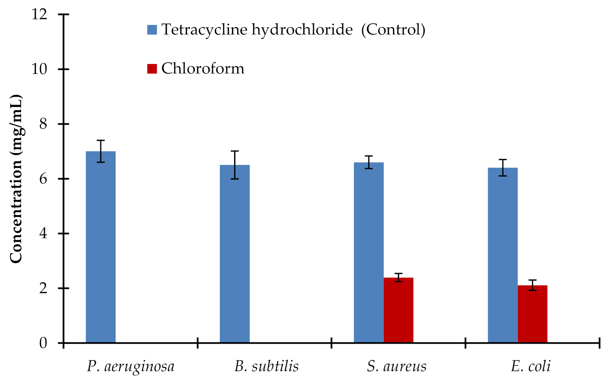
2.3. Minimal Bactericidal Concentration for Extract of the Roots of Avicennia marina
2.4. Minimal Bactericidal Concentration for Extracts of Leaves of Avicennia marina
2.5. Antifungal Activity of Extract of Different Parts of Avicennia marina
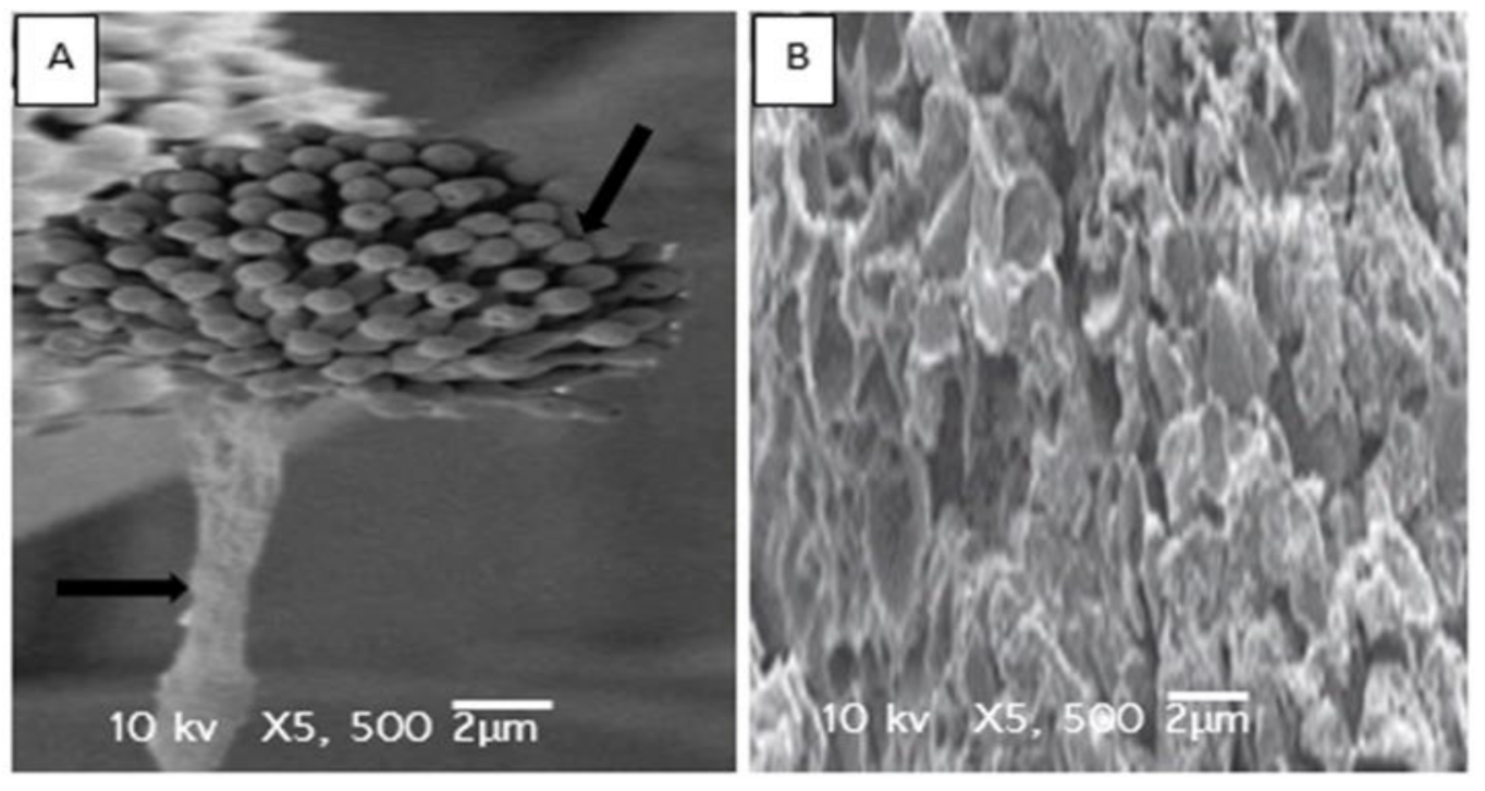
2.6. Electron Microscopic Studies
3. Discussion
4. Materials and Methods
4.1. Plant Material and Preparation of Extracts
4.2. Microbial Strains and Culture Media
4.3. Preparation of Inoculums
4.4. Analysis of Antibacterial Activity
4.5. Determination of Minimum Inhibitory Concentration (MIC)
4.6. Determination of Minimum Bactericidal Concentration (MBC)
4.7. Analysis of Antifungal Activity and Minimum Fungicidal Concentration
4.8. Scan Electron Microscopic Analysis
4.9. Statistical Analysis
5. Conclusions
Supplementary Materials
Author Contributions
Funding
Institutional Review Board Statement
Informed Consent Statement
Acknowledgments
Conflicts of Interest
References
- Nascimento, G.G.F.; Locatelli, J.; Freitas, P.C.; Silva, G.L. Antibacterial activity of plant extracts and phytochemicals on antibiotic-resistant bacteria. Braz. J. Microbiol. 2000, 31, 247–256. [Google Scholar]
- Favre-Godal, Q.; Dorsaz, S.; Queiroz, E.F.; Marcourt, L.; Ebrahimi, S.N.; Allard, P.M.; Sanglard, D. Anti-Candida Cassane-Type Diterpenoids from the Root Bark of Swartzia simplex. J. Nat. Prod. 2015, 78, 2994–3004. [Google Scholar]
- Soberón, J.R.; Lizarraga, E.F.; Sgariglia, M.A.; Juárez, M.B.C.; Sampietro, D.A.; Altabef, A.B.; Vattuone, M.A. Antifungal activity of 4-hydroxy-3-(3-methyl-2-butenyl) acetophenone against Candida albicans: Evidence for the antifungal mode of action. Antonie van Leeuwenhoek 2015, 108, 1047–1057. [Google Scholar]
- Siro, I.; Kapolna, E.; Kápolna, B.; Lugasi, A. Functional food product development, marketing and consumer acceptance—A review. Appetite 2008, 51, 456–467. [Google Scholar]
- Turner, M. Ecology: Mangrove maintenance. Nature 2015, 526, 515. [Google Scholar]
- Ser, H.L.; Tan, L.T.; Law, J.W.; Chan, K.G.; Duangjai, A.; Saokaew, S.; Pusparajah, P.; Ab-Mutalib, N.; Khan, T.M.; Goh, B.H.; et al. Focused review: Cytotoxic and antioxidant potentials of mangrove-derived Streptomyces. Front. Microbiol. 2017, 8, 2065. [Google Scholar]
- Haq, M.; Sani, W.; Hossain, A.; Taha, R.M.; Monneruzzaman, K. Total phenolic contents, antioxidant and antimicrobial activities of Bruguieragymnorrhiza. J. Med. Plants Res. 2011, 5, 4112–4118. [Google Scholar]
- Abeysinghe, P.D. Antibacterial activity of some medicinal mangroves against antibiotic resistant pathogenic bacteria. Indian J. Pharm. Sci. 2010, 72, 167–172. [Google Scholar]
- Bandaranayake, W.M. Traditional and medicinal uses of mangroves. Mangroves Salt Marshes 1998, 2, 133–148. [Google Scholar]
- Das, S.K.; Patra, J.K.; Thatoi, H. Antioxidative response to abiotic and biotic stresses in mangrove plants: A review. Int. Rev. Hydrobiol. 2016, 101, 3–19. [Google Scholar]
- Thatoi, H.; Samantaray, D.; Das, S.K. The genus Avicennia, a pioneer group of dominant mangrove plant species with potential medicinal values: A review. Front. Life Sci. 2016, 9, 267–291. [Google Scholar]
- Moore, G.E.; Grizzle, R.E.; Ward, K.M.; Alshihi, R.M. Distribution, pore-water chemistry, and stand characteristics of the mangroves of the United Arab Emirates. J. Coast. Res. 2015, 31, 957–963. [Google Scholar]
- Osman, N.A.; Abkar, F.A. Comparative evaluation of some selected bioactive constituents in the leaves and bark of Avicennia marina (Forsk.) Veirh. from the Sudanese red sea coast. J. Forest Prod. Indust. 2016, 4, 5–11. [Google Scholar]
- Rasoanaivo, P.; Petitjean, A.; Ratsimamanga-Urverg, S.; Rakoto-Ratsimamanga, A. Medicinal plants used to treat malaria in Madagascar. J. Ethnopharmacol. 1992, 37, 117–127. [Google Scholar]
- Zhu, F.; Chen, X.; Yuan, Y.; Huang, M.; Sun, H.; Xiang, W. The chemical investigations of the mangrove plant Avicennia marina and its endophytes. Open Nat. Prod. J. 2009, 2, 24–32. [Google Scholar]
- ElDohaji, L.M.; Hamoda, A.M.; Hamdy, R.; Soliman, S.S.M. Avicennia marina a natural reservoir of phytopharmaceuticals: Curative power and platform of medicines. J. Ethnopharmacol. 2020, 263, 113179. [Google Scholar]
- Namazi, R.; Zabihollahi, R.; Behbahani, M.; Rezaeic, A. Inhibitory activity of Avicennia marina, a medicinal plant in Persian folk medicine against HIV and HSV. Iran. J. Pharm. Res. 2013, 12, 435–443. [Google Scholar]
- Behbahani, M.; Zadeh, M.S.; Mohabatkar, H. Evaluation of antiherpetic activity of crude extract and fractions of Avicenna marina, in vitro. Antivir. Res. 2013, 97, 376–380. [Google Scholar]
- Behbahani, M. Evaluation of anti-HIV-1 activity of a new iridoid glycoside isolated from Avicenna marina, in vitro. Int. Immunopharmacol. 2014, 23, 262–266. [Google Scholar]
- Devi, A.S.; Rajkumar, J. In vitro antibacterial activity and stability of Avicennia marina against urinary tract infection pathogens at different parameters. Pak. J. Biol. Sci. 2013, 16, 1034–1039. [Google Scholar]
- Behbahani, A.; Shahidi, F.; Yazdi, F.T.; Mortazavi, S.A.; Mohebbi, M. Use of Plantago major seed mucilage as a novel edible coating incorporated with Anethum graveolens essential oil on shelf life extension of beef in refrigerated storage. Int. J. Biol. Macromol. 2017, 94, 515–526. [Google Scholar]
- Mahady, G.; Huang, Y.; Doyle, B.; Locklear, T. Natural products as antibacterial agents. Stud. Nat. Prod. Chem. 2008, 35, 423–444. [Google Scholar]
- Thatoi, H.N.; Patra, J.K.; Das, S.K. Free radical scavenging and antioxidant potential of mangrove plants: A review. Acta Physiol. Plant. 2014, 36, 561–579. [Google Scholar]
- Gurudeeban, S.; Ramanathan, T.; Satyavani, K. Antimicrobial and radical scavenging effects of alkaloid extracts from Rhizophora zucronata. Pharm. Chem. J. 2013, 47, 50–53. [Google Scholar]
- Mouafi, F.E.; Abdel-Aziz, S.M.; Bashir, A.A.; Fyiad, A.A. Phytochemical analysis and antimicrobial activity of mangrove leaves (Avicenna marina and Rhizophora stylosa) against some pathogens. World Appl. Sci. J. 2014, 29, 547–554. [Google Scholar]
- Aruna, K.; Pendse, A. Study on antibacterial activity of root extract from mangrove plants. Asian Sci. 2012, 7, 86–89. [Google Scholar]
- Li, Y.; Liu, J.; Yu, S.; Proksch, P.; Gu, J.; Lin, W. TNF-α inhibitory diterpenoids from the Chinese mangrove plant Excoecaria agallocha L. Phytochemistry 2010, 71, 2124–2131. [Google Scholar]
- Okla, M.K.; Alamri, S.A.; Alatar, A.A.; Hegazy, A.K.; Al-Ghamdi, A.A.; Ajarem, J.S.; Faisal, M.; Abdel-Salam, E.M.; Ali, H.M.; Salem, M.Z. Antioxidant, hypoglycemic, and neurobehavioral effects of a leaf extract of Avicennia marina on autoimmune diabetic mice. Evid. Based Complement. Alternat. Med. 2019, 2019, 1263260. [Google Scholar]
- Khattab, R.A.; Temraz, T.A. Mangrove Avicennia marina of Yanbu, Saudi Arabia: GC-MS constituents and mosquito repellent activities. Egypt. J. Aquat. Biol. Fish. 2017, 21, 45–54. [Google Scholar]
- Sutton, D.C.; Gillan, F.T.; Susic, M. Naphthofuranone phytoalexins from the grey mangrove, Avicennia marina. Phytochemistry 1985, 24, 2877–2879. [Google Scholar]
- Fauvel, M.; Taoubi, K.; Gleye, J.; Fouraste, I. Phenylpropanoid glycosides from Avicennia marina. Planta Med. 1993, 59, 387. [Google Scholar]
- Mahera, S.; Saifullah, S.; Ahmad, V.; Mohammad, F. Phytochemical studies on mangrove Avicennia marina. Pak. J. Bot. 2013, 45, 2093–2094. [Google Scholar]
- Mahera, S.; Ahmad, V.; Saifullah, S.; Mohammad, F.; Ambreen, K. Steroids and triterpenoids from grey mangrove Avicennia marina. Pak. J. Bot. 2011, 43, 1417–1422. [Google Scholar]
- Jia, R.; Guo, Y.-W.; Hou, H.X. Studies on the chemical constituents form leaves of Avicennia marina. Chin. J. Nat. Med. 2004, 2, 16–19. [Google Scholar]
- Wu, J.; Xiao, Q.; Xu, J.; Li, M.-Y.; Pana, J.-Y.; Yang, M. Natural products from true mangrove flora: Source, chemistry and Bioactivities. Nat. Prod. Rep. 2008, 25, 955–981. [Google Scholar]
- Schwarz, S.; Noble, W.C. Aspects of bacterial resistance to antimicrobials used in veterinary dermatological practice. Vet. Dermatol. 1999, 10, 163–176. [Google Scholar]
- Raut, S.V.; Anthappan, P.D. Studies on antimicrobial activity of leaves extract of Sonneratia alba. Curr. Res. Microbiol. Biotechnol. 2013, 1, 203–213. [Google Scholar]
- Khafagi, I.; Gab-Alla, A.; Salama, W.; Fouda, M. Biological activities and phytochemical constituents of the gray mangrove Avicennia marina (Forssk.) Vierh. Egypt. J. Biol. 2003, 5, 62–69. [Google Scholar]
- Mukhopadhyay, A.; Peterson, R.T. Fishing for new antimicrobials. Curr. Opin. Chem. Biol. 2006, 10, 327–333. [Google Scholar]
- Hugo, W.B.; Russell, A.D. Types of antimicrobial agents. In Principles and Practice of Disinfection, Preservation and Sterilization; Russell, A.D., Hugo, W.B., Ayliffe, G.A.J., Eds.; Blackwell Science Ltd.: Hoboken, NJ, USA, 1982; pp. 8–106. [Google Scholar]
- Neu, H.C. The crisis in antibiotic resistance. Science 1992, 257, 1064–1073. [Google Scholar]
- Tenover, F.C. Mechanisms of antimicrobial resistance in bacteria. Am. J. Med. 2006, 119, S3–S10. [Google Scholar]
- Sikkema, J.; De Bont, J.A.; Poolman, B. Mechanisms of membrane toxicity of hydrocarbons. Microbiol. Rev. 1995, 59, 201–222. [Google Scholar]
- Bajpai, V.K.; Sharma, A.; Baek, K.H. Antibacterial mode of action of Ginkgo biloba leaf essential oil: Effect on morphology and membrane permeability. Bangladesh J. Pharmacol. 2015, 1, 337–350. [Google Scholar]
- Yenna, T.W.; Khanb, M.A.; Syuhadaa, N.A.; Ringa, L.C.; Ibrahimc, D.; Tan, W.-N. Stigmasterol: An adjuvant for beta lactam antibiotics against beta-lactamase positive clinical isolates. Steroids 2017, 128, 68–71. [Google Scholar]
- Wu, H.-S.; Raza, W.; Fan, J.-Q.; Sun, Y.-G.; Bao, W.; Shen, Q.-R. Cinnamic acid inhibits growth but stimulates production of pathogenesis factors by in vitro cultures of Fusarium oxysporum f.sp. niveum. J. Agric. Food Chem. 2008, 56, 1316–1321. [Google Scholar]
- Korošec, B.; Sova, M.; Turk, S.; Kraševec, N.; Novak, M.; Lah, L.; Stojan, J.; Podobnik, B.; Berne, S.; Zupanec, N.; et al. Antifungal activity of cinnamic acid derivatives involves inhibition of benzoate 4-hydroxylase (CYP53). J. Appl. Microbiol. 2014, 116, 955–966. [Google Scholar]
- Bogdanov, S. Antibacterial substances in honey. Swiss Bee Res. Center 1997, 17, 74–76. [Google Scholar]
- Mandal, M.D.; Mandal, S. Honey: Its medicinal property and antibacterial activity. Asian Pac. J. Trop. Biomed. 2011, 1, 154–160. [Google Scholar]
- Johannes, E.; Litaay, M.; Syahribulan. The bioactivity of hexadecanoic acid compound isolated from hydroid aglaophenia cupressina lamoureoux as antibacterial agent against Salmonella typhi. Int. J. Biol. Med. Res. 2016, 7, 5469–5472. [Google Scholar]
- Radiati, L.E. Mechanism of virulence inhibition of enterophatogenic bacteria by ginger (Zingiber officinale Roscoe) rhizome extract. Ph.D. Dissertation, Postgraduate Program, Bogor Agricultural University, Bogor, Indonesia, 2002. [Google Scholar]
- Grace, O.O. Evaluation of the antimicrobial activity of citral. Lett. App. Microbiol. 1989, 9, 105–108. [Google Scholar]


Publisher’s Note: MDPI stays neutral with regard to jurisdictional claims in published maps and institutional affiliations. |
© 2021 by the authors. Licensee MDPI, Basel, Switzerland. This article is an open access article distributed under the terms and conditions of the Creative Commons Attribution (CC BY) license (http://creativecommons.org/licenses/by/4.0/).
Share and Cite
Okla, M.K.; Alatar, A.A.; Al-amri, S.S.; Soufan, W.H.; Ahmad, A.; Abdel-Maksoud, M.A. Antibacterial and Antifungal Activity of the Extracts of Different Parts of Avicennia marina (Forssk.) Vierh. Plants 2021, 10, 252. https://doi.org/10.3390/plants10020252
Okla MK, Alatar AA, Al-amri SS, Soufan WH, Ahmad A, Abdel-Maksoud MA. Antibacterial and Antifungal Activity of the Extracts of Different Parts of Avicennia marina (Forssk.) Vierh. Plants. 2021; 10(2):252. https://doi.org/10.3390/plants10020252
Chicago/Turabian StyleOkla, Mohammad K., Abdulrahman A. Alatar, Saud S. Al-amri, Walid H. Soufan, Altaf Ahmad, and Mostafa A. Abdel-Maksoud. 2021. "Antibacterial and Antifungal Activity of the Extracts of Different Parts of Avicennia marina (Forssk.) Vierh" Plants 10, no. 2: 252. https://doi.org/10.3390/plants10020252