Review

An Emerging Animal Model for Querying the Role of Whole Genome Duplication in Development, Evolution, and Disease

1
Biology Department, Brooklyn College at the City University of New York, Brooklyn, NY 11210, USA
2
Biology Department, The Graduate Center at the City University of New York, New York, NY 10016, USA
3
Biochemistry Department, The Graduate Center at the City University of New York, New York, NY 10016, USA
4
Magee-Womens Research Institute, Pittsburgh, PA 15213, USA
5
Department of Obstetrics, Gynecology, and Reproductive Sciences, University of Pittsburgh School of Medicine, Pittsburgh, PA 15213, USA
*
Author to whom correspondence should be addressed.
J. Dev. Biol. 2023, 11(2), 26; https://doi.org/10.3390/jdb11020026
Submission received: 22 April 2023 / Revised: 23 May 2023 / Accepted: 1 June 2023 / Published: 6 June 2023

Abstract

:
Whole genome duplication (WGD) or polyploidization can occur at the cellular, tissue, and organismal levels. At the cellular level, tetraploidization has been proposed as a driver of aneuploidy and genome instability and correlates strongly with cancer progression, metastasis, and the development of drug resistance. WGD is also a key developmental strategy for regulating cell size, metabolism, and cellular function. In specific tissues, WGD is involved in normal development (e.g., organogenesis), tissue homeostasis, wound healing, and regeneration. At the organismal level, WGD propels evolutionary processes such as adaptation, speciation, and crop domestication. An essential strategy to further our understanding of the mechanisms promoting WGD and its effects is to compare isogenic strains that differ only in their ploidy. Caenorhabditis elegans (C. elegans) is emerging as an animal model for these comparisons, in part because relatively stable and fertile tetraploid strains can be produced rapidly from nearly any diploid strain. Here, we review the use of Caenorhabditis polyploids as tools to understand important developmental processes (e.g., sex determination, dosage compensation, and allometric relationships) and cellular processes (e.g., cell cycle regulation and chromosome dynamics during meiosis). We also discuss how the unique characteristics of the C. elegans WGD model will enable significant advances in our understanding of the mechanisms of polyploidization and its role in development and disease.

1. Whole Genome Duplication in Development, Evolution, and Disease

Whole genome duplication (WGD) or polyploidization is vital for normal development [1], tissue homeostasis [2,3], and regeneration [4] (Figure 1). It is also a driver of evolutionary processes [5] including adaptation [6], speciation [7], and crop domestication [8]. In addition, WGD can drive pathological conditions, especially oncogenesis [1,9,10]. In nature, WGD exists at the cellular, tissue, organ, and organismal levels. Polyploid cells are normally found in most multicellular diploid organisms [11,12]. In humans, certain cells from the placenta [13], mammary glands [14], liver [15], heart [16], skin [17], and bone marrow must become polyploid to perform specialized functions [18], for tissue homeostasis [3], or as part of the wound healing process [19]. For instance, during development, megakaryocytes acquire 16 copies of the genome and thus become giant cells that are hypermetabolic. This polyploidization is a basic requirement to produce platelets in the blood, as platelets are produced by fragmenting the cytoplasm of megakaryocytes [20,21,22]. In addition, WGD is involved in oncogenesis, metastasis, and the development of resistance to chemotherapeutic drugs, and polyploid cells may accumulate in patients with neurodegenerative disorders, cardiovascular disease, and diabetes [23,24,25,26].
Polyploid cells can arise by cell fusion resulting in larger multinucleate cells, by endoduplication (alternating cycles between G and S phases without undergoing mitosis or cytokinesis) resulting in mononucleated cells, or by endomitosis (the cell enters mitosis but does not complete cell division) to generate either mononucleated giant cells or multinucleated cells [27]. In some cyanobacteria, polyploidy is established and maintained by misregulation of the replisome machinery [28,29,30].

1.1. Causes and Downstream Effects of WGD

The outcomes of WGD are diverse in nature—ranging from changes in gene expression to altered cell size and scaling and to adaptation to stress [31,32] (Figure 2). The effects of polyploidization are surprisingly similar in different biological contexts (e.g., in cells, tissues, and organisms) [4,11,33,34] and regardless of the biological process (e.g., during development, evolution, regeneration, and oncogenesis) [1,3,35,36,37,38,39,40,41] where WGD takes place (Figure 2). A common feature of WGD is that it is induced by stress, and it provides the changes that result in plasticity and thus enhances adaptation to stress in normal and pathological conditions. For instance, environmental stresses induce WGD in tissues (e.g., wounding or viral infection) [42,43,44] or organisms (e.g., large changes in temperature or water availability and salinity) [45,46,47,48,49,50]. Furthermore, stress-induced WGD is a platform for genomic diversity and versatility that provides the means for rapid adaptability (Figure 1 and Figure 2). Whereas in whole organisms, this can elicit a competitive advantage in a changing environment [32,51]; in cancer, it elicits an adaptive advantage that promotes oncogenesis, metastasis, and the development of drug resistance [32,51]. The latter is similar to how the pathogenic yeast Candida abdicans becomes resistant to antifungal drugs [6]. Therefore, it is widely expected that queries about mechanisms that give rise to WGD and its effects in both sub-organismal and organismal systems will likely reveal additional commonalities across systems and processes.
Polyploidy is often confused with aneuploidy, in part because, in humans, both phenomena are hallmarks of cancer [1,39,40,52]. Observations from numerous studies identify WGD in about 30% of human cancers [53,54] and as a precursor for many malignancies in cancer evolution [39,55,56,57,58,59]. Although WGD does not seem to require a preexisting cancer driver mutation; it most often emerges after oncogenic mutations, such as lesions in the TP53 genes [53,56]. WGD promotes diversification of copy number alterations (CNAs) and is permissive for other chromosomal aberrations and genome instability associated with poor cancer patient prognosis [39,58,59]. Interestingly, a recent study revealed a mechanism by which WGD enhances cancer-promoting alterations in DNA organization and gene expression [58]. Induction of WGD can reduce the levels of proteins involved in chromatin packaging, resulting in reorganization of the 3D DNA compartments and domains. Over time, loss of this domain structure predisposes the cell to additional cancer-promoting alterations and expression of oncogenic genes [58]. Polyploidization can also be protective, as in the liver where it both provides the basis for genetic adaptation to hepatotoxic stress and also protects from malignant transformations [21,60].
Organismal WGD is common in extant plants [7,32,61,62,63] and is also observed in protists [64], fungi [51,65,66], bacteria, and archaebacteria [67,68] and in several animal clades [2,12,35,36,40,41,69,70]. In both prokaryotes and eukaryotes, the effects of WGD are comparable with increased resistance to UV irradiation, cell size, adaptability to environmental changes, and evolvability [28,71]. Evidence for ancestral WGD events is found in the genomes of organisms of most clades, including vertebrates [36,72,73]. Polyploid metazoans arise due to errors during meiosis that result in the formation of diploid gametes that when fertilized give rise to polyploid organisms. Fertilization between diploid (2n) and haploid (n) gametes results in a triploid (3n) organism; fertilization between two diploid (2n) gametes results in a tetraploid (4n) [33,35]. When the diploid gametes of two related species fuse, the resulting organism is said to be an allopolyploid, whereas when diploid gametes from the same species fuse the result is said to be an autopolyploid [73]. Polyploidization from the cell to the organismal level most often results in lethality [32,74]. For instance, human triploid and tetraploid embryos die during gestation or soon after delivery, but triploid and tetraploid mosaic children may survive after birth [75,76]. This lethality may be caused by errors partitioning the additional chromosomes during cell division, by WGD-driven increases in cell size that interfere with tissues or organ scaling, and by epigenetic changes and gene expression changes that affect dose-dependent processes and complexes [73]. The latter may be especially true in human triploids and tetraploids which may have difficulty equalizing expression between the silenced X chromosomes and the extra sets of autosomes since variable numbers of Barr bodies may be found in these cells [75]. A newly formed polyploid may very occasionally overcome these obstacles, e.g., by establishing balanced genome expression. Then, if polyploidy provides a competitive advantage—as in a changing environment—polyploid lineages may outgrow their diploid counterparts and become established [32,41,73]. The established polyploid population will continue to evolve and then may “diploidize” to become a new species that has an adaptive advantage compared to the parental diploid species [32].

1.2. Laboratory Multicellular Organism Models for WGD

A major impediment to studying WGD and its impacts on the physiology and function of cells, tissues, and organs is the scarcity of laboratory models that permit comparison between isogenic organisms with differing ploidy [33]. Many laboratory models are sterile or embryonic lethal when polyploid [77,78,79]. Generation of autopolyploid laboratory model organisms that are not sterile (e.g., plants) frequently involves the use of chemicals such as colchicine, known to cause aneuploidy and genetic mutations. These polyploids, therefore, may not be truly isogenic with the parental diploid strains they were derived from [51,73]. The recently developed method for generating tetraploids in Caenorhabditis utilizes transient knock-down of a meiosis-specific cohesin (i.e., rec-8) by RNA interference to produce diploid gametes [80]. Therefore, the derived tetraploids are unlikely to differ significantly from the diploid strains from which they were derived (see Figure 3A for an overview of the method) [80].

2. Caenorhabditis Elegans as an Animal Laboratory Model for Understanding WGD

Several attributes of C. elegans nematodes make it an exceptional laboratory model for querying WGD: (1) it has a small number of chromosomes (five autosomes and a sex chromosome) allowing for easy visual evaluation of ploidy and differentiation between polyploidy and aneuploidy [81] (Figure 3B,C). (2) Despite C. elegans being a self-fertilizing hermaphroditic species, males can arise spontaneously in hermaphrodites by meiotic nondisjunction at a 0.2% frequency [82,83,84], allowing for genetic analysis [85]. Males are also produced by mating between hermaphrodites and males (frequency > 40%). (3) Each diploid hermaphrodite produces 200–300 offspring and has a lifecycle of three to four days from egg to egg, both of which facilitate multigenerational ecological and evolutionary studies. (4) C. elegans is normally a diploid but like vertebrates (including humans), it has polyploid tissues or cells [86]. These include the intestine (20 cells with a total of 30–34 nuclei and a ploidy of 32C (or 32 chromosome sets) per nucleus) and the hyp7 cell (a syncytium of 139 nuclei with an average ploidy of 10.7C) [86,87].
Critically, the quick and reliable method to generate fertile and viable tetraploid Caenorhabditis strains from nearly any diploid strain [80] allows direct comparison of identical cells in isogenic organisms with differing ploidy (Figure 3). The availability of hundreds of mutant strains that impinge on different aspects of development, aging, stress, and reproduction will facilitate the directed study of genes in many of the processes that polyploidy has been reported to affect. The transparency of the worms allows visualization and direct comparison of physiology and function in the live animal. Combined, these characteristics make the C. elegans WGD model unique and provide an unprecedented opportunity to answer long-standing questions about organismal polyploidization and its effects in a multicellular animal.

2.1. Polyploidy and Aneuploidy in C. elegans

In humans, all monosomies, except for the X or Y chromosomes, are lethal, whereas trisomies 13, 18, 21, X, and Y are viable. Autosomal trisomies can cause severe developmental problems; sex chromosome trisomies have comparatively mild effects, presumably because of Barr body formation (extra X chromosomes) or the dearth of active genes (Y chromosome). In C. elegans, some aneuploidies are tolerated. These include X-chromosome trisomies (2A;3X = 13 chromosomes triplo-X hermaphrodites) and monosomies (2A;1X = 11 chromosomes XO males shown in Figure 4) [88]. Compared to diploid hermaphrodites, triplo-X animals are shorter and fatter (i.e., Dumpy phenotype), lower fertility, and slower growth rate [88]. Trisomy of chromosome 4 (LGIV) has also been reported [89]. Although animals with this autosomal trisomy are viable and have a general morphological appearance that seems normal, they produce 50% fewer progeny than normal hermaphrodites [90]. Interestingly, large chromosomal fragments, generally more than an Mb in size, have been generated and preserved as free duplications and have been found for part of each of the chromosomes of C. elegans [91].
In nature, most triploids are inherently unstable, unless they reproduce asexually [92]. For instance, the parasitic nematodes of the Meloidogyne incognita group (MIG) are stable hypotriploids that reproduce by asexual parthenogenesis [93]. C. elegans triploid animals (3A;3X and 3A;2X shown in Figure 4) can be generated by crossing diploid and tetraploid animals [94]. Only 15% of the embryos produced by triploid animals hatch [94], and those that do hatch tend to be diploids or become diploid in the next generation [95,96,97]. Triploid progeny inviability is likely because the odd number of homologs results in abnormal meiotic pairing and recombination [95,98,99], yielding aneuploid gametes that produce embryos that are inviable.
Victor Nigon reported the first induction of polyploid C. elegans—the first in any animal—in the laboratory in 1949 [96]. These tetraploid strains were generated by treating spermatogenic animals with either heat shock (several hours at 25 °C) or exposure to colchicine. Both treatments were followed by screening for ‘larger than normal’ animals and cytological assessment of chromosome numbers in diakinesis-stage oocytes, when the chromosomes are highly condensed prior to the meiotic divisions (e.g., in Figure 3C). Newly formed tetraploids produced a range of phenotypes in offspring that included: progeny with variable body sizes; infertile hermaphrodites and males; intersexuality; and progeny that had reverted to diploidy [96,97]. The frequency of these aberrant phenotypes decreased with continuous passaging but did not disappear, even after 20 generations of selection for tetraploids with high fecundity. Two major types of tetraploids were identified [94,96]: about 66% of the induced tetraploids produced very low frequencies of males (0.6%) compared to the ~0.2% incidence of males in diploids; the remainder produced an average of ~42% males. Interestingly, tetraploids of both classes could interconvert and give rise to a proportion of progeny of the other type, albeit at different frequencies [97]. After 30 generations, the patterns of inheritance of high and low incidence of male progeny did not change. The tetraploid strains that produced high proportions of males are now known to correspond to a 4A;3X genotype and those that produced a low incidence of males are known to correspond to a 4A;4X genotype (see Figure 4) [94]. Even in the most stable lines, tetraploid hermaphrodites had substantially fewer self-progeny (average of ~65 progeny) than diploids (250–300 offspring in wild type, N2) and reduced hatching (decrease of 13%) [94]. Nigon’s tetraploid strains were eventually all accidentally lost after 78 generations. Several other groups have isolated fertile and relatively stable tetraploid C. elegans and C. briggsae strains [80,94,99,100,101]. Like their predecessors, each of these tetraploid strains has been reported to produce reduced numbers of self-progeny and to sporadically diploidize. Since the diploids can rapidly overtake a population due to their higher fertility, C. elegans tetraploid strains need to be maintained by continuous selection for the largest animals—at least for the first tens of generations. Interestingly, an obligate tetraploid strain has been reported (CB3911) [100]. This strain arose spontaneously from a diploid carrying a mutation in the dosage compensation gene dpy-27 which causes the lethality of XX diploids and XXXX tetraploids because of X-chromosome overexpression (Jonathan Hodgkin, personal communication). However, this strain produces viable dpy-27(rh18) 4A;3X hermaphrodites and 4A;2X males [100]. Studies of the early events post tetraploidization (e.g., in generations 1–10) therefore should shed light on how organisms acclimate to tetraploidy and become established.

2.2. Polyploid and Aneuploid Animals Uncover C. elegans Modes of Sex Determination and Dosage Compensation

One of the most critical developmental decisions that a heterogametic organism must make early in life is whether to develop as male or female and, concomitantly, whether to activate dosage compensation. From studies in C. elegans with different ploidies, it became apparent that sex is assessed by the ratio of sex chromosomes to autosomes (X/A) and that induction of hermaphrodite fate activates dosage compensation, which is achieved by reducing expression from the pair of X chromosomes [83,102]. Male phenotypes arose from animals with 2A;1X, 3A:2X, and 4A;2X, whereas 2A;2X, 3A;3X, 4A;4X, and 4A;3X gave rise to hermaphrodite phenotypes [94] (Table 1 and Figure 4).
Together, these studies led to the conclusion that an X:A ratio > 0.74 induces female/hermaphrodite identity and <0.67 promotes male development. Further support for these conclusions came from manipulation of the X/A ratio using X-linked gene duplications in the 2X;3A triploids to show at least three loci were involved in determining sex [101]. We now know the molecular details about the genes and processes involved both in assessing the X/A ratio and in executing male and hermaphrodite fates and dosage compensation [103]. Nevertheless, questions remain about how allotetraploids generated by the hybridization of different tetraploid Caenorhabditis species would assess the X/A ratio and whether manipulation or modification of this system could be used to stabilize polyploid organisms.

3. Utilizing Polyploid C. elegans as Tools to Investigate Developmental Processes

In the last ~50 years, comparisons between C. elegans strains of differing ploidy have been utilized as tools to query developmental processes including meiosis and embryonic divisions, in addition to sex determination and dosage compensation.

3.1. Understanding Early Events of Meiotic Prophase I

During meiosis, recombination between the duplicated maternal and paternal homologous chromosomes transiently connects the homologs to orderly partition them in the first division while maintaining sister chromatids together until the second division. To recombine, homologous chromosomes must first find one another in the nucleus (i.e., pair), become aligned along their lengths, and then stabilize their association by assembling the synaptonemal complex (i.e., synapse) [104]. Observations of the effects of having additional chromosomes in C. elegans triploid and tetraploid animals have helped to uncover rules underpinning homologous chromosome dynamics during meiosis [98,99]. Additional chromosomes did not significantly alter the initial steps of pairing into groups of three or four homologs in triploids and tetraploids, respectively. However, full alignment was often perturbed, and synapsis was frequently incomplete, resulting in the lengthening of the period in which synapsis normally takes place [98]. The imperfectly synapsed chromosomes became marked by di-methylation (H3K9me2), which has been proposed to prevent the synapsis defects from being detected by the quality control system [98]. Further analysis revealed that two pairs of synapsed homologous chromosomes form in tetraploids [99]. This pairwise synapsis also takes place in germ lines of mutants (i.e., spo-11) that are deficient in early steps of recombination, as in diploids. In triploid germ lines, one pair of homologs synapse, and the remaining homolog eventually folds onto itself, in a process known as self-synapse. In contrast to diploids, pairwise synapsis is disrupted in the presence of recombination-deficient mutations, suggesting that in C. elegans, as in other organisms, homologous recombination plays a role in promoting synapsis [99]. Polyploid animals heterozygous for translocation chromosomes (e.g., nT1(IV;V)) were utilized to test whether pairwise synapsis is dictated by full sequence homology along the chromosome length. Synapsis was normal in diploid translocation heterozygotes (i.e., nT1/+ or mln1/+), as long as they contained the pairing center sequence that is required for homologous chromosomes to initially find each other in the nucleus [105]. In tetraploids (nT1/nT1/+/+), there was no preference for a synapsis partner as translocation chromosomes paired with normal chromosomes with the same frequency as with themselves. In contrast, associations and synapsis between pairs of homologous chromosomes were biased towards homologs with identical sequences in the triploids [99]. These findings support the proposed model in which the initiation of synapsis depends on pairing center-mediated associations of homologs, but the completion of synapsis along the homologous pair is promoted by recombination [99].

3.2. Understanding Meiotic and Early Embryonic Cell Divisions

Female meiotic divisions are asymmetric. Each division gives rise to a large oocyte and a tiny polar body. The resulting egg contains a haploid set of chromosomes, as half of the chromosomes in each division are discarded into the polar bodies (Figure 5A,B). Therefore, C. elegans hermaphrodites trisomic for the X chromosome (2A;3X) would be expected to produce equal proportions of eggs that carry either one or two X chromosomes (one sister from the paired homologs +/− one sister from the self-synapsed chromosome). However, the observed X:XX ratio in these eggs is 2:1 [88]. Live and high-resolution visualization of the oocyte cell division in these trisomic worms showed that the self-synapsed chromosomes were preferentially partitioned into the polar body during oogenesis [95,106]. In addition, in triploid animals that have one set of homologous chromosome pairs (bivalents) and a set of unpaired chromosomes (univalents), the univalents were most often discarded into the polar body of the first oocyte division [106]. In these triploids, preferential segregation of the single sister chromatids into the polar body could also be observed in the second division. These results provide mechanistic insights into why triploid animals rapidly diploidize.
Several seminal studies have utilized tetraploid and haploid (1A;1X) C. elegans embryos to query cell division [107,108,109,110,111]. An overview of the relevant early developmental events, from fertilization to the first embryonic division, is shown in Figure 5.
Figure 5. Early development from fertilization to the first embryo division. In addition to contributing a haploid genome, the sperm also contributes a pair of centrioles to the zygote upon fertilization (A). Fertilization by the sperm triggers the completion of the oocytes’ meiotic divisions, each of which generates a polar body containing half of the chromosomes from the division (AC). The paired centrioles separate and remain in the proximity of the male pronucleus and, as they gather maternal pericentriolar material from the oocyte cytosol (B), they duplicate to form the first pair of centrosomes of the zygote (CE) [112]. Concurrently, the male pronucleus grows as DNA decondenses (B,C), and the centrosomes then become attached to its surface (C) [113]. The female pronucleus migrates towards the male pronucleus as centriole duplication is completed (C,D). The duplicated centrioles form the poles of the first bipolar spindle of the embryo (E). The centrosomes and the metaphase plate make a diamond shape (dashed and red) that defines the length (centrosome–centrosome) and width of the spindle (longer axis of the metaphase plate at the equator) (E).
Jdb 11 00026 g005
One study addressed how paternally inherited centrosomes become associated with the nucleus. Live imaging comparisons of zygotes with different ploidies suggested that centrosome attachment to the nuclear membrane depends on the size of the male pronucleus [107]. Diploid embryos with mutations in nuclear envelope genes that have aberrantly small male pronuclei initially had only one attached centrosome; the second centrosome only became attached as the male pronucleus surface increased [107]. This detached centrosome phenotype was suppressed in zygotes from tetraploid animals that had larger pronuclei. Conversely, the detached centrosome phenotype was enhanced in haploid zygotes with smaller pronuclei. It has been speculated that dynein motor accumulation on the pronuclear surface area is the limiting factor for centrosome attachment with a threshold of dynein required for both centrosomes to attach [109,114]. Of note, the haploid embryos in this study were generated utilizing conditional mutant sperm that lack nuclear DNA (i.e., emb-27(g48ts)) [109].
Another important study addressed whether the nuclear-to-cytosol ratio regulates cell cycle timing, a hypothesis that had been proposed from work in other organisms [115,116,117]. They compared diploid (N2 Bristol), tetraploid (strain SP343), and haploid embryos (generated by extracting the pronucleus from the fertilized egg before the zygote formed) [108]. In these embryos, different nuclear/cytosolic volume ratios were examined by extruding nuclei with little to no cytosol, some cytosol, or increased cytosolic volume, the latter created by laser-induced cell fusion followed by extrusion of one nucleus. Live imaging revealed that cell cycle timing was neither affected significantly by altering cell size nor by nuclear-to-cytosol ratio nor by DNA content [108]. Instead, cell fusion experiments revealed that cell cycle periods could be altered by the cytosol of a cell in a different cell cycle period. Together with other findings, these early results are consistent with studies that have posited models for the existence of “cytoplasmic factor(s)” that controls cell cycle timing [118,119,120]. The cytoplasmic factors that regulate many aspects of the cell cycle and its timing have now been identified [121].
Alterations of ploidy in embryos were also utilized to assess the scaling of the microtubule-based spindle [110]. Accurate chromosome partitioning relies on the assembly of a properly shaped spindle during cell division. The metaphase spindle forms a diamond shape, with two opposing corners at each centrosome and two at the ends of the metaphase plate (Figure 5E). The ratio between the spindle length (from the centrosome to centrosome) and width (of the metaphase plate at the equator) was constant throughout in vitro Xenopus extract studies that either molecularly or mechanically perturbed the dimensions of the metaphase spindles [122,123]. In vivo studies in C. elegans have been instrumental to our understanding of spindle shape. Initial studies showed that spindle length is affected by cell size as spindle length and width decrease and as embryogenesis progresses [110,118]. Interestingly, a reduction in the spindle width was observed when the spindle length was decreased by knocking down genes involved in spindle formation (e.g., by RNAi depletion of tpxl-1 (the C. elegans homolog of Xenopus TPX2 involved in spindle assembly) or spd-2 (involved in multiple aspects of centrosome biogenesis and thus spindle formation)) [124,125,126,127,128,129]. The opposite was not the case; changing the width of the spindle did not affect its length. In haploid embryos, the spindle width was reduced, and in polyploid embryos, it was increased [110]; yet in neither case was the embryo size or spindle length affected [124,125,126,127,128]. Correlation analysis between spindle length and width in haploid, diploid, and polyploid embryos revealed a simple equation that can predict spindle width based on the spindle length and ploidy of the embryo [110].
Another important correlation that was observed with the reduction in cell size during embryogenesis is related to chromosome condensation, a chromosome feature that is important for accurate chromosome inheritance [129,130,131,132,133]. Haploid and tetraploid embryos produced by diploid klp-18(RNAi) mothers show differences in the extent of chromosome condensation compared to diploid control embryos [130]. Chromosome condensation decreased in haploid embryos in comparison to diploid embryos, whereas condensation increased in tetraploid embryos. Differential condensation was independent of cell size, the speed of spindle elongation, and spindle length [110,130]. Therefore, an inverse correlation between nuclear DNA density and chromosome condensation was proposed and tested by observing chromosome condensation in dividing diploid and klp-18(RNAi) haploid embryos, during divisions as the size of the nucleus decreases during development [130]. The relationship between the DNA density in the nucleus and chromosome length (a measure of chromosome condensation) fits a regression line for the dividing diploid embryo. Importantly, this regression line also suitably fits the measurements of both the dividing haploid embryo and also embryos produced upon RNAi of factors that affect nuclear size including ima-3 (nuclear pore import protein) and ran-3 (ortholog of the human RCC1 regulator of chromosome condensation) [130]. These analyses revealed an allometric relationship between chromosome length/condensation and the relative ratio of DNA amount per nucleus size which is consistent with the hypothesis that chromosome condensation is regulated to adapt to fit the amount of DNA in the physical space within the nucleus.

4. Utilizing Caenorhabditis to Understand the Effects of Polyploidization

In addition to being utilized as tools to study development, C. elegans polyploids have also been used to study the process of polyploidization itself and its immediate after-effects.

4.1. Polyploid Tissues in C. elegans

At least two tissues are polyploid in C. elegans: the intestine and hypodermis (part of the nematode epidermis) (Figure 6) [86]. The intestine is a tube responsible for digestion and makes up nearly a third of the animal’s body. All intestinal cells are derived from a single blastomere (E) which is designated at the eight-cell embryo stage [134,135] (Figure 6A). E divides by mitosis, and then its progeny undergo rounds of endoduplication to allow the intestinal cells to massively increase nuclear output per unit volume which is thought to be necessary to meet the extensive enzymatic needs of this tissue.
The hypodermis has many roles in development including the establishment of the basic body plan in the embryo such as body shape and size, regulation of cell fate specification, and guidance of migrating axons and cells [136,137,138]. The hypodermal cell hyp7 is a multi-nucleated syncytium that encases most of the adult animal body (the tail and head of the animal are covered by smaller hypodermal cells) [86]. The hyp7 syncytium ultimately contains 139 nuclei in the adult which arise from cell fusions (Figure 6B) [86,139]. The embryonically derived nuclei in hyp7 remain as diploids, whereas ~98 cells generated post-embryonically endoduplicate to become tetraploids [86,87,139]. The ploidy of the hyp7 cell is key for adult animal size regulation, (see the section on allometric studies, below) [140].
In diploid cells, coordination of the centrosome and cell cycle ensures a single duplication of the centrosome per mitotic cycle, which is crucial to prevent supernumerary centrosome accumulation which can lead to genomic instability [140,141]. In cells that undergo endocycles, regulating the number of centrosome cycles is equally important for maintaining genome stability [142]. In the intestinal and hypodermal lineages of C. elegans, the centrioles become refractory to S-phase cues, and thus centriole duplication becomes uncoupled from the cell cycle, likely due to the loss of pericentriolar material, including the SPD-2 protein. After halting centriole duplication, these cells also eliminate all but one of the already duplicated centrioles [143]. Phosphorylation of the SPD-2 protein regulates centriole duplication; therefore, it represented a likely candidate for regulation of the centrioles during endocycles [144,145,146]. A CDK phospho-mutant, SPD-2S545A, prevents intestinal divisions due to a lack of centrosome duplication. In contrast, the phosphomimetic mutant, SPD-2S545E leads to supernumerary centrosomes in the intestinal cells [143]. The effect of this allele is not observed in the hypodermis, suggesting cell-type-specific specializations in centriole control. SPD-2S545E does not completely suppress the effects of cdk-2(RNAi), suggesting that additional phosphorylation sites are also involved. Indeed, SPD-2S357 was found to be regulated by Polo-Like Kinase-1 (PLK-1 in C. elegans), and a phosphomimetic mutation at this site resulted in the prolonged persistence of SPD-2 on centrioles and delayed centriole elimination in some of the intestinal nuclei. Surprisingly, PLK-1 does not seem to regulate either SPD-2 stabilization or centriole elimination [143] but appears to affect SPD-2 accumulation through its effect on the cell cycle [143]. Instead, knockdown of the proteasome b-subunit (pbs-3) results in both SPD-2 persistence indicative of a lack of centriole elimination in many of the intestinal cells. This finding implicates ubiquitin-mediated proteolysis in the uncoupling of the endocycle and centriole cycle in the intestine [143].
A central question in the WGD field has been whether there is a significant functional difference between mononucleated polyploidy (restricted to a single nucleus) and multinucleate polyploidy (in more than one nucleus) [2,147]. The developing intestine in C. elegans was used to address this question by converting the normally binucleated intestinal cells into mononucleated cells of identical ploidy using auxin-induced degradation [148] of either a regulator of mitotic entry (i.e., CDK-1) or a kinetochore protein required for chromosome partitioning in mitosis (i.e., KNL-1) to block endomitosis [38]. Whereas the absence of CDK-1 prevents entry into mitosis and essentially converts the endomitosis into an endocycle, degradation of KNL-1 allows for entry into mitosis but prevents the chromosome partitioning into two nuclei [38]. Knock-down affected neither cell size nor morphology, but the nuclear surface-to-volume ratio decreased in the mononucleated cells [38]. Single-worm transcriptome analyses revealed that the vitellogenin genes (vit-1 to vit-6) are downregulated in these mononucleated intestines. Vitellogenins are yolk proteins that are required for lipid transport to oocytes and embryos and are important determinants of progeny fitness. Accordingly, embryos from mothers with mononucleated intestines have reduced survival. Similarly, decreasing vitellogenin expression in binucleated gut cells phenocopies the impact on progeny fitness, and increasing vitellogenin expression, by overexpressing their transcriptional activators, results in mothers producing progeny with similar fitness regardless of whether they had mono- or bi-nucleated intestines [38]. Importantly, many of the genes that are downregulated in animals with mononucleated intestines normally increase in expression during the L4 to adult transition, implicating a major role in binucleation at this developmental stage [38].

4.2. Biological Size and Scaling (Allometry)

Regulation of cell size and scaling of tissues and organs are critical for development and influence homeostasis, metabolism, and function [33]. Polyploidization has been generally associated with increases in cell and organism size. For example, genome size and ploidy correlate linearly with cell volume in yeast [149,150,151]. However, the effects of polyploidy are cell-type specific in many multicellular organisms, with different cell types having a nonlinear relationship between ploidy and cell size [152]. For instance, in plants, there is a strong correlation between ploidy and cell volume in epidermal pavement cells, but the volume of palisade mesophyll cells remains at a constant size despite changes in ploidy [153]. These and other observations [154,155,156,157] make it apparent that the DNA mass does not fully explain the effect of ploidy on cell (and nuclear) size [158,159,160]. In most polyploidization models, including plants, interpreting the direct effect of ploidy is complex because the increase in cell size in tissues and organs is balanced by a reduction in cell number [33,157,160,161]. The invariant cell lineage in C. elegans simplifies the identification of mechanisms underpinning correlations between ploidy and scaling of cells, tissues, and organs.
Allometry studies related to the effects of ploidy in C. elegans have been performed for cells and animals [87,97,110,130,162,163,164,165,166]. Nigon (1949) [96] reported that compared to the 1300 µm-long diploid hermaphrodites, the 4A;3X hermaphrodites were on average 1360 µm and the 4A;4X hermaphrodites were 1560 µm long. The more profound increase in the 4A;4X hermaphrodites containing 24 chromosomes (See Figure 3) compared to the 4A;3X hermaphrodites (23 chromosomes) is surprising and suggests that gene expression from the X chromosome may contribute to an animal’s size significantly more than gene expression from autosomes.
The hypodermis secretes the cuticle forming the exoskeleton that houses the nematode’s internal organs and nervous system. There is a proportional relationship between the size of the C. elegans body and the size of the hypodermal cells (seam cells and the syncytium hyp7) [163,167,168]. Comparison of 12 nematode species from the order Rhabditida revealed a weak correlation between the number of nuclei and hypodermal volume (hyp7 in C. elegans), suggesting that body size was at least partly independent of cell number [87]. A significant correlation between animal size and the product of the number of nuclei and the ploidy was observed across species. Therefore, unlike most organisms where the evolution of body size relies on changes in cell number, in nematodes, body size evolution was likely driven by the size of the hypodermal syncytium [87]. A comparison of a tetraploid strain and its diploid revertant revealed a 39% increase in the adult body size of tetraploids [162]. The unexpected 1.4-fold increase in volume (instead of the expected 2-fold increase) in tetraploids is at least in part explained by the lower-than-expected ploidy in the hyp7 cell (16.7C instead of 22C) and the small reduction in the number of cells (by less than two nuclei) [162]. However, one should be careful in making conclusions from this comparison because it is likely that the reverted diploid differed from the original, but these were not examined. Multiple independent tetraploid strains from nearly any diploid strain provide the possibility of conducting this comparison more rigorously. Levels of endoduplication in hyp7 of C. elegans were found to be important in the regulation of the adult animal size. When C. elegans young adults were exposed to the DNA replication inhibitor hydroxyurea (HU), endoduplication of the hyp7 nuclei was inhibited, and a reduction in body size was observed [162]. Cyclin E is required for endoduplication in mice and Drosophila [169,170]. Null mutants carrying mutations of the C. elegans homolog of Cyclin E, cye-1, have an ~1.8-fold reduction in the size of the hyp7 syncytium and are 35–54% the size of wild-type worms [162].
Many signaling pathways and genes affecting diploid animal size have been characterized in C. elegans. These include pathways that only regulate embryo size, that regulate multiple developmental stages, and that influence adult body size. The TGFβ/BMP signaling pathway has a major role in the response to ploidy as it regulates both adult size and hyp7 ploidy [162,167,168,171,172,173]. The TGFβ signaling pathway that regulates body size in C. elegans includes the DAF-4 type-two receptor, the DBL-1 ligand, and the downstream SMADs (SMA-2, SMA-3, SMA-4, and SMA-6) [174]. DBL-1, the ortholog of human BMP7, is required for post-embryonic growth [87,166,175]. Whereas DBL-1 overexpression results in longer-than-normal animals (Lon phenotype) [172], mutations in the dbl-1 gene cause a dwarf phenotype (Sma) and reduced hypodermal ploidy [162,172]. Interestingly, as with the cye-1 mutant, exposure of dbl-1 mutants to hydroxyurea neither further reduced hyp7 ploidy nor affected its body size, suggesting that DBL-1 normally promotes hyp7 endoduplication [162]. Together, these and other findings [162,168] suggest that DBL-1 regulates adult growth in a dose-dependent manner [165,172] by promoting endoduplication in the adult hypodermis (hyp7) [168], likely via CYE-1 cyclin [162].
The regulation of body size by the DBL-1 pathway is complex. Many of the molecular components in the DBL-1 pathway and other pathways affecting endoduplication in the hypodermis have been identified, and their localization and molecular or genetic interactions have been described. For instance, overexpression of LON-1, a member of the conserved PR-protein superfamily [164,176], causes hypo-endoduplication, and the absence of the LON-1 protein causes hyper-endoduplication in the hypodermis [164]. Specifically, the levels of the lon-1 transcript depend on the dosage of the dbl-1 gene and TGFβ signaling. Other genes and processes affecting diploid animal size are also likely to play important roles in this process. These include cuticle collagens, β-H Spectrin, TOR kinase, MAPK signaling, Hippo–Warts signaling, and the insulin signaling pathway [166,177,178,179,180,181,182,183,184,185,186]. These genes are expected to show altered expression in polyploid animals [33,41,187,188]. Global metabolic genes, such as rRNA or tRNA genes, are likely also involved in regulating biological scaling in polyploid animals [33,189]. Whether these pathways contribute equally to growth regulation in polyploids remains an open question.

5. Potential Future Queries Utilizing C. elegans Polyploids

The ease of creating synthetic autotetraploids from any strain utilizing RNAi is shepherding in a new era. We can now expand the use of C. elegans polyploids to study cellular and developmental processes (e.g., allometric relationships of different cell types and cell division dynamics) and disease (e.g., genome instability, pathological cell divisions, and cancer) by comparing equivalent organisms, tissues, organs, and cells from isogenic diploid, triploid, and tetraploid animals. This method allows for the first time in C. elegans the ability to query the direct impact of organismal polyploidization on individual tissues across time. In addition, this polyploid model’s unique features will be of particular benefit for advancing our understanding of WGD and its effect on genome structure, transcriptome size (total RNA per unit of DNA mass) [190], specific-gene expression, regulation of gene expression by chromatin modification, and importantly how these changes affect the physiology, metabolism, and developmental character of the organism.
In the course of evolution, parts of the genomes of polyploids are lost [191,192,193], a process known as fractionation [194]. This can occur because the purifying selection—the drive to remove mutated genes—is relaxed on duplicated genes, leading to both gene and regulatory element loss [194,195]. In allotetraploids, allele dominance can facilitate gene retention/fractionation, but genes involved in multi-subunit complexes (ribosomes, polymerases, etc.) tend to be retained [194]. Previous observations in plants showed that they undergo loss of repetitive sequences, mostly transposable elements, by recombination [187,188,189]. This can reduce the genome by up to 25% within the first few generations of newly formed autopolyploids [196]. Synthetic auto- and allo-tetrapolyploid C. elegans will allow both short-term and longitudinal studies of the process of genome downsizing given the nematodes’ short life cycle and their ability to manipulate their environment. Additionally, these animals would facilitate the study of tissue-specific differences and the mitotic stability of ploidy during development. Of particular interest will be comparative analyses of the endopolyploid hyp7 and intestinal cells to determine how their DNA size/mass scales with ploidy.
Gene expression studies have been performed to assess differential gene expression in allopolyploid plants, often comparing diploids to an ancestral hybrid species [33]. An important mystery remaining is the stoichiometry of the changes in whole transcriptomes of similar cells from animals with differing ploidy [190,197]. Two competing hypotheses regarding the effect of organism polyploidization on global gene expression have been proposed: (1) each duplicated genome retains diploid transcription levels or (2) all gene loci are affected by dosage compensation to maintain a gene dose similar to that of diploids. Although very few studies have been carried out to compare the effect of differing ploidy on transcriptome size, neither global suppression nor doubling of expression is uniquely likely since allopolyploid studies have shown an intermediate transcriptome size. Importantly, single-cell transcriptomic studies have shown high variability or noise in gene expression within a cell type and between different cell types. Specific regulatory genes (e.g., RNA polymerase 2 and 5.8S rRNA) have been found to differentially increase in expression with increasing ploidy [198]. Global and specific gene expression can also be affected by chromatin remodeling (e.g., chromatin and histone modifications), and specific histone modifications, particularly histone H3K56 acetylation, are thought to buffer increases in mRNA synthesis due to an increased number of gene copies [199].
The ability to create tetraploids with any strain facilitates longitudinal studies of the tetraploid. For example, one can now create recombinant inbred strains of auto- and allotetraploids to address how the genome changes over ten to hundreds of generations. Combining these studies with exposure to environmental stressors, such as disease, temperature, infection, and DNA damage, has the potential to shed light on genomic changes that provide flexibility and drive adaptation. A greater understanding of WGD and the mechanisms of tolerance to polyploidy will also have important implications for the treatment of cancer and other human pathogens which may gain a selective advantage over endogenous cells through these mechanisms.

Funding

This research was funded by NIGMS, grant number R01GM104007 to JLY; PSC-CUNY award, 65508-00 53, and the CUNY-Research Foundation, FFPP award to MS; Tow mentoring and research program to MT and MS; and Brooklyn College’s, Samuel and Rose Goldstein award to MT.

Institutional Review Board Statement

Not applicable.

Informed Consent Statement

Not applicable.

Data Availability Statement

Not applicable.

Acknowledgments

We are grateful to Jonathan Hodgkin and the three reviewers of this manuscript for their insightful comments and suggestions. We also thank the undergraduate students at the Schvarzstein laboratory for their discussions and comments about the manuscript.

Conflicts of Interest

The authors declare no conflict of interest. The funders had no role in the design of the study; in the collection, analysis, or interpretation of the data; in the writing of the manuscript; or in the decision to publish the results.

References

  1. Davoli, T.; de Lange, T. The Causes and Consequences of Polyploidy in Normal Development and Cancer. Annu. Rev. Cell Dev. Biol. 2011, 27, 585–610. [Google Scholar] [CrossRef] [Green Version]
  2. Øvrebø, J.I.; Edgar, B.A. Polyploidy in tissue homeostasis and regeneration. Development 2018, 145, dev156034. [Google Scholar] [CrossRef] [Green Version]
  3. Donne, R.; Saroul-Aïnama, M.; Cordier, P.; Celton-Morizur, S.; Desdouets, C. Polyploidy in liver development, homeostasis and disease. Nat. Rev. Gastroenterol. Hepatol. 2020, 17, 391–405. [Google Scholar] [CrossRef]
  4. Bailey, E.C.; Kobielski, S.; Park, J.; Losick, V.P. Polyploidy in Tissue Repair and Regeneration. Cold Spring Harb. Perspect. Biol. 2021, 13, a040881. [Google Scholar] [CrossRef]
  5. Anatskaya, O.V.; Vinogradov, A.E. Polyploidy as a Fundamental Phenomenon in Evolution, Development, Adaptation and Diseases. Int. J. Mol. Sci. 2022, 23, 3542. [Google Scholar] [CrossRef]
  6. Berman, J. Ploidy plasticity: A rapid and reversible strategy for adaptation to stress. FEMS Yeast Res. 2016, 16, fow020. [Google Scholar] [CrossRef] [Green Version]
  7. Wood, T.E.; Takebayashi, N.; Barker, M.S.; Mayrose, I.; Greenspoon, P.B.; Rieseberg, L.H. The frequency of polyploid speciation in vascular plants. Proc. Natl. Acad. Sci. USA 2009, 106, 13875–13879. [Google Scholar] [CrossRef] [Green Version]
  8. Akagi, T.; Jung, K.; Masuda, K.; Shimizu, K.K. Polyploidy before and after domestication of crop species. Curr. Opin. Plant Biol. 2022, 69, 102255. [Google Scholar] [CrossRef] [PubMed]
  9. Quinton, R.J.; DiDomizio, A.; Vittoria, M.A.; Kotýnková, K.; Ticas, C.J.; Patel, S.; Koga, Y.; Vakhshoorzadeh, J.; Hermance, N.; Kuroda, T.S.; et al. Whole-genome doubling confers unique genetic vulnerabilities on tumour cells. Nature 2021, 590, 492–497. [Google Scholar] [CrossRef] [PubMed]
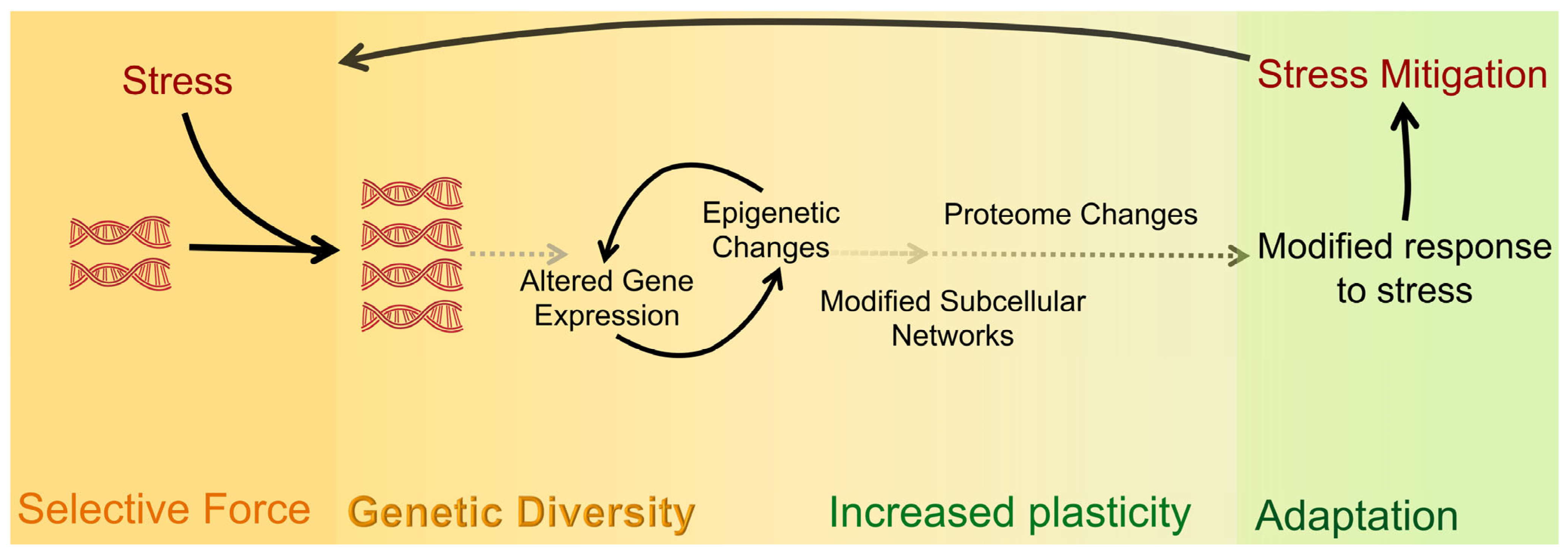
  10. Anatskaya, O.V.; Vinogradov, A.E. Polyploidy and Myc Proto-Oncogenes Promote Stress Adaptation via Epigenetic Plasticity and Gene Regulatory Network Rewiring. Int. J. Mol. Sci. 2022, 23, 9691. [Google Scholar] [CrossRef] [PubMed]
  11. Orr-Weaver, T.L. When bigger is better: The role of polyploidy in organogenesis. Trends Genet. 2015, 31, 307–315. [Google Scholar] [CrossRef] [PubMed] [Green Version]
  12. Frawley, L.E.; Orr-Weaver, T.L. Polyploidy. Curr. Biol. 2015, 25, R353–R358. [Google Scholar] [CrossRef] [PubMed] [Green Version]
  13. Chen, H.-Z.; Ouseph, M.M.; Li, J.; Pécot, T.; Chokshi, V.; Kent, L.; Bae, S.; Byrne, M.; Duran, C.; Comstock, G.; et al. Canonical and atypical E2Fs regulate the mammalian endocycle. Nature 2012, 14, 1192–1202. [Google Scholar] [CrossRef] [PubMed] [Green Version]
  14. Rios, A.C.; Fu, N.Y.; Jamieson, P.R.; Pal, B.; Whitehead, L.; Nicholas, K.R.; Lindeman, G.J.; Visvader, J.E. Essential role for a novel population of binucleated mammary epithelial cells in lactation. Nat. Commun. 2016, 7, 11400. [Google Scholar] [CrossRef] [Green Version]
  15. Pandit, S.K.; Westendorp, B.; Nantasanti, S.; van Liere, E.; Tooten, P.C.J.; Cornelissen, P.W.A.; Toussaint, M.J.M.; Lamers, W.H.; de Bruin, A. E2F8 is essential for polyploidization in mammalian cells. Nature 2012, 14, 1181–1191. [Google Scholar] [CrossRef]
  16. Senyo, S.E.; Steinhauser, M.L.; Pizzimenti, C.L.; Yang, V.K.; Cai, L.; Wang, M.; Wu, T.-D.; Guerquin-Kern, J.-L.; Lechene, C.P.; Lee, R.T. Mammalian heart renewal by pre-existing cardiomyocytes. Nature 2013, 493, 433–436. [Google Scholar] [CrossRef] [Green Version]
  17. Zanet, J.; Freije, A.; Ruiz, M.; Coulon, V.; Sanz, J.R.; Chiesa, J.; Gandarillas, A. A Mitosis Block Links Active Cell Cycle with Human Epidermal Differentiation and Results in Endoreplication. PLoS ONE 2010, 5, e15701. [Google Scholar] [CrossRef] [Green Version]
  18. Mattia, G.; Vulcano, F.; Milazzo, L.; Barca, A.; Macioce, G.; Giampaolo, A.; Hassan, H.J. Different ploidy levels of megakaryocytes generated from peripheral or cord blood CD34+ cells are correlated with different levels of platelet release. Blood 2002, 99, 888–897. [Google Scholar] [CrossRef] [Green Version]
  19. Losick, V.P. Wound-Induced Polyploidy Is Required for Tissue Repair. Adv. Wound Care 2016, 5, 271–278. [Google Scholar] [CrossRef] [Green Version]
  20. Zimmet, J.; Ravid, K. Polyploidy: Occurrence in nature, mechanisms, and significance for the megakaryocyte-platelet system. Exp. Hematol. 2000, 28, 3–16. [Google Scholar] [CrossRef]
  21. Sladky, V.C.; Eichin, F.; Reiberger, T.; Villunger, A. Polyploidy control in hepatic health and disease. J. Hepatol. 2021, 75, 1177–1191. [Google Scholar] [CrossRef]
  22. Mazzi, S.; Lordier, L.; Debili, N.; Raslova, H.; Vainchenker, W. Megakaryocyte and polyploidization. Exp. Hematol. 2018, 57, 1–13. [Google Scholar] [CrossRef]
  23. Anatskaya, O.V.; Sidorenko, N.V.; Vinogradov, A.E.; Beyer, T.V. Impact of neonatal cryptosporidial gastroenteritis on epigenetic programming of rat hepatocytes. Cell Biol. Int. 2007, 31, 420–427. [Google Scholar] [CrossRef] [PubMed]
  24. Anatskaya, O.V.; Matveev, I.V.; Sidorenko, N.V.; Kharchenko, M.V.; Kropotov, A.V.; Vinogradov, A.E. Changes in the heart of neonatal rats after cryptosporidial gastroenteritis of different degrees of severity. J. Evol. Biochem. Physiol. 2013, 49, 509–518. [Google Scholar] [CrossRef]
  25. Lazzeri, E.; Angelotti, M.L.; Conte, C.; Anders, H.-J.; Romagnani, P. Surviving Acute Organ Failure: Cell Polyploidization and Progenitor Proliferation. Trends Mol. Med. 2019, 25, 366–381. [Google Scholar] [CrossRef] [PubMed] [Green Version]
  26. Anatskaya, O.V.; Vinogradov, A.E. Whole-Genome Duplications in Evolution, Ontogeny, and Pathology: Complexity and Emergency Reserves. Mol. Biol. 2021, 55, 813–827. [Google Scholar] [CrossRef]
  27. Edgar, B.A.; Zielke, N.; Gutierrez, C. Endocycles: A recurrent evolutionary innovation for post-mitotic cell growth. Nat. Rev. Mol. Cell Biol. 2014, 15, 197–210. [Google Scholar] [CrossRef]
  28. Ohbayashi, R.; Nakamachi, A.; Hatakeyama, T.S.; Watanabe, S.; Kanesaki, Y.; Chibazakura, T.; Yoshikawa, H.; Miyagishima, S.-Y. Coordination of Polyploid Chromosome Replication with Cell Size and Growth in a Cyanobacterium. mBio 2019, 10, e00510-19. [Google Scholar] [CrossRef] [PubMed] [Green Version]
  29. Chen, A.H.; Afonso, B.; Silver, P.A.; Savage, D.F. Spatial and Temporal Organization of Chromosome Duplication and Segregation in the Cyanobacterium Synechococcus elongatus PCC 7942. PLoS ONE 2012, 7, e47837. [Google Scholar] [CrossRef] [Green Version]
  30. Watanabe, S.; Ohbayashi, R.; Shiwa, Y.; Noda, A.; Kanesaki, Y.; Chibazakura, T.; Yoshikawa, H. Light-dependent and asynchronous replication of cyanobacterial multi-copy chromosomes. Mol. Microbiol. 2012, 83, 856–865. [Google Scholar] [CrossRef]
  31. Schoenfelder, K.P.; Fox, D.T. The expanding implications of polyploidy. J. Cell Biol. 2015, 209, 485–491. [Google Scholar] [CrossRef] [PubMed]
  32. Bomblies, K. When everything changes at once: Finding a new normal after genome duplication. Proc. R. Soc. B Boil. Sci. 2020, 287, 20202154. [Google Scholar] [CrossRef] [PubMed]
  33. Doyle, J.J.; Coate, J.E. Polyploidy, the Nucleotype, and Novelty: The Impact of Genome Doubling on the Biology of the Cell. Int. J. Plant Sci. 2019, 180, 1–52. [Google Scholar] [CrossRef]
  34. Ren, R.; Wang, H.; Guo, C.; Zhang, N.; Zeng, L.; Chen, Y.; Ma, H.; Qi, J. Widespread Whole Genome Duplications Contribute to Genome Complexity and Species Diversity in Angiosperms. Mol. Plant 2018, 11, 414–428. [Google Scholar] [CrossRef] [Green Version]
  35. Fox, D.T.; Soltis, D.E.; Soltis, P.S.; Ashman, T.-L.; Van de Peer, Y. Polyploidy: A Biological Force from Cells to Ecosystems. Trends Cell Biol. 2020, 30, 688–694. [Google Scholar] [CrossRef] [PubMed]
  36. Maciver, S.K. Ancestral Eukaryotes Reproduced Asexually, Facilitated by Polyploidy: A Hypothesis. Bioessays 2019, 41, e1900152. [Google Scholar] [CrossRef]
  37. Nguyen, H.G.; Makitalo, M.; Yang, D.; Chinnappan, D.; St.Hilaire, C.; Ravid, K. Deregulated Aurora-B induced tetraploidy promotes tumorigenesis. FASEB J. 2009, 23, 2741–2748. [Google Scholar] [CrossRef] [Green Version]
  38. van Rijnberk, L.M.; Barrull-Mascaró, R.; van der Palen, R.L.; Schild, E.S.; Korswagen, H.C.; Galli, M. Endomitosis controls tissue-specific gene expression during development. PLoS Biol. 2022, 20, e3001597. [Google Scholar] [CrossRef]
  39. Storchova, Z.; Pellman, D. From polyploidy to aneuploidy, genome instability and cancer. Nat. Rev. Mol. Cell Biol. 2004, 5, 45–54. [Google Scholar] [CrossRef]
  40. Zhang, J.; Qiao, Q.; Xu, H.; Zhou, R.; Liu, X. Human cell polyploidization: The good and the evil. Semin. Cancer Biol. 2022, 81, 54–63. [Google Scholar] [CrossRef]
  41. Otto, S.P. The Evolutionary Consequences of Polyploidy. Cell 2007, 131, 452–462. [Google Scholar] [CrossRef] [PubMed] [Green Version]
  42. Lazzeri, E.; Angelotti, M.L.; Peired, A.; Conte, C.; Marschner, J.A.; Maggi, L.; Mazzinghi, B.; Lombardi, D.; Melica, M.E.; Nardi, S.; et al. Endocycle-related tubular cell hypertrophy and progenitor proliferation recover renal function after acute kidney injury. Nat. Commun. 2018, 9, 1344. [Google Scholar] [CrossRef] [PubMed] [Green Version]
  43. Losick, V.P.; Fox, D.T.; Spradling, A.C. Polyploidization and Cell Fusion Contribute to Wound Healing in the Adult Drosophila Epithelium. Curr. Biol. 2013, 23, 2224–2232. [Google Scholar] [CrossRef] [PubMed] [Green Version]
  44. Li, T.-N.; Wu, Y.-J.; Tsai, H.-W.; Sun, C.-P.; Wu, H.-L.; Pei, Y.-N.; Lu, K.-Y.; Yen, T.T.-C.; Chang, C.-W.; Chan, H.-L.; et al. Intrahepatic hepatitis B virus large surface antigen induces hepatocyte hyperploidy via failure of cytokinesis. J. Pathol. 2018, 245, 502–513. [Google Scholar] [CrossRef] [PubMed]
  45. David, K.T.; Halanych, K.M. Spatial proximity between polyploids across South American frog genera. J. Biogeogr. 2021, 48, 991–1000. [Google Scholar] [CrossRef]
  46. Glennon, K.L.; Ritchie, M.E.; Segraves, K.A. Evidence for shared broad-scale climatic niches of diploid and polyploid plants. Ecol. Lett. 2014, 17, 574–582. [Google Scholar] [CrossRef] [PubMed]
  47. Arnold, B.; Kim, S.-T.; Bomblies, K. Single Geographic Origin of a Widespread Autotetraploid Arabidopsis arenosa Lineage Followed by Interploidy Admixture. Mol. Biol. Evol. 2015, 32, 1382–1395. [Google Scholar] [CrossRef] [Green Version]
  48. Molina-Henao, Y.F.; Hopkins, R. Autopolyploid lineage shows climatic niche expansion but not divergence in Arabidopsis arenosa. Am. J. Bot. 2019, 106, 61–70. [Google Scholar] [CrossRef]
  49. Ramsey, J. Polyploidy and ecological adaptation in wild yarrow. Proc. Natl. Acad. Sci. USA 2011, 108, 7096–7101. [Google Scholar] [CrossRef] [Green Version]
  50. Martin, S.L.; Husband, B.C. Adaptation of diploid and tetraploid chamerion angustifolium to eleva-tion but not local environment: Adaptation of Chamerion angustifolium ploidies. Evolution 2013, 67, 1780–1791. [Google Scholar] [CrossRef]
  51. Selmecki, A.M.; Maruvka, Y.E.; Richmond, P.A.; Guillet, M.; Shoresh, N.; Sorenson, A.L.; De, S.; Kishony, R.; Michor, F.; Dowell, R.; et al. Polyploidy can drive rapid adaptation in yeast. Nature 2015, 519, 349–352. [Google Scholar] [CrossRef] [PubMed] [Green Version]
  52. Van De Peer, Y.; Mizrachi, Y.V.D.P.E.; Marchal, K. The evolutionary significance of polyploidy. Nat. Rev. Genet. 2017, 18, 411–424. [Google Scholar] [CrossRef] [PubMed]
  53. Bielski, C.M.; Zehir, A.; Penson, A.V.; Donoghue, M.T.A.; Chatila, W.; Armenia, J.; Chang, M.T.; Schram, A.M.; Jonsson, P.; Bandlamudi, C.; et al. Genome doubling shapes the evolution and prognosis of advanced cancers. Nat. Genet. 2018, 50, 1189–1195. [Google Scholar] [CrossRef] [PubMed]
  54. Zack, T.I.; Schumacher, S.E.; Carter, S.L.; Cherniack, A.D.; Saksena, G.; Tabak, B.; Lawrence, M.S.; Zhang, C.Z.; Wala, J.; Mermel, C.H.; et al. Pan-cancer patterns of somatic copy number alteration. Nat. Genet. 2013, 45, 1134–1140. [Google Scholar] [CrossRef] [Green Version]
  55. Sansregret, L.; Vanhaesebroeck, B.; Swanton, C. Determinants and clinical implications of chromosomal instability in cancer. Nat. Rev. Clin. Oncol. 2018, 15, 139–150. [Google Scholar] [CrossRef] [Green Version]
  56. Fujiwara, T.; Bandi, M.; Nitta, M.; Ivanova, E.V.; Bronson, R.T.; Pellman, D. Cytokinesis failure generating tetraploids promotes tumorigenesis in p53-null cells. Nature 2005, 437, 1043–1047. [Google Scholar] [CrossRef]
  57. Matsumoto, T.; Wakefield, L.; Peters, A.; Peto, M.; Spellman, P.; Grompe, M. Proliferative polyploid cells give rise to tumors via ploidy reduction. Nat. Commun. 2021, 12, 646. [Google Scholar] [CrossRef]
  58. Lambuta, R.A.; Nanni, L.; Liu, Y.; Diaz-Miyar, J.; Iyer, A.; Tavernari, D.; Katanayeva, N.; Ciriello, G.; Oricchio, E. Whole-genome doubling drives oncogenic loss of chromatin segregation. Nature 2023, 615, 925–933. [Google Scholar] [CrossRef]
  59. Dewhurst, S.M.; McGranahan, N.; Burrell, R.A.; Rowan, A.J.; Grönroos, E.; Endesfelder, D.; Joshi, T.; Mouradov, D.; Gibbs, P.; Ward, R.L.; et al. Tolerance of Whole-Genome Doubling Propagates Chromosomal Instability and Accelerates Cancer Genome Evolution. Cancer Discov. 2014, 4, 175–185. [Google Scholar] [CrossRef] [Green Version]
  60. Lin, Y.-H.; Zhang, S.; Zhu, M.; Lu, T.; Chen, K.; Wen, Z.; Wang, S.; Xiao, G.; Luo, D.; Jia, Y.; et al. Mice with Increased Numbers of Polyploid Hepatocytes Maintain Regenerative Capacity But Develop Fewer Hepatocellular Carcinomas Following Chronic Liver Injury. Gastroenterology 2020, 158, 1698–1712.e14. [Google Scholar] [CrossRef]
  61. Adams, K.L.; Wendel, J.F. Polyploidy and genome evolution in plants. Curr. Opin. Plant Biol. 2005, 8, 135–141. [Google Scholar] [CrossRef] [PubMed]
  62. Cheng, A.; Hanafiah, N.M.; Harikrishna, J.A.; Eem, L.P.; Baisakh, N.; Mispan, M.S. A Reappraisal of Polyploidy Events in Grasses (Poaceae) in a Rapidly Changing World. Biology 2022, 11, 636. [Google Scholar] [CrossRef] [PubMed]
  63. Park, C.-W.; Bhandari, G.S.; Won, H.; Park, J.H.; Park, D.S. Polyploidy and introgression in invasive giant knotweed (Fallopia sachalinensis) during the colonization of remote volcanic islands. Sci. Rep. 2018, 8, 16021. [Google Scholar] [CrossRef] [PubMed] [Green Version]
  64. Demin, S.Y.; Berdieva, M.A.; Goodkov, A.V. Cyclic Polyploidy in Obligate Agamic Amoebae. Cell Tissue Biol. 2019, 13, 242–246. [Google Scholar] [CrossRef]
  65. Storchová, Z.; Breneman, A.; Cande, J.; Dunn, J.; Burbank, K.; O’Toole, E.; Pellman, D. Genome-wide genetic analysis of polyploidy in yeast. Nature 2006, 443, 541–547. [Google Scholar] [CrossRef]
  66. Albertin, W.; Marullo, P. Polyploidy in fungi: Evolution after whole-genome duplication. Proc. R. Soc. B Boil. Sci. 2012, 279, 2497–2509. [Google Scholar] [CrossRef] [Green Version]
  67. Soppa, J. Polyploidy in Archaea and Bacteria: About Desiccation Resistance, Giant Cell Size, Long-Term Survival, Enforcement by a Eukaryotic Host and Additional Aspects. Microb. Physiol. 2015, 24, 409–419. [Google Scholar] [CrossRef]
  68. Markov, A.V.; Kaznacheev, I.S. Evolutionary consequences of polyploidy in prokaryotes and the origin of mitosis and meiosis. Biol. Direct 2016, 11, 28. [Google Scholar] [CrossRef] [Green Version]
  69. Wertheim, B.; Beukeboom, L.; van de Zande, L. Polyploidy in Animals: Effects of Gene Expression on Sex Determination, Evolution and Ecology. Cytogenet. Genome Res. 2013, 140, 256–269. [Google Scholar] [CrossRef] [Green Version]
  70. Mable, B.K.; Alexandrou, M.A.; Taylor, M.I. Genome duplication in amphibians and fish: An extended synthesis. J. Zool. 2011, 284, 151–182. [Google Scholar] [CrossRef]
  71. Mendell, J.E.; Clements, K.D.; Choat, J.H.; Angert, E.R. Extreme polyploidy in a large bacterium. Proc. Natl. Acad. Sci. USA 2008, 105, 6730–6734. [Google Scholar] [CrossRef] [PubMed] [Green Version]
  72. Sacerdot, C.; Louis, A.; Bon, C.; Berthelot, C.; Crollius, H.R. Chromosome evolution at the origin of the ancestral vertebrate genome. Genome Biol. 2018, 19, 166. [Google Scholar] [CrossRef]
  73. Comai, L. The advantages and disadvantages of being polyploid. Nat. Rev. Genet. 2005, 6, 836–846. [Google Scholar] [CrossRef] [PubMed]
  74. Monnahan, P.; Kolář, F.; Baduel, P.; Sailer, C.; Koch, J.; Horvath, R.; Laenen, B.; Schmickl, R.; Paajanen, P.; Šrámková, G.; et al. Pervasive population genomic consequences of genome duplication in Arabidopsis arenosa. Nat. Ecol. Evol. 2019, 3, 457–468. [Google Scholar] [CrossRef] [PubMed]
  75. Kelly, T.E.; Rary, J.M. Mosaic tetraploidy in a two-year-old female. Clin. Genet. 1974, 6, 221–224. [Google Scholar] [CrossRef] [PubMed]
  76. Stefanova, I.; Jenderny, J.; Kaminsky, E.; Mannhardt, A.; Meinecke, P.; Grozdanova, L.; Gillessen-Kaesbach, G. Mosaic and complete tetraploidy in live-born infants: Two new patients and review of the literature. Clin. Dysmorphol. 2010, 19, 123–127. [Google Scholar] [CrossRef]
  77. Yamazaki, W.; Takahashi, M.; Kawahara, M. Restricted development of mouse triploid fetuses with disorganized expression of imprinted genes. Zygote 2015, 23, 874–884. [Google Scholar] [CrossRef]
  78. Menon, T.; Nair, S. Experimental Manipulation of Ploidy in Zebrafish Embryos and Its Application in Genetic Screens. Vertebr. Embryog. Embryol. Cell. Genet. Methods 2019, 1920, 111–128. [Google Scholar] [CrossRef]
  79. Eakin, G.; Hadjantonakis, A.-K.; Papaioannou, V.; Behringer, R.R. Developmental potential and behavior of tetraploid cells in the mouse embryo. Dev. Biol. 2005, 288, 150–159. [Google Scholar] [CrossRef] [Green Version]
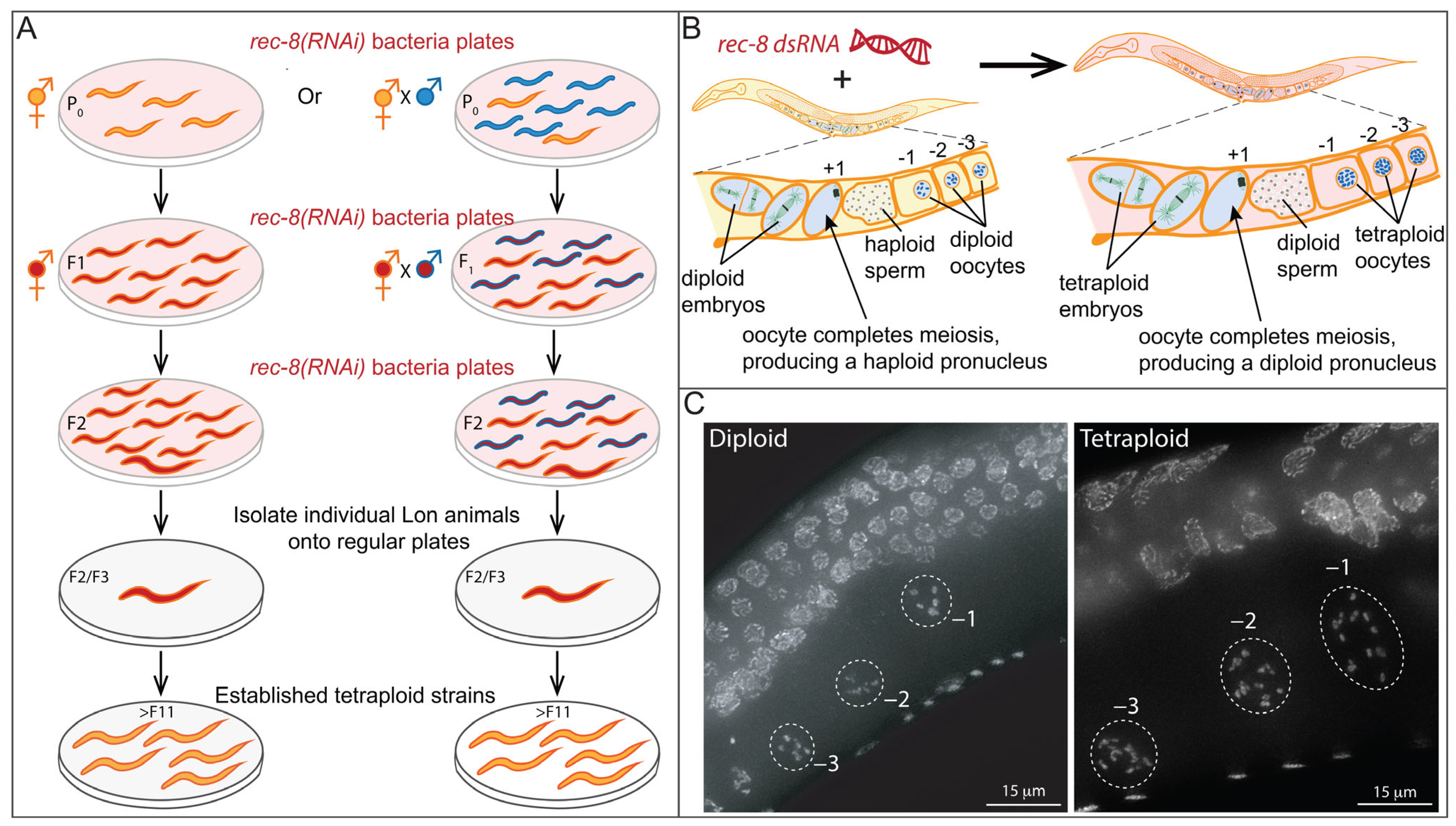
  80. Clarke, E.K.; Gomez, K.A.R.; Mustachi, Z.; Murph, M.C.; Schvarzstein, M. Manipulation of Ploidy in Caenorhabditis elegans. J. Vis. Exp. 2018, 133, 57296. [Google Scholar] [CrossRef] [Green Version]
  81. Nigon, V. Les Modalites de La Reproduction et Le Determinisme Du Sexe Chez Quelques Nematodes Libres. Ann. Sci. Nat. Zool. Biol. Anim. 1949, 11, 1–132. [Google Scholar]
  82. Hodgkin, J. Male phenotypes and mating efficiency in Caenorhabditis elegans. Genetics 1983, 103, 43–64. [Google Scholar] [CrossRef] [PubMed]
  83. Hodgkin, J. Primary sex determination in the nematode C. elegans. Development 1987, 101, 5–16. [Google Scholar] [CrossRef] [PubMed]
  84. Nigon, V.M.; Félix, M.-A. History of research on C. elegans and other free-living nematodes as model organisms. Wormbook 2017, 2017, 1–84. [Google Scholar] [CrossRef] [Green Version]
  85. Brenner, S. The Genetics of Caenorhabditis elegans. Genetics 1974, 77, 71–94. [Google Scholar] [CrossRef]
  86. Hedgecock, E.M.; White, J.G. Polyploid tissues in the nematode Caenorhabditis elegans. Dev. Biol. 1985, 107, 128–133. [Google Scholar] [CrossRef] [PubMed]
  87. Flemming, A.J.; Shen, Z.-Z.; Cunha, A.; Emmons, S.W.; Leroi, A.M. Somatic polyploidization and cellular proliferation drive body size evolution in nematodes. Proc. Natl. Acad. Sci. USA 2000, 97, 5285–5290. [Google Scholar] [CrossRef] [Green Version]
  88. Hodgkin, J.; Horvitz, H.R.; Brenner, S. Nondisjunction mutants of the nematode Caenorhabditis elegans. Genetics 1979, 91, 67–94. [Google Scholar] [CrossRef]
  89. Sigurdson, D.C.; Spanier, G.J.; Herman, R.K. Caenorhabditis elegans deficiency mapping. Genetics 1984, 108, 331–345. [Google Scholar] [CrossRef]
  90. Haack, H.; Hodgkin, J. Tests for parental imprinting in the nematode Caenorhabditis elegans. Mol. Genet. Genom. 1991, 228, 482–485. [Google Scholar] [CrossRef]
  91. Hodgkin, J. Karyotype, Ploidy, and Gene Dosage. In Wormbook. The C. elegans Research Community, WormBook. 2005. Available online: http://www.wormbook.org/chapters/www_karyotype/karyotype.html (accessed on 30 December 2022).
  92. Larkin, K.; Tucci, C.; Neiman, M. Effects of polyploidy and reproductive mode on life history trait expression. Ecol. Evol. 2016, 6, 765–778. [Google Scholar] [CrossRef] [PubMed] [Green Version]
  93. Schoonmaker, A.; Hao, Y.; Bird, D.; Conant, G.C. A Single, Shared Triploidy in Three Species of Parasitic Nematodes. G3 Genes|Genom.|Genet. 2019, 10, 225–233. [Google Scholar] [CrossRef] [Green Version]
  94. Madl, J.E.; Herman, R.K. Polyploids and sex determination in Caenorhabditis elegans. Genetics 1979, 93, 393–402. [Google Scholar] [CrossRef] [PubMed]
  95. Vargas, E.; McNally, K.; Friedman, J.A.; Cortes, D.B.; Wang, D.Y.; Korf, I.F.; McNally, F.J. Autosomal Trisomy and Triploidy Are Corrected During Female Meiosis in Caenorhabditis elegans. Genetics 2017, 207, 911–922. [Google Scholar] [CrossRef] [Green Version]
  96. Nigon, V. Effets de la polyploïdie chez un nématode libre. C. R. Acad. Sci. 1949, 228, 1161–1162. [Google Scholar]
  97. Nigon, V. Polyploidie Experimentale Chez Un Nematode Libre, Rhabditis Elegans. Maupas. Bull. Biol. Fr. Belg. 1951, 85, 187–225. [Google Scholar]
  98. Mlynarczyk-Evans, S.; Roelens, B.; Villeneuve, A.M. Evidence That Masking of Synapsis Imperfections Counterbalances Quality Control to Promote Efficient Meiosis. PLoS Genet. 2013, 9, e1003963. [Google Scholar] [CrossRef] [Green Version]
  99. Roelens, B.; Schvarzstein, M.; Villeneuve, A.M. Manipulation of Karyotype in Caenorhabditis elegans Reveals Multiple Inputs Driving Pairwise Chromosome Synapsis During Meiosis. Genetics 2015, 201, 1363–1379. [Google Scholar] [CrossRef] [Green Version]
  100. Hodgkin, J. Exploring the Envelope. Systematic Alteration in the Sex-Determination System of the Nematode Caenorhabditis elegans. Genetics 2002, 162, 767–780. [Google Scholar] [CrossRef] [PubMed]
  101. Meneely, P.M. Sex determination in polyploids of Caenorhabditis elegans. Genetics 1994, 137, 467–481. [Google Scholar] [CrossRef]
  102. Meyer, B.J. X-Chromosome Dosage Compensation. In Wormbook. The C. elegans Research Community, WormBook. 2005, pp. 1–14. Available online: http://www.wormbook.org/chapters/www_dosagecomp/dosagecomp.html (accessed on 30 December 2022).
  103. Meyer, B.J. The X chromosome in C. elegans sex determination and dosage compensation. Curr. Opin. Genet. Dev. 2022, 74, 101912. [Google Scholar] [CrossRef] [PubMed]
  104. Schvarzstein, M.; Wignall, S.M.; Villeneuve, A.M. Coordinating cohesion, co-orientation, and congression during meiosis: Lessons from holocentric chromosomes. Genes Dev. 2010, 24, 219–228. [Google Scholar] [CrossRef] [Green Version]
  105. MacQueen, A.J.; Phillips, C.; Bhalla, N.; Weiser, P.; Villeneuve, A.M.; Dernburg, A.F. Chromosome Sites Play Dual Roles to Establish Homologous Synapsis during Meiosis in C. elegans. Cell 2005, 123, 1037–1050. [Google Scholar] [CrossRef] [PubMed] [Green Version]
  106. Cortes, D.B.; McNally, K.L.; Mains, P.E.; McNally, F.J. The Asymmetry of Female Meiosis Reduces the Frequency of Inheritance of Unpaired Chromosomes. Elife 2015, 4, e06056. [Google Scholar] [CrossRef] [PubMed]
  107. Meyerzon, M.; Gao, Z.; Liu, J.; Wu, J.-C.; Malone, C.J.; Starr, D.A. Centrosome attachment to the C. elegans male pronucleus is dependent on the surface area of the nuclear envelope. Dev. Biol. 2009, 327, 433–446. [Google Scholar] [CrossRef] [Green Version]
  108. Schierenberg, E.; Wood, W.B. Control of cell-cycle timing in early embryos of Caenorhabditis elegans. Dev. Biol. 1985, 107, 337–354. [Google Scholar] [CrossRef]
  109. Sadler, P.L.; Shakes, D.C. Anucleate Caenorhabditis elegans sperm can crawl, fertilize oocytes and direct anterior-posterior polarization of the 1-cell embryo. Development 2000, 127, 355–366. [Google Scholar] [CrossRef]
  110. Hara, Y.; Kimura, A. An allometric relationship between mitotic spindle width, spindle length, and ploidy in Caenorhabditis elegans embryos. Mol. Biol. Cell 2013, 24, 1411–1419. [Google Scholar] [CrossRef]
  111. Schierenberg, E. Laser-Induced Cell Fusion. In Cell Fusion; Springer: Boston, MA, USA, 1987; pp. 409–418. [Google Scholar] [CrossRef]
  112. Oegema, K. Cell Division. In Wormbook. The C. elegans Research Community, WormBook. 2006, pp. 1–40. Available online: http://www.wormbook.org/chapters/www_celldivision/celldivision.html (accessed on 30 December 2022).
  113. Malone, C.J.; Misner, L.; Le Bot, N.; Tsai, M.-C.; Campbell, J.M.; Ahringer, J.; White, J.G. The C. elegans Hook Protein, ZYG-12, Mediates the Essential Attachment between the Centrosome and Nucleus. Cell 2003, 115, 825–836. [Google Scholar] [CrossRef] [Green Version]
  114. Gönczy, P.; Pichler, S.; Kirkham, M.; Hyman, A.A. Cytoplasmic Dynein Is Required for Distinct Aspects of Mtoc Positioning, Including Centrosome Separation, in the One Cell Stage Caenorhabditis elegans Embryo. J. Cell Biol. 1999, 147, 135–150. [Google Scholar] [CrossRef]
  115. Newport, J.; Kirschner, M. A major developmental transition in early xenopus embryos: I. characterization and timing of cellular changes at the midblastula stage. Cell 1982, 30, 675–686. [Google Scholar] [CrossRef] [PubMed]
  116. Sachsenmaier, W.; Remy, U.; Plattner-Schobel, R. Initiation of synchronous mitosis in Physarum polycephalum: A model of the control of cell division in eukariots. Exp. Cell Res. 1972, 73, 41–48. [Google Scholar] [CrossRef] [PubMed]
  117. A Fantes, P. Isolation of cell size mutants of a fission yeast by a new selective method: Characterization of mutants and implications for division control mechanisms. J. Bacteriol. 1981, 146, 746–754. [Google Scholar] [CrossRef] [PubMed] [Green Version]
  118. Matson, J.P.; Cook, J.G. Cell cycle proliferation decisions: The impact of single cell analyses. FEBS J. 2017, 284, 362–375. [Google Scholar] [CrossRef] [Green Version]
  119. Arata, Y.; Takagi, H. Quantitative Studies for Cell-Division Cycle Control. Front. Physiol. 2019, 10, 1022. [Google Scholar] [CrossRef] [PubMed] [Green Version]
  120. Koreth, J.; Heuvel, S.V.D. Cell-cycle control in Caenorhabditis elegans: How the worm moves from G1 to S. Oncogene 2005, 24, 2756–2764. [Google Scholar] [CrossRef] [Green Version]
  121. Wang, Z. Regulation of Cell Cycle Progression by Growth Factor-Induced Cell Signaling. Cells 2021, 10, 3327. [Google Scholar] [CrossRef]
  122. Dumont, S.; Mitchison, T.J. Compression Regulates Mitotic Spindle Length by a Mechanochemical Switch at the Poles. Curr. Biol. 2009, 19, 1086–1095. [Google Scholar] [CrossRef] [Green Version]
  123. Itabashi, T.; Takagi, J.; Shimamoto, Y.; Onoe, H.; Kuwana, K.; Shimoyama, I.; Gaetz, J.; Kapoor, T.M.; Ishiwata, S. Probing the mechanical architecture of the vertebrate meiotic spindle. Nat. Methods 2009, 6, 167–172. [Google Scholar] [CrossRef]
  124. E Mains, P.; Kemphues, K.J.; Sprunger, S.A.; Sulston, I.A.; Wood, W.B. Mutations affecting the meiotic and mitotic divisions of the early Caenorhabditis elegans embryo. Genetics 1990, 126, 593–605. [Google Scholar] [CrossRef]
  125. Quintin, S.; E Mains, P.; Zinke, A.; A Hyman, A. The mbk-2 kinase is required for inactivation of MEI-1/katanin in the one-cell Caenorhabditis elegans embryo. EMBO Rep. 2003, 4, 1175–1181. [Google Scholar] [CrossRef] [PubMed] [Green Version]
  126. Segbert, C.; Barkus, R.; Powers, J.; Strome, S.; Saxton, W.M.; Bossinger, O. KLP-18, a Klp2 Kinesin, Is Required for Assembly of Acentrosomal Meiotic Spindles in Caenorhabditis elegans. Mol. Biol. Cell 2003, 14, 4458–4469. [Google Scholar] [CrossRef] [PubMed] [Green Version]
  127. Maddox, A.S.; Habermann, B.; Desai, A.; Oegema, K. Distinct roles for two C. elegans anillins in the gonad and early embryo. Development 2005, 132, 2837–2848. [Google Scholar] [CrossRef] [PubMed] [Green Version]
  128. Dorn, J.F.; Zhang, L.; Paradis, V.; Edoh-Bedi, D.; Jusu, S.; Maddox, P.S.; Maddox, A.S. Actomyosin Tube Formation in Polar Body Cytokinesis Requires Anillin in C. elegans. Curr. Biol. 2010, 20, 2046–2051. [Google Scholar] [CrossRef] [Green Version]
  129. Fankhauser, G. Cytological studies on egg fragments of the salamander Triton. V. Chromosome number and chromosome individuality in the cleavage mitoses of merogonic fragments. J. Exp. Zool. 1934, 68, 18. [Google Scholar] [CrossRef]
  130. Hara, Y.; Iwabuchi, M.; Ohsumi, K.; Kimura, A. Intranuclear DNA density affects chromosome condensation in metazoans. Mol. Biol. Cell 2013, 24, 2442–2453. [Google Scholar] [CrossRef]
  131. Conklin, E.G. Cell size and nuclear size. J. Exp. Zool. 1912, 12, 1–98. [Google Scholar] [CrossRef] [Green Version]
  132. Di Berardino, M.A. The karyotype of Rana pipiens and investigation of its stability during embryonic differentiation. Dev. Biol. 1962, 5, 101–126. [Google Scholar] [CrossRef]
  133. Belmont, A.S.; Sedat, J.W.; Agard, D.A. A three-dimensional approach to mitotic chromosome structure: Evidence for a complex hierarchical organization. J. Cell Biol. 1987, 105, 77–92. [Google Scholar] [CrossRef]
  134. Deppe, U.; Schierenberg, E.; Cole, T.; Krieg, C.; Schmitt, D.; Yoder, B.; von Ehrenstein, G. Cell lineages of the embryo of the nematode Caenorhabditis elegans. Proc. Natl. Acad. Sci. USA 1978, 75, 376–380. [Google Scholar] [CrossRef] [Green Version]
  135. Sulston, J.E.; Schierenberg, E.; White, J.G.; Thomson, J.N. The embryonic cell lineage of the nematode Caenorhabditis elegans. Dev. Biol. 1983, 100, 64–119. [Google Scholar] [CrossRef]
  136. Greenwald, I. Development of the Vulva, 2nd ed.; C. elegans II.; Cold Spring Harbor Laboratory Press: New York, NY, USA, 1997; Chapter 19; pp. 519–541. [Google Scholar]
  137. Simske, J.S.; Hardin, J. Getting into Shape: Epidermal Morphogenesis in Caenorhabditis elegans Embryos. Bioessays 2001, 23, 12–23. [Google Scholar] [CrossRef] [PubMed]
  138. Wong, M.-C.; Schwarzbauer, J.E. Gonad morphogenesis and distal tip cell migration in the Caenorhabditis elegans hermaphrodite. Wiley Interdiscip. Rev. Dev. Biol. 2012, 1, 519–531. [Google Scholar] [CrossRef] [Green Version]
  139. Shemer, G.; Podbilewicz, B. Fusomorphogenesis: Cell fusion in organ formation. Dev. Dyn. 2000, 218, 30–51. [Google Scholar] [CrossRef]
  140. Tsou, M.-F.B.; Stearns, T. Mechanism limiting centrosome duplication to once per cell cycle. Nature 2006, 442, 947–951. [Google Scholar] [CrossRef] [PubMed]
  141. Tsou, M.-F.B.; Stearns, T. Controlling centrosome number: Licenses and blocks. Curr. Opin. Cell Biol. 2006, 18, 74–78. [Google Scholar] [CrossRef] [PubMed]
  142. Duensing, A.; Duensing, S. Centrosomes, Polyploidy and Cancer. Adv. Exp. Med. Biol. 2010, 676, 93–103. [Google Scholar] [CrossRef]
  143. Lu, Y.; Roy, R. Centrosome/Cell Cycle Uncoupling and Elimination in the Endoreduplicating Intestinal Cells of C. elegans. PLoS ONE 2014, 9, e110958-17. [Google Scholar] [CrossRef] [Green Version]
  144. Song, M.H.; Liu, Y.; Anderson, D.E.; Jahng, W.J.; O’Connell, K.F. Protein Phosphatase 2A-SUR-6/B55 Regulates Centriole Duplication in C. elegans by Controlling the Levels of Centriole Assembly Factors. Dev. Cell 2011, 20, 563–571. [Google Scholar] [CrossRef] [Green Version]
  145. Kitagawa, D.; Flückiger, I.; Polanowska, J.; Keller, D.; Reboul, J.; Gönczy, P. PP2A Phosphatase Acts upon SAS-5 to Ensure Centriole Formation in C. elegans Embryos. Dev. Cell 2011, 20, 550–562. [Google Scholar] [CrossRef]
  146. Decker, M.; Jaensch, S.; Pozniakovsky, A.; Zinke, A.; O’Connell, K.F.; Zachariae, W.; Myers, E.; Hyman, A.A. Limiting Amounts of Centrosome Material Set Centrosome Size in C. elegans Embryos. Curr. Biol. 2011, 21, 1259–1267. [Google Scholar] [CrossRef] [Green Version]
  147. Lee, H.O.; Davidson, J.M.; Duronio, R.J. Endoreplication: Polyploidy with purpose. Genes Dev. 2009, 23, 2461–2477. [Google Scholar] [CrossRef] [Green Version]
  148. Zhang, L.; Ward, J.D.; Cheng, Z.; Dernburg, A.F. The auxin-inducible degradation (AID) system enables versatile conditional protein depletion in C. elegans. Development 2015, 142, 4374–4384. [Google Scholar] [CrossRef] [PubMed] [Green Version]
  149. Jorgensen, P.; Edgington, N.P.; Schneider, B.L.; Rupeš, I.; Tyers, M.; Futcher, B. The Size of the Nucleus Increases as Yeast Cells Grow. Mol. Biol. Cell 2007, 18, 3523–3532. [Google Scholar] [CrossRef] [PubMed] [Green Version]
  150. Yahya, G.; Menges, P.; Amponsah, P.S.; Ngandiri, D.A.; Schulz, D.; Wallek, A.; Kulak, N.; Mann, M.; Cramer, P.; Savage, V.; et al. Sublinear scaling of the cellular proteome with ploidy. Nat. Commun. 2022, 13, 6182. [Google Scholar] [CrossRef] [PubMed]
  151. Neumann, F.R.; Nurse, P. Nuclear size control in fission yeast. J. Cell Biol. 2007, 179, 593–600. [Google Scholar] [CrossRef] [PubMed] [Green Version]
  152. Gillooly, J.F.; Hein, A.; Damiani, R. Nuclear DNA Content Varies with Cell Size across Human Cell Types. Cold Spring Harb. Perspect. Biol. 2015, 7, a019091. [Google Scholar] [CrossRef] [Green Version]
  153. Katagiri, Y.; Hasegawa, J.; Fujikura, U.; Hoshino, R.; Matsunaga, S.; Tsukaya, H. The coordination of ploidy and cell size differs between cell layers in leaves. Development 2016, 143, 1120–1125. [Google Scholar] [CrossRef] [Green Version]
  154. Anatskaya, O.V.; E Vinogradov, A. Paraoxical relationship between protein content and nucleolar activity in mammalian cardiomyocytes. Genome 2004, 47, 565–578. [Google Scholar] [CrossRef]
  155. Robinson, D.O.; Coate, J.E.; Singh, A.; Hong, L.; Bush, M.; Doyle, J.J.; Roeder, A.H. Ploidy and Size at Multiple Scales in the Arabidopsis Sepal. Plant Cell 2018, 30, 2308–2329. [Google Scholar] [CrossRef] [Green Version]
  156. Hisanaga, T.; Kawade, K.; Tsukaya, H. Compensation: A key to clarifying the organ-level regulation of lateral organ size in plants. J. Exp. Bot. 2015, 66, 1055–1063. [Google Scholar] [CrossRef] [Green Version]
  157. Kawade, K.; Horiguchi, G.; Tsukaya, H. Non-cell-autonomously coordinated organ size regulation in leaf development. Development 2010, 137, 4221–4227. [Google Scholar] [CrossRef] [Green Version]
  158. Cadart, C.; Heald, R. Scaling of biosynthesis and metabolism with cell size. Mol. Biol. Cell 2022, 33, pe5. [Google Scholar] [CrossRef]
  159. Cantwell, H.; Dey, G. Nuclear size and shape control. Semin. Cell Dev. Biol. 2022, 130, 90–97. [Google Scholar] [CrossRef]
  160. Tsukaya, H. Re-examination of the role of endoreduplication on cell-size control in leaves. J. Plant Res. 2019, 132, 571–580. [Google Scholar] [CrossRef] [PubMed] [Green Version]
  161. Horiguchi, G.; Tsukaya, H. Organ Size Regulation in Plants: Insights from Compensation. Front. Plant Sci. 2011, 2, 24. [Google Scholar] [CrossRef] [PubMed] [Green Version]
  162. Lozano, E.; Sáez, A.G.; Flemming, A.J.; Cunha, A.; Leroi, A.M. Regulation of Growth by Ploidy in Caenorhabditis elegans. Curr. Biol. 2006, 16, 493–498. [Google Scholar] [CrossRef] [PubMed]
  163. Nyström, J.; Shen, Z.-Z.; Aili, M.; Flemming, A.J.; Leroi, A.; Tuck, S. Increased or decreased levels of Caenorhabditis elegans lon-3, a gene encoding a collagen, cause reciprocal changes in body length. Genetics 2002, 161, 83–97. [Google Scholar] [CrossRef]
  164. Morita, K.; Flemming, A.J.; Sugihara, Y.; Mochii, M.; Suzuki, Y.; Yoshida, S.; Wood, W.B.; Kohara, Y.; Leroi, A.M.; Ueno, N. A Caenorhabditis elegans TGF-β, DBL-1, controls the expression of LON-1, a PR-related protein, that regulates polyploidization and body length. EMBO J. 2002, 21, 1063–1073. [Google Scholar] [CrossRef] [PubMed] [Green Version]
  165. Morita, K.; Chow, K.L.; Ueno, N. Regulation of body length and male tail ray pattern formation of Caenorhabditis elegans by a member of TGF-β family. Development 1999, 126, 1337–1347. [Google Scholar] [CrossRef]
  166. Hirose, T.; Nakano, Y.; Nagamatsu, Y.; Misumi, T.; Ohta, H.; Ohshima, Y. Cyclic GMP-dependent protein kinase EGL-4 controls body size and lifespan in C. elegans. Development 2003, 130, 1089–1099. [Google Scholar] [CrossRef] [Green Version]
  167. Nagamatsu, Y.; Ohshima, Y. Mechanisms for the control of body size by a G-kinase and a downstream TGFbeta signal pathway in Caenorhabditis elegans. Genes Cells 2004, 9, 39–47. [Google Scholar] [CrossRef] [Green Version]
  168. Wang, J.; Tokarz, R.; Savage-Dunn, C. The expression of TGFβ signal transducers in the hypodermis regulates body size in C. elegans. Development 2002, 129, 4989–4998. [Google Scholar] [CrossRef] [PubMed]
  169. Parisi, T.; Beck, A.R.; Rougier, N.; McNeil, T.; Lucian, L.; Werb, Z.; Amati, B. Cyclins E1 and E2 are required for endoreplication in placental trophoblast giant cells. EMBO J. 2003, 22, 4794–4803. [Google Scholar] [CrossRef] [Green Version]
  170. Geng, Y.; Yu, Q.; Sicinska, E.; Das, M.; E Schneider, J.; Bhattacharya, S.; Rideout, W.M.; Bronson, R.T.; Gardner, H.; Sicinski, P. Cyclin E Ablation in the Mouse. Cell 2003, 114, 431–443. [Google Scholar] [CrossRef] [PubMed] [Green Version]
  171. Tuck, S. The control of cell growth and body size in Caenorhabditis elegans. Exp. Cell Res. 2014, 321, 71–76. [Google Scholar] [CrossRef] [PubMed]
  172. Suzuki, Y.; Yandell, M.D.; Roy, P.J.; Krishna, S.; Savage-Dunn, C.; Ross, R.M.; Padgett, R.W.; Wood, W.B. A BMP homolog acts as a dose-dependent regulator of body size and male tail patterning in Caenorhabditis elegans. Development 1999, 126, 241–250. [Google Scholar] [CrossRef]
  173. Savage-Dunn, C.; Yu, L.; Gill, K.; Awan, M.; Fernando, T. Non-stringent tissue-source requirements for BMP ligand expression in regulation of body size in Caenorhabditis elegans. Genet. Res. 2011, 93, 427–432. [Google Scholar] [CrossRef] [Green Version]
  174. Savage-Dunn, C. Targets of TGFβ-related signaling in Caenorhabditis elegans. Cytokine Growth Factor Rev. 2001, 12, 305–312. [Google Scholar] [CrossRef]
  175. Savage-Dunn, C.; Tokarza, R.; Wangb, H.; Cohenb, S.; Giannikasa, C.; Padgett, R.W. SMA-3 Smad Has Specific and Critical Functions in DBL-1/SMA-6 TGFβ-Related Signaling. Dev. Biol. 2000, 223, 70–76. [Google Scholar] [CrossRef] [Green Version]
  176. Szyperski, T.; Fernández, C.; Mumenthaler, C.; Wüthrich, K. Structure comparison of human glioma pathogenesis-related protein GliPR and the plant pathogenesis-related protein P14a indicates a functional link between the human immune system and a plant defense system. Proc. Natl. Acad. Sci. USA 1998, 95, 2262–2266. [Google Scholar] [CrossRef] [PubMed] [Green Version]
  177. Cai, Q.; Wang, W.; Gao, Y.; Yang, Y.; Zhu, Z.; Fan, Q. Ce-wts-1 plays important roles in Caenorhabditis elegans development. FEBS Lett. 2009, 583, 3158–3164. [Google Scholar] [CrossRef] [PubMed] [Green Version]
  178. Jones, K.T.; Greer, E.R.; Pearce, D.; Ashrafi, K. Rictor/TORC2 Regulates Caenorhabditis elegans Fat Storage, Body Size, and Development through sgk-1. PLoS Biol. 2009, 7, e1000060. [Google Scholar] [CrossRef] [Green Version]
  179. Oldham, S.; Böhni, R.; Stocker, H.; Brogiolo, W.; Hafen, E. Genetic control of size in Drosophila. Philos. Trans. R. Soc. B Biol. Sci. 2000, 355, 945–952. [Google Scholar] [CrossRef] [PubMed] [Green Version]
  180. Schmelzle, T.; Hall, M.N. TOR, a Central Controller of Cell Growth. Cell 2000, 103, 253–262. [Google Scholar] [CrossRef] [PubMed] [Green Version]
  181. Zhang, W.; Liu, H.T. MAPK signal pathways in the regulation of cell proliferation in mammalian cells. Cell Res. 2002, 12, 9–18. [Google Scholar] [CrossRef]
  182. Watanabe, N.; Nagamatsu, Y.; Gengyo-Ando, K.; Mitani, S.; Ohshima, Y. Control of body size by SMA-5, a homolog of MAP kinase BMK1/ERK5, in C. elegans. Development 2005, 132, 3175–3184. [Google Scholar] [CrossRef] [Green Version]
  183. Hyun, S. Body size regulation and insulin-like growth factor signaling. Cell. Mol. Life Sci. 2013, 70, 2351–2365. [Google Scholar] [CrossRef]
  184. McKeown, C.; Praitis, V.; Austin, J. sma-1 encodes a βH-spectrin homolog required for Caenorhabditis elegans morphogenesis. Development 1998, 125, 2087–2098. [Google Scholar] [CrossRef]
  185. Hayashi, S.; Yokoyama, H.; Tamura, K.; Hayashi, S.; Yokoyama, H.; Tamura, K. Roles of Hippo signaling pathway in size control of organ regeneration. Dev. Growth Differ. 2015, 57, 341–351. [Google Scholar] [CrossRef]
  186. Goodman, M.B.; Savage-Dunn, C. Reciprocal interactions between transforming growth factor beta signaling and collagens: Insights from C aenorhabditis elegans. Dev. Dyn. 2022, 251, 47–60. [Google Scholar] [CrossRef] [PubMed]
  187. Song, Q.; Ando, A.; Jiang, N.; Ikeda, Y.; Chen, Z.J. Single-cell RNA-seq analysis reveals ploidy-dependent and cell-specific transcriptome changes in Arabidopsis female gametophytes. Genome Biol. 2020, 21, 178. [Google Scholar] [CrossRef] [PubMed]
  188. Wu, C.-Y.; Rolfe, P.A.; Gifford, D.K.; Fink, G.R. Control of Transcription by Cell Size. PLoS Biol. 2010, 8, e1000523. [Google Scholar] [CrossRef] [Green Version]
  189. Marguerat, S.; Bähler, J. Coordinating genome expression with cell size. Trends Genet. 2012, 28, 560–565. [Google Scholar] [CrossRef] [PubMed] [Green Version]
  190. Coate, J.E.; Doyle, J.J. Variation in transcriptome size: Are we getting the message? Chromosoma 2015, 124, 27–43. [Google Scholar] [CrossRef] [PubMed]
  191. Leitch, I.J.; Bennett, M.D. Genome downsizing in polyploid plants. Biol. J. Linn. Soc. 2004, 82, 651–663. [Google Scholar] [CrossRef] [Green Version]
  192. Zenil-Ferguson, R.; Ponciano, J.M.; Burleigh, J.G. Evaluating the role of genome downsizing and size thresholds from genome size distributions in angiosperms. Am. J. Bot. 2016, 103, 1175–1186. [Google Scholar] [CrossRef] [Green Version]
  193. Pellicer, J.; Hidalgo, O.; Dodsworth, S.; Leitch, I.J. Genome Size Diversity and Its Impact on the Evolution of Land Plants. Genes 2018, 9, 88. [Google Scholar] [CrossRef] [Green Version]
  194. Cheng, F.; Wu, J.; Cai, X.; Liang, J.; Freeling, M.; Wang, X. Gene retention, fractionation and subgenome differences in polyploid plants. Nat. Plants 2018, 4, 258–268. [Google Scholar] [CrossRef]
  195. Baduel, P.; Quadrana, L.; Hunter, B.; Bomblies, K.; Colot, V. Relaxed purifying selection in autopolyploids drives transposable element over-accumulation which provides variants for local adaptation. Nat. Commun. 2019, 10, 5818. [Google Scholar] [CrossRef] [Green Version]
  196. Raina, S.N.; Parida, A.; Koul, K.K.; Salimath, S.S.; Bisht, M.S.; Raja, V.; Khoshoo, T.N. Associated chromosomal DNA changes in polyploids. Genome 1994, 37, 560–564. [Google Scholar] [CrossRef] [PubMed]
  197. Lovén, J.; Orlando, D.A.; Sigova, A.A.; Lin, C.Y.; Rahl, P.B.; Burge, C.B.; Levens, D.L.; Lee, T.I.; Young, R.A. Revisiting Global Gene Expression Analysis. Cell 2012, 151, 476–482. [Google Scholar] [CrossRef] [PubMed] [Green Version]
  198. Bourdon, M.; Pirrello, J.; Cheniclet, C.; Coriton, O.; Bourge, M.; Brown, S.; Moïse, A.; Peypelut, M.; Rouyère, V.; Renaudin, J.-P.; et al. Evidence for karyoplasmic homeostasis during endoreduplication and a ploidy-dependent increase in gene transcription during tomato fruit growth. Development 2012, 139, 3817–3826. [Google Scholar] [CrossRef] [PubMed] [Green Version]
  199. Voichek, Y.; Bar-Ziv, R.; Barkai, N. Expression homeostasis during DNA replication. Science 2016, 351, 1087–1090. [Google Scholar] [CrossRef] [PubMed]
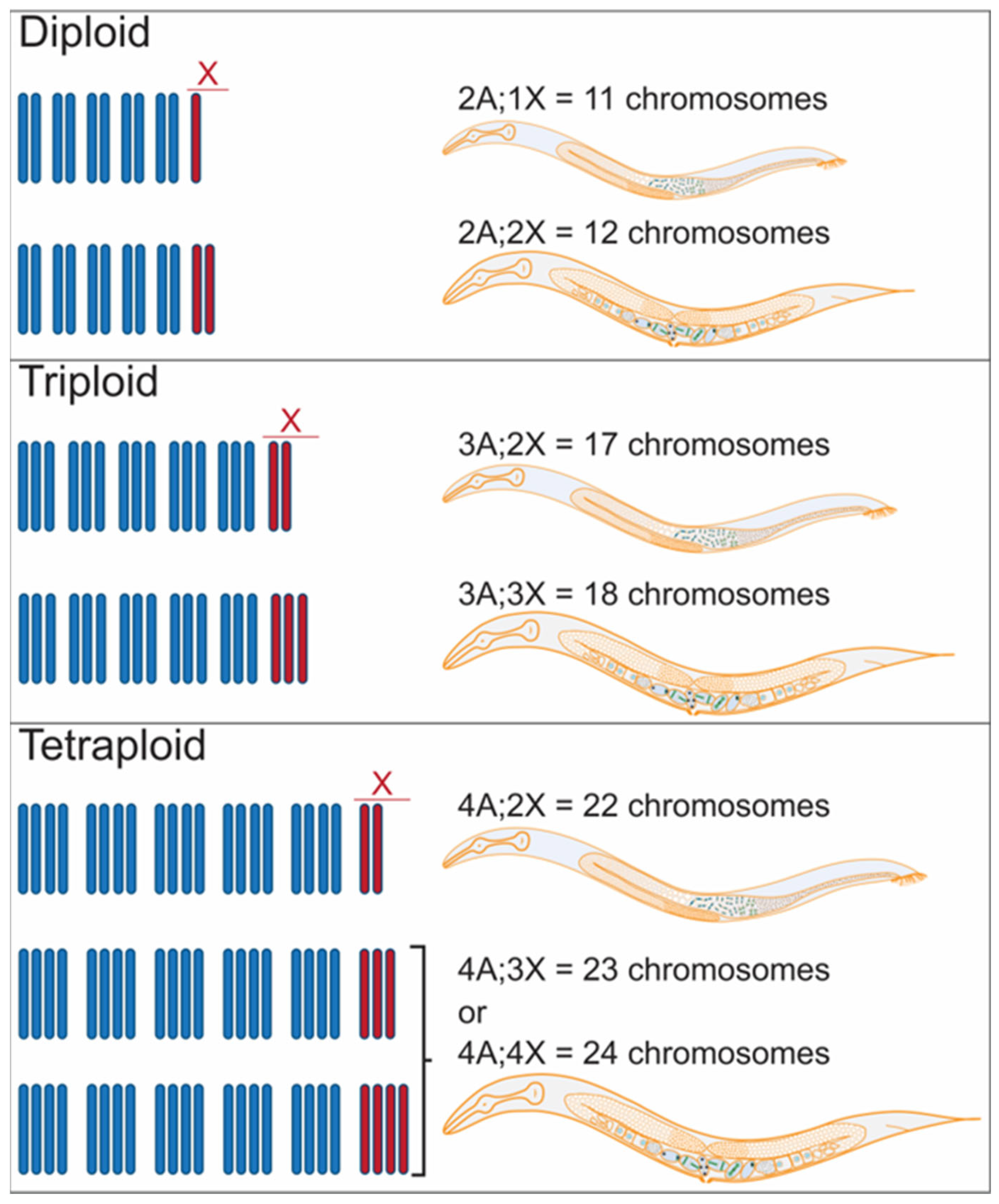
Figure 1. Roles of whole genome duplication (WGD). Diagram depicting key examples of processes affected by polyploidization/WGD. Polyploidy can occur at the cellular, tissue, or organismal levels. It is a key step in important biological processes affecting cellular metabolism and function, normal development, tissue homeostasis, regeneration, wound healing, organismal adaptation to the environment, and speciation. In addition, polyploidization can cause and/or prevent disease and repair injuries.
Jdb 11 00026 g001
Figure 2. Shared causes and downstream effects of WGD across biological settings. Many forms of stress can lead an organism to undergo WGD. Whether as a response to the stress itself or as a way to adapt to the changing environment and/or different selective pressures, gene and epigenome changes lead to an altered proteome that supports an adaptive response to the initial stress exposure.
Jdb 11 00026 g002
Figure 3. Method for rapid isolation of C. elegans tetraploids. (A). Diagram of the published method for generating tetraploid strains from diploids. This protocol relies on the transient knockdown of the meiosis-specific cohesin rec-8 which results in the production of diploid instead of haploid gametes. Thus, upon fertilization, tetraploid progeny (which are longer and bigger) are produced. The method works both during the selfing of the hermaphrodites (left) and during outcrossing (right). Crossing allows for the generation of tetraploids with two different genetic backgrounds. After two to three generations of exposure to rec-8 dsRNA, the larger hermaphrodites are individually plated on normal growth media. Strains that consistently produce large (Lon) hermaphrodites are then screened to confirm that these strains are tetraploids. (B). Diploid hermaphrodites (left) produce haploid gametes. Haploid sperm are stored in the spermatheca. Oocytes prior to the meiotic divisions have six pairs of connected homologous chromosomes that can be visualized by DAPI in the diakinesis-stage oocytes (Figure 3C, below) just adjacent to the spermatheca (−1, −2, and −3 oocytes based on position). The oocyte moves through the spermatheca and is fertilized and completes the meiotic divisions to produce a haploid pronucleus. The tetraploid worms derived from exposure to rec-8 dsRNA, (right) produce diploid gametes with 12 DAPI bodies. (C) Examples of fixed hermaphrodites stained with DAPI to screen for ploidy by counting chromosome pairs in the −1, −2, and −3 diakinesis oocytes.
Jdb 11 00026 g003
Figure 4. Sexual fate in C. elegans is determined by the X-chromosome/autosome ratio. C. elegans has six chromosomes, six autosomes, and one X chromosome. Diploid males and hermaphrodites have two sets of autosomes and one or two X chromosomes, respectively. Triploid males and hermaphrodites have 3 sets of autosomes and 2 or 3 X chromosomes making 17 or 18 total chromosomes, respectively. Tetraploid males have 22 chromosomes: 4 sets of autosomes (A) and 2 X chromosomes. In contrast, tetraploid hermaphrodites have four sets of autosomes and either three or four X chromosomes. See Table 1 for a summary of the X/A ratios that determine sex in C. elegans.
Jdb 11 00026 g004
Figure 6. Polyploid tissues in C. elegans. (A) During embryogenesis, the E blastomere undergoes mitotic divisions to give rise to 20 intestinal diploid cells [86]. During the L1 stage, six of these cells neither divide nor replicate their DNA, four cells may or may not replicate their DNA or divide, and ten cells replicate their DNA and divide by endomitosis, producing cells with two diploid nuclei each. Therefore, by the end of the L1 stage, the 20 cells of the intestine have 30–34 diploid nuclei. Prior to the transition molt to the L2 stage, all nuclei of the intestine endoduplicate their DNA (4C each nucleus). Nuclei continue to endoduplicate prior to the molts between larval stages: the L2 (8C each nucleus), the L3 (16C), and finally the L4 (32C) larval stages. Thus, the adult intestine is composed of 20 cells with a total of 30–34 nuclei, each with 32C ploidy [86]. (B) Formation of the hypodermis. During embryogenesis, 23 cells fuse to the hyp7 cell. Post-embryonically, 116 cells fuse to the hyp7 syncytium. About 96 nuclei from these cells undergo one round of endoduplication. Most of these cells undergo two more rounds of endoduplication that take place post-embryonically to give rise to the adult hyp7 syncytium that contains 139 nuclei with 10.7C on average by the fully grown adult stage [87].
Jdb 11 00026 g006
Table 1. X/A ratio determines sex.
KaryotypeX/A RatioSexual Fate
2A;2X1female
2A;1X0.5male
3A;3X1female
3A;2X0.67 *Male
4A;4X1female
4A;3X0.75 *female
4A;2X0.5male
* X/A limits for female and male development. An X/A ratio ≥ 0.75 yields female fates and an X/A ratio ≤ 0.67 promotes male fates.
Disclaimer/Publisher’s Note: The statements, opinions and data contained in all publications are solely those of the individual author(s) and contributor(s) and not of MDPI and/or the editor(s). MDPI and/or the editor(s) disclaim responsibility for any injury to people or property resulting from any ideas, methods, instructions or products referred to in the content.

