
The Next Frontier in Health Disparities—A Closer Look at Exploring Sex Differences in Glioma Data and Omics Analysis, from Bench to Bedside and Back
 , ,
, ,
Abstract
: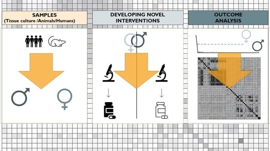
1. Introduction
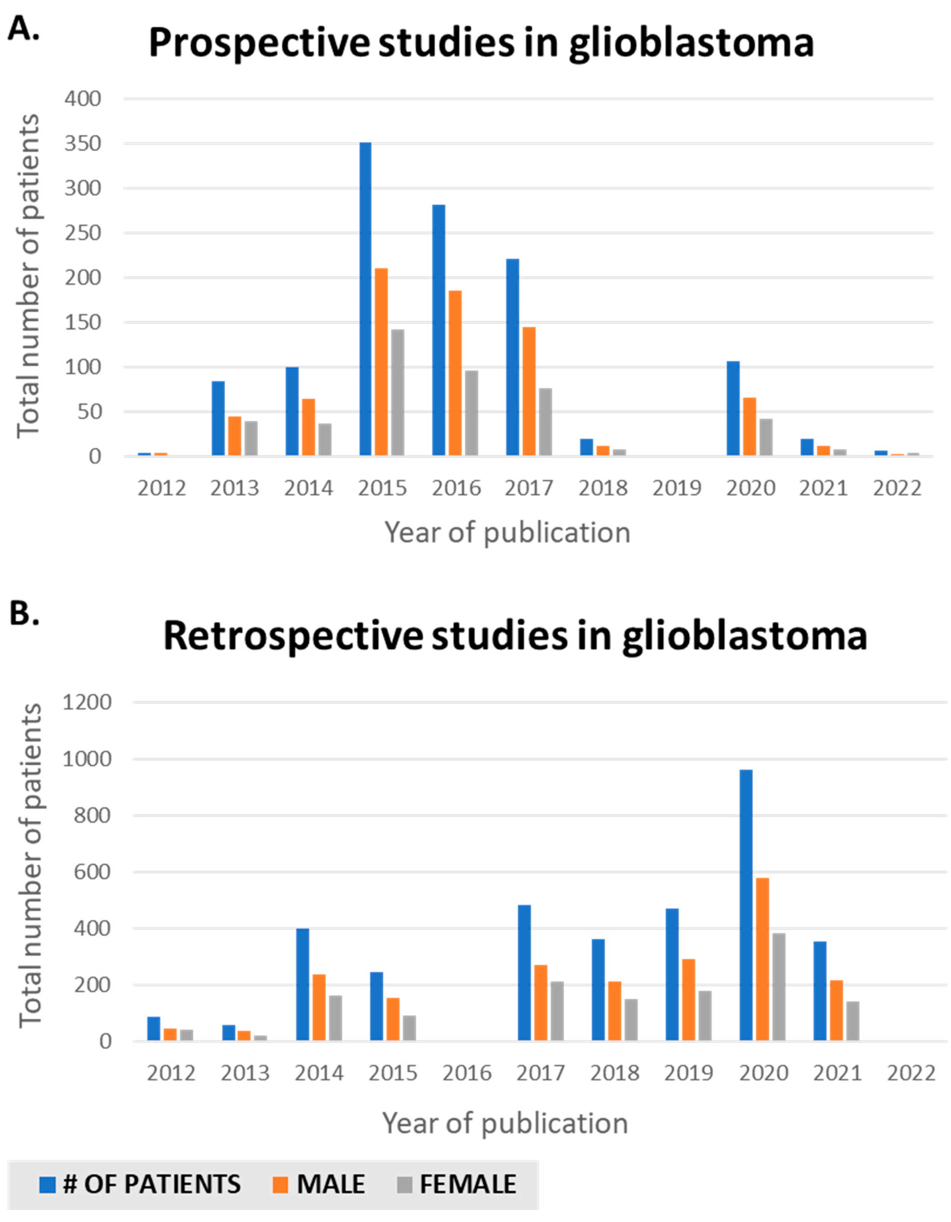
2. Examining Prospective and Retrospective Literature Regarding Male and Female Representation
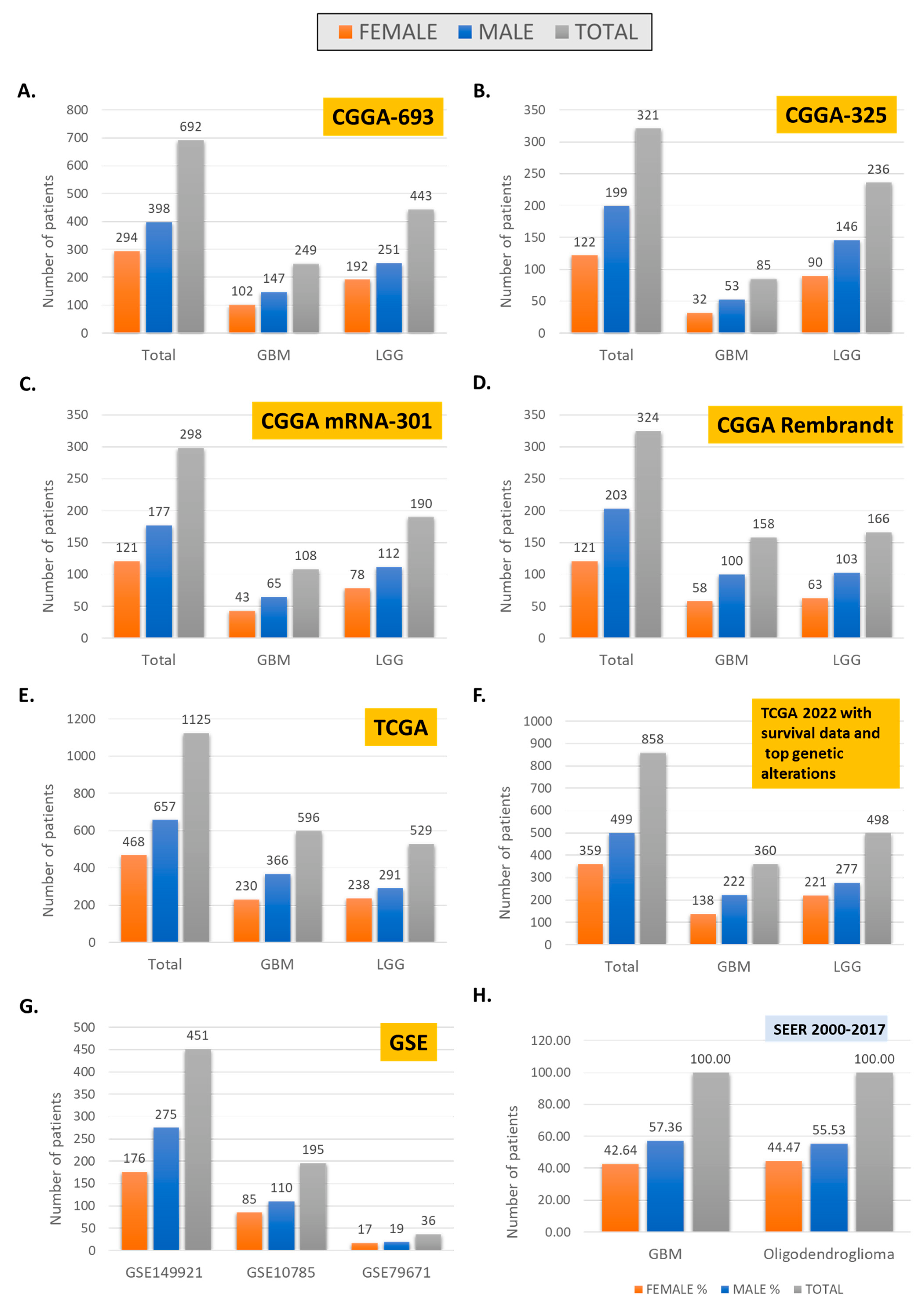
3. Large-Scale Data Sets and Male/Female Representation
4. Omics and Biospecimen Analysis
5. Conclusions
Author Contributions
Funding
Institutional Review Board Statement
Data Availability Statement
Conflicts of Interest
Abbreviations
| CBTRUS | Central Brain Tumor Registry of the United States |
| CGGA | Chinese Glioma Genome Atlas +CNS Central Nervous System |
| GBM | Glioblastoma Multiforme |
| MGMT | Management Science Replication Project. |
| NPCR | National Program of Cancer Registries |
| PDX | Patient-Derived Xenograft |
| SEER | Surveillance, Epidemiology, and End Results |
| TCGA | The Cancer Genome Atlas |
| TCIA | The Cancer Imaging Archive |
| TMB | Tumor Mutational burden |
References
- Paulsen, E. Recognizing, Addressing Unintended Gender Bias in Patient Care. Duke Health Ref. Physicians 2020, 8, 2021. [Google Scholar]
- Feldman, S.; Ammar, W.; Lo, K.; Trepman, E.; Van Zuylen, M.; Etzioni, O. Quantifying Sex Bias in Clinical Studies at Scale With Automated Data Extraction. JAMA Netw. Open 2019, 2, e196700. [Google Scholar] [CrossRef] [PubMed]
- Patil, N.; Somasundaram, E.; Waite, K.A.; Lathia, J.D.; Machtay, M.; Gilbert, M.R.; Connor, J.R.; Rubin, J.B.; Berens, M.E.; Buerki, R.A.; et al. Independently validated sex-specific nomograms for predicting survival in patients with newly diagnosed glioblastoma: NRG Oncology RTOG 0525 and 0825. J. Neuro-Oncol. 2021, 155, 363–372. [Google Scholar] [CrossRef]
- Sorajja, N.; Moore, K.J.; Sample, J.M.; Hubbard, A.K.; Williams, L.A. Global variation in young adult central nervous system tumor incidence by region, age, and sex from 1988 to 2012. Cancer Epidemiol. 2022, 78, 102151. [Google Scholar] [CrossRef]
- Moore, K.J.; Moertel, C.L.; Williams, L.A. Young adult males have worse survival than females that is largely independent of treatment received for many types of central nervous system tumors: A National Cancer Database analysis. Cancer 2022, 128, 1616–1625. [Google Scholar] [CrossRef] [PubMed]
- Tomita, Y.; Tanaka, Y.; Takata, N.; A Hibler, E.; Hashizume, R.; Becher, J.O. Fifteen-year trends and differences in mortality rates across sex, age, and race/ethnicity in patients with brainstem tumors. Neuro-Oncol. Adv. 2021, 3, vdab137. [Google Scholar] [CrossRef] [PubMed]
- Wang, G.-M.; Cioffi, G.; Patil, N.; A Waite, K.; Lanese, R.; Ostrom, Q.T.; Kruchko, C.; E Berens, M.; Connor, J.R.; Lathia, J.D.; et al. Importance of the intersection of age and sex to understand variation in incidence and survival for primary malignant gliomas. Neuro-Oncology 2021, 24, 302–310. [Google Scholar] [CrossRef]
- Lee, J.; Troike, K.; Fodor, R.; Lathia, J.D. Unexplored functions of sex hormones in glioblastoma cancer stem cells. Endocrinology 2022, 163, bqac002. [Google Scholar] [CrossRef]
- Mondragón, J.A.; Serrano, Y.; Torres, A.; Orozco, M.; Segovia, J.; Manjarrez, G.; Romano, M.C. Glioblastoma cells express crucial enzymes involved in androgen synthesis: 3β-hydroxysteroid dehydrogenase, 17-20α-hydroxylase, 17β-hydroxysteroid dehydrogenase and 5α-reductase. Endocrinol. Diabetes Metab. 2021, 4, e00289. [Google Scholar] [CrossRef]
- Tavelin, B.; Malmström, A. Sex Differences in Glioblastoma—Findings from the Swedish National Quality Registry for Primary Brain Tumors between 1999–2018. J. Clin. Med. 2022, 11, 486. [Google Scholar] [CrossRef]
- Lee, J.; Kay, K.; Troike, K.; Ahluwalia, M.S.; Lathia, J.D. Sex Differences in Glioblastoma Immunotherapy Response. NeuroMolecular Med. 2021, 24, 50–55. [Google Scholar] [CrossRef] [PubMed]
- Marcu, L.G. Gender and Sex-Related Differences in Normal Tissue Effects Induced by Platinum Compounds. Pharmaceuticals 2022, 15, 255. [Google Scholar] [CrossRef] [PubMed]
- Özdemir, B.; Oertelt-Prigione, S.; Adjei, A.; Borchmann, S.; Haanen, J.; Letsch, A.; Mir, O.; Quaas, A.; Verhoeven, R.; Wagner, A. Investigation of sex and gender differences in oncology gains momentum: ESMO announces the launch of a Gender Medicine Task Force. Ann. Oncol. 2022, 33, 126–128. [Google Scholar] [CrossRef]
- Garcia, C.A.; Bhargav, A.G.; Brooks, M.; Suárez-Meade, P.; Mondal, S.K.; Zarco, N.; ReFaey, K.; Jentoft, M.; Middlebrooks, E.H.; Snuderl, M.; et al. Functional Characterization of Brain Tumor-Initiating Cells and Establishment of GBM Preclinical Models that Incorporate Heterogeneity, Therapy, and Sex Differences. Mol. Cancer Ther. 2021, 20, 2585–2597. [Google Scholar] [CrossRef]
- Hallaert, G.; Pinson, H.; Broecke, C.V.D.; Van Roost, D.; Kalala, J.; Boterberg, T. Sex-based survival differences in IDH-wildtype glioblastoma: Results from a retrospective cohort study. J. Clin. Neurosci. 2021, 91, 209–213. [Google Scholar] [CrossRef] [PubMed]
- Carrano, A.; Juarez, J.; Incontri, D.; Ibarra, A.; Cazares, H.G. Sex-Specific Differences in Glioblastoma. Cells 2021, 10, 1783. [Google Scholar] [CrossRef]
- Shireman, J.M.; Ammanuel, S.; Eickhoff, J.C.; Dey, M. Sexual dimorphism of the immune system predicts clinical outcomes in glioblastoma immunotherapy: A systematic review and meta-analysis. Neuro-Oncol. Adv. 2022, 4, vdac082. [Google Scholar] [CrossRef]
- Fitt, B.; Loy, G.; Christopher, E.; Brennan, P.M.; Poon, M.T.C. Analytic approaches to clinical validation of results from preclinical models of glioblastoma: A systematic review. PLoS ONE 2022, 17, e0264740. [Google Scholar] [CrossRef]
- Labots, G.; Jones, A.; de Visser, S.J.; Rissmann, R.; Burggraaf, J. Gender differences in clinical registration trials: Is there a real problem? Br. J. Clin. Pharmacol. 2018, 84, 700–707. [Google Scholar] [CrossRef]
- Gispen-de Wied, C.; de Boer, A. Commentary on ‘Gender differences in clinical registration trials; is there a real problem?’ by Labots et al. Br. J. Clin. Pharmacol. 2018, 84, 1639–1640. [Google Scholar] [CrossRef]
- Mata, D.A.; Benhamida, J.K.; Lin, A.L.; Vanderbilt, C.M.; Yang, S.-R.; Villafania, L.B.; Ferguson, D.C.; Jonsson, P.; Miller, A.M.; Tabar, V.; et al. Genetic and epigenetic landscape of IDH-wildtype glioblastomas with FGFR3-TACC3 fusions. Acta Neuropathol. Commun. 2020, 8, 186. [Google Scholar] [CrossRef] [PubMed]
- Kim, S.-H.; Yoo, H.; Chang, J.H.; Kim, C.-Y.; Chung, D.S.; Kim, S.H.; Park, S.-H.; Lee, Y.S.; Yang, S.H. Procarbazine and CCNU Chemotherapy for Recurrent Glioblastoma with MGMT Promoter Methylation. J. Korean Med. Sci. 2018, 33, e167. [Google Scholar] [CrossRef] [PubMed]
- Burgenske, D.M.; Yang, J.; A Decker, P.; Kollmeyer, T.M.; Kosel, M.L.; Mladek, A.C.; A Caron, A.; A Vaubel, R.; Gupta, S.K.; Kitange, G.J.; et al. Molecular profiling of long-term IDH-wildtype glioblastoma survivors. Neuro-Oncology 2019, 21, 1458–1469. [Google Scholar] [CrossRef]
- Håvik, A.B.; Brandal, P.; Honne, H.; Dahlback, H.-S.S.; Scheie, D.; Hektoen, M.; Meling, T.R.; Helseth, E.; Heim, S.; Lothe, R.A.; et al. MGMT promoter methylation in gliomas-assessment by pyrosequencing and quantitative methylation-specific PCR. J. Transl. Med. 2012, 10, 36. [Google Scholar] [CrossRef]
- Wick, W.; Steinbach, J.P.; Platten, M.; Hartmann, C.; Wenz, F.; Von Deimling, A.; Shei, P.; Moreau-Donnet, V.; Stoffregen, C.; Combs, S.E. Enzastaurin before and concomitant with radiation therapy, followed by enzastaurin maintenance therapy, in patients with newly diagnosed glioblastoma without MGMT promoter hypermethylation. Neuro-Oncology 2013, 15, 1405–1412. [Google Scholar] [CrossRef] [PubMed]
- Galia, A.; Calogero, A.; Condorelli, R.A.; Fraggetta, F.; La Corte, A.; Ridolfo, F.; Bosco, P.; Castiglione, R.; Salemi, M. PARP-1 protein expression in glioblastoma multiforme. Eur. J. Histochem. 2012, 56, e9. [Google Scholar] [CrossRef] [PubMed]
- Song, A.; Bar-Ad, V.; Martinez, N.; Glass, J.; Andrews, D.W.; Judy, K.; Evans, J.J.; Farrell, C.J.; Werner-Wasik, M.; Chervoneva, I.; et al. Initial experience with scalp sparing radiation with concurrent temozolomide and tumor treatment fields (SPARE) for patients with newly diagnosed glioblastoma. J. Neuro-Oncol. 2020, 147, 653–661. [Google Scholar] [CrossRef] [PubMed]
- Kaley, T.; Touat, M.; Subbiah, V.; Hollebecque, A.; Rodon, J.; Lockhart, A.C.; Keedy, V.; Bielle, F.; Hofheinz, R.D.; Joly, F.; et al. BRAF Inhibition in BRAF(V600)-Mutant Gliomas: Results From the VE-BASKET Study. J. Clin. Oncol. 2018, 36, 3477–3484. [Google Scholar] [CrossRef]
- Nishii, R.; Higashi, T.; Kagawa, S.; Arimoto, M.; Kishibe, Y.; Takahashi, M.; Yamada, S.; Saiki, M.; Arakawa, Y.; Yamauchi, H.; et al. Differential Diagnosis between Low-Grade and High-Grade Astrocytoma Using System A Amino Acid Transport PET Imaging with C-11-MeAIB: A Comparison Study with C-11-Methionine PET Imaging. Contrast Media Mol. Imaging 2018, 2018, 1292746. [Google Scholar] [CrossRef]
- Schaff, L.R.; Yan, D.; Thyparambil, S.; Tian, Y.; Cecchi, F.; Rosenblum, M.; Reiner, A.S.; Panageas, K.S.; Hembrough, T.; Lin, A.L. Characterization of MGMT and EGFR protein expression in glioblastoma and association with survival. J. Neuro-Oncol. 2019, 146, 163–170. [Google Scholar] [CrossRef]
- Tanguturi, S.K.; Trippa, L.; Ramkissoon, S.H.; Pelton, K.; Knoff, D.; Sandak, D.; Lindeman, N.I.; Ligon, A.H.; Beroukhim, R.; Parmigiani, G.; et al. Leveraging molecular datasets for biomarker-based clinical trial design in glioblastoma. Neuro-Oncology 2017, 19, 908–917. [Google Scholar] [CrossRef] [PubMed] [Green Version]
- Jang, B.-S.; Kim, I.A. A Radiosensitivity Gene Signature and PD-L1 Status Predict Clinical Outcome of Patients with Glioblastoma Multiforme in The Cancer Genome Atlas Dataset. Cancer Res. Treat. 2020, 52, 530–542. [Google Scholar] [CrossRef] [PubMed]
- Nowosielski, M.; Ellingson, B.M.; Chinot, O.L.; Garcia, J.; Revil, C.; Radbruch, A.; Nishikawa, R.; Mason, W.P.; Henriksson, R.; Saran, F.; et al. Radiologic progression of glioblastoma under therapy—an exploratory analysis of AVAglio. Neuro-Oncology 2017, 20, 557–566. [Google Scholar] [CrossRef] [PubMed]
- Patrizz, A.; Dono, A.; Zhu, P.; Tandon, N.; Ballester, L.Y.; Esquenazi, Y. Tumor recurrence or treatment-related changes following chemoradiation in patients with glioblastoma: Does pathology predict outcomes? J. Neuro-Oncol. 2021, 152, 163–172. [Google Scholar] [CrossRef]
- Malmström, A.; Poulsen, H.S.; Grønberg, B.H.; Stragliotto, G.; Hansen, S.; Asklund, T.; Holmlund, B.; Lysiak, M.; Dowsett, J.; Kristensen, B.W.; et al. Postoperative neoadjuvant temozolomide before radiotherapy versus standard radiotherapy in patients 60 years or younger with anaplastic astrocytoma or glioblastoma: A randomized trial. Acta Oncol. 2017, 56, 1776–1785. [Google Scholar] [CrossRef]
- E Piccioni, D.; Achrol, A.S.; A Kiedrowski, L.; Banks, K.; Boucher, N.; Barkhoudarian, G.; Kelly, D.F.; Juarez, T.; Lanman, R.B.; Raymond, V.M.; et al. Analysis of cell-free circulating tumor DNA in 419 patients with glioblastoma and other primary brain tumors. CNS Oncol. 2019, 8, CNS34. [Google Scholar] [CrossRef]
- Etcheverry, A.; Aubry, M.; Idbaih, A.; Vauleon, E.; Marie, Y.; Menei, P.; Boniface, R.; Figarella-Branger, M.; Karayan-Tapon, L.; Quillien, V.; et al. DGKI Methylation Status Modulates the Prognostic Value of MGMT in Glioblastoma Patients Treated with Combined Radio-Chemotherapy with Temozolomide. PLoS ONE 2014, 9, e104455. [Google Scholar] [CrossRef]
- Weller, M.; Tabatabai, G.; Kästner, B.; Felsberg, J.; Steinbach, J.P.; Wick, A.; Schnell, O.; Hau, P.; Herrlinger, U.; Sabel, M.C.; et al. MGMT Promoter Methylation Is a Strong Prognostic Biomarker for Benefit from Dose-Intensified Temozolomide Rechallenge in Progressive Glioblastoma: The Director Trial. Clin. Cancer Res. 2015, 21, 2057–2064. [Google Scholar] [CrossRef]
- Faria, G.M.; Soares, I.D.P.; Salazar, M.D.; Amorim, M.R.; Pessoa, B.L.; Da Fonseca, C.O.; Quirico-Santos, T. Intranasal perillyl alcohol therapy improves survival of patients with recurrent glioblastoma harboring mutant variant for MTHFR rs1801133 polymorphism. BMC Cancer 2020, 20, 294. [Google Scholar] [CrossRef]
- Egaña, L.; Auzmendi-Iriarte, J.; Andermatten, J.; Villanua, J.; Ruiz, I.; Elua-Pinin, A.; Aldaz, P.; Querejeta, A.; Sarasqueta, C.; Zubia, F.; et al. Methylation of MGMT promoter does not predict response to temozolomide in patients with glioblastoma in Donostia Hospital. Sci. Rep. 2020, 10, 18445. [Google Scholar] [CrossRef]
- Sanai, N.; Li, J.; Boerner, J.; Stark, K.; Wu, J.; Kim, S.; Derogatis, A.; Mehta, S.; Dhruv, H.D.; Heilbrun, L.K.; et al. Phase 0 Trial of AZD1775 in First-Recurrence Glioblastoma Patients. Clin. Cancer Res. 2018, 24, 3820–3828. [Google Scholar] [CrossRef] [PubMed] [Green Version]
- Geletneky, K.; Hajda, J.; Angelova, A.L.; Leuchs, B.; Capper, D.; Bartsch, A.J.; Neumann, J.-O.; Schöning, T.; Hüsing, J.; Beelte, B.; et al. Oncolytic H-1 Parvovirus Shows Safety and Signs of Immunogenic Activity in a First Phase I/IIa Glioblastoma Trial. Mol. Ther. 2017, 25, 2620–2634. [Google Scholar] [CrossRef]
- Wick, A.; Bähr, O.; Schuler, M.; Rohrberg, K.S.; Chawla, S.P.; Janku, F.; Schiff, D.; Heinemann, V.; Narita, Y.; Lenz, H.-J.; et al. Phase I Assessment of Safety and Therapeutic Activity of BAY1436032 in Patients with IDH1-Mutant Solid Tumors. Clin. Cancer Res. 2021, 27, 2723–2733. [Google Scholar] [CrossRef]
- Chinnaiyan, P.; Chowdhary, S.; Potthast, L.; Prabhu, A.; Tsai, Y.-Y.; Sarcar, B.; Kahali, S.; Brem, S.; Yu, H.M.; Rojiani, A.; et al. Phase I trial of vorinostat combined with bevacizumab and CPT-11 in recurrent glioblastoma. Neuro-Oncology 2011, 14, 93–100. [Google Scholar] [CrossRef] [PubMed]
- Cloughesy, T.; Finocchiaro, G.; Belda-Iniesta, C.; Recht, L.; Brandes, A.A.; Pineda, E.; Mikkelsen, T.; Chinot, O.L.; Balana, C.; Macdonald, D.R.; et al. Randomized, Double-Blind, Placebo-Controlled, Multicenter Phase II Study of Onartuzumab Plus Bevacizumab Versus Placebo Plus Bevacizumab in Patients With Recurrent Glioblastoma: Efficacy, Safety, and Hepatocyte Growth Factor and O6-Methylguanine–DNA Methyltransferase Biomarker Analyses. J. Clin. Oncol. 2017, 35, 343–351. [Google Scholar] [CrossRef] [PubMed]
- Maraka, S.; Groves, M.D.; Mammoser, A.G.; Melguizo-Gavilanes, I.; Conrad, C.A.; Tremont-Lukats, I.W.; Loghin, M.E.; Brien, B.J.O.; Puduvalli, V.K.; Sulman, E.P.; et al. Phase 1 lead-in to a phase 2 factorial study of temozolomide plus memantine, mefloquine, and metformin as postradiation adjuvant therapy for newly diagnosed glioblastoma. Cancer 2018, 125, 424–433. [Google Scholar] [CrossRef]
- Nabors, L.B.; Fink, K.L.; Mikkelsen, T.; Grujicic, D.; Tarnawski, R.; Nam, D.H.; Mazurkiewicz, M.; Salacz, M.; Ashby, L.; Zagonel, V.; et al. Two cilengitide regimens in combination with standard treatment for patients with newly diagnosed glioblastoma and unmethylated MGMT gene promoter: Results of the open-label, controlled, randomized phase II CORE study. Neuro-Oncology 2015, 17, 708–717. [Google Scholar] [CrossRef]
- Herrlinger, U.; Schäfer, N.; Steinbach, J.P.; Weyerbrock, A.; Hau, P.; Goldbrunner, R.; Friedrich, F.; Rohde, V.; Ringel, F.; Schlegel, U.; et al. Bevacizumab Plus Irinotecan Versus Temozolomide in Newly Diagnosed O6-Methylguanine–DNA Methyltransferase Nonmethylated Glioblastoma: The Randomized GLARIUS Trial. J. Clin. Oncol. 2016, 34, 1611–1619. [Google Scholar] [CrossRef]
- Tewari, S.; Tom, M.C.; Park, D.Y.; Wei, W.; Chao, S.T.; Jennifer, S.Y.; Suh, J.H.; Kilic, S.; Peereboom, D.M.; Stevens, G.H.; et al. Sex-Specific Differences in Low Grade Glioma Presentation and Outcome. Int. J. Radiat. Oncol. Biol. Phys. 2022, in press. [Google Scholar]
- Montemurro, N.; Fanelli, G.N.; Scatena, C.; Ortenzi, V.; Pasqualetti, F.; Mazzanti, C.M.; Morganti, R.; Paiar, F.; Naccarato, A.G.; Perrini, P. Surgical outcome and molecular pattern characterization of recurrent glioblastoma multiforme: A single-center retrospective series. Clin. Neurol. Neurosurg. 2021, 207, 106735. [Google Scholar] [CrossRef]
- Fontanilles, M.; Marguet, F.; Ruminy, P.; Basset, C.; Noel, A.; Beaussire, L.; Viennot, M.; Viailly, P.-J.; Cassinari, K.; Chambon, P.; et al. Simultaneous detection of EGFR amplification and EGFRvIII variant using digital PCR-based method in glioblastoma. Acta Neuropathol. Commun. 2020, 8, 52. [Google Scholar] [CrossRef] [PubMed]
- Massey, S.C.; White, H.; Whitmire, P.; Doyle, T.; Johnston, S.K.; Singleton, K.W.; Jackson, P.R.; Hawkins-Daarud, A.; Bendok, B.R.; Porter, A.B.; et al. Image-based metric of invasiveness predicts response to adjuvant temozolomide for primary glioblastoma. PLoS ONE 2020, 15, e0230492. [Google Scholar] [CrossRef] [Green Version]
- Beije, N.; Kraan, J.; Taal, W.; Van Der Holt, B.; Oosterkamp, H.M.; Walenkamp, A.M.; Beerepoot, L.; Hanse, M.; E Van Linde, M.; Otten, A.; et al. Prognostic value and kinetics of circulating endothelial cells in patients with recurrent glioblastoma randomised to bevacizumab plus lomustine, bevacizumab single agent or lomustine single agent. A report from the Dutch Neuro-Oncology Group BELOB trial. Br. J. Cancer 2015, 113, 226–231. [Google Scholar] [CrossRef]
- Mohan, R.; Liu, A.Y.; Brown, P.D.; Mahajan, A.; Dinh, J.; Chung, C.; McAvoy, S.; McAleer, M.F.; Lin, S.H.; Li, J.; et al. Proton therapy reduces the likelihood of high-grade radiation-induced lymphopenia in glioblastoma patients: Phase II randomized study of protons vs photons. Neuro-Oncology 2020, 23, 284–294. [Google Scholar] [CrossRef] [PubMed]
- Guan, Y.; Xiong, J.; Pan, M.; Shi, W.; Li, J.; Zhu, H.; Gong, X.; Li, C.; Mei, G.; Liu, X.; et al. Safety and efficacy of Hypofractionated stereotactic radiosurgery for high-grade Gliomas at first recurrence: A single-center experience. BMC Cancer 2021, 21, 123. [Google Scholar] [CrossRef]
- Biau, J.; Chautard, E.; de Schlichting, E.; Dupic, G.; Pereira, B.; Fogli, A.; Müller-Barthélémy, M.; Dalloz, P.; Khalil, T.; Dillies, A.F.; et al. Radiotherapy plus temozolomide in elderly patients with glioblastoma: A “real-life” report. Radiat. Oncol. 2017, 12, 197. [Google Scholar] [CrossRef]
- Omuro, A.; Beal, K.; Gutin, P.; Karimi, S.; Correa, D.D.; Kaley, T.J.; DeAngelis, L.M.; Chan, T.A.; Gavrilovic, I.T.; Nolan, C.; et al. Phase II Study of Bevacizumab, Temozolomide, and Hypofractionated Stereotactic Radiotherapy for Newly Diagnosed Glioblastoma. Clin. Cancer Res. 2014, 20, 5023–5031. [Google Scholar] [CrossRef]
- Miller, K.E.; Cassady, K.A.; Roth, J.C.; Clements, J.; Schieffer, K.M.; Leraas, K.; Miller, A.R.; Prasad, N.; Leavenworth, J.W.; Aban, I.B.; et al. Immune Activity and Response Differences of Oncolytic Viral Therapy in Recurrent Glioblastoma: Gene Expression Analyses of a Phase IB Study. Clin. Cancer Res. 2022, 28, 498–506. [Google Scholar] [CrossRef]
- A Thomas, A.; E Abrey, L.; Terziev, R.; Raizer, J.; Martinez, N.; Forsyth, P.; Paleologos, N.; Matasar, M.; Sauter, C.S.; Moskowitz, C.; et al. Multicenter phase II study of temozolomide and myeloablative chemotherapy with autologous stem cell transplant for newly diagnosed anaplastic oligodendroglioma. Neuro-Oncology 2017, 19, 1380–1390. [Google Scholar] [CrossRef]
- Norden, A.D.; Lesser, G.J.; Drappatz, J.; Ligon, K.L.; Hammond, S.N.; Lee, E.Q.; Reardon, D.R.; Fadul, C.E.; Plotkin, S.R.; Batchelor, T.T.; et al. Phase 2 study of dose-intense temozolomide in recurrent glioblastoma. Neuro-Oncology 2013, 15, 930–935. [Google Scholar] [CrossRef]
- Han, S.J.; Rolston, J.D.; Molinaro, A.M.; Clarke, J.L.; Prados, M.D.; Chang, S.M.; Berger, M.S.; DeSilva, A.; Butowski, N.A. Phase II trial of 7 days on/7 days off temozolomide for recurrent high-grade glioma. Neuro-Oncology 2014, 16, 1255–1262. [Google Scholar] [CrossRef] [PubMed]
- Wick, W.; Gorlia, T.; Bady, P.; Platten, M.; Van Den Bent, M.J.; Taphoorn, M.J.; Steuve, J.; Brandes, A.A.; Hamou, M.-F.; Wick, A.; et al. Phase II Study of Radiotherapy and Temsirolimus versus Radiochemotherapy with Temozolomide in Patients with Newly Diagnosed Glioblastoma without MGMT Promoter Hypermethylation (EORTC 26082). Clin. Cancer Res. 2016, 22, 4797–4806. [Google Scholar] [CrossRef] [PubMed] [Green Version]
- Lombardi, G.; Rumiato, E.; Bertorelle, R.; Saggioro, D.; Farina, P.; della Puppa, A.; Zustovich, F.; Berti, F.; Sacchetto, V.; Marcato, R.; et al. Clinical and Genetic Factors Associated With Severe Hematological Toxicity in Glioblastoma Patients During Radiation Plus Temozolomide Treatment: A Prospective Study. Am. J. Clin. Oncol. 2015, 38, 514–519. [Google Scholar] [CrossRef] [PubMed]
- Pitz, M.W.; Eisenhauer, E.A.; MacNeil, M.V.; Thiessen, B.; Easaw, J.C.; Macdonald, D.R.; Eisenstat, D.D.; Kakumanu, A.S.; Salim, M.; Chalchal, H.; et al. Phase II study of PX-866 in recurrent glioblastoma. Neuro-Oncology 2015, 17, 1270–1274. [Google Scholar] [CrossRef] [PubMed]
- Hu, J.; Wen, P.Y.; Abrey, L.E.; Fadul, C.E.; Drappatz, J.; Salem, N.; Supko, J.G.; Hochberg, F. A phase II trial of oral gimatecan for recurrent glioblastoma. J. Neuro-Oncol. 2012, 111, 347–353. [Google Scholar] [CrossRef] [PubMed]
- SEER. The Surveillance, Epidemiology, and End Results (SEER) Program. 2022. Available online: https://seer.cancer.gov/ (accessed on 7 July 2022).
- TCGA. The Cancer Genome Atlas Program. 2022. Available online: https://portal.gdc.cancer.gov/ (accessed on 7 July 2022).
- CGGA. Chinese Glioma Genoma Atlas. 2022. Available online: http://www.cgga.org.cn/ (accessed on 7 July 2022).
- Tasci, E.; Zhuge, Y.; Camphausen, K.; Krauze, A.V. Bias and Class Imbalance in Oncologic Data—Towards Inclusive and Transferrable AI in Large Scale Oncology Data Sets. Cancers 2022, 14, 2897. [Google Scholar] [CrossRef]
- Cucchiara, F.; Luci, G.; Giannini, N.; Giorgi, F.S.; Orlandi, P.; Banchi, M.; Di Paolo, A.; Pasqualetti, F.; Danesi, R.; Bocci, G. Association of plasma levetiracetam concentration, MGMT methylation and sex with survival of chemoradiotherapy-treated glioblastoma patients. Pharmacol. Res. 2022, 181, 106290. [Google Scholar] [CrossRef]
- Smits, A.; Lysiak, M.; Magnusson, A.; Rosell, J.; Söderkvist, P.; Malmström, A. Sex Disparities in MGMT Promoter Methylation and Survival in Glioblastoma: Further Evidence from Clinical Cohorts. J. Clin. Med. 2021, 10, 556. [Google Scholar] [CrossRef]
- James, B.D.; Guerin, P.; Allen, J.B. Let’s Talk About Sex—Biological Sex Is Underreported in Biomaterial Studies. Adv. Healthc. Mater. 2020, 10, e2001034. [Google Scholar] [CrossRef]
- Shah, K.; McCormack, C.E.; Bradbury, N.A. Do you know the sex of your cells? Am. J. Physiol. Cell Physiol. 2014, 306, C3–C18. [Google Scholar] [CrossRef]
- Torsvik, A.; Stieber, D.; Enger, P.; Golebiewska, A.; Molven, A.; Svendsen, A.; Westermark, B.; Niclou, S.P.; Olsen, T.K.; Enger, M.C.; et al. U-251 revisited: Genetic drift and phenotypic consequences of long-term cultures of glioblastoma cells. Cancer Med. 2014, 3, 812–824. [Google Scholar] [CrossRef] [PubMed]
- Haddad, A.F.; Young, J.S.; Amara, D.; Berger, M.S.; Raleigh, D.R.; Aghi, M.K.; A Butowski, N. Mouse models of glioblastoma for the evaluation of novel therapeutic strategies. Neuro-Oncol. Adv. 2021, 3, vdab100. [Google Scholar] [CrossRef] [PubMed]
- da Hora, C.C.; Schweiger, M.W.; Wurdinger, T.; Tannous, B.A. Patient-Derived Glioma Models: From Patients to Dish to Animals. Cells 2019, 8, 1177. [Google Scholar] [CrossRef] [PubMed]
- Vaubel, R.A.; Tian, S.; Remonde, D.; Schroeder, M.A.; Mladek, A.C.; Kitange, G.J.; Caron, A.; Kollmeyer, T.M.; Grove, R.; Peng, S.; et al. Genomic and Phenotypic Characterization of a Broad Panel of Patient-Derived Xenografts Reflects the Diversity of Glioblastoma. Clin. Cancer Res. 2020, 26, 1094–1104. [Google Scholar] [CrossRef] [PubMed]
- Li, L.; Bai, L.; Lin, H.; Dong, L.; Zhang, R.; Cheng, X.; Liu, Z.; Ouyang, Y.; Ding, K. Multiomics analysis of tumor mutational burden across cancer types. Comput. Struct. Biotechnol. J. 2021, 19, 5637–5646. [Google Scholar] [CrossRef]
- Wang, W.; Howard, D.; Giglio, P.; Thomas, D.; Otero, J.J. Bioethical implications of current state practices of molecular diagnostics in neuropathology. Neuro-Oncology 2022, 24, 853–854. [Google Scholar] [CrossRef]
- ClinicalTrials.gov. 2022. Available online: https://clinicaltrials.gov/ct2/results?term=biospecimen&cond=glioma&Search=Apply&recrs=a&age_v=&gndr=&type=&rslt= (accessed on 7 July 2022).


| Publication | Title | Number of Patients | Conclusions |
|---|---|---|---|
| Schaff et al., 2020 [30] | Characterization of MGMT and EGFR protein expression in glioblastoma and association with survival | 51 patients 17 females (33%) | A weak association was seen between MGMT protein expression and promoter methylation. Quantification of MGMT protein expression was inferior to MGMT methylation for prognostication in GBM. |
| Tanguturi et al., 2017 [31] | Leveraging molecular data sets for biomarker-based clinical trial design in glioblastoma | 233 patients 107 females (46%) | There were associations between GBM genomic subgroups and clinical or molecular prognostic covariates, demonstrating potential impacts on clinical trial design and interpretation. |
| Mata et al., 2020 [21] | Genetic and epigenetic landscape of IDH-wildtype glioblastomas with FGFR3-TACC3 fusions | 37 patients 15 females (40.50%) | Patients with FGFR3-TACC3 fusions demonstrated characteristic associated mutational, copy-number, and methylation profiles, and patients with F3T3-positive tumors had clinical outcomes slightly better than patients with F3T3-wildtype tumors. |
| Montemurro et al., 2021 [50] | Surgical outcome and molecular pattern characterization of recurrent glioblastoma multiforme: A single-center retrospective series | 63 patients 24 females (38.10%) | This study confirmed the extent of resection (EOR) at first and at recurrence as a significant predictor of outcome in patients with recurrent GBM. |
| Se- Hyuk et al. 2018 [22] | Procarbazine and CCNU Chemotherapy for Recurrent Glioblastoma with MGMT Promoter Methylation | 8 patients 4 females (50%) | The efficacy of procarbazine and CCNU chemotherapy is not satisfactory. |
| Bum-Sup et al., 2020 [32] | A Radiosensitivity Gene Signature and PD-L1 Status Predict Clinical Outcome of Patients with Glioblastoma Multiforme in The Cancer Genome Atlas Dataset | 277 patients 112 females (40%) | Taken together, PD-L1-high-RR group could potentially benefit from radiotherapy combined with PD-1/PD-L1 blockade and angiogenesis inhibition. |
| Burgenske et al., 2019 [23] | Molecular profiling of long-term IDH-wildtype glioblastoma survivors | 49 patients 17 females (35%) | Unique attributes were observed regarding altered gene expression and pathway enrichment. These attributes may be valuable prognostic markers and are worth further examination. |
| Nowosielski et al., 2018 [33] | Radiologic progression of glioblastoma under therapy—an exploratory analysis of AVAglio | 299 patients 114 females (38%) | Progression of glioblastoma under therapy can be characterized radiologically. These radiologic phenotypes are influenced by treatment and develop differently over time with differential outcomes. Complete resolution of contrast enhancement during treatment is a favorable factor for outcome. |
| Patrizz et al., 2021 [34] | Tumor recurrence or treatment-related changes following chemoradiation in patients with glioblastoma: does pathology predict outcomes? | 137 patients 48 females (35%) | Histopathologic findings following chemoradiation do not correlate with clinical outcomes. Such findings should be considered during patient management and clinical trial enrollment. |
| Fontanilles et al., 2020 [51] | Simultaneous detection of EGFR amplification and EGFRvIII variant using digital PCR-based method in glioblastoma | 62 patients 28 females (45%) | The results highlight that the dPCR assay using LNA-hydrolysis probes allowed the simultaneous detection of the EGFR amplification and EGFRvIII variant and may be used routinely in patients treated for glioblastoma. |
| Massey et al., 2020 [52] | Image-based metric of invasiveness predicts response to adjuvant temozolomide for primary glioblastoma | 90 patients 30 females (33%) | Factors like patient age, cycles of TMZ received, time to nadir volume, and tumor nodularity are associated with volumetric response during adjuvant TMZ in GBM patients receiving standard of care treatment. Most notably, nodular tumors have a cycle-dependent and more favorable image-based response to TMZ than diffuse tumors. |
| Galia et al., 2012 [26] | PARP-1 protein expression in glioblastoma multiforme | 27 patients 13 females (48%) | PARP-1 gene is expressed in GBM. This finding may be envisioned as an attempt to trigger apoptosis in this tumor and in many other malignancies. The presence of the protein exclusively at the nucleus further supports the function played by this gene in genome integrity maintenance and apoptosis. Finally, PARP-1 staining may be used as GBM cell marker. |
| Faria et al., 2020 [39] | Intranasal perillyl alcohol therapy improves the survival of patients with recurrent glioblastoma harboring mutant variant for MTHFR rs1801133 polymorphism | 100 patients 38 females (38%) | rGBM patients under POH-based therapy harboring hypermethylated phenotype and TT variant for rs1801133 had longer survival. |
| Egaña et al., 2020 [40] | Methylation of MGMT promoter does not predict response to temozolomide in patients with glioblastoma in Donostia Hospital | 334 patients 139 females (42%) | No association was detected between methylation of MGMT promoter and molecular markers such as ATRX, IDH, p53, and Ki67 |
| Beije et al., 2015 [53] | Prognostic value and kinetics of circulating endothelial cells in patients with recurrent glioblastoma randomized to bevacizumab plus lomustine, bevacizumab single agent, or lomustine single agent. A report from the Dutch Neuro-Oncology Group BELOB trial | 141 patients 55 females (39%) | CEC numbers increased during treatment with bevacizumab plus lomustine but not during treatment with either agent alone, suggesting that this combination induced the most significant vascular damage |
| Malmström et al., 2017 [35] | Postoperative neoadjuvant temozolomide before radiotherapy versus standard radiotherapy in patients 60 years or younger with anaplastic astrocytoma or glioblastoma: a randomized trial | 144 patients 55 females (38%) | No advantage of NeoTMZ was noted for the overall study population or subgroup of GBM, while NeoTMZ resulted in 5 years longer median survival for patients diagnosed as AA. |
| Piccioni et al., 2019 [36] | Analysis of cell-free circulating tumor DNA in 419 patients with glioblastoma and other primary brain tumors | 419 patients 160 females (38%) | Contrary to previous studies with very low yields, we found that half of PBT patients had detectable ctDNA with genomically targetable off-label or clinical trial options for almost 50%. |
| Håvik et al., 2012 [24] | MGMT promoter methylation in gliomas assessment by pyrosequencing and quantitative methylation-specific PCR | 58 patients 27 females (47%) | MGMT promoter methylation analysis gives sufficient prognostic information to merit its inclusion in the standard management of patients with high-grade gliomas, and in this study, pyrosequencing came across as the better analytical method. |
| Wick et al., 2013 [25] | Enzastaurin before and concomitant with radiation therapy, followed by enzastaurin maintenance therapy, in patients with newly diagnosed glioblastoma without MGMT promoter hypermethylation | 57 patients 21 females (37%) | PFS-6 missed the primary planned outcome of 55%. The secondary exploratory analysis of resection status of the different subgroups of patients with biopsies, partial resection, and complete resection demonstrates the strong prognostic influence of resection on overall survival. |
| Etcheverry et al., 2014 [37] | DGKI Methylation Status Modulates the Prognostic Value of MGMT in Glioblastoma Patients Treated with Combined Radio-Chemotherapy with Temozolomide | 399 patients 161 females (40%) | The study results improve the conventional MGMT stratification of GBM patients receiving standard treatment. These results could help interpret published or ongoing clinical trial outcomes and refine patient recruitment in the future. |
| Weller et al., 2015 [38] | MGMT Promoter Methylation Is a Strong Prognostic Biomarker for Benefit from Dose-Intensified Temozolomide Rechallenge in Progressive Glioblastoma: The DIRECTOR Trial | 105 patients 36 females (34%) | Temozolomide rechallenge is a treatment option for MGMT promoter-methylated recurrent glioblastoma. Alternative strategies need to be considered for patients with progressive glioblastoma without MGMT promoter methylation. |
| Mohan et al., 2021 [54] | Proton therapy reduces the likelihood of high-grade radiation-induced lymphopenia in glioblastoma patients: phase II randomized study of protons vs photons | 84 patients 37 females (44%) | Sex, baseline ALC, and whole-brain V20 were the strongest predictors of G3+L for patients with GBM treated with radiation and temozolomide. PT reduced brain volumes receiving low and intermediate doses and, consequently, reduced G3+L. |
| Guan et al., 2021 [55] | Safety and efficacy of Hypofractionated stereotactic radiosurgery for high-grade Gliomas at first recurrence: a single-center experience | 70 patients 30 females (43%) | Salvage HSRS showed a favorable outcome and acceptable toxicity for rHGG |
| Song et al., 2020 [27] | Initial experience with scalp sparing radiation with concurrent temozolomide and tumor treatment fields (SPARE) for patients with newly diagnosed glioblastoma | 10 patients 2 females (20%) | Concurrent TTFields with scalp-sparing chemoradiation is a safe and feasible treatment option with limited toxicity. |
| Kaley et al., 2018 [28] | BRAF Inhibition in BRAFV600-Mutant Gliomas: Results From the VE-BASKET Study | 24 patients 18 females (75%) | Vemurafenib demonstrated evidence of durable antitumor activity in some patients with BRAFV600-mutant gliomas, although efficacy seemed to vary qualitatively by histologic subtype. |
| Nishii et al., 2018 [29] | Differential Diagnosis between Low-Grade and High-Grade Astrocytoma Using System A Amino Acid Transport PET Imaging with C-11-MeAIB: A Comparison Study with C-11-Methionine PET Imaging | 31 patients 15 females (48%) | MeAIB, a system A amino acid transport-specific radiolabeled agents, could provide better assessments for detecting malignant type brain tumors. |
| Biau et al., 2017 [56] | Radiotherapy plus temozolomide in elderly patients with glioblastoma: a “real-life” report | 104 patients 51 females (49%) | These outcomes agree with the literature regarding optimal surgery and HFRT as a standard treatment for elderly GBM patients. |
| Publication | Title | Number of Patients | Conclusions |
|---|---|---|---|
| Sanai et al., 2018 [41] | Phase 0 Trial of AZD1775 in First-Recurrence Glioblastoma Patients | 20 patients 8 females (40%) | In contrast to recent preclinical data, this phase 0 study of AZD 1775 in recurrent glioblastoma indicates good human brain tumor penetration, provides the first evidence of clinical, biological activity in human glioblastoma and confirms the utility of phase 0 trials as part of an accelerated paradigm for drug development in patients with glioma. |
| Geletneky et al., 2017 [42] | Oncolytic H-1 Parvovirus Shows Safety and Signs of Immunogenic Activity in a First Phase I/IIa Glioblastoma Trial | 18 patients 4 females (22%) | The ParvOryx01 trial data confirm H-1PV safety and tolerability. This trial points to H-1PV capacity for establishing an immunogenic tumor microenvironment, making H-1PV an interesting candidate for further clinical development. |
| Wick et al., 2021 [43] | Phase I Assessment of Safety and Therapeutic Activity of BAY1436032 in Patients with IDH1-Mutant Solid Tumors | 4 patients 0 females (0%) | BAY1436032 was well tolerated and showed evidence of target inhibition and durable objective responses in a small subset of subjects with LGG. |
| Chinnaiyan et al., 2012 [44] | Phase I trial of vorinostat combined with bevacizumab and CPT-11 in recurrent glioblastoma. | 19 patients 7 females (37%) | Based on the intimate cross-talk and coregulation between VEGF and PDGF signaling, it can be hypothesized that continued VEGF inhibition may modulate PDGF-AA expression through regulatory feedback inhibition, thereby attenuating inhibitory signaling contributing toward VEGF-independent progression. |
| Cloughesy et al., 2017 [45] | Randomized, Double-Blind, Placebo-Controlled, Multicenter Phase II Study of Onartuzumab Plus Bevacizumab Versus Placebo Plus Bevacizumab in Patients with Recurrent Glioblastoma: Efficacy, Safety, and Hepatocyte Growth Factor and O6-Methylguanine-DNA Methyltransferase Biomarker Analyses | 129 patients 46 females (36%) | There was no evidence of further clinical benefit with the addition of onartuzumab to bevacizumab compared with bevacizumab plus placebo in unselected patients with recurrent glioblastoma in this phase II study. |
| Maraka et al., 2020 [46] | Phase 1 Lead-in to a Phase 2 Factorial Study of Temozolomide Plus Memantine, Mefloquine, and Metformin as Postradiation Adjuvant Therapy for Newly Diagnosed Glioblastoma | 107 patients 42 females (40%) | Memantine, mefloquine, and metformin can be combined safely with TMZ in patients with newly diagnosed glioblastoma. |
| Nabors et al., 2015 [47] | Two cilengitide regimens in combination with standard treatment for patients with newly diagnosed glioblastoma and unmethylated MGMT gene promoter: results of the open-label, controlled, randomized phase II CORE study | 265 patients 110 females (42%) | Standard and intensive cilengitide dose regimens were well tolerated in combination with TMZ/RT→TMZ. Inconsistent overall survival and progression-free survival outcomes and limited sample size did not allow firm conclusions regarding clinical efficacy. |
| Omuro et al., 2014 [57] | Phase II Study of Bevacizumab, Temozolomide, and Hypofractionated Stereotactic Radiotherapy for Newly Diagnosed Glioblastoma | 40 patients 14 females (35%) | This aggressive radiotherapy schedule was safe and more convenient for patients, achieving an OS comparable to historical controls. Analysis of advanced neuro-imaging parameters suggests ADC and FDG-PET as potentially valuable biomarkers, whereas tissue correlatives uncovered the poor prognosis associated with the proneural signature in non-IDH-1 mutated glioblastoma. |
| Miller et al., 2022 [58] | Immune activity and response differences of oncolytic viral therapy in recurrent glioblastoma: Gene expression analyses of a Phase IB study | 6 patients 4 females (67%) | The data supports that the oHSV-induced type I IFN production and the subsequent recruitment of an adaptive immune response differed between enrolled patients and showed an association with survival duration in patients with recurrent malignant glioma after treatment with an early generation oHSV. |
| Thomas et al., 2017 [59] | Multicenter phase II study of temozolomide and myeloablative chemotherapy with autologous stem cell transplant for newly diagnosed anaplastic oligodendroglioma | 41 patients 14 females (34%) | TMZ followed by HDC-ASCT can be safely administered to patients with newly diagnosed 1p/19q co deleted AO. |
| Norden et al., 2013 [60] | Phase 2 study of dose-intense temozolomide in recurrent glioblastoma | 55 patients 22 females (40%) | Dose-intense temozolomide on this schedule is safe in recurrent GBM. However, efficacy is marginal and predictive biomarkers are needed. |
| Han et al., 2014 [61] | Phase II trial of 7 days on/7 days off temozolomide for recurrent high-grade glioma | 60 patients 22 females (37%) | The dose-dense temozolomide regimen was well tolerated, although it has no significant activity in this population. |
| Herrlinger et al., 2016 [48] | Bevacizumab Plus Irinotecan Versus Temozolomide in Newly Diagnosed O6-Methylguanine-DNA Methyltransferase Non methylated Glioblastoma: The Randomized GLARIUS Trial | 170 patients 56 females (33%) | BEV+IRI resulted in a superior PFS-6 rate and median PFS compared with TMZ. However, BEV+IRI did not improve OS, potentially because of the high crossover rate. BEV+IRI did not alter QOL compared with TMZ. |
| Wick et al., 2016 [62] | Phase II Study of Radiotherapy and Temsirolimus versus Radiochemotherapy with Temozolomide in Patients with Newly Diagnosed Glioblastoma without MGMT Promoter Hypermethylation (EORTC 26082) | 111 patients 40 females (36%) | Temsirolimus was not superior to temozolomide in patients with an unmethylated MGMT promoter. Phosphorylation of mTORSer2448 in the pretreatment tumor tissue may define a subgroup benefitting from mTOR inhibition. |
| Lombardi et al., 2015 [63] | Clinical and Genetic Factors Associated with Severe Hematological Toxicity in Glioblastoma Patients During Radiation Plus Temozolomide Treatment: A Prospective Study | 87 patients 32 females (37%) | Although we studied a small population, we suggest clinical and genetic factors might simultaneously be associated with severe myelosuppression developed during TMZ plus RT. |
| Pitz et al., 2015 [64] | Phase II study of PX-866 in recurrent glioblastoma | 33 patients 12 females (36%) | PX-866 was relatively well tolerated. The overall response rate was low, and the study did not meet its primary endpoint; however, 21% of participants obtained durable, stable disease. |
| Hu et al., 2013 [65] | A phase II trial of oral gimatecan for recurrent glioblastoma | 29 patients 17 females (59%) | Treatment with gimatecan 1.0 mg/m2/day for 5 days, repeated every 28 days, showed minimal efficacy. |
| Publication | Title | Cohort Origin | Number of Patients | Conclusion |
|---|---|---|---|---|
| Tewari et al., 2022 [49] | Sex-Specific Differences in Low-Grade Glioma Presentation and Outcome | Single institution | 372/792 (47% female) 291 with molecularly avail status | Female sex independently associated with improved outcomes in pts with avail molecular status. |
| Tavelin et al., 2022 [10] | Sex Differences in Glioblastoma-Findings from the Swedish National Quality Registry for Primary Brain Tumors between 1999–2018 | Swedish National Quality Registry for Primary Brain Tumors | 2083/5243 (40% female) | Sex-related differences in clinical factors could be identified in a population-based cohort. In this data set, for survival, the only advantage noted was for women who had undergone radical surgery, although this was clinically almost negligible. |
| Tomita et al., 2021 [6] | Fifteen-year trends and differences in mortality rates across sex, age, and race/ethnicity in patients with brainstem tumors | SEER (2004–2018) | 395/838 (47%) female (younger< 14 yro) 520/1201 (43%) female (older> 15 yro) | The age-adjusted mortality rate is higher for 5–9 years of age, with a reverse trend seen for 50–79 years of age. |
| Wang et al., 2022 [7] | Importance of the intersection of age and sex to understand variation in incidence and survival for primary malignant gliomas | CBTRUS (data NPCR and SEER) | 130 051/294 886 (44.1%) female | Females had worse survival for ages 0–9, male survival worse for all other age groups, with the difference highest in 20–29 years. |
| Data Set | Molecular Data | Samples # | Gender Distribution | GBM/LGG Samples # |
|---|---|---|---|---|
| CGGA—693 mRNA—RNAseq | mRNA | 693 | Total—693 Male—398 Female—295 | GBM—249 LGG—443 |
| CGGA—mRNA 325 samples RNAseq | mRNA | 325 | Total—325: Male—203 Female—122 | GBM—109 LGG—212 |
| CGGA—mRNA microarray 301 samples | mRNA | 301 | Total—301 Male—180 Female—121 | GBM—113 LGG—185 |
| mRNA | ||||
| CGGA—miRNA micro-array 198 | miRNA | 198 | Total—198 Male—123 Female—75 | GBM—85 LGG—113 |
| CGGA—methylation micro-array | Methyl | 159 | Total—159 Male—89 Female—62 NA—8 | GBM—43, Normal—8 LGG—108 |
| CGGA—Mutation Data | mutation | 286 | Total—286 Male—168 Female—118 | GBM—102 LGG—184 |
| CGGA—Normal RNA-seq | mRNA | 20 | NA | Normal—20 |
| CGGA—Rembrandt mRNA Array 475 samples | mRNA | 475 | Total—475: Male—203 Female—121 NA—151 | GBM—183 LGG—221 |
| TCGA—LGG | mRNA | 530 | Total—530 Male—291 Female—238 | LGG—530 |
| meth | 530 | LGG—530 | ||
| miRNA | 526 | LGG—526 | ||
| CNV | 514 | LGG—514 | ||
| TCGA—GBM | mRNA | 166 | Total—595 Male—365 Female—230 | GBM—166 |
| meth | 285 | GBM—285 | ||
| miRNA | 5 | GBM—5 | ||
| CNV | 595 | GBM—595 |
| Study | Number of Patients | Anticipated Completion |
|---|---|---|
| ALBATROSS Study: International Multicenter Study for Prospective Validation of Imaging Biomarkers Calculated at Vascular Habitats of High-grade Gliomas (ALBATROSS) | 300 | 1 June 2022 |
| The circTeloDIAG: Liquid Biopsy for Glioma Tumor (circTeloDIAG) | 150 | August 2023 |
| Visual Study of Molecular Genotype in Glioma Evolution | 1000 | 31 December 2021 |
| Glioma Brain Tumours-E12513-SensiScreen Glioma | 220 | 31 December 2022 |
| Studying the Biology of IDH-mutant Gliomas Via Longitudinal Observation of 2-hydroxyglutarate (2-HG) Using MR Spectroscopy | 270 | 31 December 2025 |
| Survival Significance of Molecular Pathology and Genetic Variation in Brain Gliomas | 3000 | 1 January 2025 |
| Evaluating the Expression Levels of MicroRNA-10b in Patients With Gliomas | 200 | May 2022 |
Publisher’s Note: MDPI stays neutral with regard to jurisdictional claims in published maps and institutional affiliations. |
© 2022 by the authors. Licensee MDPI, Basel, Switzerland. This article is an open access article distributed under the terms and conditions of the Creative Commons Attribution (CC BY) license (https://creativecommons.org/licenses/by/4.0/).
Share and Cite
Diaz Rosario, M.; Kaur, H.; Tasci, E.; Shankavaram, U.; Sproull, M.; Zhuge, Y.; Camphausen, K.; Krauze, A. The Next Frontier in Health Disparities—A Closer Look at Exploring Sex Differences in Glioma Data and Omics Analysis, from Bench to Bedside and Back. Biomolecules 2022, 12, 1203. https://doi.org/10.3390/biom12091203
Diaz Rosario M, Kaur H, Tasci E, Shankavaram U, Sproull M, Zhuge Y, Camphausen K, Krauze A. The Next Frontier in Health Disparities—A Closer Look at Exploring Sex Differences in Glioma Data and Omics Analysis, from Bench to Bedside and Back. Biomolecules. 2022; 12(9):1203. https://doi.org/10.3390/biom12091203
Chicago/Turabian StyleDiaz Rosario, Maria, Harpreet Kaur, Erdal Tasci, Uma Shankavaram, Mary Sproull, Ying Zhuge, Kevin Camphausen, and Andra Krauze. 2022. "The Next Frontier in Health Disparities—A Closer Look at Exploring Sex Differences in Glioma Data and Omics Analysis, from Bench to Bedside and Back" Biomolecules 12, no. 9: 1203. https://doi.org/10.3390/biom12091203