
Plant Extracts Mediated Metal-Based Nanoparticles: Synthesis and Biological Applications


Abstract
:
1. Introduction
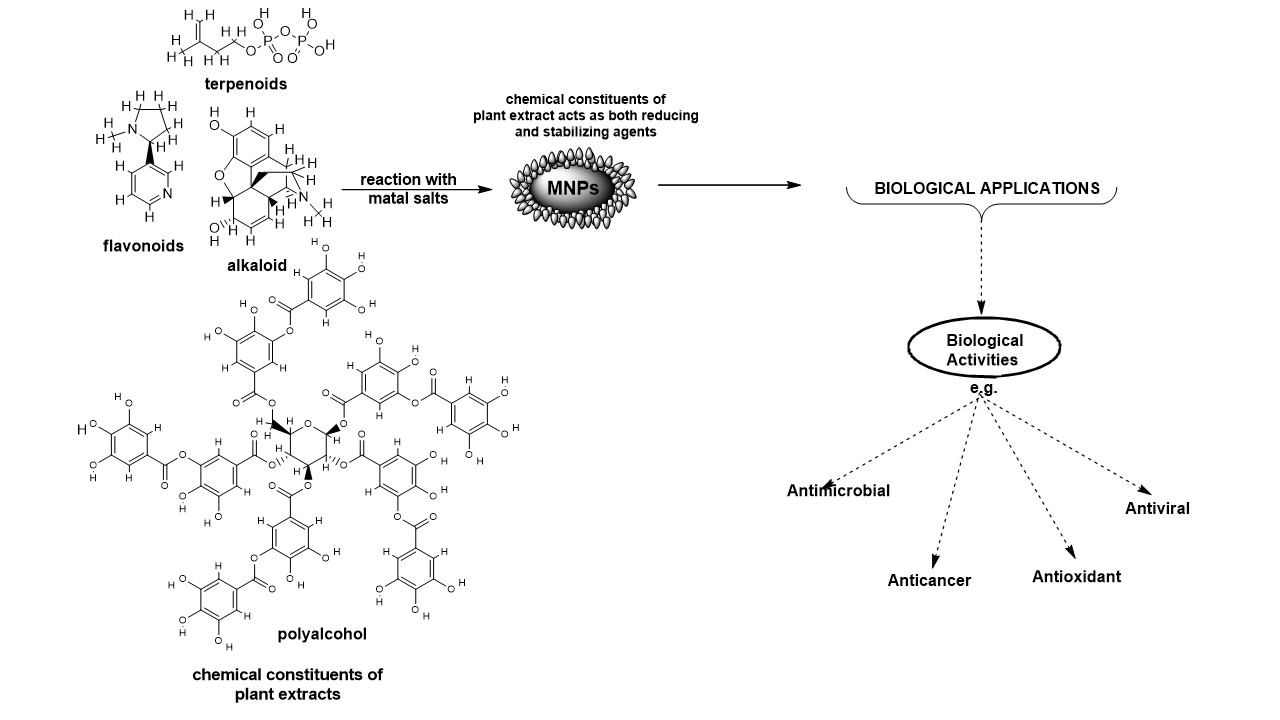
2. Phytochemical-Induced Synthesis of Metal-Based Nanoparticles
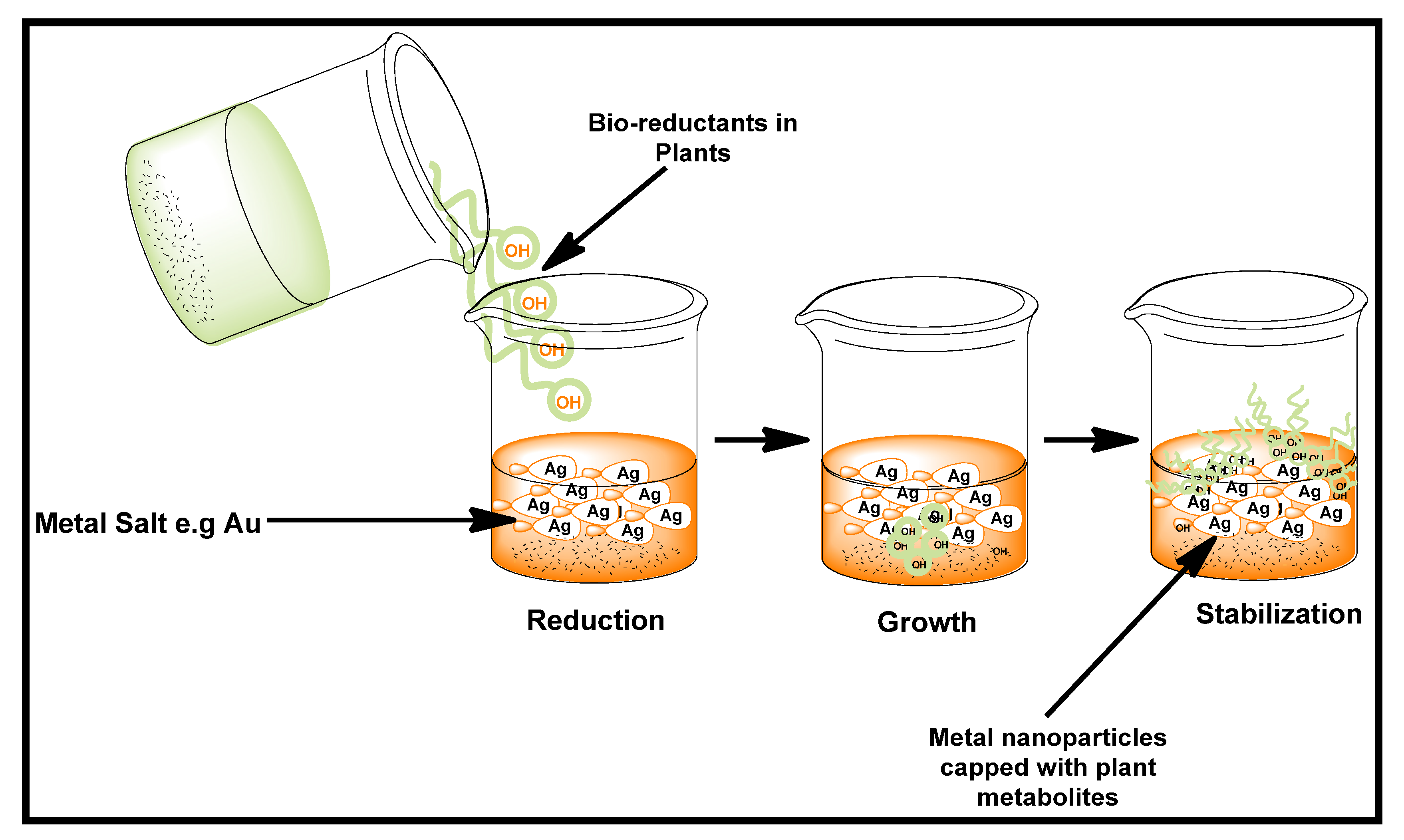
2.1. Possible Mechanism for Synthesis of Nanoparticles Using Plant Extract
2.2. Determination of Physicochemical Properties of Metal-Based Nanomaterials
3. Biological Importance of Biogenic Metal-Based Nanoparticles
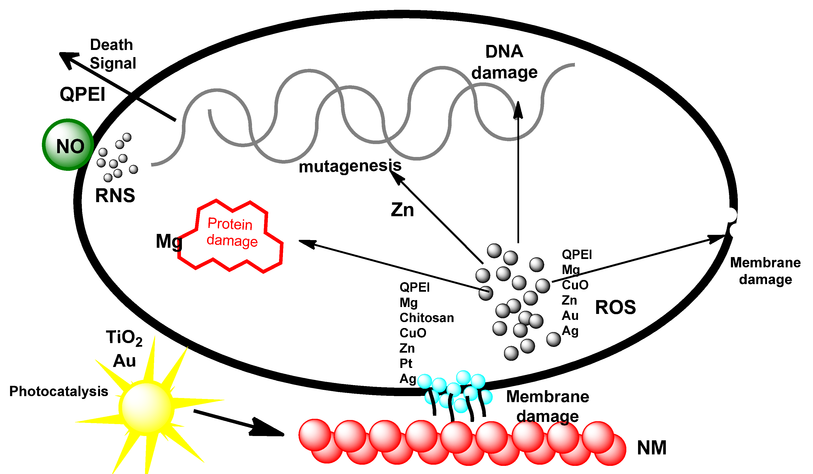
3.1. Plant-Mediated Metal-Based Nanoparticles as Antimicrobial Agents
3.2. Plant Mediated Metal-Based Nanoparticles as Anticancer Agents
3.3. Plant Mediated Metal-Based Nanoparticles as Antioxidant Agents
3.4. Plant Mediated Metal-Based Nanoparticles as Anti-Inflammatory Agents
3.5. Plant Mediated Metal-Based Nanoparticles as Antiviral Agents
4. Challenges and Future Prospects
5. Conclusions
Author Contributions
Funding
Institutional Review Board Statement
Informed Consent Statement
Data Availability Statement
Conflicts of Interest
References
- Hoet, P.H.; Brüske-Hohlfeld, I.; Salata, O.V. Nanoparticles—Known and unknown health risks. J. Nanobiotechnol. 2004, 6, 12. [Google Scholar] [CrossRef] [PubMed] [Green Version]
- Padil, V.V.T.; Černík, M. Green Synthesis of Copper Oxide Nanoparticles Using Gum Karaya as a Biotemplate and Their Antibacterial Application. Int. J. Nanomed. 2013, 8, 889–898. [Google Scholar] [CrossRef] [Green Version]
- Hussein, A.K. Applications of Nanotechnology in Renewable Energies—A Comprehensive Overview and Understanding. Renew. Sustain. Energy Rev. 2015, 42, 460–476. [Google Scholar] [CrossRef]
- Ahmed, E.S.A.; Sohal, H.S. Nanotechnology in Communication Engineering: Issues, Applications and Future Possibilities. World Sci. News 2017, 66, 134–148. [Google Scholar]
- Imran, K.; Mohd, F.; Pratichi, S.; Padma, T. Nanotechnology for Environmental Remediation. Res. J. Pharm. Biol. Chem. Sci. 2014, 5, 1916–1927. [Google Scholar]
- Adeyemi, J.O.; Ajiboye, T.; Onwudiwe, D.C. Mineralization of Antibiotics in Wastewater Via Photocatalysis. Water Air Soil Pollut. 2021, 232, 219. [Google Scholar] [CrossRef]
- Murthy, S.K. Nanoparticles in Modern Medicine: State of the Art and Future Challenges. Int. J. Nanomed. 2007, 2, 129–141. [Google Scholar]
- Logothetidis, S. Nanomedicine and Nanobiotechnology. In Nanomedicine: The Medicine of Tomorrow; Springer: Berlin, Germany, 2006; Volume 10, pp. 1–26. [Google Scholar]
- Roco, M.C. Nanotechnology: Convergence with Modern Biology and Medicine. Curr. Opin. Biotechnol. 2003, 14, 337–346. [Google Scholar] [CrossRef] [Green Version]
- Mehta, D.; Guvva, S.; Patil, M. Future Impact of Nanotechnology on Medicine and Dentistry. J. Indian Soc. Periodontol. 2008, 12, 34. [Google Scholar] [CrossRef]
- Heiligtag, F.J.; Niederberger, M. The Fascinating World of Nanoparticle Research. Mater. Today 2013, 16, 262–271. [Google Scholar] [CrossRef]
- Sánchez-López, E.; Gomes, D.; Esteruelas, G.; Bonilla, L.; Lopez-Machado, A.L.; Galindo, R.; Cano, A.; Espina, M.; Ettcheto, M.; Camins, A.; et al. Metal-Based Nanoparticles as Antimicrobial Agents: An Overview. Nanomaterials 2020, 10, 292. [Google Scholar] [CrossRef] [PubMed] [Green Version]
- Rahim, M.; Iram, S.; Khan, M.S.; Khan, M.S.; Shukla, A.R.; Srivastava, A.K.; Ahmad, S. Glycation-Assisted Synthesized Gold Nanoparticles Inhibit Growth of Bone Cancer Cells. Colloids Surf. B Biointerfaces 2014, 117, 473–479. [Google Scholar] [CrossRef]
- Wang, Y.; Wang, H.; Khan, M.S.; Husain, F.M.; Ahmad, S.; Bian, L. Bioconjugation of Gold Nanoparticles with Aminoguanidine as a Potential Inhibitor of Non-Enzymatic Glycation Reaction. J. Biomol. Struct. Dyn. 2021, 39, 2014–2020. [Google Scholar] [CrossRef] [PubMed]
- Ashraf, J.M.; Ansari, M.A.; Fatma, S.; Abdullah, S.M.S.; Iqbal, J.; Madkhali, A.; Hamali, A.H.; Ahmad, S.; Jerah, A.; Echeverria, V.; et al. Inhibiting Effect of Zinc Oxide Nanoparticles on Advanced Glycation Products and Oxidative Modifications: A Potential Tool to Counteract Oxidative Stress in Neurodegenerative Diseases. Mol. Neurobiol. 2018, 55, 7438–7452. [Google Scholar] [CrossRef] [PubMed]
- Gahlawat, G.; Choudhury, A.R. A Review on the Biosynthesis of Metal and Metal Salt Nanoparticles by Microbes. RSC Adv. 2019, 9, 12944–12967. [Google Scholar] [CrossRef] [Green Version]
- Ovais, M.; Khalil, A.T.; Islam, N.U.; Ahmad, I.; Ayaz, M.; Saravanan, M.; Shinwari, Z.K.; Mukherjee, S. Role of Plant Phytochemicals and Microbial Enzymes in Biosynthesis of Metallic Nanoparticles. Appl. Microbiol. Biotechnol. 2018, 102, 6799–6814. [Google Scholar] [CrossRef]
- Watt, J.; Cheong, S.; Tilley, R.D. How to Control the Shape of Metal Nanostructures in Organic Solution Phase Synthesis for Plasmonics and Catalysis. Nano Today 2013, 8, 198–215. [Google Scholar] [CrossRef]
- Kandi, V.; Kandi, S. Antimicrobial Properties of Nanomolecules: Potential Candidates as Antibiotics in the Era of Multi-Drug Resistance. Epidemiol. Health 2015, 37, e2015020. [Google Scholar] [CrossRef] [Green Version]
- Wang, Y.; Xia, Y. Bottom-up and Top-down Approaches to the Synthesis of Monodispersed Spherical Colloids of Low Melting-Point Metals. Nano Lett. 2004, 4, 2047–2050. [Google Scholar] [CrossRef]
- Mittal, A.K.; Chisti, Y.; Banerjee, U.C. Synthesis of Metallic Nanoparticles Using Plant Extracts. Biotechnol. Adv. 2013, 31, 346–356. [Google Scholar] [CrossRef]
- Cao, G. Nanostructures and Nanomaterials; Imperial College Press: London, UK, 2004; ISBN 978-1-86094-415-4. [Google Scholar]
- Singh, J.; Dutta, T.; Kim, K.-H.; Rawat, M.; Samddar, P.; Kumar, P. “Green” Synthesis of Metals and Their Oxide Nanoparticles: Applications for Environmental Remediation. J. Nanobiotechnol. 2018, 16, 84. [Google Scholar] [CrossRef] [PubMed]
- Das, R.K.; Pachapur, V.L.; Lonappan, L.; Naghdi, M.; Pulicharla, R.; Maiti, S.; Cledon, M.; Dalila, L.M.A.; Sarma, S.J.; Brar, S.K. Biological Synthesis of Metallic Nanoparticles: Plants, Animals and Microbial Aspects. Nanotechnol. Environ. Eng. 2017, 2, 18. [Google Scholar] [CrossRef] [Green Version]
- Husen, A. Gold Nanoparticles from Plant System: Synthesis, Characterization and Their Application. In Nanoscience and Plant–soil Systems; Springer: Cham, Switzerland; Berlin, Germany, 2017; pp. 455–479. [Google Scholar] [CrossRef]
- Ankamwar, B. Biosynthesis of Gold Nanoparticles (Green-Gold) Using Leaf Extract of Terminalia Catappa. E-J. Chem. 2010, 7, 1334–1339. [Google Scholar] [CrossRef] [Green Version]
- Adeyemi, J.O.; Elemike, E.E.; Onwudiwe, D.C. ZnO Nanoparticles Mediated by Aqueous Extracts of Dovyalis Caffra Fruits and the Photocatalytic Evaluations. Mater. Res. Express 2019, 6, 125091. [Google Scholar] [CrossRef]
- Li, X.; Xu, H.; Chen, Z.-S.; Chen, G. Biosynthesis of Nanoparticles by Microorganisms and Their Applications. J. Nanomater. 2011, 2011, 270974. [Google Scholar] [CrossRef] [Green Version]
- Zhang, X.-F.; Liu, Z.-G.; Shen, W.; Gurunathan, S. Silver Nanoparticles: Synthesis, Characterization, Properties, Applications, and Therapeutic Approaches. Int. J. Mol. Sci. 2016, 17, 1534. [Google Scholar] [CrossRef]
- Narayanan, K.B.; Sakthivel, N. Phytosynthesis of Gold Nanoparticles Using Leaf Extract of Coleus Amboinicus Lour. Mater. Charact. 2010, 61, 1232–1238. [Google Scholar] [CrossRef]
- Nagajyothi, P.C.; Lee, K.D. Synthesis of Plant-Mediated Silver Nanoparticles Using Dioscorea Batatas Rhizome Extract and Evaluation of Their Antimicrobial Activities. J. Nanomater. 2011, 2011, 573429. [Google Scholar] [CrossRef] [Green Version]
- Ahmad, N.; Sharma, S.; Singh, V.N.; Shamsi, S.F.; Fatma, A.; Mehta, B.R. Biosynthesis of Silver Nanoparticles from Desmodium Triflorum: A Novel Approach Towards Weed Utilization. Biotechnol. Res. Int. 2011, 2011, 454090. [Google Scholar] [CrossRef] [Green Version]
- Kesharwani, J.; Yoon, K.Y.; Hwang, J.; Rai, M. Phytofabrication of Silver Nanoparticles by Leaf Extract of Datura Metel: Hypothetical Mechanism Involved in Synthesis. J. Bionanosci. 2009, 3, 39–44. [Google Scholar] [CrossRef]
- Huang, J.; Li, Q.; Sun, D.; Lu, Y.; Su, Y.; Yang, X.; Wang, H.; Wang, Y.; Shao, W.; He, N.; et al. Biosynthesis of Silver and Gold Nanoparticles by Novel Sundried Cinnamomum Camphoraleaf. Nanotechnology 2007, 18, 105104. [Google Scholar] [CrossRef]
- Dubey, M.; Bhadauria, S.; Kushwah, B. Green Synthesis of Nanosilver Particles from Extract of Eucalyptus Hybrida (Safeda) Leaf. Dig. J. Nanomater Biostr. 2009, 4, 537–543. [Google Scholar]
- Hayat, M.A. Colloidal Gold: Principles, Methods, and Applications; Colloidal Gold Series; Academic Press: Cambridge, MA, USA, 1989; ISBN 9780123339270. [Google Scholar]
- Daisy, P.; Saipriya, K. Biochemical Analysis of Cassia Fistula Aqueous Extract and Phytochemically Synthesized Gold Nanoparticles as Hypoglycemic Treatment for Diabetes Mellitus. Int. J. Nanomed. 2012, 7, 1189–1202. [Google Scholar] [CrossRef] [PubMed] [Green Version]
- Liu, Q.; Liu, H.; Yuan, Z.; Wei, D.; Ye, Y. Evaluation of Antioxidant Activity of Chrysanthemum Extracts and Tea Beverages by Gold Nanoparticles-Based Assay. Colloids Surf. B Biointerfaces 2012, 92, 348–352. [Google Scholar] [CrossRef] [PubMed]
- Castro, L.; Blázquez, M.L.; Muñoz, J.A.; González, F.; García-Balboa, C.; Ballester, A. Biosynthesis of Gold Nanowires Using Sugar Beet Pulp. Process Biochem. 2011, 46, 1076–1082. [Google Scholar] [CrossRef]
- Srinoi, P.; Chen, Y.T.; Vittur, V.; Marquez, M.D.; Lee, T.R. Bimetallic Nanoparticles: Enhanced Magnetic and Optical Properties for Emerging Biological Applications. Appl. Sci. 2018, 8, 1106. [Google Scholar] [CrossRef] [Green Version]
- Sharma, G.; Kumar, A.; Sharma, S.; Naushad, M.; Dwivedi, R.P.; ALOthman, Z.A.; Mola, G.T. Novel Development of Nanoparticles to Bimetallic Nanoparticles and Their Composites: A Review. J. King Saud Univ.-Sci. 2019, 31, 257–269. [Google Scholar] [CrossRef]
- Burda, C.; Chen, X.; Narayanan, R.; El-Sayed, M.A. Chemistry and Properties of Nanocrystals of Different Shapes. Chem. Rev. 2005, 105, 1025–1102. [Google Scholar] [CrossRef]
- Logunov, S.L.; Ahmadi, T.S.; El-Sayed, M.A.; Khoury, J.T.; Whetten, R.L. Electron Dynamics of Passivated Gold Nanocrystals Probed by Subpicosecond Transient Absorption Spectroscopy. J. Phys. Chem. B 1997, 101, 3713–3719. [Google Scholar] [CrossRef]
- Gao, J.; Ren, X.; Chen, D.; Tang, F.; Ren, J. Bimetallic Ag–Pt Hollow Nanoparticles: Synthesis and Tunable Surface Plasmon Resonance. Scr. Mater. 2007, 57, 687–690. [Google Scholar] [CrossRef]
- Toshima, N.; Yonezawa, T. Bimetallic Nanoparticles—Novel Materials for Chemical and Physical Applications. New J. Chem. 1998, 22, 1179–1201. [Google Scholar] [CrossRef]
- Sun, S.; Murray, C.B.; Weller, D.; Folks, L.; Moser, A. Monodisperse FePt Nanoparticles and Ferromagnetic FePt Nanocrystal Superlattices. Science 2000, 287, 1989–1992. [Google Scholar] [CrossRef]
- Arora, N.; Thangavelu, K.; Karanikolos, G.N. Bimetallic Nanoparticles for Antimicrobial Applications. Front. Chem. 2020, 8, 41. [Google Scholar] [CrossRef]
- Shankar, S.S.; Rai, A.; Ahmad, A.; Sastry, M. Rapid Synthesis of Au, Ag, and Bimetallic Au Core–Ag Shell Nanoparticles Using Neem (Azadirachta Indica) Leaf Broth. J. Colloid Interface Sci. 2004, 275, 496–502. [Google Scholar] [CrossRef]
- Acad, I. Spectral and Thermal Studies on Mixed Figand Complexes of Zinc (Ll) and Cadmium (II) with Diethyldithiocarbamate and 2, 2’-Bipyridyl/1, 10-phenanthroline. Proc. Indian Acad. Sci.-Chem. Sci. 1993, 105, 87–94. [Google Scholar]
- Adeyemi, J.O.; Elemike, E.E.; Onwudiwe, D.C.; Singh, M. Bio-Inspired Synthesis and Cytotoxic Evaluation of Silver-Gold Bimetallic Nanoparticles Using Kei-Apple (Dovyalis Caffra) Fruits. Inorg. Chem. Commun. 2019, 109, 107569. [Google Scholar] [CrossRef]
- Naseem, T.; Durrani, T. The Role of Some Important Metal Oxide Nanoparticles for Wastewater and Antibacterial Applications: A Review. Environ. Chem. Ecotoxicol. 2021, 3, 59–75. [Google Scholar] [CrossRef]
- Ezhilarasi, A.A.; Vijaya, J.J.; Kaviyarasu, K.; Kennedy, L.J.; Ramalingam, R.J.; Al-Lohedan, H.A. Green Synthesis of NiO Nanoparticles Using Aegle Marmelos Leaf Extract for the Evaluation of In-Vitro Cytotoxicity, Antibacterial and Photocatalytic Properties. J. Photochem. Photobiol. B Biol. 2018, 180, 39–50. [Google Scholar] [CrossRef] [PubMed]
- Andreescu, S.; Ornatska, M.; Erlichman, J.S.; Estevez, A.; Leiter, J.C. Biomedical Applications of Metal Oxide Nanoparticles; Springer: Boston, MA, USA, 2012; pp. 57–100. ISBN 9781461403791. [Google Scholar]
- Song, J.Y.; Kim, B.S. Rapid Biological Synthesis of Silver Nanoparticles Using Plant Leaf Extracts. Bioprocess Biosyst. Eng. 2009, 32, 79–84. [Google Scholar] [CrossRef]
- Arundoss, T.; Arulkumar, S.; Senthilkumar, K.; Sabesan, M.; Vasudevan, K. A Novel Approach for the Synthesis and Characterization of Ginkgo Biloba Gold Nanoparticles-An Alternative Approach to Chemical Synthesis. Asian J. Sci. Technol. 2013, 4, 140–144. [Google Scholar]
- Vankar, P.S.; Bajpai, D. Preparation of Gold Nanoparticles from Mirabilis Jalapa Flowers. Indian J. Biochem. Biophys. 2010, 47, 157–160. [Google Scholar] [PubMed]
- Armendariz, V.; Herrera, I.; Peralta-Videa, J.R.; Jose-Yacaman, M.; Troiani, H.; Santiago, P.; Gardea-Torresdey, J.L. Size Controlled Gold Nanoparticle Formation by Avena Sativa Biomass: Use of Plants in Nanobiotechnology. J. Nanoparticle Res. 2004, 6, 377–382. [Google Scholar] [CrossRef]
- Velmurugan, P.; Lee, S.-M.; Iydroose, M.; Lee, K.-J.; Oh, B.-T. Pine Cone-Mediated Green Synthesis of Silver Nanoparticles and Their Antibacterial Activity against Agricultural Pathogens. Appl. Microbiol. Biotechnol. 2013, 97, 361–368. [Google Scholar] [CrossRef] [PubMed]
- Aromal, S.A.; Philip, D. Green Synthesis of Gold Nanoparticles Using Trigonella Foenum-Graecum and Its Size-Dependent Catalytic Activity. Spectrochim. Acta A Mol. Biomol. Spectrosc. 2012, 97, 1–5. [Google Scholar] [CrossRef]
- Niraimathi, K.L.; Sudha, V.; Lavanya, R.; Brindha, P. Biosynthesis of Silver Nanoparticles Using Alternanthera Sessilis (Linn.) Extract and Their Antimicrobial, Antioxidant Activities. Colloids Surf. B Biointerfaces 2013, 102, 288–291. [Google Scholar] [CrossRef]
- Suriyakalaa, U.; Antony, J.J.; Suganya, S.; Siva, D.; Sukirtha, R.; Kamalakkannan, S.; Pichiah, P.B.T.; Achiraman, S. Hepatocurative Activity of Biosynthesized Silver Nanoparticles Fabricated Using Andrographis paniculata. Colloids Surf. B Biointerfaces 2013, 102, 189–194. [Google Scholar] [CrossRef]
- Krishnaraj, C.; Jagan, E.G.; Rajasekar, S.; Selvakumar, P.; Kalaichelvan, P.T.; Mohan, N. Synthesis of Silver Nanoparticles Using Acalypha Indica Leaf Extracts and Its Antibacterial Activity against Water Borne Pathogens. Colloids Surf. B Biointerfaces 2010, 76, 50–56. [Google Scholar] [CrossRef]
- Krishnaraj, C.; Muthukumaran, P.; Ramachandran, R.; Balakumaran, M.D.; Kalaichelvan, P.T. Acalypha Indica Linn: Biogenic Synthesis of Silver and Gold Nanoparticles and Their Cytotoxic Effects against MDA-MB-231, Human Breast Cancer Cells. Biotechnol. Rep. 2014, 4, 42–49. [Google Scholar] [CrossRef] [Green Version]
- Song, J.Y.; Kwon, E.Y.; Kim, B.S. Biological Synthesis of Platinum Nanoparticles Using Diopyros Kaki Leaf Extract. Bioprocess Biosyst. Eng. 2010, 33, 159–164. [Google Scholar] [CrossRef]
- Mondal, S.; Roy, N.; Laskar, R.A.; Sk, I.; Basu, S.; Mandal, D.; Begum, N.A. Biogenic Synthesis of Ag, Au and Bimetallic Au/Ag Alloy Nanoparticles Using Aqueous Extract of Mahogany (Swietenia Mahogani JACQ.) Leaves. Colloids Surf. B Biointerfaces 2011, 82, 497–504. [Google Scholar] [CrossRef]
- Nasrollahzadeh, M.; Sajadi, S.M.; Khalaj, M. Green Synthesis of Copper Nanoparticles Using Aqueous Extract of the Leaves of Euphorbia Esula L and Their Catalytic Activity for Ligand-Free Ullmann-Coupling Reaction and Reduction of 4-Nitrophenol. RSC Adv. 2014, 4, 47313–47318. [Google Scholar] [CrossRef]
- Coccia, F.; Tonucci, L.; Bosco, D.; Bressan, M.; d’Alessandro, N. One-Pot Synthesis of Lignin-Stabilised Platinum and Palladium Nanoparticles and Their Catalytic Behaviour in Oxidation and Reduction Reactions. Green Chem. 2012, 14, 1073–1078. [Google Scholar] [CrossRef]
- Njagi, E.C.; Huang, H.; Stafford, L.; Genuino, H.; Galindo, H.M.; Collins, J.B.; Hoag, G.E.; Suib, S.L. Biosynthesis of Iron and Silver Nanoparticles at Room Temperature Using Aqueous Sorghum Bran Extracts. Langmuir 2011, 27, 264–271. [Google Scholar] [CrossRef]
- Phokha, S.; Seraphin, S.; Maensiri, S.; Laokul, P.; Klinkaewnarong, J.; Phokha, S.; Seraphin, S. Indium Oxide (In2O3) Nanoparticles Using Aloe Vera Plant Extract: Synthesis and Optical Properties Indium Oxide (In2O3) Nanoparticles Using Aloe Vera Plant Extract: Synthesis and Optical Properties. J. Optoelectron. Adv. Mater 2008, 2, 161–165. [Google Scholar]
- Roopan, S.M.; Bharathi, A.; Kumar, R.; Khanna, V.G.; Prabhakarn, A. Acaricidal, Insecticidal, and Larvicidal Efficacy of Aqueous Extract of Annona Squamosa L Peel as Biomaterial for the Reduction of Palladium Salts into Nanoparticles. Colloids Surf. B Biointerfaces 2012, 92, 209–212. [Google Scholar] [CrossRef]
- Singh, R.P.; Shukla, V.K.; Yadav, R.S.; Sharma, P.K.; Singh, P.K.; Pandey, A.C. Biological Approach of Zinc Oxide Nanoparticles Formation and Its Characterization. Adv. Mater. Lett. 2011, 2, 313–317. [Google Scholar] [CrossRef]
- Haider, A.; Ijaz, M.; Ali, S.; Haider, J.; Imran, M.; Majeed, H.; Shahzadi, I.; Ali, M.M.; Khan, J.A.; Ikram, M. Green Synthesized Phytochemically (Zingiber Officinale and Allium Sativum) Reduced Nickel Oxide Nanoparticles Confirmed Bactericidal and Catalytic Potential. Nanoscale Res. Lett. 2020, 15, 50. [Google Scholar] [CrossRef]
- Khandel, P.; Yadaw, R.K.; Soni, D.K.; Kanwar, L.; Shahi, S.K. Biogenesis of Metal Nanoparticles and Their Pharmacological Applications: Present Status and Application Prospects; Springer: Berlin, Germany, 2018; Volume 8, ISBN 4009701802674. [Google Scholar]
- Sathishkumar, M.; Sneha, K.; Yun, Y.S. Immobilization of Silver Nanoparticles Synthesized Using Curcuma Longa Tuber Powder and Extract on Cotton Cloth for Bactericidal Activity. Bioresour. Technol. 2010, 101, 7958–7965. [Google Scholar] [CrossRef]
- Gardea-Torresdey, J.L.; Tiemann, K.J.; Gamez, G.; Dokken, K.; Tehuacanero, S.; José-Yacamán, M. Gold Nanoparticles Obtained by Bio-Precipitation from Gold(III) Solutions. J. Nanoparticle Res. 1999, 1, 397–404. [Google Scholar] [CrossRef]
- Kuppusamy, P.; Yusoff, M.M.; Maniam, G.P.; Govindan, N. Biosynthesis of Metallic Nanoparticles Using Plant Derivatives and Their New Avenues in Pharmacological Applications-An Updated Report. Saudi Pharm. J. 2016, 24, 473–484. [Google Scholar] [CrossRef]
- Singh, A.K.; Talat, M.; Singh, D.P.; Srivastava, O.N. Biosynthesis of Gold and Silver Nanoparticles by Natural Precursor Clove and Their Functionalization with Amine Group. J. Nanoparticle Res. 2010, 12, 1667–1675. [Google Scholar] [CrossRef]
- Pirtarighat, S.; Ghannadnia, M.; Baghshahi, S. Biosynthesis of Silver Nanoparticles Using Ocimum Basilicum Cultured under Controlled Conditions for Bactericidal Application. Mater. Sci. Eng. C 2019, 98, 250–255. [Google Scholar] [CrossRef] [PubMed]
- Latif, H.H.; Ghareib, M.; Tahon, M.A. Photosynthese von Silber-Nanopartikeln Mit Blattextrakt von Ocimum Basilicum Und Mangifira Indica Und Deren Auswirkung Auf Bestimmte Biochemische Eigenschaften von Triticum Aestivum. Gesunde Pflanz. 2017, 69, 39–46. [Google Scholar] [CrossRef]
- Khan, M.; Khan, M.; Adil, S.F.; Tahir, M.N.; Tremel, W.; Alkhathlan, H.Z.; Al-Warthan, A.; Siddiqui, M.R.H. Green Synthesis of Silver Nanoparticles Mediated by Pulicaria Glutinosa Extract. Int. J. Nanomed. 2013, 8, 1507–1516. [Google Scholar] [CrossRef] [Green Version]
- Kumar, S.A.; Peter, Y.A.; Nadeau, J.L. Facile Biosynthesis, Separation and Conjugation of Gold Nanoparticles to Doxorubicin. Nanotechnology 2008, 19, 495101. [Google Scholar] [CrossRef] [PubMed] [Green Version]
- Song, J.Y.; Jang, H.K.; Kim, B.S. Biological Synthesis of Gold Nanoparticles Using Magnolia Kobus and Diopyros Kaki Leaf Extracts. Process Biochem. 2009, 44, 1133–1138. [Google Scholar] [CrossRef]
- Begum, N.A.; Mondal, S.; Basu, S.; Laskar, R.A.; Mandal, D. Biogenic Synthesis of Au and Ag Nanoparticles Using Aqueous Solutions of Black Tea Leaf Extracts. Colloids Surf. B Biointerfaces 2009, 71, 113–118. [Google Scholar] [CrossRef]
- Osuntokun, J.; Onwudiwe, D.C.; Ebenso, E.E. Aqueous Extract of Broccoli Mediated Synthesis of CaO Nanoparticles and Its Application in the Photocatalytic Degradation of Bromocrescol Green. IET Nanobiotechnol. 2018, 12, 888–894. [Google Scholar] [CrossRef]
- Vijayaraghavan, K.; Nalini, S.P.K. Biotemplates in the Green Synthesis of Silver Nanoparticles. Biotechnol. J. 2010, 5, 1098–1110. [Google Scholar] [CrossRef]
- Lukman, A.I.; Gong, B.; Marjo, C.E.; Roessner, U.; Harris, A.T. Facile Synthesis, Stabilization, and Anti-Bacterial Performance of Discrete Ag Nanoparticles Using Medicago Sativa Seed Exudates. J. Colloid Interface Sci. 2011, 353, 433–444. [Google Scholar] [CrossRef]
- Lin, P.-C.; Lin, S.; Wang, P.C.; Sridhar, R. Techniques for Physicochemical Characterization of Nanomaterials. Biotechnol. Adv. 2014, 32, 711–726. [Google Scholar] [CrossRef] [Green Version]
- Farokhzad, O.C.; Langer, R. Nanomedicine: Developing Smarter Therapeutic and Diagnostic Modalities. Adv. Drug Deliv. Rev. 2006, 58, 1456–1459. [Google Scholar] [CrossRef]
- Akhter, S.; Ahmad, I.; Ahmad, M.Z.; Ramazani, F.; Singh, A.; Rahman, Z.; Ahmad, F.J.; Storm, G.; Kok, R.J. Nanomedicines as Cancer Therapeutics: Current Status. Curr. Cancer Drug Targets 2013, 13, 362–378. [Google Scholar] [CrossRef] [PubMed] [Green Version]
- Kim, T.H.; Lee, S.; Chen, X. Nanotheranostics for Personalized Medicine. Expert Rev. Mol. Diagn. 2013, 13, 257–269. [Google Scholar] [CrossRef] [PubMed]
- Schaffer, B.; Hohenester, U.; Trügler, A.; Hofer, F. High-Resolution Surface Plasmon Imaging of Gold Nanoparticles by Energy-Filtered Transmission Electron Microscopy. Phys. Rev. B 2009, 79, 041401. [Google Scholar] [CrossRef] [Green Version]
- Pal, S.; Tak, Y.K.; Song, J.M. Does the Antibacterial Activity of Silver Nanoparticles Depend on the Shape of the Nanoparticle? A Study of the Gram-Negative Bacterium Escherichia Coli. Appl. Environ. Microbiol. 2007, 73, 1712–1720. [Google Scholar] [CrossRef] [PubMed] [Green Version]
- Strasser, P.; Koh, S.; Anniyev, T.; Greeley, J.; More, K.; Yu, C.; Liu, Z.; Kaya, S.; Nordlund, D.; Ogasawara, H.; et al. Lattice-Strain Control of the Activity in Dealloyed Core-Shell Fuel Cell Catalysts. Nat. Chem. 2010, 2, 454–460. [Google Scholar] [CrossRef] [PubMed]
- Veeresham, C. Natural Products Derived from Plants as a Source of Drugs. J. Adv. Pharm. Technol. Res. 2012, 3, 200. [Google Scholar] [CrossRef]
- Drummer, S.; Madzimbamuto, T.; Chowdhury, M. Green Synthesis of Transition-Metal Nanoparticles and Their Oxides: A Review. Materials 2021, 14, 2700. [Google Scholar] [CrossRef]
- Lu, W.; Yao, J.; Zhu, X.; Qi, Y. Nanomedicines: Redefining Traditional Medicine. Biomed. Pharmacother. 2021, 134, 111103. [Google Scholar] [CrossRef]
- Malik, S.; Ghosh, A.; Kerry, R.G.; Rout, J.R. Nanotechnology in Preclinical Pharmacokinetics. In Advances in Pharmaceutical Biotechnology; Springer: Singapore, 2020; pp. 461–478. [Google Scholar] [CrossRef]
- Alhadrami, H.A.; Alkhatabi, H.; Abduljabbar, F.H.; Abdelmohsen, U.R.; Sayed, A.M. Anticancer Potential of Green Synthesized Silver Nanoparticles of the Soft Coral Cladiella Pachyclados Supported by Network Pharmacology and In Silico Analyses. Pharmaceutics 2021, 13, 1846. [Google Scholar] [CrossRef] [PubMed]
- Hadrup, N.; Lam, H.R. Oral Toxicity of Silver Ions, Silver Nanoparticles and Colloidal Silver-A Review. Regul. Toxicol. Pharmacol. 2014, 68, 1–7. [Google Scholar] [CrossRef] [PubMed]
- Patil, S.; Chandrasekaran, R. Biogenic Nanoparticles: A Comprehensive Perspective in Synthesis, Characterization, Application and Its Challenges. J. Genet. Eng. Biotechnol. 2020, 18, 67. [Google Scholar] [CrossRef] [PubMed]
- Marston, H.D.; Dixon, D.M.; Knisely, J.M.; Palmore, T.N.; Fauci, A.S. Antimicrobial Resistance. JAMA 2016, 316, 1193–1204. [Google Scholar] [CrossRef] [PubMed] [Green Version]
- Batalha, I.L.; Bernut, A.; Schiebler, M.; Ouberai, M.M.; Passemar, C.; Klapholz, C.; Kinna, S.; Michel, S.; Sader, K.; Castro-Hartmann, P.; et al. Polymeric Nanobiotics as a Novel Treatment for Mycobacterial Infections. J. Control. Release 2019, 314, 116–124. [Google Scholar] [CrossRef] [PubMed]
- van Duin, D.; Paterson, D.L. Multidrug Resistant Bacteria in the Community: Trends and Lessons Learned. Infect. Dis. Clin. N. Am. 2016, 30, 377. [Google Scholar] [CrossRef] [Green Version]
- Fox, M.E.; Szoka, F.C.; Fréchet, J.M.J. Soluble Polymer Carriers for the Treatment of Cancer: The Importance of Molecular Architecture. Acc. Chem. Res. 2009, 42, 1141–1151. [Google Scholar] [CrossRef] [Green Version]
- Li, H.; Chen, Q.; Zhao, J.; Urmila, K. Enhancing the Antimicrobial Activity of Natural Extraction Using the Synthetic Ultrasmall Metal Nanoparticles. Sci. Rep. 2015, 5, 11033. [Google Scholar] [CrossRef] [Green Version]
- Bresee, J.; Bond, C.M.; Worthington, R.J.; Smith, C.A.; Gifford, J.C.; Simpson, C.A.; Carter, C.J.; Wang, G.; Hartman, J.; Osbaugh, N.A.; et al. Nanoscale Structure-Activity Relationships, Mode of Action, and Biocompatibility of Gold Nanoparticle Antibiotics. J. Am. Chem. Soc. 2014, 136, 5295–5300. [Google Scholar] [CrossRef]
- Beyth, N.; Houri-Haddad, Y.; Domb, A.; Khan, W.; Hazan, R. Alternative Antimicrobial Approach: Nano-Antimicrobial Materials. Evid.-Based Complement. Altern. Med. 2015, 2015, 1–16. [Google Scholar] [CrossRef] [Green Version]
- Sadalage, P.S.; Patil, R.V.; Havaldar, D.V.; Gavade, S.S.; Santos, A.C.; Pawar, K.D. Optimally Biosynthesized, PEGylated Gold Nanoparticles Functionalized with Quercetin and Camptothecin Enhance Potential Anti-Inflammatory, Anti-Cancer and Anti-Angiogenic Activities. J. Nanobiotechnol. 2021, 19, 84. [Google Scholar] [CrossRef] [PubMed]
- Sathishkumar, G.; Jha, P.K.; Vignesh, V.; Rajkuberan, C.; Jeyaraj, M.; Selvakumar, M.; Jha, R.; Sivaramakrishnan, S. Cannonball Fruit (Couroupita Guianensis, Aubl.) Extract Mediated Synthesis of Gold Nanoparticles and Evaluation of Its Antioxidant Activity. J. Mol. Liq. 2016, 215, 229–236. [Google Scholar] [CrossRef]
- Saratale, R.G.; Benelli, G.; Kumar, G.; Kim, D.S.; Saratale, G.D. Bio-Fabrication of Silver Nanoparticles Using the Leaf Extract of an Ancient Herbal Medicine, Dandelion (Taraxacum Officinale), Evaluation of Their Antioxidant, Anticancer Potential, and Antimicrobial Activity against Phytopathogens. Environ. Sci. Pollut. Res. Int. 2018, 25, 10392–10406. [Google Scholar] [CrossRef] [PubMed]
- Lee, S.Y.; Krishnamurthy, S.; Cho, C.W.; Yun, Y.S. Biosynthesis of Gold Nanoparticles Using Ocimum Sanctum Extracts by Solvents with Different Polarity. ACS Sustain. Chem. Eng. 2016, 4, 2651–2659. [Google Scholar] [CrossRef]
- Saravanan, M.; Vahidi, H.; Medina Cruz, D.; Vernet-Crua, A.; Mostafavi, E.; Stelmach, R.; Webster, T.J.; Ali Mahjoub, M.; Rashedi, M.; Barabadi, H. Emerging Antineoplastic Biogenic Gold Nanomaterials for Breast Cancer Therapeutics: A Systematic Review. Int. J. Nanomed. 2020, 15, 3577. [Google Scholar] [CrossRef]
- Sreekanth, T.V.M.; Nagajyothi, P.C.; Muthuraman, P.; Enkhtaivan, G.; Vattikuti, S.V.P.; Tettey, C.O.; Kim, D.H.; Shim, J.; Yoo, K. Ultra-Sonication-Assisted Silver Nanoparticles Using Panax Ginseng Root Extract and Their Anti-Cancer and Antiviral Activities. J. Photochem. Photobiol. B Biol. 2018, 188, 6–11. [Google Scholar] [CrossRef]
- Ajitha, B.; Ashok Kumar Reddy, Y.; Shameer, S.; Rajesh, K.M.; Suneetha, Y.; Sreedhara Reddy, P. Lantana Camara Leaf Extract Mediated Silver Nanoparticles: Antibacterial, Green Catalyst. J. Photochem. Photobiol. B Biol. 2015, 149, 84–92. [Google Scholar] [CrossRef]
- Ishwarya, R.; Vaseeharan, B.; Shanthi, S.; Ramesh, S.; Manogari, P.; Dhanalakshmi, K.; Vijayakumar, S.; Benelli, G. Green Synthesized Silver Nanoparticles: Toxicity Against Poecilia Reticulata Fishes and Ceriodaphnia Cornuta Crustaceans. J. Clust. Sci. 2017, 28, 519–527. [Google Scholar] [CrossRef]
- Goodarzi, V.; Zamani, H.; Bajuli, L.; Moradshahi, A. Evaluation of Antioxidant Potential and Reduction Capacity of Some Plant Extracts in Silver Nanoparticles’ Synthesis. Mol. Biol. Res. Commun. 2014, 3, 165. [Google Scholar] [CrossRef]
- Aboyewa, J.A.; Sibuyi, N.R.S.; Meyer, M.; Oguntibeju, O.O. Gold Nanoparticles Synthesized Using Extracts of Cyclopia Intermedia, Commonly Known as Honeybush, Amplify the Cytotoxic Effects of Doxorubicin. Nanomaterials 2021, 11, 132. [Google Scholar] [CrossRef]
- Al-Ani, L.A.; Yehye, W.A.; Kadir, F.A.; Hashim, N.M.; AlSaadi, M.A.; Julkapli, N.M.; Hsiao, V.K.S. Hybrid Nanocomposite Curcumin-Capped Gold Nanoparticle-Reduced Graphene Oxide: Anti-Oxidant Potency and Selective Cancer Cytotoxicity. PLoS ONE 2019, 14, e0216725. [Google Scholar] [CrossRef] [PubMed] [Green Version]
- Chinnasamy, G.; Chandrasekharan, S.; Bhatnagar, S. Biosynthesis of Silver Nanoparticles from Melia Azedarach: Enhancement of Antibacterial, Wound Healing, Antidiabetic and Antioxidant Activities. Int. J. Nanomed. 2019, 14, 9823–9836. [Google Scholar] [CrossRef] [PubMed] [Green Version]
- Saratale, R.G.; Shin, H.S.; Kumar, G.; Benelli, G.; Kim, D.S.; Saratale, G.D. Exploiting Antidiabetic Activity of Silver Nanoparticles Synthesized Using Punica Granatum Leaves and Anticancer Potential against Human Liver Cancer Cells (HepG2). Artif. Cells Nanomed. Biotechnol. 2018, 46, 211–222. [Google Scholar] [CrossRef] [PubMed] [Green Version]
- Reddy, N.V.; Li, H.; Hou, T.; Bethu, M.S.; Ren, Z.; Zhang, Z. Phytosynthesis of Silver Nanoparticles Using Perilla Frutescens Leaf Extract: Characterization and Evaluation of Antibacterial, Antioxidant, and Anticancer Activities. Int. J. Nanomed. 2021, 16, 15–29. [Google Scholar] [CrossRef]
- Rahman, A.U.; Khan, A.U.; Yuan, Q.; Wei, Y.; Ahmad, A.; Ullah, S.; Khan, Z.U.H.; Shams, S.; Tariq, M.; Ahmad, W. Tuber Extract of Arisaema Flavum Eco-Benignly and Effectively Synthesize Silver Nanoparticles: Photocatalytic and Antibacterial Response against Multidrug Resistant Engineered E. Coli QH4. J. Photochem. Photobiol. B 2019, 193, 31–38. [Google Scholar] [CrossRef]
- Saratale, R.G.; Shin, H.S.; Kumar, G.; Benelli, G.; Ghodake, G.S.; Jiang, Y.Y.; Kim, D.S.; Saratale, G.D. Exploiting Fruit Byproducts for Eco-Friendly Nanosynthesis: Citrus × Clementina Peel Extract Mediated Fabrication of Silver Nanoparticles with High Efficacy against Microbial Pathogens and Rat Glial Tumor C6 Cells. Environ. Sci. Pollut. Res. Int. 2018, 25, 10250–10263. [Google Scholar] [CrossRef]
- Patra, J.K.; Das, G.; Shin, H.S. Facile Green Biosynthesis of Silver Nanoparticles Using Pisum Sativum L. Outer Peel Aqueous Extract and Its Antidiabetic, Cytotoxicity, Antioxidant, and Antibacterial Activity. Int. J. Nanomed. 2019, 14, 6679–6690. [Google Scholar] [CrossRef] [Green Version]
- Küp, F.Ö.; Çoşkunçay, S.; Duman, F. Biosynthesis of Silver Nanoparticles Using Leaf Extract of Aesculus Hippocastanum (Horse Chestnut): Evaluation of Their Antibacterial, Antioxidant and Drug Release System Activities. Mater. Sci. Eng. C 2020, 107, 11027. [Google Scholar] [CrossRef]
- Kumar, C.S.; Mahesh, A.; Antoniraj, M.G.; Vaidevi, S.; Ruckmani, K. Ultrafast Synthesis of Stabilized Gold Nanoparticles Using Aqueous Fruit Extract of Limonia Acidissima L. and Conjugated Epirubicin: Targeted Drug Delivery for Treatment of Breast Cancer. RSC Adv. 2016, 6, 26874–26882. [Google Scholar] [CrossRef]
- Mmola, M.; Le Roes-Hill, M.; Durrell, K.; Bolton, J.J.; Sibuyi, N.; Meyer, M.E.; Beukes, D.R.; Antunes, E. Enhanced Antimicrobial and Anticancer Activity of Silver and Gold Nanoparticles Synthesised Using Sargassum Incisifolium Aqueous Extracts. Molecules 2016, 21, 1633. [Google Scholar] [CrossRef] [Green Version]
- Devi, G.K.; Sathishkumar, K. Synthesis of Gold and Silver Nanoparticles Using Mukia Maderaspatna Plant Extract and Its Anticancer Activity. IET Nanobiotechnology 2017, 11, 143–151. [Google Scholar] [CrossRef]
- Fatima, M.; Zaidi, N. us S.S.; Amraiz, D.; Afzal, F. In Vitro Antiviral Activity of Cinnamomum Cassia and Its Nanoparticles against H7N3 Influenza a Virus. J. Microbiol. Biotechnol. 2015, 26, 151–159. [Google Scholar] [CrossRef]
- Haggag, E.G.; Elshamy, A.M.; Rabeh, M.A.; Gabr, N.M.; Salem, M.; Youssif, K.A.; Samir, A.; Bin Muhsinah, A.; Alsayari, A.; Abdelmohsen, U.R. Antiviral Potential of Green Synthesized Silver Nanoparticles of Lampranthus Coccineus and Malephora Lutea. Int. J. Nanomed. 2019, 14, 6217–6229. [Google Scholar] [CrossRef] [PubMed] [Green Version]
- Dhayalan, M.; Denison, M.I.J.; Anitha Jegadeeshwari, L.; Krishnan, K.; Nagendra Gandhi, N. In Vitro Antioxidant, Antimicrobial, Cytotoxic Potential of Gold and Silver Nanoparticles Prepared Using Embelia Ribes. Nat. Prod. Res. 2016, 31, 465–468. [Google Scholar] [CrossRef] [PubMed]
- Divakaran, D.; Lakkakula, J.R.; Thakur, M.; Kumawat, M.K.; Srivastava, R. Dragon Fruit Extract Capped Gold Nanoparticles: Synthesis and Their Differential Cytotoxicity Effect on Breast Cancer Cells. Mater. Lett. 2019, 236, 498–502. [Google Scholar] [CrossRef]
- Soshnikova, V.; Kim, Y.J.; Singh, P.; Huo, Y.; Markus, J.; Ahn, S.; Castro-Aceituno, V.; Kang, J.; Chokkalingam, M.; Mathiyalagan, R.; et al. Cardamom Fruits as a Green Resource for Facile Synthesis of Gold and Silver Nanoparticles and Their Biological Applications. Artif. Cells Nanomed. Biotechnol. 2018, 46, 108–117. [Google Scholar] [CrossRef] [PubMed] [Green Version]
- Vijayakumar, S. Eco-Friendly Synthesis of Gold Nanoparticles Using Fruit Extracts and in Vitro Anticancer Studies. J. Saudi Chem. Soc. 2019, 23, 753–761. [Google Scholar] [CrossRef]
- Safarpoor, M.; Ghaedi, M.; Yousefinejad, M.; Javadian, H.; Asfaram, A.; Ghasemi, Z.; Jaberi, H.; Rahimi, D. Podophyllotoxin Extraction from Linum Usitatissimum Plant and Its Anticancer Activity against HT-29, A-549 and MDA-MB-231 Cell Lines with and without the Presence of Gold Nanoparticles. Appl. Organomet. Chem. 2018, 32, e4024. [Google Scholar] [CrossRef]
- Patra, N.; Dehury, N.; Pal, A.; Behera, A.; Patra, S. Preparation and Mechanistic Aspect of Natural Xanthone Functionalized Gold Nanoparticle. Mater. Sci. Eng. C 2018, 90, 439–445. [Google Scholar] [CrossRef]
- Oueslati, M.H.; Tahar, L.B.; Harrath, A.H. Catalytic, Antioxidant and Anticancer Activities of Gold Nanoparticles Synthesized by Kaempferol Glucoside from Lotus Leguminosae. Arab. J. Chem. 2020, 13, 3112–3122. [Google Scholar] [CrossRef]
- Oh, K.H.; Soshnikova, V.; Markus, J.; Kim, Y.J.; Lee, S.C.; Singh, P.; Castro-Aceituno, V.; Ahn, S.; Kim, D.H.; Shim, Y.J.; et al. Biosynthesized Gold and Silver Nanoparticles by Aqueous Fruit Extract of Chaenomeles Sinensis and Screening of Their Biomedical Activities. Artif. Cells Nanomed. Biotechnol. 2018, 46, 599–606. [Google Scholar] [CrossRef] [PubMed] [Green Version]
- Mehmood, Y.; Farooq, U.; Yousaf, H.; Riaz, H.; Mahmood, R.K.; Nawaz, A.; Abid, Z.; Gondal, M.; Malik, N.S.; Barkat, K.; et al. Antiviral Activity of Green Silver Nanoparticles Produced Using Aqueous Buds Extract of Syzygium Aromaticum. Pak. J. Pharm. Sci. 2020, 33, 839–845. [Google Scholar] [CrossRef] [PubMed]
- Khandanlou, R.; Murthy, V.; Saranath, D.; Damani, H. Synthesis and Characterization of Gold-Conjugated Backhousia Citriodora Nanoparticles and Their Anticancer Activity against MCF-7 Breast and HepG2 Liver Cancer Cell Lines. J. Mater. Sci. 2018, 53, 3106–3118. [Google Scholar] [CrossRef]
- Ismail, E.H.; Saqer, A.M.A.; Assirey, E.; Naqvi, A.; Okasha, R.M. Successful Green Synthesis of Gold Nanoparticles Using a Corchorus Olitorius Extract and Their Antiproliferative Effect in Cancer Cells. Int. J. Mol. Sci. 2018, 19, 2612. [Google Scholar] [CrossRef] [Green Version]
- Huo, Y.; Singh, P.; Kim, Y.J.; Soshnikova, V.; Kang, J.; Markus, J.; Ahn, S.; Castro-Aceituno, V.; Mathiyalagan, R.; Chokkalingam, M.; et al. Biological Synthesis of Gold and Silver Chloride Nanoparticles by Glycyrrhiza Uralensis and in Vitro Applications. Artif. Cells Nanomed. Biotechnol. 2017, 46, 303–312. [Google Scholar] [CrossRef] [PubMed] [Green Version]
- Sulaiman, G.M.; Tawfeeq, A.T.; Jaaffer, M.D. Biogenic Synthesis of Copper Oxide Nanoparticles Using Olea Europaea Leaf Extract and Evaluation of Their Toxicity Activities: An in Vivo and in Vitro Study. Biotechnol. Prog. 2018, 34, 218–230. [Google Scholar] [CrossRef] [PubMed]
- Sankar, R.; Maheswari, R.; Karthik, S.; Shivashangari, K.S.; Ravikumar, V. Anticancer Activity of Ficus Religiosa Engineered Copper Oxide Nanoparticles. Mater. Sci. Eng. C Mater. Biol. Appl. 2014, 44, 234–239. [Google Scholar] [CrossRef] [PubMed]
- Kasi, S.D.; Ramasamy, J.M.; Nagaraj, D.; Santiyagu, V.; Ponraj, J.S. Biogenic Synthesis of Copper Oxide Nanoparticles Using Leaf Extracts of Cissus Quadrangularis and Piper Betle and Its Antibacterial Effects. Micro Nano Lett. 2021, 16, 419–424. [Google Scholar] [CrossRef]
- Hashemi, S.; Asrar, Z.; Pourseyedi, S.; Nadernejad, N. Green Synthesis of ZnO Nanoparticles by Olive (Olea Europaea). IET Nanobiotechnology 2016, 10, 400–404. [Google Scholar] [CrossRef]
- Zangeneh, M.M.; Ghaneialvar, H.; Akbaribazm, M.; Ghanimatdan, M.; Abbasi, N.; Goorani, S.; Pirabbasi, E.; Zangeneh, A. Novel Synthesis of Falcaria Vulgaris Leaf Extract Conjugated Copper Nanoparticles with Potent Cytotoxicity, Antioxidant, Antifungal, Antibacterial, and Cutaneous Wound Healing Activities under in Vitro and in Vivo Condition. J. Photochem. Photobiol. B 2019, 197, 111556. [Google Scholar] [CrossRef]
- Firdhouse, M.J.; Lalitha, P. Cytotoxicity of Spherical Gold Nanoparticles Synthesised Using Aqueous Extracts of Aerial Roots of Rhaphidophora Aurea (Linden Ex Andre) Intertwined over Lawsonia Inermis and Areca Catechu on MCF-7 Cell Line. IET Nanobiotechnology 2017, 11, 2–11. [Google Scholar] [CrossRef] [PubMed]
- Tyavambiza, C.; Elbagory, A.M.; Madiehe, A.M.; Meyer, M.; Meyer, S. The Antimicrobial and Anti-Inflammatory Effects of Silver Nanoparticles Synthesised from Cotyledon Orbiculata Aqueous Extract. Nanomaterials 2021, 11, 1343. [Google Scholar] [CrossRef] [PubMed]
- Ranjith, D.S.; Kumar, S.; Senthilkumar, P.; Surendran, L.; Sudhagar, B. Ganoderma lucidum-oriental mushroom mediated synthesis of gold nanoparticles conjugated with doxorubicin and evaluation of its anticancer potential on human breast cancer mcf-7/dox cells. Int. J. Pharm. Pharm. Sci. 2017, 9, 267–274. [Google Scholar] [CrossRef] [Green Version]
- Chahardoli, A.; Karimi, N.; Sadeghi, F.; Fattahi, A. Green Approach for Synthesis of Gold Nanoparticles from Nigella Arvensis Leaf Extract and Evaluation of Their Antibacterial, Antioxidant, Cytotoxicity and Catalytic Activities. Artif. Cells Nanomed. Biotechnol. 2017, 46, 579–588. [Google Scholar] [CrossRef] [Green Version]
- Govindaraju, K.; Kiruthiga, V.; Kumar, V.G.; Singaravelu, G. Extracellular Synthesis of Silver Nanoparticles by a Marine Alga, Sargassum Wightii Grevilli and Their Antibacterial Effects. J. Nanosci. Nanotechnol. 2009, 9, 5497–5501. [Google Scholar] [CrossRef] [PubMed]
- Vivek, M.; Kumar, P.S.; Steffi, S.; Sudha, S. Biogenic Silver Nanoparticles by Gelidiella Acerosa Extract and Their Antifungal Effects. Avicenna J. Med. Biotechnol. 2011, 3, 143–148. [Google Scholar]
- Chen, X.; Zhao, X.; Gao, Y.; Yin, J.; Bai, M.; Wang, F. Green Synthesis of Gold Nanoparticles Using Carrageenan Oligosaccharide and Their In Vitro Antitumor Activity. Mar. Drugs 2018, 16, 277. [Google Scholar] [CrossRef] [Green Version]
- Kathiresan, K.; Manivannan, S.; Nabeel, M.A.; Dhivya, B. Studies on Silver Nanoparticles Synthesized by a Marine Fungus, Penicillium Fellutanum Isolated from Coastal Mangrove Sediment. Colloids Surf. B Biointerfaces 2009, 71, 133–137. [Google Scholar] [CrossRef]
- Gnanakani, P.E.; Santhanam, P.; Premkumar, K.; Kumar, K.E.; Dhanaraju, M.D. Nannochloropsis Extract–Mediated Synthesis of Biogenic Silver Nanoparticles, Characterization and In Vitro Assessment of Antimicrobial, Antioxidant and Cytotoxic Activities. Asian Pac. J. Cancer Prev. 2019, 20, 2353. [Google Scholar] [CrossRef]
- Ferlay, J.; Colombet, M.; Soerjomataram, I.; Parkin, D.M.; Piñeros, M.; Znaor, A.; Bray, F. Cancer Statistics for the Year 2020: An Overview. Int. J. Cancer 2021, 149, 778–789. [Google Scholar] [CrossRef]
- Thun, M.J.; DeLancey, J.O.; Center, M.M.; Jemal, A.; Ward, E.M. The Global Burden of Cancer: Priorities for Prevention. Carcinogenesis 2010, 31, 100. [Google Scholar] [CrossRef] [PubMed] [Green Version]
- Chakraborty, S.; Rahman, T. The Difficulties in Cancer Treatment. Ecancermedicalscience 2012, 6, ed16. [Google Scholar] [CrossRef] [PubMed]
- Mansouri, V.; Beheshtizadeh, N.; Gharibshahian, M.; Sabouri, L.; Varzandeh, M.; Rezaei, N. Recent Advances in Regenerative Medicine Strategies for Cancer Treatment. Biomed. Pharmacother. 2021, 141, 111875. [Google Scholar] [CrossRef] [PubMed]
- Rossi, F.; Noren, H.; Jove, R.; Beljanski, V.; Grinnemo, K.H. Differences and Similarities between Cancer and Somatic Stem Cells: Therapeutic Implications. Stem Cell Res. Ther. 2020, 11, 1–16. [Google Scholar] [CrossRef] [PubMed]
- Sutradhar, K.B.; Amin, M.L. Nanotechnology in Cancer Drug Delivery and Selective Targeting. ISRN Nanotechnol. 2014, 2014, 1–12. [Google Scholar] [CrossRef] [Green Version]
- Zulkipli, I.N.; David, S.R.; Rajabalaya, R.; Idris, A. Medicinal Plants: A Potential Source of Compounds for Targeting Cell Division. Drug Target Insights 2015, 9, 9. [Google Scholar] [CrossRef]
- Cox, P.A.; King, S. Bioprospecting. In Encyclopedia of Biodiversity, 2nd ed.; Elsevier: Amsterdam, The Netherlands, 2013; Volume 1, pp. 588–599. ISBN 9780123847195. [Google Scholar] [CrossRef]
- Che, E.; Gao, Y.; Wan, L.; Zhang, Y.; Han, N.; Bai, J.; Li, J.; Sha, Z.; Wang, S. Paclitaxel/Gelatin Coated Magnetic Mesoporous Silica Nanoparticles: Preparation and Antitumor Efficacy in Vivo. Microporous Mesoporous Mater. 2015, 204, 226–234. [Google Scholar] [CrossRef]
- Bharali, D.J.; Mousa, S.A. Emerging Nanomedicines for Early Cancer Detection and Improved Treatment: Current Perspective and Future Promise. Pharmacol. Ther. 2010, 128, 324–335. [Google Scholar] [CrossRef]
- Goel, A.; Bhatia, A.K. Phytosynthesized Nanoparticles for Effective Cancer Treatment: A Review. Nanosci. Nanotechnology-Asia 2018, 9, 437–443. [Google Scholar] [CrossRef]
- Peng, J.; Liang, X.; Calderon, L. Progress in Research on Gold Nanoparticles in Cancer Management. Medicine 2019, 98, e15311. [Google Scholar] [CrossRef]
- Cheng, K.; Kang, Q.; Zhao, X. Biogenic Nanoparticles as Immunomodulator for Tumor Treatment. Wiley Interdiscip. Rev. Nanomed. Nanobiotechnology 2020, 12, e1646. [Google Scholar] [CrossRef]
- Kurutas, E.B. The Importance of Antioxidants Which Play the Role in Cellular Response against Oxidative/Nitrosative Stress: Current State. Nutr. J. 2016, 15, 71. [Google Scholar] [CrossRef] [Green Version]
- Lourenço, S.C.; Moldão-Martins, M.; Alves, V.D. Antioxidants of Natural Plant Origins: From Sources to Food Industry Applications. Molecules 2019, 24, 4132. [Google Scholar] [CrossRef] [PubMed] [Green Version]
- Xu, D.P.; Li, Y.; Meng, X.; Zhou, T.; Zhou, Y.; Zheng, J.; Zhang, J.J.; Li, H. Bin Natural Antioxidants in Foods and Medicinal Plants: Extraction, Assessment and Resources. Int. J. Mol. Sci. 2017, 18, 96. [Google Scholar] [CrossRef] [PubMed]
- Datkhile, K.D.; Patil, S.R.; Durgavale, P.P.; Patil, M.N.; Jagdale, N.J.; Deshmukh, V.N. Studies on Antioxidant and Antimicrobial Potential of Biogenic Silver Nanoparticles Synthesized Using Nothapodytes Foetida Leaf Extract (Wight) Sleumer. Biomed. Pharmacol. J. 2020, 13, 441–448. [Google Scholar] [CrossRef]
- Kalia, A.; Kaur, M.; Shami, A.; Jawandha, S.K.; Alghuthaymi, M.A.; Thakur, A.; Abd-Elsalam, K.A. Nettle-Leaf Extract Derived ZnO/CuO Nanoparticle-Biopolymer-Based Antioxidant and Antimicrobial Nanocomposite Packaging Films and Their Impact on Extending the Post-Harvest Shelf Life of Guava Fruit. Biomolecules 2021, 11, 224. [Google Scholar] [CrossRef] [PubMed]
- Jadhav, M.S.; Kulkarni, S.; Raikar, P.; Barretto, D.A.; Vootla, S.K.; Raikar, U.S. Green Biosynthesis of CuO & Ag–CuO Nanoparticles from Malus Domestica Leaf Extract and Evaluation of Antibacterial, Antioxidant and DNA Cleavage Activities. New J. Chem. 2018, 42, 204–213. [Google Scholar] [CrossRef]
- Nethravathi, P.C.; Kumar, M.A.P.; Suresh, D.; Lingaraju, K. Materials Science in Semiconductor Processing Tinospora Cordifolia Mediated Facile Green Synthesis of Cupric Oxide Nanoparticles and Their Photocatalytic, Antioxidant and Antibacterial Properties. Mater. Sci. Semicond. Process. 2015, 33, 81–88. [Google Scholar] [CrossRef]
- Singh, R.; Hano, C.; Tavanti, F.; Sharma, B. Biogenic Synthesis and Characterization of Antioxidant and Antimicrobial Silver Nanoparticles Using Flower Extract of Couroupita Guianensis Aubl. Materials 2021, 14, 6854. [Google Scholar] [CrossRef]
- Michels, N.; van Aart, C.; Morisse, J.; Mullee, A.; Huybrechts, I. Chronic Inflammation towards Cancer Incidence: A Systematic Review and Meta-Analysis of Epidemiological Studies. Crit. Rev. Oncol. Hematol. 2021, 157, 103177. [Google Scholar] [CrossRef]
- Chen, L.; Deng, H.; Cui, H.; Fang, J.; Zuo, Z.; Deng, J.; Li, Y.; Wang, X.; Zhao, L. Inflammatory Responses and Inflammation-Associated Diseases in Organs. Oncotarget 2018, 9, 7204. [Google Scholar] [CrossRef] [Green Version]
- Zhou, Y.; Hong, Y.; Huang, H. Triptolide Attenuates Inflammatory Response in Membranous Glomerulo-Nephritis Rat via Downregulation of NF-ΚB Signaling Pathway. Kidney Blood Press. Res. 2016, 41, 901–910. [Google Scholar] [CrossRef] [PubMed]
- Furman, D.; Campisi, J.; Verdin, E.; Carrera-Bastos, P.; Targ, S.; Franceschi, C.; Ferrucci, L.; Gilroy, D.W.; Fasano, A.; Miller, G.W.; et al. Chronic Inflammation in the Etiology of Disease across the Life Span. Nat. Med. 2019, 25, 1822. [Google Scholar] [CrossRef] [PubMed]
- Sánchez, A.B.; Calpena, A.C.; Soriano, J.L.; Gálvez, P.; Clares, B. Anti-Inflammatory Nanomedicines: What Does the Future Hold? Nanomedicine 2020, 15, 1357–1360. [Google Scholar] [CrossRef] [PubMed]
- Tolba, R. Nonsteroidal Anti-Inflammatory Drugs (NSAIDs). In Treatment of Chronic Pain Conditions; Springer: New York, NY, USA, 2017; pp. 77–79. ISBN 9781493969760. [Google Scholar]
- Tabas, I.; Glass, C.K. Anti-Inflammatory Therapy in Chronic Disease: Challenges and Opportunities. Science 2013, 339, 166–172. [Google Scholar] [CrossRef] [Green Version]
- Brusini, R.; Varna, M.; Couvreur, P. Advanced Nanomedicines for the Treatment of Inflammatory Diseases. Adv. Drug Deliv. Rev. 2020, 157, 161. [Google Scholar] [CrossRef]
- Rehman, A.; John, P.; Bhatti, A. Biogenic Selenium Nanoparticles: Potential Solution to Oxidative Stress Mediated Inflammation in Rheumatoid Arthritis and Associated Complications. Nanomaterials 2021, 11, 2005. [Google Scholar] [CrossRef] [PubMed]
- Agarwal, H.; Nakara, A.; Shanmugam, V.K. Anti-Inflammatory Mechanism of Various Metal and Metal Oxide Nanoparticles Synthesized Using Plant Extracts: A Review. Biomed. Pharmacother. 2019, 109, 2561–2572. [Google Scholar] [CrossRef]
- Chen, L.; Liang, J. An Overview of Functional Nanoparticles as Novel Emerging Antiviral Therapeutic Agents. Mater. Sci. Eng. C Mater. Biol. Appl. 2020, 112, 110924. [Google Scholar] [CrossRef]
- Chue-Gonçalves, M.; Pereira, G.N.; Faccin-Galhardi, L.C.; Kobayashi, R.K.T.; Nakazato, G. Metal Nanoparticles against Viruses: Possibilities to Fight SARS-CoV-2. Nanomaterials 2021, 11, 3118. [Google Scholar] [CrossRef]
- Narkhede, R.R.; Pise, A.V.; Cheke, R.S.; Shinde, S.D. Recognition of Natural Products as Potential Inhibitors of COVID-19 Main Protease (Mpro): In-Silico Evidences. Nat. Products Bioprospect. 2020, 10, 297–306. [Google Scholar] [CrossRef] [PubMed]
- Rai, M.; Ingle, A.P.; Gupta, I.; Brandelli, A. Bioactivity of Noble Metal Nanoparticles Decorated with Biopolymers and Their Application in Drug Delivery. Int. J. Pharm. 2015, 496, 159–172. [Google Scholar] [CrossRef] [PubMed]
- Capek, I. Viral Nanoparticles, Noble Metal Decorated Viruses and Their Nanoconjugates. Adv. Colloid Interface Sci. 2015, 222, 119–134. [Google Scholar] [CrossRef] [PubMed]
- Lamprecht, A. Nanomedicines in Gastroenterology and Hepatology. Nat. Rev. Gastroenterol. Hepatol. 2015, 12, 195–204. [Google Scholar] [CrossRef]
- Huang, R.H.; Nayeem, N.; He, Y.; Morales, J.; Graham, D.; Klajn, R.; Contel, M.; O’Brien, S.; Ulijn, R.V. Self-Complementary Zwitterionic Peptides Direct Nanoparticle Assembly and Enable Enzymatic Selection of Endocytic Pathways. Adv. Mater. 2021, 34, 2104962. [Google Scholar] [CrossRef] [PubMed]


| Type of MNPs | Conditions | Properties | Plants | Refs. |
|---|---|---|---|---|
| Ag Au | 25 to 95 °C | 15 to 50 nm, cubic 5 to 40 nm, spherical | Ginkgo biloba leaves, | [54] [55] |
| Au | 25 °C | ~100 nm spherical | Mirabilis jalapa flowers | [56] |
| Au | 25 °C | 5–85 irregular, rod shape | Avena sativa stem | [57] |
| Ag | 25 °C | 35 nm, triangular | Pinus thunbergii | [58] |
| Au | 25 to 95 °C | 15 to 25 nm, spherical | Trigonella-foenum graecum seeds | [59] |
| Ag | 25 to 95 °C | 40 nm spherical | Alternanthera sessilis whole plant | [60] |
| Ag | 30 to 95 °C | 13 to 27 nm spherical | Andrographis paniculata leaves | [61] |
| Ag Ag, Au | 37 °C | 20 to 30 nm, spherical 20 to 30 nm, spherical | Acalypha indica leaves | [62] [63] |
| Ag Au/Ag | 95 oC | 15 to 90 nm spherical 50 to 500 nm cubic | Diospyros kaki | [64] |
| Ag/Au | 25 °C | 50 nm | Swietenia mahogani leaves | [65] |
| Cu | 25 | 20 to 110 spherical | Euphorbia esula leaves | [66] |
| Pb Pt | 80 °C | 16 to 20 nm, spherical 6 to 8 nm, irregular | Pinus resinosa bark | [67] |
| Fe2+ Ag | 25 to 95 °C | 50 nm spherical | Sorghum bran | [68] |
| In2O3 | 60°C | 5 to 50 nm, spherical | Aloe vera leaves | [69] |
| TiO2 | 60 °C | 100 to 150 nm spherical | Annona squamosa peel | [70] |
| ZnO | 60 °C | 5 to 40 nm Spherical | Calotropis procera | [71] |
| NiO | 60 °C | 16 to 52 nm spherical 11 to 59 nm | Zingiber officinale (ginger) Allium sativum (garlic) | [72] |
| CuO | 75 °C | 4.8 nm spherical | Sterculia urens | [2] |
| Physiochemical Properties | Analytical Technique | Advantages | Disadvantages |
|---|---|---|---|
| Phase, size, shape, and structure of crystalline materials | XRD | Widely recognized technique. Resolutions at the atomic scale are very spatial. | Limited to only crystalline materials. In comparison to electron diffraction, possess low intensity. Only a single conformation/binding state of the sample is accessible. |
| Structure and conformation of bioconjugate surface properties | Infrared spectroscopy (IR). Attenuated total reflection Fourier transform infrared (ATR–FTIR) | Cheap and Fast measurement. Modern ones (ATR-FTIR) require no sample preparation, which makes them easily reproducible. Regardless of sample thickness, measurement can be acquired. | The older version may possess complicated sample preparation (IR) procedure. Interference and strong absorbance of H2O (IR). Sensitivity may be low at the nanoscale. |
| Hydrodynamic size distribution. | Dynamic light scattering (DLS) | Measurement can be achieved in any solvent of interest. Results are easily reproducible. Materials can be easily collected after analysis (non-destructive and invasive). For monodisperse samples, their hydrodynamic sizes are accurately measured. Equipment is not too expensive. | Measurement is influenced by small numbers of large particles. For polydisperse samples, measurements are limited. Size resolutions are limited. Techniques assume that all samples are spherical. Insensitive size correlations. |
| Stability referring to surface charge | Zeta potential | Many samples can be measured simultaneously. | Measurements are not easily reproducible. Electro-osmotic effect. |
| Aggregation/agglomeration Dispersion Shape Size and distribution | Scanning electron microscopy (SEM) Environmental SEM (ESEM) | Images of material are obtained in high resolutions. Possibilities for direct size measurement and their distribution. The size of the material can be easily observed. Biomolecules in their natural state can be easily captured using ESEM. | Only dry samples are required. Sample analysis is in non-physiological conditions (except ESEM). In a heterogeneous sample, there is potential for a biased statistic in the allocation of size distribution. The instrument is very expensive. |
| Aggregation/agglomeration Dispersion Shape Size and distribution | Transmission electron microscopy (TEM) | The shape of material with higher spatial resolution than SEM can be easily observed and measured. Size and size distribution can be directly measured. | Very thin sample is required in non-physiological conditions. Possibility for poor sampling, and damage. Equipment is expensive. |
| Chemical and electronic properties. Hydrodynamic size and size distribution (indirect analysis). Conformation changes of protein–metallic NP conjugate structural. | Raman scattering (RS) Surface-enhanced Raman (SERS) Tip-enhanced Raman spectroscopy (TERS) | Does not require sample preparation. Increases spatial resolution (SERS). Gives topological information (SERS, TERS). Potential for detecting tissue abnormality. Enhanced RS signal (SERS). | Measurements are not reproducible. Fluorescence interferences. Cross-sections are extremely small. Resolutions are limited. Signals are weak compared to Rayleigh scattering. |
| Hydrodynamic dimension Binding kinetics. | Fluorescence correlation spectroscopy (FCS) | Many particles can be measured simultaneously (using ELS). Can study chemical kinetics, molecular diffusion, the effect of concentration, and conformation dynamics. Possess high spatial and temporal resolution. Uses up small samples for fluorescent probes. | Limit in fluorophore species. Limited applications and inaccuracy due to a lack of appropriate models. |
| Size/size distribution Shape Structure | Small-angle X-ray scattering (SAXS) | Simple sample preparation is required. Non-destructive method. Amorphous samples can be easily measured. | - |
| Aggregation/agglomeration Dispersion Shape Size and distribution Surface properties (Modified AFM) | Atomic force microscopy (AFM) | Mapping of the sample surface in 3D. Direct measurement of samples in aqueous, ambient, and dry environments. Sub-nanoscale topographic resolution. | Lateral dimensions are usually overestimated. Time-consuming. Poor sampling. Only exterior properties are measured. |
| Biological Source | Natural Extract/Compound | Type of MNPs | Biological Activity | Reference |
|---|---|---|---|---|
| Plant | Catechin | CuO-NPs | Antibacterial | [105] |
| Almond seed extract | AuNPs-Quercetin AuNPs-Camptothecin | Anti-inflammatory, anticancer, anti-angiogenic | [108] | |
| Fruit extract of Couroupita guianensis Aubl. | AuNPs | Antioxidant | [109] | |
| Extract of Taraxacum officinale leaf | AgNPs | Antioxidant, anticancer, antimicrobial | [110] | |
| Extract of Ocimum sanctum leaf | AuNPs | Antioxidant, reducing ability | [111] | |
| Dragon fruit from the genus Hylocereus | AuNPs | Anticancer (Breast cancer) | [112] | |
| Extract of Panax ginseng root | AgNPs | Anticancer, antiviral | [113] | |
| Extract of Lantana camara leaf | AgNPs | Antibacterial, catalytic | [114] | |
| Cissus quadrangularis | AgNPs | Antimicrobial, Larvicidal, Cytotoxicity | [115] | |
| Extracts of Rosmarinus sp. and Zataria multiflora aerial parts | AgNPs | Antioxidant and reducing capacities | [116] | |
| Extract of Cyclopia intermedia | AuNP | Anticancer | [117] | |
| Extract of Curcuma longa rhizomes | AuNP-conjugated graphene oxide | Antioxidant and anticancer | [118] | |
| Aqueous extract of Melia azedarach leaf | AgNPs | Antioxidant, antibacterial, wound healing effect, antidiabetic | [119] | |
| Punica granatum leaf extract | AgNPs | Antidiabetic and anticancer | [120] | |
| Perilla frutescens leaf extract | AgNPs | Antioxidant, antibacterial and anticancer | [121] | |
| Arisaema flavum tuber extract | AgNPs | Antibacterial | [122] | |
| Citrus clementina peel extract | AgNPs | Antimicrobial, anticancer | [123] | |
| Pisum sativum outer peel aqueous extract | AgNPs | Antidiabetic, anticancer, antioxidant, antibacterial | [124] | |
| Aesculus hippocastanum | AgNPs | Antibacterial, antioxidant, drug release system | [125] | |
| Fruit extract of Limonia acidissima and conjugated epirubicin | AuNPs | Targeted drug delivery against breast cancer | [126] | |
| Sargassum incisifolium Aqueous Extracts | AgNPs AuNPs | Antimicrobial, anticancer | [127] | |
| Mukia maderaspatna fresh leaf extract | AuNPs AgNPs | Anticancer | [128] | |
| Cinnamomum cassia | AgNPs | Antiviral | [129] | |
| Lampranthus coccineus and Malephora lutea | AgNPs | Antiviral | [130] | |
| Seed extract of Embelia ribes | AuNPs AgNPs | Antioxidant, antimicrobial, anticancer | [131] | |
| Extract of Anacardium occidentale | AuNPs | Cytotoxic (breast cancer) | [112] | |
| Extract of Lycium chinensis | AuNPs | Anticancer | [132] | |
| Dried fruit extract of Amomum villosum | AuNPs | Antioxidant, antimicrobial, anticancer | [133] | |
| Fruit extracts of Aegle marmelos, Eugenia jambolana, and Soursop | AuNPs | Anticancer | [134] | |
| Podophyllotoxin extract from Linum usitassimum | AuNPs | Anticancer | [135] | |
| Xanthone derivative (mangiferin) from Mangifera indica leaves | AuNPs | Non-toxic to normal human breast cell line | [136] | |
| Citrus macroptera | AuNPs | Anticancer | [112] | |
| Kaempferol glucoside from Lotus leguminosae | AuNPs | Antioxidant, anticancer | [137] | |
| Aqueous fruit extract of Chaenomeles sinensis | AuNPs, AgNPs | Antioxidant, antimicrobial, anticancer | [138] | |
| Syzygium aromaticum | AgNps | Antiviral | [139] | |
| Backhousia citriodora leaf extract | AuNPs | Antioxidant, anticancer | [140] | |
| Corchorus olitorius extract | AuNPs | Anticancer | [141] | |
| Aqueous root extract of Glycyrrhiza uralensis | AuNPs AgCl-NPs | Antimicrobial, antioxidant, anticancer | [142] | |
| Olea europaea leaf extract | CuO-NPs | Anticancer, non-toxicity to normal cells | [143] | |
| Ficus religiosa leaf extract | CuO-NPs | Anticancer | [144] | |
| Leaf extracts of Cissus quadrangularis and Piper betle | CuO-NPs | Antibacterial | [145] | |
| leaf extracts of olive (Olea europaea) | ZnO-NPs | Antioxidant | [146] | |
| Falcaria vulgaris leaf extract | CuO-NPs | Anticancer, antioxidant, antifungal, antibacterial, cutaneous wound healing | [147] | |
| Aqueous extracts of aerial roots of Rhaphidophora aurea intertwined over Lawsonia inermis and Areca catechu | AuNPs | Anticancer | [148] | |
| Cotyledon orbiculata fresh leaf extract | AgNPs | Anti-inflammatory | [149] | |
| Ganoderma lucidum -oriental Mushroom extract | AuNP-Doxorubicin conjugate | Anticancer | [150] | |
| Nigella arvensis leaf extract | AuNPs | Antibacterial, antioxidant, anticancer, catalytic | [151] | |
| Marine plants | Seaweed (Sargassum wightii) extract | AgNPs | Antibacterial | [152] |
| Seaweed (Gelidiella acerosa) extract | AgNPs | Antifungal | [153] | |
| Carrageenan oligosaccharide derived from marine red alga | AuNPs | Antitumour | [154] | |
| Penicillium fellutanum | AgNPs | Antimicrobial | [155] | |
| Algae | n-hexane and ethyl acetate fractions of Nannochloropsis sp. | AgNPs | Antioxidant, antimicrobial, anticancer | [156] |
| Dunaliella salina | AuNPs | Anticancer (Breast cancer) | [112] |
Publisher’s Note: MDPI stays neutral with regard to jurisdictional claims in published maps and institutional affiliations. |
© 2022 by the authors. Licensee MDPI, Basel, Switzerland. This article is an open access article distributed under the terms and conditions of the Creative Commons Attribution (CC BY) license (https://creativecommons.org/licenses/by/4.0/).
Share and Cite
Adeyemi, J.O.; Oriola, A.O.; Onwudiwe, D.C.; Oyedeji, A.O. Plant Extracts Mediated Metal-Based Nanoparticles: Synthesis and Biological Applications. Biomolecules 2022, 12, 627. https://doi.org/10.3390/biom12050627
Adeyemi JO, Oriola AO, Onwudiwe DC, Oyedeji AO. Plant Extracts Mediated Metal-Based Nanoparticles: Synthesis and Biological Applications. Biomolecules. 2022; 12(5):627. https://doi.org/10.3390/biom12050627
Chicago/Turabian StyleAdeyemi, Jerry O., Ayodeji O. Oriola, Damian C. Onwudiwe, and Adebola O. Oyedeji. 2022. "Plant Extracts Mediated Metal-Based Nanoparticles: Synthesis and Biological Applications" Biomolecules 12, no. 5: 627. https://doi.org/10.3390/biom12050627