Article

Zinc Metalloprotease ProA from Legionella pneumophila Inhibits the Pro-Inflammatory Host Response by Degradation of Bacterial Flagellin

1
Institut für Mikrobiologie, Technische Universität Braunschweig, Spielmannstr. 7, 38106 Braunschweig, Germany
2
Institut für Pathologie, Städtisches Klinikum Braunschweig, Celler Straße 38, 38114 Braunschweig, Germany
3
Helmholtz Center for Infection Research, Inhoffenstraße 7, 38124 Braunschweig, Germany
*
Author to whom correspondence should be addressed.
These authors contributed equally to this work.
Biomolecules 2022, 12(5), 624; https://doi.org/10.3390/biom12050624
Submission received: 17 March 2022 / Revised: 8 April 2022 / Accepted: 9 April 2022 / Published: 22 April 2022

Abstract

:
The environmental bacterium Legionella pneumophila is an intracellular pathogen of various protozoan hosts and able to cause Legionnaires’ disease, a severe pneumonia in humans. By encoding a wide selection of virulence factors, the infectious agent possesses several strategies to manipulate its host cells and evade immune detection. In the present study, we demonstrate that the L. pneumophila zinc metalloprotease ProA functions as a modulator of flagellin-mediated TLR5 stimulation and subsequent activation of the pro-inflammatory NF-κB pathway. We found ProA to be capable of directly degrading immunogenic FlaA monomers but not the polymeric form of bacterial flagella. These results indicate a role of the protease in antagonizing immune stimulation, which was further substantiated in HEK-BlueTM hTLR5 Detection assays. Addition of purified proteins, bacterial suspensions of L. pneumophila mutant strains as well as supernatants of human lung tissue explant infection to this reporter cell line demonstrated that ProA specifically decreases the TLR5 response via FlaA degradation. Conclusively, the zinc metalloprotease ProA serves as a powerful regulator of exogenous flagellin and presumably creates an important advantage for L. pneumophila proliferation in mammalian hosts by promoting immune evasion.

1. Introduction

Legionella pneumophila, the causative agent of Legionnaires’ disease, was discovered in 1976 after an epidemic outbreak of atypical pneumonia at the American Legion convention in Philadelphia [1,2]. Via inhalation of Legionella-containing aerosols, the environmental bacterium is able to enter the human lung and replicate within macrophages or lung epithelial cells [3,4,5]. However, mammalian host cells have evolved many strategies to recognize and defend invading pathogens. The innate immune system plays an indispensable role in detecting conserved pathogen associated molecular patterns (PAMPs) with germline-encoded receptors [6,7,8]. Two key groups of these so-called pattern recognition receptors (PRRs) are the Toll-like receptors (TLRs) and the nucleotide-binding oligomerization domain (Nod)-like receptors (NLRs) [9]. TLRs detect a broad spectrum of PAMPs, mostly complex molecules consisting of lipids, carbohydrates and peptides [10].
A very specific pathway is the recognition of extracellular bacterial flagellin by membrane-bound TLR5 [11]. Flagellin is the major component of the surface-exposed flagellar filament, which is attached via the flagellar hook to the basal body [12,13,14]. The flagellin monomer consists of four globular domains (D0, D1, D2 and D3), whereby the D1 domain is highly conserved within both Gram-positive and Gram-negative bacteria. Especially this D1, but also the D0 polymerization domain, are crucial for TLR5 recognition [15,16,17]. TLR5 stimulation by flagellin initiates a complex signaling cascade, where the phosphorylated Interleukin-1 receptor-associated kinase-1 (IRAK-1) is released from the TLR5/MyD88 (Myeloid differentiation primary response 88) complex. IRAK-1 causes the activation of IκB kinase IKK as well as degradation of the inhibitor IκB, which, in turn, results in activation of the transcription factor NF-κB (nuclear factor kappa-light-chain-enhancer of activated B cells) and cytokine expression [18]. In L. pneumophila, the expression of a single monopolar flagellum is life-cycle-dependent and correlates with the virulent post-exponential growth phase. Especially the major subunit FlaA enhances the invasion capacity and mediates toxicity to host cells. Non-flagellated mutants of L. pneumophila are hence less infective for amoeba and macrophages [19,20,21]. However, free not-assembled FlaA induces innate immunity of alveolar macrophages and lung epithelial cells. This leads, for example, to the expression of IL-8, which attracts and activates especially polymorphonuclear neutrophils (PMNs) in inflammatory regions of the alveolar space [22,23,24,25,26]. Recognition of L. pneumophila flagellin by extracellular TLR5 thus plays an important role for bacterial clearance by the host immune response. A common stop-codon polymorphism in the ligand-binding domain of TLR5 even correlates with susceptibility to Legionnaires’ disease [23].
Nevertheless, pathogenic bacteria have evolved many strategies to evade host cell-mediated immune recognition, which should be addressed in this study [27,28,29]. Some pathogens developed mechanisms to prevent TLR5 activation like Pseudomonas aeruginosa utilizing its virulence proteases AprA and pseudolysin to attack exogenous flagellin monomers [30,31,32]. The major secretory protein ProA of L. pneumophila represents an M4 zinc metalloprotease highly homologous to the elastase pseudolysin [33,34,35]. The expression of ProA is, accordingly to the virulent phenotype of L. pneumophila, especially induced during the stationary growth phase [36]. Initially, the protease is translated as a preproenzyme and matures to a 38 kDa peptidase after release into the extracellular space via the type II secretion system [34]. It shows hemolytic activity and furthermore hydrolyzes a versatile spectrum of substrates comprising host factors like collagen IV or human serum proteins [35,37,38,39,40]. Additionally, the protease is associated with lung lesions and tissue necrosis, and can hinder different types of immune cells [41,42,43]. In a human lung tissue explant model, we earlier demonstrated how effectively L. pneumophila can use the lung as a habitat and lead to substantial tissue destruction [44,45,46]. In this case, ProA massively contributed to the observed damage during infections and induced a phenotype with significantly increased alveolar septal thickness [40]. A very specific function of the protease is the direct activation of the glycerophospholipid:cholesterol acyltransferase PlaC from L. pneumophila, leading to the suggestion that ProA also acts as a regulator in host infection processes [47].
In this study, we revealed that the zinc metalloprotease ProA is able to regulate FlaA, another bacterial substrate, and clarified its role as a specific TLR5 signaling inhibitor. Thereby, we demonstrated that L. pneumophila, actually known as an environmental-associated bacterium and, per se, not adapted to the human host, exerts strategies which explicitly influence mammalian host inflammation pathways.

2. Materials and Methods

2.1. Cultivation of Bacteria and Eukaryotic Cell Lines

All strains, plasmids and primers used for the experiments of this study are listed in Table 1. The L. pneumophila Corby wild type (WT) was cultivated on buffered charcoal yeast extract (BCYE) agar for 3–5 days at 37 °C. Liquid cultures were grown in yeast extract broth (YEB) at 200 rpm and 37 °C to the stationary phase. All media were supplemented with 20 µg/mL kanamycin (Km) or 12 µg/mL chloramphenicol (Cml) for the cultivation of L. pneumophila Corby mutant strains. HEK-Blue™ hTLR5 cells were cultured in DMEM with 4.5 g/L glucose, 10% (v/v) FBS, 2 mM glutamine, 30 µg/mL blasticidin and 100 µg/mL Zeocin™ at 37 °C and 5% CO2.

2.2. Isolation and Purification of Zinc Metalloprotease ProA

Native ProA production and purification were performed as recently described [40]. L. pneumophila Corby WT was grown overnight in 1 L YEB. Culture supernatant was obtained by centrifugation at 5000× g and 4 °C for 20 min, and subsequent filtration to eliminate cells. Secreted proteins were precipitated with 60% (w/v) ammonium sulfate. After incubation at 4 °C overnight, the suspension was centrifuged for 30 min at 20,000× g and 4 °C. The resulting protein pellet was resuspended in 20 mM Tris-HCl pH 7.5 and dialyzed against 2 L of the same buffer. ProA was purified via anion exchange chromatography (DEAE-Sepharose®, Sigma-Aldrich, Taufkirchen, Germany) and eluted with 20 mM Tris-HCl pH 7.5 and 1 M NaCl. Pure protease fractions (eluates 12–33) were identified by SDS-PAGE (Figure S1). They were pooled and concentrated via Amicon Ultra-15 filter centrifugal units (30,000 MWCO, Merck, Darmstadt, Germany). Additionally, ProA was rebuffered in 50 mM Tris-HCl pH 7.5 with 100 mM NaCl and 5% (v/v) glycerin for storage at −20 °C. Protein concentration was determined by Roti®-Nanoquant (Carl Roth, Karlsruhe, Germany) Bradford assay, and protease activity was measured by azocasein assay [51].

2.3. Isolation of Native Flagellin

Flagellin was isolated from L. pneumophila (FlaA) and P. aeruginosa (FliC) similar to Montie et al. [52]. The L. pneumophila Corby proA deletion mutant was cultivated in YEB overnight and plated on BCYE. After 5 d at 30 °C, the bacteria were rinsed from the agar plate with 0.01 M potassium phosphate buffer pH 7.0 and centrifuged for 15 min at 5000× g and 4 °C. The resulting pellet was resuspended in phosphate buffer, and the cells were homogenized for 3 min with a blender. After centrifugation at 16,000× g and 4 °C for 15 min, the supernatant was collected and again centrifuged at 40,000× g and 4 °C for 3 h. The resulting protein pellet was suspended in a small amount of 50 mM Tris-HCl pH 7.5 with 100 mM NaCl and 1% (v/v) glycerin. Protein concentration was measured via Bradford assay, purity was determined by SDS-PAGE.

2.4. Flagellin Degradation Assay

FlaA or FliC degradation assays were performed according to Bardoel et al. by incubating different concentrations of natively purified ProA with 10 µg/mL monomeric or polymeric flagellin diluted in PBS [31]. For depolymerization, flagella were previously heated at 70 °C for 20 min. Cleavage was analyzed after 1 h at 37 °C via a silver-stained 12% (v/v) SDS gel and immunoblotting. Following an SDS-PAGE, the proteins were transferred to a PVDF membrane by semi-dry method. After blocking for 1 h with Tris-buffered saline with 0.05% (v/v) Tween 20 (TBS-T) and 5% (w/v) milk powder, the blot was incubated with a polyclonal rabbit α-FlaA antibody overnight [53]. After washing, the membrane was incubated with the alkaline phosphatase (AP)-linked goat α-rabbit IgG (Fisher Scientific, Schwerte, Germany) secondary antibody for 1 h, followed by colorimetric protein visualization with NBT/BCIP. Relative band intensities of FlaA in stained SDS gels were determined using ImageJ [54].

2.5. Collection of Human Lung Tissue Explant Supernatant

Tumor-free human lung tissue explants (HLTEs) were obtained from surgery patients and infected as described previously [44,45,46]. RPMI 1640 with 10% (v/v) FBS, 20 mM HEPES and 1 mM sodium pyruvate was inoculated with L. pneumophila Corby from stationary phase cultures at a density of 107 bacteria/mL. The vital lung specimen was cut into pieces of 0.1 g and transferred into the bacterial suspension. Tissue samples were incubated up to 24 h at 37 °C and 5% CO2 in a 24-well format. For inflammatory response analysis, supernatant was isolated at indicated time points and stored at −80 °C until measurement.

2.6. HEK-Blue™ hTLR5 Detection Assay

HEK-Blue™ hTLR5 cells (InvivoGen) were seeded in 96-well cell culture plates 24 h prior to stimulation at a density of 2.52 × 10⁴ cells/well in cell culture medium. Before adding the proteins, bacteria or infection supernatant samples, cells were washed once with sterile medium. From each sample and negative control, 20 µL were applied. In bacterial approaches, stationary cultures adjusted to 108 CFU/mL were used. Protein samples were derived from the native source and used in final concentrations of 0.05–100 ng/mL for L. pneumophila FlaA and 0.1–5000 ng/mL for ProA. After adding 180 µL of HEK-Blue™ Detection medium (InvivoGen), the eukaryotic cells were incubated for 16 h at 37 °C and 5% CO2. The cell line contains an SEAP (secreted embryonic alkaline phosphatase) reporter gene, which is induced by the transcription factors NF-κB and AP-1 in the downstream signaling process after receptor stimulation of hTLR5. SEAP activity was measured at OD620.

2.7. Statistical Analyses

All experiments were carried out in duplicates and reproduced at least three times. Statistical analyses were performed using a one-way ANOVA for repeated measurements and Dunnett’s post hoc test for simple effect analyses in GraphPad Prism version 8.2.0 for Windows, GraphPad Software, San Diego, California, CA, USA, www.graphpad.com. Beforehand, normal distribution of the data was reviewed via Shapiro-Wilk test, and residual analyses were graphically evaluated via QQ plots. Differences were considered significant at p ≤ 0.05.

3. Results

3.1. Zinc Metalloprotease ProA Degrades Monomeric Flagellin

In a previous study, Casilag and colleagues presented that the elastase pseudolysin from P. aeruginosa, a ProA homologue of the same enzyme family, prevents flagellin-mediated immune recognition via cleavage of flagella subunits [32,55]. To analyze if L. pneumophila ProA is also able to degrade flagellin, the purified protease was incubated in defined concentrations with polymeric (complete flagellar filaments) and monomeric forms of FlaA (Figure 1). An SDS-PAGE as well as Western blot analyses with specific immunostaining showed that 10 µg/mL polymeric flagellin is even stable during co-incubation with 3 µg/mL of ProA (Figure 1A). In contrast, monomeric FlaA was efficiently cleaved by small amounts of the L. pneumophila protease (Figure 1B). Relative band intensities from three independent experiments supported stability of FlaA polymers as well as gradual digestion of monomers. It revealed that monomeric flagellin inoculated with 0.1 µg/mL of ProA was reduced to one third of the initial concentration after 1 h, while polymeric FlaA was still stable to ten times more ProA (Figure S2). These results demonstrated that ProA only degrades depolymerized flagellin and not entire filaments, which retains the functionality of bacterial flagella. Control experiments with heat-inactivated protease additionally verified that observed FlaA cleavage is directly dependent on the ProA enzyme activity (Figure S3).
Moreover, similar degradation experiments with the filament subunit FliC from P. aeruginosa and the L. pneumophila protease indicate a rather conserved cleavage site of the flagellin molecules, which enables ProA to even recognize monomers of other bacterial species (Figure S4). Our observations were strongly supported by the fact that depolymerization of samples isolated from the wild type of L. pneumophila or P. aeruginosa led to a decisive loss of flagellin. This is most likely due to residual protease activity after purification, which is only able to degrade monomeric but not polymeric forms. Consistently, FlaA purified from the proA-deficient L. pneumophila strain was not altered during the process of monomerization and was therefore used in this study (Figure S5).

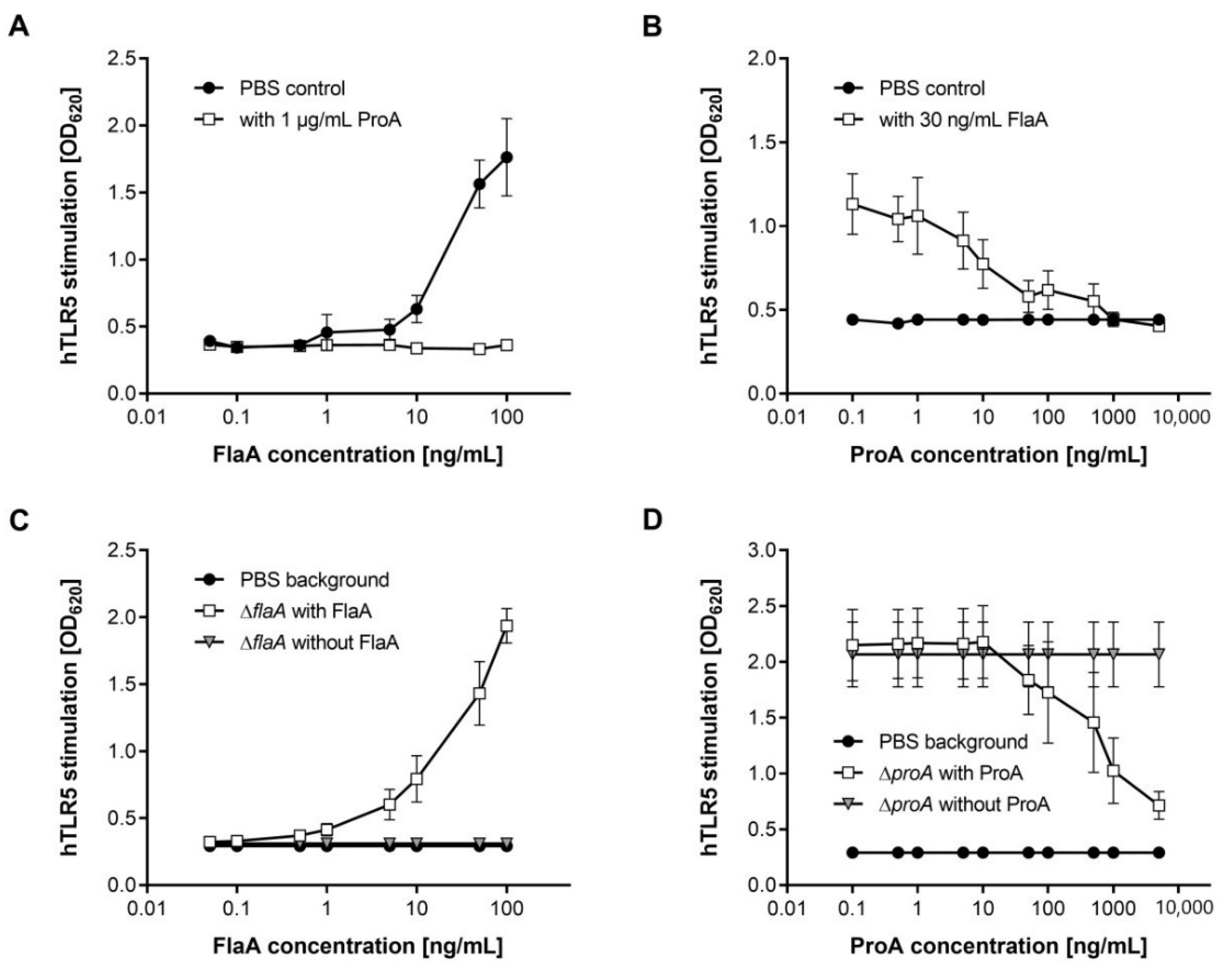
3.2. Flagellin-Mediated TLR5 Activation Is Prevented by ProA

Bacterial flagellin is a potent inducer of pro-inflammatory host responses and is extracellularly detected by TLR5 on host cell surfaces [11,56]. To elucidate if ProA-dependent FlaA degradation alters the innate immune recognition by mammalian cells, we performed a specific detection assay with the hTLR5-expressing cell line HEK-Blue™. In substrate-containing medium, SEAP reporter gene activity induced by NF-κB signaling after TLR5 activation was measured at OD620. Co-incubation of L. pneumophila flagellin with HEK-Blue™ cells accordingly showed concentration-dependent hTLR5 stimulation (Figure 2A). The more FlaA was added, the more SEAP was expressed, and thus its colorimetric product led to higher absorbance at OD620.
Interestingly, such TLR5 activation was completely inhibited by adding 1 µg/mL of purified ProA to the experimental setup. Even at concentrations of 100 ng/mL FlaA, no TLR5 activation was observed in the presence of ProA. This was directly dependent on the proteolytic activity, since a control reaction with heat-inactivated protease was not able to reduce TLR5 stimulation by purified L. pneumophila flagellin. It was additionally validated that ProA does not exhibit activity against host proteins like TLR5 on the cell surface, and observed effects are thus due to FlaA cleavage (Figure S6). Different doses of ProA were tested in the HEK-Blue™ Detection assay to determine which concentration of the Legionella protease is sufficient for preventing TLR5 detection of 30 ng/mL FlaA (Figure 2B). The reduction of OD620 values correlated with increasing ProA concentrations, and TLR5 stimulation was again abolished by using 1 µg/mL of ProA. These data clearly demonstrate that L. pneumophila ProA is able to diminish TLR5 activation by FlaA effectively.
To further analyze these effects, hTLR5 on HEK-Blue™ cells was activated by whole bacterial cells. Co-incubation with a L. pneumophila Corby flaA deletion mutant only resulted in SEAP activity similar to the negative PBS control (Figure 2C). By additionally adding purified FlaA in different concentrations, the TLR5 stimulation was clearly increased. Consequently, downstream pathways seem not to be unspecifically triggered by other bacterial factors besides flagellin. In contrast, a proA-negative mutant immediately induced high TLR5 response as measured by absorbance at OD620 after co-inoculation with HEK-Blue™ cells (Figure 2D). Reduction of the observed TLR5 activation was provoked by the addition of at least 50 ng/mL of purified ProA. With higher protease concentrations, the SEAP activity decreased in a concentration-dependent manner of ProA. An addition of 10 µg/mL of purified protease presumably abolishes the TLR5 activation induced by a proA deletion mutant. All these data demonstrate that extracellular ProA, which is also secreted by L. pneumophila during infections, is able to decrease TLR5 activation by cleavage of free monomeric flagellin in the surrounding medium of bacterial cells.

3.3. Immune Recognition of the L. pneumophila WT via TLR5 Is Reduced by ProA

In further bacterial analyses, the HEK-Blue™ Detection assay was performed to directly differentiate the TLR5 responses evoked by the L. pneumophila Corby WT compared to a proA-negative and a flaA-negative mutant, as well as the functional complementation strains ∆proA proA and ∆flaA flaA (Figure 3). Measurements with 107 bacterial CFU/mL revealed OD620 values of 1.4 for the Legionella WT. ProA deletion resulted in a significant increase of TLR5 activation to an OD620 of 1.7, whereas stimulation by the flaA-deficient mutant is almost abolished to the background level of the medium control. Additionally, these phenotypes were fully restored by using the corresponding complemented strains. FlaA overexpression in ∆flaA even led to enhanced TLR5 stimulation compared to the wild type. Altogether, these results demonstrate a repression of L. pneumophila flagellin-induced TLR5 activation by ProA, which is also detectable in comparison of ProA-expressing or non-expressing bacterial strains.

3.4. ProA Diminishes Extracellular FlaA Stimuli during Human Lung Tissue Infections

For analyzing the ability of L. pneumophila to evade the TLR5-dependent immune response on tissue level, we infected human lung tissue explants (HLTEs) with different Legionella mutant strains. This model was previously demonstrated to be capable of revealing intra- and extracellular aspects of the host–pathogen interaction during Legionnaires’ disease via bacterial growth kinetics, transmigration and localization as well as histopathological alterations of the lung tissue architecture [44,45,46]. Therefore, it is well suitable to compare pro-inflammatory host cell responses in infections with the wild type (WT), a proA- and a flaA-negative mutant (Figure 4). Furthermore, potential phenotypes were reconstituted by the addition of purified proteins (30 ng/mL FlaA and 1 µg/mL ProA) or the functional complementation strains ∆proA proA and ∆flaA flaA. Infected human lung tissue pieces were incubated for different time intervals at 37 °C in cell culture medium. To investigate TLR5 stimulation in the different samples, HEK-Blue™ cells were co-incubated with HLTE infection supernatants from 2 h (Figure 4A) or 24 h (Figure 4B) post inoculation.
The results showed significant differences in the hTLR5 activation potential of all used Legionella deletion strains. At both time points, infection with a proA-negative mutant resulted in significantly higher TLR5 activation than with the wild type. In contrast to that, tissue supernatant of a flaA-deficient mutant strain showed lower OD620 values comparable to the medium control. By the addition of purified ProA, the L. pneumophila ΔproA phenotype could be restored. The enhanced immune response stimulation of this deletion mutant was also significantly reduced using the ΔproA proA complementant. Equivalently, reconstitution was observed for co-incubation of HLTEs with the flaA-deficient mutant and corresponding protein supplementation or complementation strain samples. Our results demonstrate that L. pneumophila Corby flagellin triggers TLR5-dependent immune response during infections of human lung tissue, which can be significantly reduced by expression of ProA. Interestingly, this effect seems to be most important during the first hours of infection, whereas an established infection generally leads to a stronger activation of the NF-κB pathway in exposed human cells.

4. Discussion

In this study, we were able to identify L. pneumophila flagellin FlaA as a physiological substrate of the bacterial zinc metalloprotease ProA. Interestingly, the enzyme only targets the immunogenic monomeric form and leaves polymerized FlaA intact. Our immunological experiments clarified the role of ProA as a regulator of another bacterial virulence factor. In HEK-BlueTM Detection assays, the FlaA-mediated stimulation of TLR5 exposed on host cells was measured with purified proteins, bacterial suspensions of deletion and complementation mutants but also with supernatants of infected HLTEs. Receptor stimulation by a flaA-deficient mutant equated to the medium control and was significantly reduced compared to the L. pneumophila WT or other FlaA-containing samples. Furthermore, the absorbance at 620 nm due to exogenous flagellin was decreased by supplementation with the protease ProA. Bacterial suspensions or infection supernatants containing a proA deletion strain showed an increased TLR5 activation, indicating an important role of the zinc metalloprotease in counteracting pro-inflammatory properties of bacterial flagellin. This supports the notion that recognition of FlaA and subsequent TLR5-mediated immune response plays an important role for pathogen clearance during human Legionella infections. Patients with a TLR5392STOP polymorphism in the ligand-binding domain, accordingly, exhibit an increased susceptibility for Legionnaires’ disease [23].
Since the TLR5 receptor as well as the protease ProA exclusively target monomeric flagellin, in both cases, the recognition sites are most likely hidden inside the polymerized flagellum. In this context, it was already published that amino acid residues of the conserved D1 domain, but also the flagellin polymerization domain D0, are specifically involved in TLR5 binding [16,17]. This would allow ProA to induce immune inhibitory effects without influencing bacterial motility. Similar roles were already reported for the homologous M4 protease pseudolysin as well as AprA of P. aeruginosa, which reduces flagellin-mediated TLR5 activation completely. Thus, an aprA-negative mutant causes a 100-fold enhancement in receptor stimulation compared to the WT, even exceeding the deletion effect of ProA in L. pneumophila Corby [31,32].
TLR5 is predominantly expressed on alveolar macrophages but also on type II pneumocytes, epithelial, endothelial and dendritic cells. A decreasing TLR5 activation alters expression and secretion of chemokines as a part of the pro-inflammatory immune response [57,58]. Via the NF-κB pathway, TLR5-mediated recognition of FlaA provokes, for example, the production of TNF-α, which plays a protective role during L. pneumophila infections. This important immune mediator induces apoptosis of infected host cells, hence reducing proliferation and bacterial load in the lung [24,59]. In this context, it was already shown that L. pneumophila ProA is able to dampen the chemokine and cytokine output from infected macrophages [60]. The protease also exhibits direct proteolytic activity against TNF-α, and the interleukins IL-2 and IL-6 [60,61,62]. Moreover, mice experiments with P. aeruginosa additionally revealed a TLR5-dependent expression of the chemokines MIP-2, IL-17, and IL-22 as well as antimicrobial peptides such as β-defensin 2 or CRAMP (cathelin-dependent antimicrobial peptide) [63]. Among different chemokines, IL-8 is one of the most important pro-inflammatory host messengers, which is expressed in a TLR5-dependent, and thus also in a FlaA-dependent, manner [64,65,66]. It participates in the recruitment of immune cells, especially PMNs, for the clearance of the pathogen [24,67]. Moreover, IL-8 was the cytokine with the highest level in culture supernatants of L. pneumophila-infected macrophages [60]. Results of this study point to a reduced secretion of pro-inflammatory host factors like IL-8 and TNF-α due to a repression of the TLR5-NF-κB pathway by ProA-expressing Legionella strains (Figure 5). We therefore hypothesize that the zinc metalloprotease is able to diminish the IL-8-dependent recruitment and cellular immune response of PMNs during human lung tissue infection.
Altogether, our results indicate that cleavage of free flagellin by ProA promotes immune evasion and, in turn, might represent an important advantage for replication of the pathogen in the human host. Moreover, the immune inhibitory function of the protease is not affecting the polymerized flagellum of the bacterium, and thus its role during the proliferation inside pulmonary lung tissue. Conclusively, we revealed a new component and mechanism of the extensive spectrum of targets by which ProA contributes to different aspects of L. pneumophila pathogenesis. Besides the extracellular detection of FlaA by TLR5 on host cells, flagellin is also sensed in the cytosol by the Naip5/Nlrc4 inflammasome [68]. It is likely that ProA is also able to degrade flagellin, which is inadvertently translocated into the host cell depending on the L. pneumophila dot/icm type IV secretion system (T4SS), since the protease was previously found to assemble at the cytoplasmic site of the LCV. Similar to FlaA, the zinc metalloprotease might be translocated into the host cell cytoplasm due to unspecific pore formation by the T4SS or another bacterial factor of the LCV lumen. However, it is also possible that the semipermeability of the LCV results from host cell membrane transporters, which are acquired via ER-derived vesicles during the first hours after bacterial invasion [69,70,71]. Hence, in the future, it will be particularly interesting to investigate to which extent ProA might additionally influence the intracellular activation of a FlaA-mediated immune response.

Supplementary Materials

The following supporting information can be downloaded at: https://www.mdpi.com/article/10.3390/biom12050624/s1, Figure S1: SDS gels of the ProA purification via anion exchange chromatography; Figure S2: Relative amounts of monomeric and polymeric FlaA in degradation assays with the zinc metalloprotease ProA from L. pneumophila; Figure S3: SDS gel of FlaA degradation experiments with inactive ProA; Figure S4: SDS gels of degradation experiments with FliC from P. aeruginosa and L. pneumophila ProA; Figure S5: SDS gel of flagellin polymers and monomers purified from the L. pneumophila WT, the deletion mutant ∆proA and P. aeruginosa; Figure S6: HEK-BlueTM hTLR5 Detection assay with purified L. pneumophila FlaA and ProA.

Author Contributions

Conceptualization, M.S.; methodology, formal analysis and investigation, L.S. and S.T.; resources, A.D. and M.S.; writing—original draft preparation, L.S. and S.T.; writing—review and editing, M.S.; visualization, L.S.; supervision, C.M.Ü., A.D. and M.S.; project administration and funding acquisition, M.S. All authors have read and agreed to the published version of the manuscript.

Funding

This research was funded by the “Rahmenprogramm Gesundheitsforschung” of the Bundesministerium für Bildung und Forschung (BMBF), grant number 16GW0213.

Institutional Review Board Statement

The study was conducted according to the guidelines of the Declaration of Helsinki, and approved on 10 February 2017 by the Ethics Committee of the Hannover Medical School (No. 2235-2014).

Informed Consent Statement

Informed consent was obtained from all subjects involved in the study.

Data Availability Statement

The data that support the findings of this study are contained within the article or Supplementary Materials.

Acknowledgments

The L. pneumophila Corby flaA deletion mutant was kindly provided by Klaus Heuner, Robert Koch-Institut, Berlin, Germany. We cordially thank Myriam Friemelt and Jolanda Selmar for the technical assistance with HEK-BlueTM hTLR5 Detection assays.

Conflicts of Interest

The authors declare no conflict of interest.

References

  1. Fraser, D.W.; Tsai, T.R.; Orenstein, W.; Parkin, W.E.; Beecham, H.J.; Sharrar, R.G.; Harris, J.; Mallison, G.F.; Martin, S.M.; McDade, J.E.; et al. Legionnaires’ disease: Description of an epidemic of pneumonia. N. Engl. J. Med. 1977, 297, 1189–1197. [Google Scholar] [CrossRef]
  2. McDade, J.E.; Brenner, D.J.; Bozeman, F.M. Legionnaires’ disease bacterium isolated in 1947. Ann. Intern. Med. 1979, 90, 659–661. [Google Scholar] [CrossRef]
  3. Cianciotto, N.P.; Stamos, J.K.; Kamp, D.W. Infectivity of Legionella pneumophila Mip Mutant for Alveolar Epithelial Cells. Curr. Microbiol. 1995, 30, 247–250. [Google Scholar] [CrossRef]
  4. Steinert, M.; Hentschel, U.; Hacker, J. Legionella pneumophila: An Aquatic Microbe Goes Astray. FEMS Microbiol. Rev. 2002, 26, 149–162. [Google Scholar] [CrossRef]
  5. Swanson, M.S.; Hammer, B.K. Legionella pneumophila pathogenesis: A fateful journey from amoebae to macrophages. Annu. Rev. Microbiol. 2000, 54, 567–613. [Google Scholar] [CrossRef]
  6. Akira, S. Toll-like receptor signaling. J. Biol. Chem. 2003, 278, 38105–38108. [Google Scholar] [CrossRef] [Green Version]
  7. Janeway Jr, C.A.; Medzhitov, R. Innate immune recognition. Annu. Rev. Immunol. 2002, 20, 197–216. [Google Scholar] [CrossRef] [Green Version]
  8. Pedra, J.H.; Cassel, S.L.; Sutterwala, F.S. Sensing pathogens and danger signals by the inflammasome. Curr. Opin. Immunol. 2009, 21, 10–16. [Google Scholar] [CrossRef] [Green Version]
  9. Vinzing, M.; Eitel, J.; Lippmann, J.; Hocke, A.C.; Zahlten, J.; Slevogt, H.; Dje N’Guessan, P.; Günther, S.; Schmeck, B.; Hippenstiel, S.; et al. NAIP and Ipaf control Legionella pneumophila replication in human cells. J. Immunol. 2008, 180, 6808–6815. [Google Scholar] [CrossRef] [Green Version]
  10. Smith, K.D.; Andersen-Nissen, E.; Hayashi, F.; Strobe, K.; Bergman, M.A.; Barrett, S.L.R.; Cookson, B.T.; Aderem, A. Toll-like Receptor 5 Recognizes a Conserved Site on Flagellin Required for Protofilament Formation and Bacterial Motility. Nat. Immunol. 2003, 4, 1247–1253. [Google Scholar] [CrossRef]
  11. Gewirtz, A.T.; Navas, T.A.; Lyons, S.; Godowski, P.J.; Madara, J.L. Cutting Edge: Bacterial Flagellin Activates Basolaterally Expressed TLR5 to Induce Epithelial Proinflammatory Gene Expression. J. Immunol. 2001, 167, 1882–1885. [Google Scholar] [CrossRef] [PubMed] [Green Version]
  12. Chevance, F.F.; Hughes, K.T. Coordinating assembly of a bacterial macromolecular machine. Nat. Rev. Microbiol. 2008, 6, 455–465. [Google Scholar] [CrossRef] [PubMed] [Green Version]
  13. Samatey, F.A.; Imada, K.; Nagashima, S.; Vonderviszt, F.; Kumasaka, T.; Yamamoto, M.; Namba, K. Structure of the bacterial flagellar protofilament and implications for a switch for supercoiling. Nature 2001, 410, 331–337. [Google Scholar] [CrossRef] [PubMed]
  14. Yonekura, K.; Maki-Yonekura, S.; Namba, K. Complete atomic model of the bacterial flagellar filament by electron cryomicroscopy. Nature 2003, 424, 643–650. [Google Scholar] [CrossRef]
  15. Donnelly, M.A.; Steiner, T.S. Two Nonadjacent Regions in Enteroaggregative Escherichia coli Flagellin Are Required for Activation of Toll-like Receptor 5. J. Biol. Chem. 2002, 277, 40456–40461. [Google Scholar] [CrossRef] [Green Version]
  16. Forstnerič, V.; Ivičak-Kocjan, K.; Plaper, T.; Jerala, R.; Benčina, M. The role of the C-terminal D0 domain of flagellin in activation of Toll like receptor 5. PLoS Pathog. 2017, 13, e1006574. [Google Scholar]
  17. Song, W.S.; Jeon, Y.J.; Namgung, B.; Hong, M.; Yoon, S. A conserved TLR5 binding and activation hot spot on flagellin. Sci. Rep. 2017, 7, 40878. [Google Scholar] [CrossRef] [Green Version]
  18. Gohda, J.; Matsumura, T.; Inoue, J.I. Cutting Edge: TNFR-Associated Factor (TRAF) 6 Is Essential for MyD88-Dependent Pathway but Not Toll/IL-1 Receptor Domain-Containing Adaptor-Inducing IFN-Beta (TRIF)-Dependent Pathway in TLR Signaling. J. Immunol. 2004, 173, 2913–2917. [Google Scholar] [CrossRef] [Green Version]
  19. Dietrich, C.; Heuner, K.; Brand, B.C.; Hacker, J.; Steinert, M. Flagellum of Legionella pneumophila positively affects the early phase of infection of eukaryotic host cells. Infect. Immun. 2001, 69, 2116–2122. [Google Scholar] [CrossRef] [Green Version]
  20. Hammer, B.K.; Tateda, E.S.; Swanson, M.S. A two-component regulator induces the transmission phenotype of stationary-phase Legionella pneumophila. Mol. Microbiol. 2002, 44, 107–118. [Google Scholar] [CrossRef] [Green Version]
  21. Heuner, K.; Steinert, M. The Flagellum of Legionella pneumophila and Its Link to the Expression of the Virulent Phenotype. Int. J. Med. Microbiol. 2003, 293, 133–143. [Google Scholar] [CrossRef] [PubMed]
  22. Bickel, M. The Role of Interleukin-8 in Inflammation and Mechanisms of Regulation. J. Periodontol. 1993, 64, 456–460. [Google Scholar]
  23. Hawn, T.R.; Verbon, A.; Lettinga, K.D.; Zhao, L.P.; Li, S.S.; Laws, R.J.; Skerrett, S.J.; Beutler, B.; Schroeder, L.; Nachman, A.; et al. A Common Dominant TLR5 Stop Codon Polymorphism Abolishes Flagellin Signaling and Is Associated with Susceptibility to Legionnaires’ Disease. J. Exp. Med. 2003, 198, 1563–1572. [Google Scholar] [CrossRef] [PubMed]
  24. Hawn, T.R.; Berrington, W.R.; Smith, I.A.; Uematsu, S.; Akira, S.; Aderem, A.; Smith, K.D.; Skerrett, S.J. Altered inflammatory responses in TLR5-deficient mice infected with Legionella pneumophila. J. Immunol. 2007, 179, 6981–6987. [Google Scholar] [CrossRef] [Green Version]
  25. Mukaida, N.; Harada, A.; Matsushima, K. Interleukin-8 (IL-8) and Monocyte Chemotactic and Activating Factor (MCAF/MCP-1), Chemokines Essentially Involved in Inflammatory and Immune Reactions. Cytokine Growth Factor Rev. 1998, 9, 9–23. [Google Scholar] [CrossRef]
  26. Takamatsu, R.; Teruya, H.; Takeshima, E.; Ishikawa, C.; Matsumoto, K.; Mukaida, N.; Li, J.-D.; Heuner, K.; Higa, F.; Fujita, J.; et al. Retraction: Molecular characterization of Legionella pneumophila-induced interleukin-8 expression in T cells. BMC Microbiol. 2011, 10, 1–18. [Google Scholar] [CrossRef] [Green Version]
  27. Alhede, M.; Bjarnsholt, T.; Givskov, M.; Alhede, M. Pseudomonas aeruginosa biofilms: Mechanisms of immune evasion. Adv. Appl. Microbiol. 2014, 86, 1–40. [Google Scholar] [CrossRef]
  28. Mejías-Luque, R.; Gerhard, M. Immune Evasion Strategies and Persistence of Helicobacter Pylori. Curr. Top. Microbiol. Immunol. 2017, 400, 53–71. [Google Scholar] [CrossRef]
  29. Zecconi, A.; Scali, F. Staphylococcus aureus Virulence Factors in Evasion from Innate Immune Defenses in Human and Animal Diseases. Immunol. Lett. 2013, 150, 12–22. [Google Scholar] [CrossRef]
  30. Andersen-Nissen, E.; Smith, K.D.; Strobe, K.L.; Barrett, S.L.R.; Cookson, B.T.; Logan, S.M.; Aderem, A. Evasion of Toll-like receptor 5 by flagellated bacteria. Proc. Natl. Acad. Sci. USA 2005, 102, 9247–9252. [Google Scholar] [CrossRef] [Green Version]
  31. Bardoel, B.W.; van der Ent, S.; Pel, M.J.C.; Tommassen, J.; Pieterse, C.M.J.; van Kessel, K.P.M.; van Strijp, J.A.G. Pseudomonas evades immune recognition of flagellin in both mammals and plants. PLoS Pathog. 2011, 7, e1002206. [Google Scholar] [CrossRef] [PubMed] [Green Version]
  32. Casilag, F.; Lorenz, A.; Krueger, J.; Klawonn, F.; Weiss, S.; Häussler, S. The LasB Elastase of Pseudomonas aeruginosa Acts in Concert with Alkaline Protease AprA To Prevent Flagellin-Mediated Immune Recognition. Infect. Immun. 2015, 84, 162–171. [Google Scholar] [CrossRef] [Green Version]
  33. Dreyfus, L.A.; Iglewski, B.H. Purification and Characterization of an Extracellular Protease of Legionella pneumophila. Infect. Immun. 1986, 51, 736–743. [Google Scholar] [CrossRef] [PubMed] [Green Version]
  34. Hales, L.M.; Shuman, H.A. Legionella pneumophila contains a type II general secretion pathway required for growth in amoebae as well as for secretion of the Msp protease. Infect. Immun. 1999, 67, 3662–3666. [Google Scholar] [CrossRef] [PubMed] [Green Version]
  35. Thompson, M.R.; Miller, R.D.; Iglewski, B.H. In vitro production of an extra-cellular protease by Legionella pneumophila. Infect. Immun. 1981, 34, 299–302. [Google Scholar] [CrossRef] [Green Version]
  36. Tiaden, A.; Spirig, T.; Weber, S.S.; Brüggemann, H.; Bosshard, R.; Buchrieser, C.; Hilbi, H. The Legionella pneumophila response regulator LqsR promotes host cell interactions as an element of the virulence regulatory network controlled by RpoS and LetA. Cell. Microbiol. 2007, 9, 2903–2920. [Google Scholar] [CrossRef]
  37. Keen, M.G.; Hoffman, P.S. Characterization of a Legionella pneumophila Extracellular Protease Exhibiting Hemolytic and Cytotoxic Activities. Infect. Immun. 1989, 57, 732–738. [Google Scholar] [CrossRef] [Green Version]
  38. Müller, H.E. Proteolytic Action of Legionella pneumophila on Human Serum Proteins. Infect. Immun. 1980, 27, 51–53. [Google Scholar] [CrossRef] [Green Version]
  39. Quinn, F.D.; Tompkins, L.S. Analysis of a cloned sequence of Legionella pneumophila encoding a 38 kD metalloprotease possessing haemolytic and cytotoxic activities. Mol. Microbiol. 1989, 3, 797–805. [Google Scholar] [CrossRef]
  40. Scheithauer, L.; Thiem, S.; Schmelz, S.; Dellmann, A.; Büssow, K.; Brouwer, R.M.H.J.; Ünal, C.M.; Blankenfeldt, W.; Steinert, M. Zinc metalloprotease ProA of Legionella pneumophila increases alveolar septal thickness in human lung tissue explants by collagen IV degradation. Cell. Microbiol. 2021, 23, e13313. [Google Scholar] [CrossRef]
  41. Conlan, J.W.; Baskerville, A.; Ashworth, L.A. Separation of Legionella pneumophila proteases and purification of a protease which produces lesions like those of Legionnaires’ disease in guinea pig lung. J. Gen. Microbiol. 1986, 132, 1565–1574. [Google Scholar] [CrossRef] [PubMed] [Green Version]
  42. Rechnitzer, C.; Diamant, M.; Pedersen, B.K. Inhibition of Human Natural Killer Cell Activity by Legionella pneumophila Protease. Eur. J. Clin. Microbiol. Infect. Dis. Off. Publ. Eur. Soc. Clin. Microbiol. 1989, 8, 989–992. [Google Scholar] [CrossRef] [PubMed]
  43. Williams, A.; Baskerville, A.; Dowsett, A.B.; Conlan, J.W. Immunocytochemical demonstration of the association between Legionella pneumophila, its tissue-destructive protease, and pulmonary lesions in experimental Legionnaires’ disease. J. Pathol. 1987, 153, 257–264. [Google Scholar] [CrossRef] [PubMed]
  44. Hoppe, J.; Ünal, C.M.; Thiem, S.; Grimpe, L.; Goldmann, T.; Gaßler, N.; Richter, M.; Shevchuk, O.; Steinert, M. PilY1 Promotes Legionella pneumophila Infection of Human Lung Tissue Explants and Contributes to Bacterial Adhesion, Host Cell Invasion, and Twitching Motility. Front. Cell. Infect. Microbiol. 2017, 7, 63. [Google Scholar] [CrossRef] [Green Version]
  45. Jäger, J.; Marwitz, S.; Tiefenau, J.; Rasch, J.; Shevchuk, O.; Kugler, C.; Goldmann, T.; Steinert, M. Human lung tissue explants reveal novel interactions during Legionella pneumophila infections. Infect. Immun. 2014, 82, 275–285. [Google Scholar] [CrossRef] [Green Version]
  46. Scheithauer, L.; Steinert, M. Infection of Human Lung Tissue Explants (HLTEs) with Legionella pneumophila. In Legionella, 2nd ed.; Buchrieser, C., Hilbi, H., Eds.; Humana Press: New York, NY, USA, 2019; pp. 323–331. ISBN 978-1-4939-9047-4. [Google Scholar]
  47. Lang, C.; Rastew, E.; Hermes, B.; Siegbrecht, E.; Ahrends, R.; Banerji, S.; Flieger, A. Zinc metalloproteinase ProA directly activates Legionella pneumophila PlaC glycerophospholipid:cholesterol acyltransferase. J. Biol. Chem. 2012, 287, 23464–23478. [Google Scholar] [CrossRef] [Green Version]
  48. Wintermeyer, E.; Ludwig, B.; Steinert, M.; Schmidt, B.; Fischer, G.; Hacker, J. Influence of site specifically altered Mip proteins on intracellular survival of Legionella pneumophila in eukaryotic cells. Infect. Immun. 1995, 63, 4576–4583. [Google Scholar] [CrossRef] [Green Version]
  49. Jacobi, S.; Schade, R.; Heuner, K. Characterization of the Alternative Sigma Factor sigma54 and the Transcriptional Regulator FleQ of Legionella pneumophila, Which Are Both Involved in the Regulation Cascade of Flagellar Gene Expression. J. Bacteriol. 2004, 186, 2540–2547. [Google Scholar] [CrossRef] [Green Version]
  50. Rossier, O.; Starkenburg, S.R.; Cianciotto, N.P. Legionella pneumophila type II protein secretion promotes virulence in the A/J mouse model of Legionnaires’ disease pneumonia. Infect. Immun. 2004, 72, 310–321. [Google Scholar] [CrossRef] [Green Version]
  51. Secades, P.; Guijarro, J.A. Purification and Characterization of an Extracellular Protease from the Fish Pathogen Yersinia ruckeri and Effect of Culture Conditions on Production. Appl. Environ. Microbiol. 1999, 65, 3969–3975. [Google Scholar] [CrossRef] [Green Version]
  52. Montie, T.C.; Craven, R.C.; Holder, I.A. Flagellar Preparations from Pseudomonas aeruginosa: Isolation and Characterization. Infect. Immun. 1982, 35, 281–288. [Google Scholar] [CrossRef] [PubMed] [Green Version]
  53. Heuner, K.; Bender-Beck, L.; Brand, B.C.; Lück, P.C.; Mann, K.H.; Marre, R.; Ott, M.; Hacker, J. Cloning and Genetic Characterization of the Flagellum Subunit Gene (flaA) of Legionella pneumophila Serogroup 1. Infect. Immun. 1995, 63, 2499–2507. [Google Scholar] [CrossRef] [PubMed] [Green Version]
  54. Gallagher, S.R. Digital image processing and analysis with ImageJ. Curr. Protoc. Essent. Lab. Tech. 2010, 3, 3A–3C. [Google Scholar]
  55. Black, W.J.; Quinn, F.D.; Tompkins, L.S. Legionella pneumophila zinc metalloprotease is structurally and functionally homologous to Pseudomonas aeruginosa elastase. J. Bacteriol. 1990, 172, 2608–2613. [Google Scholar] [CrossRef] [PubMed] [Green Version]
  56. Cobb, L.M.; Mychaleckyj, J.C.; Wozniak, D.J.; López-Boado, Y.S. Pseudomonas aeruginosa Flagellin and Alginate Elicit Very Distinct Gene Expression Patterns in Airway Epithelial Cells: Implications for Cystic Fibrosis Disease. J. Immunol. 2004, 173, 5659–5670. [Google Scholar] [CrossRef] [PubMed] [Green Version]
  57. Honko, A.N.; Mizel, S.B. Effects of flagellin on innate and adaptive immunity. Immunol. Res. 2005, 33, 83–101. [Google Scholar] [CrossRef]
  58. Shikhagaie, M.; Bergqvist, A.; Andersson, C.; Mori, M.; Krohn, K.; Dahl, R.; Hoffman, H.J.; Bjermer, L.; Erjefält, J. Altered distribution patterns of TLR5 and TLR7 in the central airways of severe asthmatics (P4427). J. Immunol. 2013, 190, 206.2. [Google Scholar]
  59. Kawamoto, Y.; Morinaga, Y.; Kimura, Y.; Kaku, N.; Kosai, K.; Uno, N.; Hasegawa, H.; Yanagihara, K. TNF-α inhibits the growth of Legionella pneumophila in airway epithelial cells by inducing apoptosis. J. Infect. Chemother. 2017, 23, 51–55. [Google Scholar] [CrossRef]
  60. McCoy-Simandle, K.; Stewart, C.R.; Dao, J.; DebRoy, S.; Rossier, O.; Bryce, P.J.; Cianciotto, N.P. Legionella pneumophila type II secretion dampens the cytokine response of infected macrophages and epithelia. Infect. Immun. 2011, 79, 1984–1997. [Google Scholar] [CrossRef] [Green Version]
  61. Hell, W.; Essig, A.; Bohnet, S.; Gatermann, S.; Marre, R. Cleavage of tumor necrosis factor-alpha by Legionella exoprotease. APMIS 1993, 101, 120–126. [Google Scholar] [CrossRef]
  62. Mintz, C.S.; Miller, R.D.; Gutgsell, N.S.; Malek, T. Legionella pneumophila protease inactivates interleukin-2 and cleaves CD4 on human T cells. Infect. Immun. 1993, 61, 3416–3421. [Google Scholar] [CrossRef] [PubMed] [Green Version]
  63. Yu, F.; Cornicelli, M.D.; Kovach, M.A.; Newstead, M.W.; Zeng, X.; Kumar, A.; Gao, N.; Yoon, S.G.; Gallo, R.L.; Standiford, T.J. Flagellin stimulates protective lung mucosal immunity: Role of cathelicidin-related antimicrobial peptide. J. Immunol. 2010, 185, 1142–1149. [Google Scholar] [CrossRef] [PubMed]
  64. Im, J.; Jeon, J.H.; Cho, M.K.; Woo, S.S.; Kang, S.S.; Yun, C.H.; Lee, K.; Chung, D.K.; Han, S.H. Induction of IL-8 expression by bacterial flagellin is mediated through lipid raft formation and intracellular TLR5 activation in A549 cells. Mol. Immunol. 2009, 47, 614–622. [Google Scholar] [CrossRef] [PubMed]
  65. Schmeck, B.; N’Guessan, P.D.; Ollomang, M.; Lorenz, J.; Zahlten, J.; Opitz, B.; Flieger, A.; Suttorp, N.; Hippenstiel, S. Legionella pneumophila-induced NF-κB- and MAPK-dependent cytokine release by lung epithelial cells. Eur. Respir. J. 2007, 29, 25–33. [Google Scholar] [CrossRef] [Green Version]
  66. Yu, Y.; Zeng, H.; Lyons, S.; Carlson, A.; Merlin, D.; Neish, A.S.; Gewirtz, A.T. TLR5-mediated activation of p38 MAPK regulates epithelial IL-8 expression via posttranscriptional mechanism. Am. J. Physiol. Gastrointest. Liver Physiol. 2003, 285, G282–G290. [Google Scholar] [CrossRef]
  67. Kunkel, S.L.; Standiford, T.; Kasahara, K.; Strieter, R.M. Interleukin-8 (IL-8): The major neutrophil chemotactic factor in the lung. Exp. Lung Res. 1991, 17, 17–23. [Google Scholar] [CrossRef]
  68. Akhter, A.; Gavrilin, M.A.; Frantz, L.; Washington, S.; Ditty, C.; Limoli, D.; Day, C.; Sarkar, A.; Newland, C.; Butchar, J.; et al. Caspase-7 activation by the Nlrc4/Ipaf inflammasome restricts Legionella pneumophila infection. PLoS Pathog. 2009, 5, e1000361. [Google Scholar] [CrossRef] [Green Version]
  69. Miao, E.A.; Andersen-Nissen, E.; Warren, S.E.; Aderem, A. TLR5 and Ipaf: Dual sensors of bacterial flagellin in the innate immune system. Semin. Immunopathol. 2007, 29, 275–288. [Google Scholar] [CrossRef]
  70. Molofsky, A.B.; Byrne, B.G.; Whitfield, N.N.; Madigan, C.A.; Fuse, E.T.; Tateda, K.; Swanson, M.S. Cytosolic recognition of flagellin by mouse macrophages restricts Legionella pneumophila infection. J. Exp. Med. 2006, 203, 1093–1104. [Google Scholar] [CrossRef]
  71. Truchan, H.K.; Christman, H.D.; White, R.C.; Rutledge, N.S.; Cianciotto, N.P. Type II Secretion Substrates of Legionella pneumophila Translocate Out of the Pathogen-Occupied Vacuole via a Semipermeable Membrane. mBio 2017, 8, e00870-17. [Google Scholar] [CrossRef] [Green Version]
Figure 1. ProA degrades monomeric flagellin. Flagella were isolated from L. pneumophila Corby and heated for 20 min to 70 °C for depolymerization. Polymeric (A) and monomeric (B) flagellin (10 µg/mL) was incubated for 1 h at 37 °C with indicated concentrations of purified ProA. Samples were separated in an SDS gel with subsequent protein silver staining. M: 1–5 µL of the PageRulerTM Prestained Protein Ladder by Thermo Scientific were used as a standard. Additionally, the samples were immunoblotted with a primary antibody against L. pneumophila FlaA and detected with an alkaline phosphatase antibody and NBT/BCIP substrate solution. While the polymeric form of flagellin is resistant against the proteolytic degradation by ProA, FlaA monomers were efficiently cleaved by the protease. Not more than one third of the initial substrate concentration was left after inoculation of FlaA monomers with 0.1 µg/mL ProA.
Biomolecules 12 00624 g001
Figure 2. ProA inhibits TLR5 activation by FlaA in HEK-Blue™ cells. HEK-Blue™ hTLR5 cells were seeded at a density of 2.52 × 10⁴ cells/well. After adherence, they were inoculated with HEK-Blue™ Detection medium and 20 µL samples containing the indicated bacterial strains or proteins. PBS served as a negative or background control (black dots). After incubation at 37 °C and 5% CO2 for 16 h, SEAP activity, and hence hTLR5 stimulation, were determined at OD620. Means of measurements are shown with ±SEM from three (A,B) or four (C,D) independent experiments. (A) HEK-Blue™ cells were treated with PBS or 1 µg/mL ProA (white squares) and different concentrations of purified FlaA, only leading to increasing hTLR5 activation in the negative control. (B) 30 ng/mL FlaA per well (white squares) were added to HEK-Blue™ cells, which were additionally treated with different ProA concentrations between 0.1 ng/mL and 5000 ng/mL. TLR5 stimulation was thereby inhibited in a concentration-dependent manner. (C) HEK-Blue™ cells were inoculated with the L. pneumophila Corby ΔflaA mutant (grey triangles). Co-incubation with purified flagellin reconstituted the lacking TLR5 stimulation of the eukaryotic cells (white squares). (D) L. pneumophila Corby ΔproA inoculation of HEK-Blue™ cells resulted in high hTLR5 activation (grey triangles), but was diminished by adding natively purified ProA in increasing concentrations (white squares).
Biomolecules 12 00624 g002
Figure 3. Specific TLR5 activation in HEK-Blue™ cells by different L. pneumophila strains. HEK-Blue™ hTLR5 cells at a density of 2.52 × 10⁴ cells/well were incubated with 180 µL detection medium and 20 µL culture of the L. pneumophila Corby wild type (WT), the negative mutant strains ΔproA and ΔflaA or complementants ΔproA proA and ΔflaA flaA in a 96-well format. Prior to inoculation, bacterial strains were grown to stationary phase in YEB, and the cell count was adjusted to 108 CFU/mL. After incubation of the assay plate at 37 °C and 5% CO2 for 16 h, TLR5-dependent NF-κB activation was measured by SEAP activity at OD620. The results are depicted in a scatter plot with means and reveal significantly diminished values in the case of the flaA-negative strain but even stronger induced TLR5 stimulation by the proA deletion mutant compared to the WT. Complementation strains of ΔproA and ΔflaA were able to completely reconstitute these phenotypes. Statistics were performed using a repeated measurement one-way ANOVA with Dunnett’s post hoc test for simple effect analysis. Significant results of ten biological replicates are indicated with asterisks (* p ≤ 0.05, *** p ≤ 0.001, **** p ≤ 0.0001).
Biomolecules 12 00624 g003
Figure 4. TLR5 activation in HEK-Blue™ cells by supernatants of infected lung tissue explants (HLTEs). Explanted human lung tissue specimens were infected with the L. pneumophila Corby WT, a proA-negative mutant ΔproA and a flaA-negative mutant ΔflaA. Loss of ProA or FlaA was reconstituted either by addition of purified proteins (1 µg/mL ProA and 100 ng/mL monomeric FlaA) (A) or use of the specific complementation strains ΔproA proA and ΔflaA flaA (B). Untreated tissue samples served as a negative control. Infected tissue pieces were weighed, and supernatants were isolated and incubated with HEK-Blue™ hTLR5 cells for 16 h at 37 °C and 5% CO2. Activation of the TLR5-NF-κB pathway was measured by SEAP activity at OD620 and is shown in scatter plots with means. Compared to the L. pneumophila WT, a HEK-Blue™ Detection assay revealed significantly reduced TLR5 activation by ΔflaA-infected tissue supernatant and elevated stimulation with the ΔproA mutant strain. These effects were apparent after 2 h (A) and 24 h (B) post infection and were successfully restored by complementation with respective proteins or gene sequences. Significance of at least twelve replicates was evaluated by repeated measurement one-way ANOVA with Dunnett’s post hoc test for simple effect analysis (* p ≤ 0.05, *** p ≤ 0.001, **** p ≤ 0.0001).
Biomolecules 12 00624 g004
Figure 5. Proposed mechanism of counteracting flagellin-mediated host immune response by the L. pneumophila protease ProA. (A) During lung infection with L. pneumophila, bacterial FlaA monomers are recognized via TLR5 presented on the surface of lung epithelial cells or alveolar macrophages. Stimulation of the receptor initiates a signaling cascade which results in NF-κB activation and IL-8 production. IL-8 is a main chemoattractant of PMNs triggering recruitment into the infected region and subsequent pathogen clearance. Alveolar macrophages additionally express pro-inflammatory cytokines like IL-6 and TNF-α upon TLR5 stimulation. (B) L. pneumophila secretes the zinc metalloprotease ProA which is able to cleave immunogenic FlaA monomers, while the intact flagellum, and hence bacterial motility, are not affected by its proteolytic activity. Efficient degradation of monomeric flagellin results in a strong reduction of TLR5 activation and the following cytokine production. Consequently, stimulation, recruitment and bacterial killing of PMNs will be reduced. Thus, ProA may function as an important modulator of the FlaA-mediated pro-inflammatory host response (created with BioRender.com).
Biomolecules 12 00624 g005
Table 1. List of bacterial strains, cell lines, plasmids and primers.
Cell LineDescriptionReferences
HEK-BlueTM hTLR5HEK 293 cell line expressing human TLR5, SEAP reporterInvivoGen, Toulouse, France
StrainDescriptionReferences
L. pneumophila Corby WTWT reference strain[48]
L. pneumophila Corby ΔproAproA::nptI, proA deletion mutant, Kmr[40]
L. pneumophila Corby ΔproA proAproA deletion mutant with pMMB2002-proA, Kmr, Cmlr[40]
L. pneumophila Corby ΔflaAflaA::nptI, flaA deletion mutant, Kmr[49]
L. pneumophila Corby ΔflaA flaAflaA deletion mutant with pMMB2002-flaA, Kmr, CmlrThis study
P. aeruginosa PAO1WT strainATCC 15692
PlasmidDescriptionReferences
pMMB2002pMMB207-derived expression plasmid for L. pneumophila, deleted mobA gene, Cmlr[50]
pMMB2002-proApMMB2002-derived vector expressing the proA gene, Cmlr[40]
pMMB2002-flaApMMB2002-derived vector expressing the flaA gene, CmlrThis study
Primer5′-3′-SequenceReferences
FlaA-Pr_fwCATGAGCTCTCGACTTGATAACCCGAACCThis study
FlaA+_rvAAGGTACCCTATCGACCTAACAATGATAATACThis study
Publisher’s Note: MDPI stays neutral with regard to jurisdictional claims in published maps and institutional affiliations.

