Article

Characterization of a Novel Regulator of Biofilm Formation in the Pathogen Legionella pneumophila

1
Department of Microbiology and Immunology, Drexel University College of Medicine, Philadelphia, PA 19129, USA
2
Kinneret Limnological Laboratory (KLL), Israel Oceanographic and Limnological Research (IOLR), Migdal 14950, Israel
*
Author to whom correspondence should be addressed.
Biomolecules 2022, 12(2), 225; https://doi.org/10.3390/biom12020225
Submission received: 31 December 2021 / Revised: 23 January 2022 / Accepted: 24 January 2022 / Published: 27 January 2022

Abstract

:
Legionella pneumophila is a Gram-negative, facultative intracellular pathogen that causes severe pneumonia known as Legionnaires’ disease. The bacterium causes disease when contaminated water is aerosolized and subsequently inhaled by individuals, which allows the bacteria to gain access to the lungs, where they infect alveolar macrophages. L. pneumophila is ubiquitous in the environment, where it survives by growing in biofilms, intracellularly within protozoa, and planktonically. Biofilms are a major concern for public health because they provide a protective niche that allows for the continuous leaching of bacteria into the water supply. In addition, biofilms enhance the survival of the bacteria by increasing resistance to temperature fluctuations and antimicrobial agents. Currently, there is little known about biofilm formation and regulation by L. pneumophila. Here, we present evidence of a specific gene, bffA, which appears to be involved in the regulation of motility, biofilm formation, cellular replication, and virulence of L. pneumophila. A strain lacking bffA has an enhanced biofilm formation phenotype, forming biofilms that are both faster and thicker than wild type. Additionally, the knockout strain has significantly reduced motility, enhanced uptake into amoebae, and altered growth kinetics on solid media. Our data suggest a potential role for bffA in signaling pathways that govern changes in growth rate and motility in response to environmental conditions.

1. Introduction

Legionella pneumophila is an opportunistic pathogen and the causative agent of a severe form of pneumonia known as Legionnaires’ Disease. Legionnaires’ Disease is responsible for 18,000 hospitalizations per year in the United States, has a case mortality rate of 5–30%, and is particularly problematic for elderly and immunocompromised individuals [1,2,3,4,5,6]. The disease is caused when a contaminated water source becomes aerosolized and is inhaled by humans, thereby providing bacteria access to the lungs, where they infect alveolar macrophages [2,7,8]. L. pneumophila is ubiquitous in aquatic environments, where it can be found planktonically, intracellularly within protozoa, or within biofilms [9].
In recent years, biofilms have become a major public health concern with the increased awareness of the potential of biofilms to provide a continuous source of bacterial contamination in freshwater supply systems. Importantly, association with biofilms has been shown to enhance the survival of residing bacteria [4,10,11] by providing protection against antibiotics, antimicrobial agents, temperature fluctuations, and nutrient and oxygen limitations [1,4,12,13,14,15]. The presence of biofilms containing L. pneumophila in domestic water supplies has been identified as a potential cause of drinking water-acquired Legionellosis [10]. L. pneumophila is responsible for up to 30% of drinking water-borne diseases each year, and up to 80% of drinking water deaths in the United States [16]. Additionally, a large percentage of nosocomial-acquired Legionellosis is due to inadequate sanitization of water supply systems and the inability to eliminate biofilms within the system [5].
The identification of key regulators could prove useful in the development of novel antibiofilm strategies that would reduce the incidence of disease by eradicating biofilms from water supplies. While there has been some research into Legionella mono-and multispecies biofilms [6,9,10,12,15,17,18,19,20,21,22,23,24], the majority of the genes and proteins involved in the biofilm pathway of L. pneumophila remain to be identified. Of special interest is the second messenger cyclic diguanosine monophosphate (c-di-GMP), which has been shown in other bacteria to play a role in regulating the biofilm lifestyle [25,26,27]. A few of the L. pneumophila genes involved in the metabolism of c-di-GMP have been identified [18,19,28,29]. In a paper by Levi et al., the authors presented data showing that deletion of putative diguanylate cyclase (DGC) and phosphodiesterase (PDE) genes did not have any effect on intracellular growth; however, the overexpression of some of the genes led to an intracellular growth defect. This work provides evidence that c-di-GMP levels affect the virulence of L. pneumophila. Carlson et al. show that a heme nitric oxide (H-NOX) regulates the activity of a DGC, lpg1057. Upon deletion of the hnox1 gene, the bacteria display a hyper-biofilm phenotype, suggesting that hnox1 is inhibiting the activity of lpg1057 to block biofilm formation. Thus, when hnox1 is deleted, intracellular levels of c-di-GMP increase, leading to the hyper-biofilm phenotype observed. These findings demonstrate the importance of c-di-GMP pathways in L. pneumophila, linking the pathway to biofilm regulation and intracellular survival of this pathogen.
In this work, we sought to investigate the function of a previously uncharacterized Legionella gene lpg1387 containing a putative phosphodiesterase (PDE) domain. We found that a deletion mutant of lpg1387 has altered growth kinetics on solid media, enhanced uptake into amoebae, decreased flagellar motility, and enhanced biofilm formation compared to wild type L. pneumophila. Our data suggest that the gene, which we named bffA, is involved in the regulation of motility and biofilm formation in L. pneumophila.

2. Materials and Methods

2.1. Strains, Plasmids, and Primers

The strains used in this study are outlined in Table 1. We used both L. pneumophila strains, CR39 and JR32, and generated a ΔbffA mutant in both backgrounds. In-frame deletion of bffA was performed by homologous recombination after the introduction of the suicide vector pSR47s (pCM4) containing a fusion of the 800 bp upstream and 800 bp downstream of bffA generated using primers SN252, SN253, SN254 and SN255 and ligated with plasmid pSR47s digested with XbaI and SacI. Plasmid pMMB207-Cam with full-length bffA (pCM2) was generated by digesting the plasmid pCM1 with BamHI and HindIII and ligating with a PCR fragment generated using primer SN256 and SN257. The plasmid pMMB207-empty (pCM1) was generated by digesting pMMB207-M45NT with BamHI and HindIII and treating with T4 DNA ligase to fill the HindIII site, followed by blunt-end ligation and transformation. All vectors were confirmed by PCR and digest. See Table 1 for full list of strains, primers, and plasmids.

2.2. Media and Antibiotics

For Legionella strains: all were grown in either AYE broth [1% yeast extract [HIMEDIA], 1% N-(2-acetamido)-2-aminoethanesulphonic acid (ACES; pH 6.9) [Amresco], 3.3 mM L-cysteine [Sigma], 0.33 mM Fe(NO3)3] or on charcoal yeast extract agar (CYE) (AYE containing 1.5% bacto-agar [HIMEDIA], 0.2% activated charcoal [Sigma]). Antibiotics, all purchased from Sigma unless stated otherwise, used for Legionella strains were added to the media at the following concentrations: streptomycin 100 μg/mL, kanamycin 20 μg/mL, chloramphenicol 10 μg/mL.
E. coli strains were grown in LB medium (LB) 1% bacto-tryptone, 0.5% yeast extract, 1% NaCl [Sigma] or on LB-agar plates (LB containing 1.5% bacto-agar). Antibiotics were added to the media at the following concentrations: ampicillin 100 μg/mL, kanamycin 40 μg/mL, chloramphenicol 25 μg/mL.
Acanthamoeba castellanii (ATCC 30234) were grown at room temperature in ATCC medium 712 (PYG). One hour before infection, A. castellanii cultures were incubated at 37 °C in a 5% CO2 atmosphere in PYG medium without glucose, protease peptone, and yeast extract (amoebae buffer). If indicated, after infection, A. castellanii was washed with sterile phosphate-buffered saline (PBS).

2.3. Broth Growth Curve

Broth growth curves were started from plate-grown bacteria that had been incubated for 48 h. Overnight cultures grown in liquid AYE medium were diluted to OD600 = 0.05 and grown shaking at 37 °C in glass culture tubes, with OD600 measurements taken approximately every 45 min.

2.4. Intracellular Growth Curve in A. Castellanii

A. castellanii was plated at a concentration of approximately 1 × 10⁵ cells per well of a 24-well plate, 24 h prior to infection. One hour before infection, each well was washed twice with sterile PBS and the media was replaced with amoebae buffer, and the plates were incubated at 37 °C. Plate-grown bacteria that had been incubated for 48 h prior to infection were suspended in distilled water to OD600 = 1.0 and then diluted to OD600 = 0.05. Each well was infected with 5.0 μL of the OD600 = 0.05 suspension, a multiplicity of infection (MOI) of 1, and incubated for two hours at 37 °C. Time-zero wells were processed by removing the media, washing the wells three times with sterile PBS to remove extracellular bacteria, and lysing the amoebae in 1.0 mL distilled water, then diluting and plating for enumeration of colony-forming units (CFU). The rest of the wells were washed three times with sterile PBS and 1.0 mL fresh media was added to each well. The plates were returned to the incubator and processed at the specified times. To process the subsequent time points, the media from each well was combined with the lysate to account for extracellular, as well as intracellular, bacteria.

2.5. Flagellar Motility Assay

Overnight cultures of each strain were serial diluted 6:7 from an initial OD600 of 0.5, (37 °C), or 0.65 (30 °C), and grown shaking at 37 °C or 30 °C. The OD600 of each culture was read in the morning, and the motility was observed using a Nikon Ti Eclipse microscope. Each culture was given a motility score based upon the percentage of the observable culture performing directional motility. To calculate the motility score, each culture was observed for several minutes over five fields of view per sample, to enumerate a percentage of the overall culture that was either stationary or moving in a directional manner. Bacteria were manually counted, with an average of 50–100 bacteria per field of view to generate the percentage of motile bacteria per culture. The experiment was repeated three independent times and statistical analysis was performed using an unpaired t-test.

2.6. 96-Well Biofilm Assay

Plate-grown strains were resuspended to OD600 = 0.3 in AYE. 200 μL of each strain was plated per well of the 96-well plate and then incubated at 30 °C. The wells were stained with 200 μL of 0.1% crystal violet on the indicated days and resolubilized in 30% acetic acid, and absorbance was measured in OD600.

2.7. Coverslip Biofilm Assay

Cultures of each strain were grown overnight shaking at 37 °C. The OD600 in the morning was adjusted to OD = 1.0, and 250 μL was applied to coverslips placed inside each well of a 24-well plate. The plate was then incubated at 30 °C on a slant to allow the air interface to run across the center of the coverslip, according to the method by Merritt et al. [35]. The coverslips were then stained with 4,6-diamidino-2-phenylindole (DAPI), mounted using Mowiol, and imaged on a Nikon Ti Eclipse microscope.

2.8. Biofilm Binding Assay

To test for enhanced binding of ΔbffA to the wells of a 96-well plate, we grew wild-type and ΔbffA strains overnight shaking at 37 °C. In the morning, the cultures were diluted to OD600 = 1.0 and 200 μL was plated per well in 9 wells for each strain. For T0, the bacteria were allowed to adhere for 15 min, 2 h, or 4 h before the media was removed and the wells were washed with distilled water. The adherent bacteria were collected from the well using a sterile cotton-tipped applicator, resuspended in sterile PBS, and plated for CFUs.

2.9. Gentamicin Protection Assay

To measure uptake into A. castellanii, amoebae were plated at a concentration of 1 × 10⁵ cells per well of a 24-well plate one day prior to infection. One hour prior to infection, the wells were washed with PBS and the amoebae were incubated with amoebae buffer at 37 °C for one hour. The wells were then infected with an MOI = 100 for 2 h, following a 5 min centrifugation at 1000 rpm. After 2 h, the amoebae were washed with PBS containing 100 μg/mL gentamicin and subsequently incubated for 30 min at 37 °C with buffer containing 100 μg/mL gentamicin. The lysate was collected and plated for CFUs. Additionally, the input was plated for CFUs to calculate the percentage uptake of each strain. Statistical analysis was performed using an unpaired t-test.

3. Results

3.1. L. pneumophila ΔbffA Exhibits Altered Growth Kinetics on Solid Medium, but Not in Broth, Compared to Wild-Type

bffA is annotated as a putative phosphodiesterase, however, it appears to be lacking key residues that would be involved in the cleavage of c-di-GMP (Figure S1), suggesting it lacks the ability to cleave c-di-GMP but may still be involved in a signaling pathway that responds to fluctuations in intracellular levels of the second messenger. Phosphodiesterases (PDEs) cleave c-di-GMP to produce 5’-phosphoguanylyl-(3’,5’)-guanosine (pGpG), which is then hydrolyzed to produce two GMP molecules. PDEs are known to be involved in lifestyle decisions in many bacterial species, including Pseudomonas aeruginosa and Vibrio cholerae, where c-di-GMP signaling regulates both planktonic and biofilm growth [13,26,27,36,37,38,39]. While we have no evidence that bffA is acting as a PDE, the first observation we made with the ΔbffA strain, once it was constructed, was that it forms single colonies in 48 h, compared to 72–96 h required for wild-type bacteria to form single colonies (Figure 1A). This phenotype is also observed when the ΔbffA strain is grown from a frozen stock, where it takes an additional 24 h for the wild-type strain to grow. Furthermore, we were able to revert the ΔbffA strain to a wild-type phenotype using a pMMB207 derived expression vector, where the expression of bffA is driven off of the icmR promoter (Figure 1B) when the proper selection was present. The 3′ end of the bffA gene, encoding C-terminal residues 367–469 of the BffA protein, is not necessary for the observed growth regulation activity of bffA, as a vector encoding for a BffA truncation is sufficient to revert the mutant phenotype back to wild-type growth rates (Figure 1B). The complementation observed is specifically due to the expression of bffA, and not merely the presence of the plasmid since the empty vector pCM1 did not complement the growth phenotype. These results suggest that bffA has a role in the regulation of L. pneumophila growth. Additionally, these results indicate that the C-terminus of BffA is not required for the observed growth phenotype.
The ΔbffA mutant was also characterized in liquid broth to test whether the growth rate phenotype we observed was specific to solid media. The ΔbffA strain did not display altered growth kinetics in broth when compared to wild-type (Figure 1C). This suggests that bffA is important to growth on solid surfaces, but not during the planktonic growth phase of the bacteria.

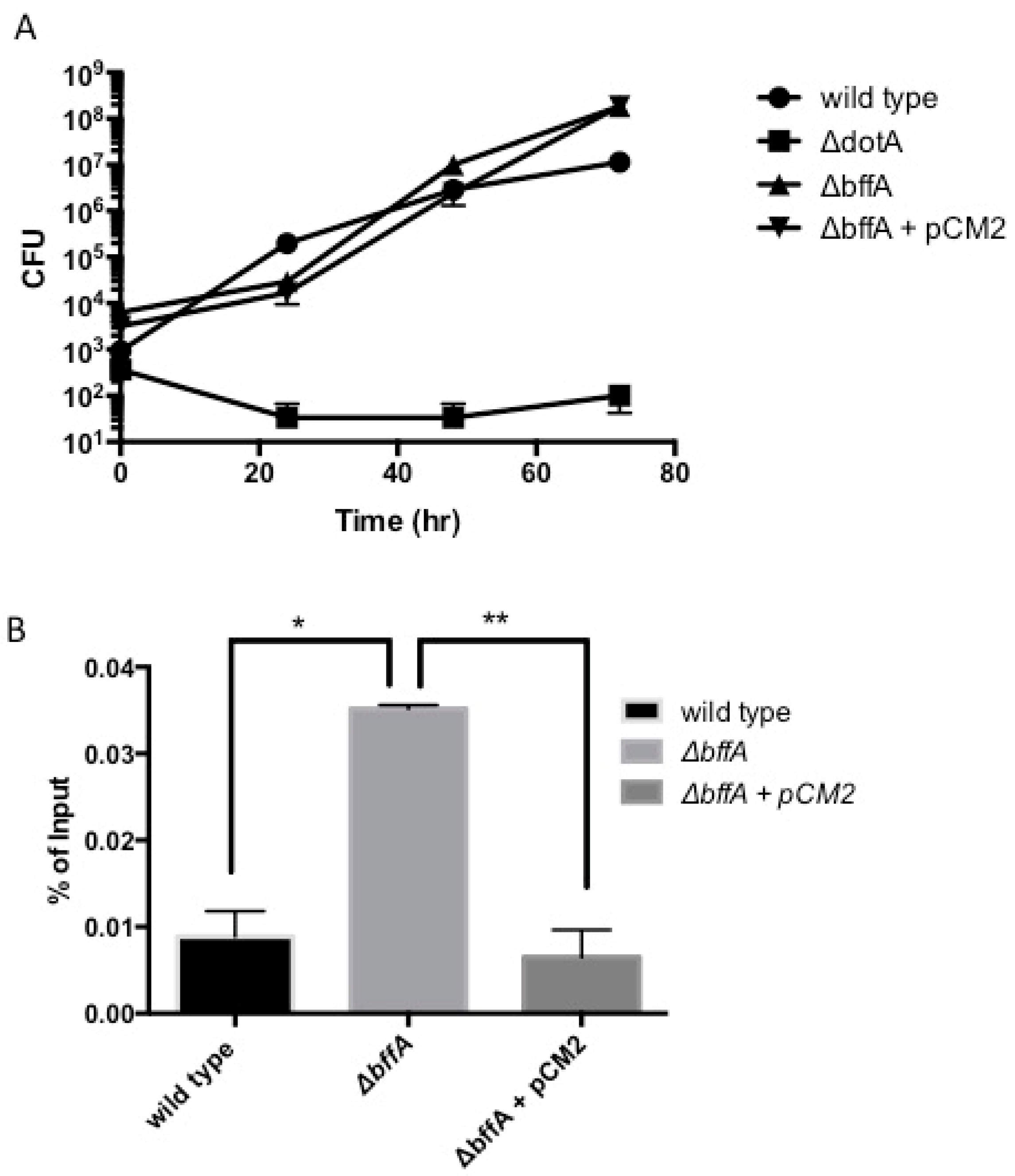
3.2. The ΔbffA Strain Has Normal Rates of Intracellular Growth in A. Castellanii

L. pneumophila is an intracellular pathogen, with a large portion of its lifecycle spent replicating within a Legionella-containing vacuole inside of protozoans in the environment, or macrophages during human infection [2,40,41,42,43,44,45,46,47,48]. Since we observed altered growth kinetics on solid media, we determined the intracellular growth rate of ΔbffA in A. castellanii, which is considered a natural host for L. pneumophila in the environment. We found that in A. castellanii, ΔbffA has a slight enhancement in intracellular growth rate compared to wild-type (Figure 2A). We were unable to see the reversal of the ΔbffA phenotype to a wild-type phenotype in this assay due to the inability to maintain adequate levels of antibiotics in the media over the three-day time course. The complementation strain rapidly loses the vector carrying bffA within 48 h of inadequate antibiotic levels, making complementation impossible. We have observed that, when grown on CYE without antibiotics, the complemented strain forms single colonies in two days, and these colonies no longer possess the ability to grow on antibiotic-containing plates, indicating they have lost the complementation vector (not shown). Additionally, through these experiments, we observed a large increase in the uptake of the ΔbffA strain into the amoebae at time zero, which is reverted to wild-type levels with the expression of bffA in trans. We further tested uptake levels using a gentamicin protection assay, in which bacteria that are not taken up by A. castellanii are killed by the addition of the antibiotic gentamicin to the amoebae medium. For this assay, amoebae are infected with a high multiplicity of infection (MOI), and the output, i.e., what is collected from the lysed amoebae after antibiotic treatment, is normalized to the input to generate a percentage of the input that is protected from antibiotic treatment. Figure 2B shows that ΔbffA has significantly enhanced uptake into A. castellanii and the addition of bffA in trans reverted the uptake rate back to wild-type levels, indicating a phenotype specific to the deletion of bffA. The increased uptake phenotype of ΔbffA could be a manifestation of the altered growth rate during the short infection. Alternatively, bffA may function in the downregulation of uptake into amoebae through a yet-unknown mechanism.

3.3. ΔbffA Forms Biofilms Faster than the Wild-Type Strain

In many bacterial species, including P. aeruginosa and V. cholerae, planktonic growth and biofilm growth are regulated in an opposing manner [49,50,51]. Based upon this and on our previous experiments with the ΔbffA strain, we hypothesized that the deletion of bffA would lead to a shift in the growth of the ΔbffA strain to a biofilm lifestyle, as opposed to a motile, planktonic lifestyle. Since we have already observed alterations in growth on solid media and virulence, we sought to establish several biofilm assays to test the ability of ΔbffA to form biofilms. In a 96-well biofilm formation assay, plate grown bacteria were resuspended to an OD600 = 0.3 and plated at 200 μL per well of a 96-well plate. For each time point during the 12 days of the experiment, washed wells were stained with 0.5% crystal violet to measure the degree of biofilm formation. The crystal violet was then resolubilized from the stained biofilms in 30% acetic acid, and the OD600 was measured. We observed that all biofilms formed at the air-liquid interface, and the ΔbffA strain formed biofilms more rapidly and densely than the wild-type strain (Figure 3A). This indicates that bffA may be involved in the regulation of biofilm formation. Attempts to complement this phenotype were unsuccessful due to the instability of the complementation vector in the ΔbffA strain.
We next established an assay for the qualitative visualization of the biofilms. For this assay, we grew the biofilms on glass coverslips in a 24-well plate, tilted to ensure the air interface of the culture was situated across the center of the coverslip. The coverslips were removed from the plate and stained to observe the structure of the biofilm and measure biofilm thickness by fluorescence microscopy. We observed that ΔbffA biofilms formed earlier and appeared thicker than wild-type biofilms (Figure 3B). These results are in line with the results obtained using the 96-well plate assay.
To test whether the enhanced biofilm formation may be a result of differences in the capacity of the strains to bind to the wells of the 96-well plate, we performed a plate-binding assay. Bacteria were plated at 200 μL of OD600 = 1.0 per well of a 96-well plate and allowed to adhere to the wells for 15 min, 2 h, and 4 h. We did not observe any difference in the ability of ΔbffA to bind to the wells compared to the wild-type strain (Figure 3C). We concluded that the ΔbffA strain is able to form biofilms faster than the wild-type, in a mechanism that is independent of the initial adherence phase of biofilm formation.

3.4. ΔbffA Is Less Motile than Wild-Type Bacteria at 37 °C, but Not at 30 °C

Motility and biofilm formation are inversely regulated in bacteria [26,27,49,52], so we sought to determine if we could observe reduced motility in the ΔbffA strain since we previously observed enhanced biofilm formation compared to wild-type. With the lack of a plate-derived motility assay, we used light microscopy to measure the percentage of the population displaying flagellar motility in several overnight cultures. Using this approach, we determined the level of motility in the wild-type and ΔbffA strains, as well as in wild-type and ΔbffA harboring pCM2. We set up 12 cultures and 6:7 serial dilutions of each strain, starting with an OD600 = 0.5. By setting up a series of cultures, we expected that we would be able to capture the bacteria at peak motility. The cultures were grown overnight at 37 °C, and the OD600 was measured again in the morning, followed by subsequent microscopic analysis for motility. We observed that wild-type, wild-type-pCM2, and ΔbffA–pCM2 were able to reach peak motility levels of 90% (Figure 4A), independent of culture density. On the other hand, the ΔbffA strain had very limited motility, regardless of culture density, and the highest level of motility we were able to record for this strain was 20% of the culture displaying flagellar motility (Figure 4A).
Interestingly, when repeating this assay with bacteria grown at 30 °C, we observed no difference in flagellar motility between the ΔbffA strain and the other strains (Figure 4B). All of the strains tested showed 80–90% motility of the culture, with no significant difference in motility detected between the strains (p < 0.005). This suggests a temperature-dependent role for bffA in the regulation of flagellar motility.

4. Discussion

In recent years, the bacterial second messenger, c-di-GMP, has become the focus of much research and found to influence biofilm formation, motility, cellular replication, and virulence in many bacterial species. Work aimed at identifying proteins involved in the metabolism of c-di-GMP has led to the discovery of degenerate DGCs and PDEs that still bind c-di-GMP, though they lack enzymatic activity [38,50,53,54]. In these studies, proteins were found to be missing specific motifs and/or residues required for the production of c-di-GMP or its breakdown. While degenerate c-di-GMP metabolizing genes would appear to be non-functional, their deletion mutants are often found to have strong phenotypes [38,53,54]. Here, we investigate an L. pneumophila protein we termed BffA, which contains a putative PDE domain but lacks conserved residues found in known proteins from this family. In our work, we show that the deletion of bffA results in strong phenotypes linked to the regulation of motility, virulence, cellular replication, and biofilm formation. Our work suggests that BffA may be functioning within the c-di-GMP pathway, despite lacking key PDE active-site residues.
It has been previously demonstrated that degenerate c-di-GMP metabolism genes can still affect c-di-GMP levels without being enzymatically active. Studies with PelD and FimX from P. aeruginosa [38,54] and LapD from Pseudomonas fluorescens [53] provide evidence that proteins possessing degenerate DGC and PDE domains maintain the ability to bind c-di-GMP and affect downstream signaling of the molecule. In the case of PelD, the protein possesses a degenerate DGC domain, but still maintains the ability to bind c-di-GMP at the I-site [54]. FimX has a degenerate DGC and PDE domain and can bind c-di-GMP through its PDE domain. Deletion of fimX leads to a complete abrogation of biofilm formation, suggesting a major role for c-di-GMP binding proteins in the regulation of motility and biofilm formation [38]. LapD from P. fluorescens is also a dual DGC–PDE domain protein, with both domains degenerate and enzymatically inactive. LapD binds c-di-GMP through its degenerate PDE domain and induces biofilm formation through the production and localization of LapA, an adhesin that is essential for the attachment of the bacteria to surfaces [53].
In a recent study, it was reported that an ATPase gene (mshE) in V. cholerae and its homolog in P. aeruginosa lacked all conserved motifs of PDEs, but still functioned as c-di-GMP binding proteins. Upon deletion of mshE from V. cholerae, the strain had reduced motility and enhanced biofilm formation, much like ΔbffA. This suggests that, while it does not appear to have a functional PDE domain, it is possible that BffA is still exerting an effect on c-di-GMP levels within L. pneumophila. In a different study with V. cholerae, researchers deleted a PDE encoding gene, cdgJ, which resulted in reduced motility and enhanced biofilm formation in the knockout strain [49]. However, they were unable to detect any change in global c-di-GMP levels with the deletion of cdgJ, suggesting that changes in local levels of the dinucleotide can lead to significant changes within the bacterial cell [49]. A similar finding is reported in a study by Pécastaings et al. [29]—their work characterizes the GGDEF-EAL domain, which contains proteins from the Legionella strain Lens—demonstrating that in some cases deletion mutants resulted in hyper biofilm formation without the expected elevation in c-di-GMP levels and suggesting that alternative mechanisms of biofilm regulation exist that do not involve the elevation of global c-di-GMP levels.
The bffA deletion mutant displays a phenotype resembling that of the hnox1 deletion characterized by Carlson et al. [18], both resulting in hyper biofilm phenotypes. The Hnox1 protein was shown to inhibit the diguanylate cyclase activity of Lpg1057 in response to nitric oxide, leading to lower c-di-GMP levels and less biofilm formation [18]. Similarly, BffA may also be responding to a yet-unidentified specific signal within the cell, providing a regulatory pathway-controlling biofilm formation. In fact, a genome-wide transcription analysis carried out by Hochstrasser et al. [28] points at bffA as one of a few hundred genes that are regulated by the L. pneumophila Lqs–LvbR quorum-sensing regulatory network. Similar to ΔbffA, the ΔLvbR mutant also results in hyper biofilm formation. Interestingly, in deletion, mutants of both lvbR and lqsR downregulation of bffA are observed in sessile bacteria [28]. This data provides a possible link between quorum sensing and the function of bffA in the downregulation of biofilm formation.
The bffA gene has not been identified in a screen previously performed for the identification of c-di-GMP metabolizing genes in L. pneumophila [55]. The screen was designed to identify L. pneumophila genes that possess conserved signature sequences linking them to the c-di-GMP pathway and not meant to identify degenerate genes [18,19,55]. To date, only one degenerate PDE gene has been identified in L. pneumophila, as part of an operon believed to be orthologous to the lapD-lapG system of P. fluorescens. The Legionella LapD-like protein was studied in vitro, but the effect of the protein has not been demonstrated in vivo [19]. The work presented here is the first example of a degenerate PDE gene in L. pneumophila, with a phenotype suggesting a link to the c-di-GMP second messenger signaling pathway. While the mechanism of action of BffA remains to be determined, our data strongly suggest that BffA is a regulatory protein, as we observe inverse regulation of motility and biofilm formation in a bffA deletion mutant. Moreover, the expression of bffA in trans is able to rescue the motility defect seen in the mutant, indicating that the observed phenotype is specific to the loss of bffA, and ruling out the possibility of a genetic polar effect. It remains to be determined whether bffA is functioning within the c-di-GMP signaling pathway or through a different regulatory network. Future work will determine the mechanism by which bffA regulates lifestyle decisions in the pathogen L. pneumophila.

Supplementary Materials

The following are available online at https://www.mdpi.com/article/10.3390/biom12020225/s1, Figure S1: Schematic diagram showing the location of the putatitive phosphodiesterase (PDE) domain of bffA.

Author Contributions

Conceptualization, S.N. and C.M.; investigation, C.M. and O.K.K.; writing—original draft preparation, C.M.; writing—review and editing, S.N. All authors have read and agreed to the published version of the manuscript.

Funding

This research was supported by a grant from the United States-Israel Binational Science Foundation (BSF), Jerusalem, Israel, grant number 2013463 (S.N.).

Institutional Review Board Statement

Not applicable.

Informed Consent Statement

Not applicable.

Data Availability Statement

Not applicable.

Conflicts of Interest

The authors declare no conflict of interest.

References

  1. Donlan, R.M.; Costerton, J.W. Biofilms: Survival mechanisms of clinically relevant microorganisms. Clin. Microbiol. Rev. 2002, 15, 167–193. [Google Scholar] [CrossRef] [PubMed] [Green Version]
  2. Molofsky, A.B.; Shetron-Rama, L.M.; Swanson, M.S. Components of the Legionella pneumophila flagellar regulon contribute to multiple virulence traits, including lysosome avoidance and macrophage death. Infect. Immun. 2005, 73, 5720–5734. [Google Scholar] [CrossRef] [PubMed] [Green Version]
  3. Cristino, S.; Legnani, P.P.; Leoni, E. Plan for the control of Legionella infections in long-term care facilities: Role of environmental monitoring. Int. J. Hyg. Environ. Health 2012, 215, 279–285. [Google Scholar] [CrossRef] [PubMed]
  4. Farhat, M.; Moletta-Denat, M.; Frère, J.; Onillon, S.; Trouilhé, M.-C.; Robine, E. Effects of disinfection on Legionella spp., eukarya, and biofilms in a hot water system. Appl. Environ. Microbiol. 2012, 78, 6850–6858. [Google Scholar] [CrossRef] [Green Version]
  5. Ferranti, G.; Marchesi, I.; Favale, M.; Borella, P.; Bargellini, A. Aetiology, source and prevention of waterborne healthcare-associated infections: A review. J. Med. Microbiol. 2014, 63, 1247–1259. [Google Scholar] [CrossRef] [Green Version]
  6. Lu, J.; Buse, H.Y.; Gomez-Alvarez, V.; Struewing, I.; Santo Domingo, J.; Ashbolt, N.J. Impact of drinking water conditions and copper materials on downstream biofilm microbial communities and Legionella pneumophila colonization. J. Appl. Microbiol. 2014, 117, 905–918. [Google Scholar] [CrossRef]
  7. Albert-Weissenberger, C.; Sahr, T.; Sismeiro, O.; Hacker, J.; Heuner, K.; Buchrieser, C. Control of flagellar gene regulation in Legionella pneumophila and its relation to growth phase. J. Bacteriol. 2010, 192, 446–455. [Google Scholar] [CrossRef] [Green Version]
  8. Schulz, T.; Rydzewski, K.; Schunder, E.; Holland, G.; Bannert, N.; Heuner, K. FliA expression analysis and influence of the regulatory proteins RpoN, FleQ and FliA on virulence and in vivo fitness in Legionella pneumophila. Arch. Microbiol. 2012, 194, 977–989. [Google Scholar] [CrossRef] [Green Version]
  9. Abdel-Nour, M.; Duncan, C.; Prashar, A.; Rao, C.; Ginevra, C.; Jarraud, S.; Low, D.E.; Ensminger, A.W.; Terebiznik, M.R.; Guyard, C. The Legionella pneumophila collagen-like protein mediates sedimentation, autoaggregation, and pathogen-phagocyte interactions. Appl. Environ. Microbiol. 2014, 80, 1441–1454. [Google Scholar] [CrossRef] [Green Version]
  10. Declerck, P.; Behets, J.; Margineanu, A.; van Hoef, V.; De Keersmaecker, B.; Ollevier, F. Replication of Legionella pneumophila in biofilms of water distribution pipes. Microbiol. Res. 2009, 164, 593–603. [Google Scholar] [CrossRef]
  11. Wingender, J.; Flemming, H.-C. Biofilms in drinking water and their role as reservoir for pathogens. Int. J. Hyg. Environ. Health 2011, 214, 417–423. [Google Scholar] [CrossRef] [PubMed]
  12. Cooper, I.R.; Hanlon, G.W. Resistance of Legionella pneumophila serotype 1 biofilms to chlorine-based disinfection. J. Hosp. Infect. 2010, 74, 152–159. [Google Scholar] [CrossRef] [PubMed]
  13. Gupta, K.; Liao, J.; Petrova, O.E.; Cherny, K.E.; Sauer, K. Elevated levels of the second messenger c-di-GMP contribute to antimicrobial resistance of Pseudomonas aeruginosa. Mol. Microbiol. 2014, 92, 488–506. [Google Scholar] [CrossRef] [PubMed] [Green Version]
  14. Saby, S.; Suty, H. Resistance of Legionella to disinfection in hot water distribution systems. Water Sci. Technol. 2005, 52, 15–28. [Google Scholar] [CrossRef]
  15. Loret, J.F.; Levi, Y. Comparison of disinfectants for biofilm, protozoa and Legionella control. J. Water Health 2005, 3, 423–433. [Google Scholar] [CrossRef]
  16. Craun, G.F.; Brunkard, J.M.; Yoder, J.S.; Roberts, V.A.; Carpenter, J.; Wade, T.; Calderon, R.L.; Roberts, J.M.; Beach, M.J.; Roy, S.L. Causes of Outbreaks Associated with Drinking Water in the United States from 1971 to 2006. Clin. Microbiol. Rev. 2010, 23, 507–528. [Google Scholar] [CrossRef] [Green Version]
  17. Buse, H.Y.; Lu, J.; Struewing, I.T.; Ashbolt, N.J. Preferential colonization and release of Legionella pneumophila from mature drinking water biofilms grown on copper versus unplasticized polyvinylchloride coupons. Int. J. Hyg. Environ. Health 2014, 217, 219–225. [Google Scholar] [CrossRef]
  18. Carlson, H.K.; Vance, R.E.; Marletta, M.A. H-NOX regulation of c-di-GMP metabolism and biofilm formation in Legionella pneumophila. Mol. Microbiol. 2010, 77, 930–942. [Google Scholar] [CrossRef] [Green Version]
  19. Chatterjee, D.; Boyd, C.D.; O’Toole, G.A.; Sondermann, H. Structural characterization of a conserved, calcium-dependent periplasmic protease from Legionella pneumophila. J. Bacteriol. 2012, 194, 4415–4425. [Google Scholar] [CrossRef] [Green Version]
  20. Hindre, T.; Bruggemann, H.; Buchrieser, C.; Hechard, Y. Transcriptional profiling of Legionella pneumophila biofilm cells and the influence of iron on biofilm formation. Microbiol. 2008, 154, 30–41. [Google Scholar] [CrossRef] [Green Version]
  21. Mallegol, J.; Duncan, C.; Prashar, A.; So, J.; Low, D.E.; Terebeznik, M.; Guyard, C. Essential Roles and Regulation of the Legionella pneumophila Collagen-Like Adhesin during Biofilm Formation. PLoS ONE 2012, 7, e46462. [Google Scholar] [CrossRef] [PubMed] [Green Version]
  22. Pécastaings, S.; Bergé, M.; Dubourg, K.M.; Roques, C. Sessile Legionella pneumophila is able to grow on surfaces and generate structured monospecies biofilms. Biofouling 2010, 26, 809–819. [Google Scholar] [CrossRef] [PubMed] [Green Version]
  23. Piao, Z.; Sze, C.C.; Barysheva, O.; Iida, K.-I.; Yoshida, S.-I. Temperature-regulated formation of mycelial mat-like biofilms by Legionella pneumophila. Appl. Environ. Microbiol. 2006, 72, 1613–1622. [Google Scholar] [CrossRef] [PubMed] [Green Version]
  24. Stewart, C.R.; Muthye, V.; Cianciotto, N.P. Legionella pneumophila Persists within Biofilms Formed by Klebsiella pneumoniae, Flavobacterium sp., and Pseudomonas fluorescens under Dynamic Flow Conditions. PLoS ONE 2012, 7, e50560. [Google Scholar] [CrossRef] [PubMed] [Green Version]
  25. Tamayo, R.; Pratt, J.T.; Camilli, A. Roles of Cyclic Diguanylate in the Regulation of Bacterial Pathogenesis. Annu. Rev. Microbiol. 2007, 61, 131–148. [Google Scholar] [CrossRef] [PubMed] [Green Version]
  26. Hengge, R. Principles of c-di-GMP signalling in bacteria. Nat. Rev. Micro. 2009, 7, 263–273. [Google Scholar] [CrossRef]
  27. Mills, E.; Pultz, I.S.; Kulasekara, H.D.; Miller, S.I. The bacterial second messenger c-di-GMP: Mechanisms of signalling. Cell Microbiol. 2011, 13, 1122–1129. [Google Scholar] [CrossRef]
  28. Hochstrasser, R.; Kessler, A.; Sahr, T.; Simon, S.; Schell, U.; Gomez-Valero, L.; Buchrieser, C.; Hilbi, H. The pleiotropic Legionella transcription factor LvbR links the Lqs and c-di-GMP regulatory networks to control biofilm architecture and virulence. Environ. Microbiol. 2019, 21, 1035–1053. [Google Scholar] [CrossRef]
  29. Pécastaings, S.; Allombert, J.; Lajoie, B.; Doublet, P.; Roques, C.; Vianney, A. New insights into Legionella pneumophila biofilm regulation by c-di-GMP signaling. Biofouling 2016, 32, 935–948. [Google Scholar] [CrossRef] [Green Version]
  30. Berger, K.H.I.; Ralph, R. Two Distinct defects in intracellular growth complemented by a single genetic locus in Legionella pneumophila. Mol. Microbiol. 1993, 7, 7–19. [Google Scholar] [CrossRef]
  31. Zuckman, D.M.; Hung, J.B.; Roy, C.R. Pore-forming activity is not sufficient for Legionella pneumophila phagosome trafficking and intracellular growth. Mol. Microbiol. 1999, 32, 990–1001. [Google Scholar] [CrossRef] [PubMed]
  32. Marra, A.; Shuman, H.A. Isolation a Legionella pneumophila restriction mutant with increased ability to act as a recipient in heterospecific matings. J. Bacteriol. 2007, 171, 2238–2240. [Google Scholar] [CrossRef] [PubMed] [Green Version]
  33. Coers, J.; Kagan, J.C.; Matthews, M.; Nagai, H.; Zuckman, D.M.; Roy, C.R. Identification of Icm protein complexes that play distinct roles in the biogenesis of an organelle permissive for Legionella pneumophila intracellular growth. Mol. Microbiol. 2000, 38, 719–736. [Google Scholar] [CrossRef] [PubMed]
  34. Merriam, J.J.; Mathur, R.; Maxfield-Boumil, R.; Isberg, R.R. Analysis of the Legionella pneumophila fliI gene: Intracellular Growth of a Defined Mutant defective for flagellum biosynthesis. Infect. Immun. 1997, 65, 2497–2501. [Google Scholar] [CrossRef] [Green Version]
  35. Merritt, J.H.; Kadouri, D.E.; O’Toole, G.A. Growing and Analyzing Static Biofilms. Curr Protoc Microbiol. 2005, 22, 1B.1.1–1B.1.18. [Google Scholar]
  36. Kevin, G.; Roelofs, C.J.J.; Helman, S.R.; Shang, X.; Orr, M.W.; Goodson, J.R.; Galperin, M.Y.; Yildiz, F.H.; Lee, V.T. Systematic Identification of Cyclic-di-GMP Binding Proteins in Vibrio cholerae Reveals a Novel Class of Cyclic-di-GMP-Binding ATPases Associated with Type II Secretion Systems. PLoS Pathog. 2015, 11, e1005232. [Google Scholar]
  37. Lee, V.T.; Matewish, J.M.; Kessler, J.L.; Hyodo, M.; Hayakawa, Y.; Lory, S. A cyclic-di-GMP receptor required for bacterial exopolysaccharide production. Mol. Microbiol. 2007, 65, 1474–1484. [Google Scholar] [CrossRef] [Green Version]
  38. Navarro, M.V.A.S.; De, N.; Bae, N.; Wang, Q.; Sondermann, H. Structural Analysis of the GGDEF-EAL Domain-Containing c-di-GMP Receptor FimX. Structure 2009, 17, 1104–1116. [Google Scholar] [CrossRef] [Green Version]
  39. Paul, K.; Nieto, V.; Carlquist, W.C.; Blair, D.F.; Harshey, R.M. The c-di-GMP Binding Protein YcgR Controls Flagellar Motor Direction and Speed to Affect Chemotaxis by a Backstop Brake Mechanism. Mol. Cell 2010, 38, 128–139. [Google Scholar] [CrossRef] [Green Version]
  40. Creasey, E.A.; Isberg, R.R. The protein SdhA maintains the integrity of the Legionella-containing vacuole. Proc. Natl. Acad. Sci. USA 2012, 109, 3481–3486. [Google Scholar] [CrossRef] [Green Version]
  41. Harding, C.R.; Stoneham, C.A.; Schuelein, R.; Newton, H.; Oates, C.V.; Hartland, E.L.; Schroeder, G.N.; Frankel, G. The Dot/Icm effector SdhA is necessary for virulence of Legionella pneumophila in Galleria mellonella and A/J mice. Infect. Immun. 2013, 81, 2598–2605. [Google Scholar] [CrossRef] [PubMed] [Green Version]
  42. Ninio, S.; Roy, C.R. Effector proteins translocated by Legionella pneumophila: Strength in numbers. Trends Microbiol. 2007, 15, 372–380. [Google Scholar] [CrossRef] [PubMed]
  43. Diederen, B.M. Legionella spp. and Legionnaires disease. J. Infect. 2008, 56, 1–12. [Google Scholar] [CrossRef] [PubMed]
  44. Franco, I.S.; Shohdy, N.; Shuman, H.A. The Legionella pneumophila Effector VipA Is an Actin Nucleator That Alters Host Cell Organelle Trafficking. PLoS Pathog. 2012, 8, e1002546. [Google Scholar] [CrossRef] [Green Version]
  45. Gaspar, A.H.; Machner, M.P. VipD is a Rab5-activated phospholipase A1 that protects Legionella pneumophila from endosomal fusion. Proc. Natl. Acad. Sci. USA 2014, 111, 4560–4565. [Google Scholar] [CrossRef] [PubMed] [Green Version]
  46. Ku, B.; Lee, K.-H.; Park, W.S.; Yang, C.-S.; Ge, J.; Lee, S.-G.; Cha, S.-S.; Shao, F.; Heo, W.D.; Jung, J.U.; et al. VipD of Legionella pneumophila Targets Activated Rab5 and Rab22 to Interfere with Endosomal Trafficking in Macrophages. PLoS Pathog. 2012, 8, e1003082. [Google Scholar] [CrossRef] [PubMed] [Green Version]
  47. Molofsky, A.B.; Swanson, M.S. Differentiate to thrive: Lessons from the Legionella pneumophila life cycle. Mol. Microbiol. 2004, 53, 29–40. [Google Scholar] [CrossRef] [Green Version]
  48. Swanson, M.S.; Isberg, R.R. Association of Legionella pneumophila with the Macrophage Endoplasmic Reticulum. Infect. Immun. 1995, 63, 3609–3620. [Google Scholar] [CrossRef] [Green Version]
  49. Liu, X.; Beyhan, S.; Lim, B.; Linington, R.G.; Yildiz, F.H. Identification and Characterization of a Phosphodiesterase That Inversely Regulates Motility and Biofilm Formation in Vibrio cholerae. J. Bacteriol. 2010, 192, 4541–4552. [Google Scholar] [CrossRef] [Green Version]
  50. Ahmad, I.; Wigren, E.; Le Guyon, S.; Vekkeli, S.; Blanka, A.; el Mouali, Y.; Anwar, N.; Chuah, M.L.; Lünsdorf, H.; Frank, R.; et al. The EAL-like protein STM1697 regulates virulence phenotypes, motility and biofilm formation in Salmonella typhimurium. Mol. Microbiol. 2013, 90, 1216–1232. [Google Scholar] [CrossRef]
  51. Guttenplan, S.B.; Kearns, D.B. Regulation of flagellar motility during biofilm formation. FEMS Microbiol. Rev. 2013, 37, 849–871. [Google Scholar] [CrossRef] [PubMed] [Green Version]
  52. Römling, U. Cyclic di-GMP, an established secondary messenger still speeding up. Environ. Microbiol. 2011, 14, 1817–1829. [Google Scholar] [CrossRef] [PubMed] [Green Version]
  53. Newell, P.D.; Monds, R.D.; O’Toole, G.A. LapD is a bis-(3,5)-cyclic dimeric GMP-bindingprotein that regulates surface attachment by Pseudomonas fluorescens Pf0-1. Proc. Natl. Acad. Sci. USA 2009, 106, 3461–3466. [Google Scholar]
  54. Whitney, J.C.; Colvin, K.M.; Marmont, L.S.; Robinson, H.; Parsek, M.R.; Howell, P.L. Structure of the Cytoplasmic Region of PelD, a Degenerate Diguanylate Cyclase Receptor That Regulates Exopolysaccharide Production in Pseudomonas aeruginosa. J. Biol. Chem. 2012, 287, 23582–23593. [Google Scholar] [CrossRef] [Green Version]
  55. Levi, A.; Folcher, M.; Jenal, U.; Shuman, H.A. Cyclic diguanylate signaling proteins control intracellular growth of Legionella pneumophila. mBio 2011, 2, e00310–e00316. [Google Scholar] [CrossRef] [Green Version]
Figure 1. ΔbffA forms single colonies in two days compared to wild-type and ΔbffA +pBffA (pCM2) and shows no difference in planktonic growth compared to wild-type. (A) Wild-type, ΔbffA, and ΔdotA strains after two days of growth at 37 °C on CYE agar. (B) ΔbffA was also grown with the addition of empty vector (pCM1), pBffA (pCM2), or pBffA(1-366) (pCM3). Both plates were incubated in the same incubator simultaneously, and all strains are in the CR39 genetic background. The image is of a representative experiment. (C) Broth growth curves performed with wild-type and ΔbffA strains of the CR39 background display no difference in growth kinetics by the ΔbffA strain. 10 h growth curve. The graph is an average of six replicates.
Biomolecules 12 00225 g001
Figure 2. ΔbffA has normal intracellular growth but enhanced uptake into amoebae. Intracellular growth in amoebae was tested using a 72-hour amoebae growth curve (A). To test the enhanced uptake, we performed a gentamicin protection assay (B). * p < 0.05, ** p < 0.005 Experiments are an average of three repetitions.
Biomolecules 12 00225 g002
Figure 3. ΔbffA forms biofilms faster than wild type. Biofilms grown in 96-well plates (A) and on glass coverslips and stained with DAPI (B). Binding assay (C) to test the ability of the bacteria to adhere to the 96-well plate. All experiments were repeated 3 times, with A and B a representative of each, and C the average of the three repetitions.
Biomolecules 12 00225 g003
Figure 4. ΔbffA has reduced flagellar motility at 37 °C, but not 30 °C. Flagellar motility assays performed at 37 °C with the JR32 strains (A) and 30 °C (B) as detailed in the materials and methods. ** p < 0.005, N = 3.
Biomolecules 12 00225 g004
Table 1. Strains, Plasmids, and Primers.
SN#GenotypeReference
CR39L. pneumophila serogroup 1, strain LP01 rpsL[30]
CR58LP01 rpsL ΔdotA[31]
JR32L. pneumophila serogroup 1, rpsL[32]
SN300LP01 rpsL ΔbffAThis study
SN306LP01 rpsL ΔbffA +pCM1This study
SN307LP01 rpsL ΔbffA+ pCM2This study
SN308LP01 rpsL ΔbffA + pCM3This study
SN278JR32 ΔbffAThis study
SN283JR32 ΔbffA + pCM2This study
SN284JR32 ΔbffA + pCM3This study
SN286JR32 ΔbffA + pCM1This study
PlasmidImportant propertiesMarkerReference
pMMB207-M45NTAmino-terminal M45 epitope tag vectorCm[33]
pSR47sGene replacement vectorKan[34]
pCM1pMMB207 empty vector-lacking PstI site in MCSCmThis study
pCM2pCM1 with full length bffACmThis study
pCM3pMMB207-M45T with bffA(1-366)CmThis study
pCM4pSR47s-bffAKanThis study
PrimersSequenceSites
SN252GCGTCTAGAATTTATTGTCCTTATTTTTATAGTCXbaI
SN253CGGGTAGGAGCAAATATTTACTTCAATCATAACG
SN254TAAATATTTGCTCCTACCCGAGGTGCTTAA
SN255CGCGAGCTCAACCTGCTTTGCTAAAACAAGASacI
SN256GCGGGATCCttATGATTGAAGTAAATATTTGGTTABamHI
SN257gcgaagcttTTAAGCACCTCGGGTAGGAHindIII
Publisher’s Note: MDPI stays neutral with regard to jurisdictional claims in published maps and institutional affiliations.

