Article

TNP Analogues Inhibit the Virulence Promoting IP3-4 Kinase Arg1 in the Fungal Pathogen Cryptococcus neoformans

by
Desmarini Desmarini
1,2,†,
Daniel Truong
3,†,
Lorna Wilkinson-White
4,
Chandrika Desphande
4,5,
Mario Torrado
4,
Joel P. Mackay
5,
Jacqueline M. Matthews
5,
Tania C. Sorrell
2,
Sophie Lev
1,2,
Philip E. Thompson
3,* and
Julianne Teresa Djordjevic
1,2,6,*
1
Centre for Infectious Diseases and Microbiology, The Westmead Institute for Medical Research, Westmead, NSW 2145, Australia
2
Sydney Institute for Infectious Diseases, Faculty of Medicine and Health, University of Sydney, Sydney, NSW 2006, Australia
3
Medicinal Chemistry, Monash Institute of Pharmaceutical Sciences, Monash University, 381 Royal Parade, Parkville, VIC 3052, Australia
4
Sydney Analytical, Core Research Facilities, The University of Sydney, Sydney, NSW 2006, Australia
5
School of Life & Environmental Sciences, The University of Sydney, Sydney, NSW 2006, Australia
6
Western Sydney Local Health District, Westmead, NSW 2145, Australia
*
Authors to whom correspondence should be addressed.
These authors contributed equally to this work.
Biomolecules 2022, 12(10), 1526; https://doi.org/10.3390/biom12101526
Submission received: 26 September 2022 / Revised: 17 October 2022 / Accepted: 18 October 2022 / Published: 20 October 2022

Abstract

:
New antifungals with unique modes of action are urgently needed to treat the increasing global burden of invasive fungal infections. The fungal inositol polyphosphate kinase (IPK) pathway, comprised of IPKs that convert IP3 to IP8, provides a promising new target due to its impact on multiple, critical cellular functions and, unlike in mammalian cells, its lack of redundancy. Nearly all IPKs in the fungal pathway are essential for virulence, with IP3-4 kinase (IP3-4K) the most critical. The dibenzylaminopurine compound, N2-(m-trifluorobenzylamino)-N6-(p-nitrobenzylamino)purine (TNP), is a commercially available inhibitor of mammalian IPKs. The ability of TNP to be adapted as an inhibitor of fungal IP3-4K has not been investigated. We purified IP3-4K from the human pathogens, Cryptococcus neoformans and Candida albicans, and optimised enzyme and surface plasmon resonance (SPR) assays to determine the half inhibitory concentration (IC50) and binding affinity (KD), respectively, of TNP and 38 analogues. A novel chemical route was developed to efficiently prepare TNP analogues. TNP and its analogues demonstrated inhibition of recombinant IP3-4K from C. neoformans (CnArg1) at low µM IC50s, but not IP3-4K from C. albicans (CaIpk2) and many analogues exhibited selectivity for CnArg1 over the human equivalent, HsIPMK. Our results provide a foundation for improving potency and selectivity of the TNP series for fungal IP3-4K.

1. Introduction

Invasive fungal diseases affect over 300 million people and cause greater than 1.5 million deaths annually around the world, matching deaths from tuberculosis and exceeding those from malaria [1,2]. Cryptococcus neoformans and drug-resistant Candida species are high on a list of a soon-to-be-released fungal priority pathogens established by the WHO. C. neoformans is an environment yeast that initially infects the lungs but has a predilection for the central nervous system where it causes meningoencephalitis. C. albicans is a component of the human mycobiota and causes candidemia.
Despite the high rates of morbidity and mortality due to invasive fungal diseases, our antifungal drug armamentarium has limitations and is confined to four classes that predominantly inhibit fungal sterols (ergosterol) and/or their synthesis, and synthesis of the cell wall. Prolonged use of the polyenes, which target fungal ergosterol (e.g., Amphotericin B), can cause kidney failure [3]. The azoles, the most advanced class, inhibit fungal growth but do not kill fungi and drug resistance is a major concern [4]. Increased human exposure to azoles used in agriculture and farming, coupled with prolonged azole treatment in the clinic, are thought to be major drivers of resistance and azole cross-resistance [4,5]. The echinocandins (target cell wall synthesis) have a limited antifungal spectrum with no activity against C. neoformans [6,7]. Furthermore, echinocandin-resistant strains, including the multi-drug resistant C. auris [8], are emerging and pose a serious global health threat. Thus, there is an urgent need to develop new antifungal drug classes with novel targets.
Recent studies in C. neoformans and C. albicans have shown that the inositol polyphosphate (IP) kinase (IPK) pathway is a promising target for antifungal drug development due to its involvement in numerous critical cellular processes (reviewed in [9,10,11,12]). The fungal IPK pathway is comprised of a series of sequentially acting IPKs that convert IP3 to IP8, with IP3-4K the most critical for virulence and cellular function in both C. neoformans [13,14,15,16] and C. albicans [17,18,19]. IP6K (Kcs1) and its product, IP7, are also essential for the virulence of C. neoformans [15].
In contrast to fungi, the product of IP3-4K, IP5, can be generated by more than one route in human and involves 4 different enzymes: inositol polyphosphate multikinase (HsIPMK), IP3K, INPP5 and ITPK1 (reviewed in [10,11]). In addition to having IP3-4K activity, HsIPMK functions as a phosphoinositide 3-kinase [20,21,22] and as a scaffold protein in the target of rapamycin (TOR) where its enzymatic activity is not required [23] (reviewed in [24]). HsIP6K has been implicated in various health conditions including cancer [25,26], aging [27] and diabetes and obesity [28].
Based on the critical role of the IPK pathway in fungal virulence and the redundancy in the human pathway, targeting fungal IP3-4K is a promising strategy for developing a novel class of antifungal drug. Firstly, IP3-4K is a different target to the current antifungal drug targets and impacts numerous cellular functions critical for virulence rather than a single function. Secondly, fungal and human IP3-4Ks share a low amino acid sequence homology, with commonality restricted to a few key catalytic residues [11]. Thirdly, IPK function is non-redundant in fungi, with each step catalysed by a single enzyme. This is in contrast to mammalian cells where the pathway is more branched, with at least 3 IPKs catalysing the conversion of IP3 to IP5. The latter points suggest that inhibitors specific for the fungal enzyme can be generated without detrimentally impacting the human IPK pathway.
The search for human IPK inhibitors, especially IP6K inhibitors, to serve as metabolic regulators has been ongoing for over 20 years with several promising potent molecules produced (reviewed in [29]). The spiro-oxindole-based compounds SC-919 [30], LI-2242 and LI-2172 [31] possess nanomolar potencies against all HsIP6K isoforms in vitro. The benzoisoxazole-based UNC7467 achieved selectivity for IP6K1 and IP6K2 with nanomolar potencies compared to IP6K3 [32]. Currently, N2-(m-trifluorobenzylamino)-N6-(p-nitrobenzylamino) purine (TNP), is the only commercially available IPK inhibitor (reviewed in [29]). TNP has a dibenzylaminopurine scaffold in which two benzyl rings are attached at positions N2 and N6 of the purine structure. TNP is an ATP-competitive inhibitor of all three IP6K isoforms in mammalian cells [33] and was discovered through a purine-based compound library screen against human IP3K [34]. However, TNP was later found to be more potent against IP6K, with an IC50 of 0.47 µM [33] compared to an IC50 of 10 µM against IP3K [34], and demonstrated anti-obesity and anti-diabetic effects in a mouse model of high fat diet-induced obesity [28,35]. Although TNP is a potent inhibitor of human IP6K, it does not inhibit rat IPMK at a concentration of 10 µM [36] and its inhibitor properties against human IPMK have not been determined.
As a probe and drug, TNP has low water solubility, which negatively impacts its potency, and has off-target effects [28,35]. Using medicinal chemistry approaches, it was shown that modifications could be introduced to TNP to improve its solubility and selectivity for the different IP6K isoforms, with a preference for IP6K1 [37], providing proof-of-principle that selectivity of TNP for other IPKs in the same family, including IP3-4Ks, can be achieved. In a separate study, the modification of TNP was able to reduce off-target effects by reducing inhibition of the cytochrome P450 enzyme CYP3A4 [35].
TNP treatment of the non-pathogenic yeast, Saccharomyces cerevisiae, reduces IP7 levels and phenocopies an IP6K deletion mutant by causing vacuolar fragmentation [33,38], consistent with TNP targeting yeast IP6K. However, the potency of TNP as an inhibitor of fungal IPKs in vitro was not assessed. Here, we purify tag-free IP3-4K from C. neoformans (CnArg1) and C. albicans (CaIpk2), and HsIPMK and assess the potency and selectivity of inhibition of TNP and 38 derivatives using an optimised enzyme assay and a newly developed surface plasmon resonance (SPR) assay.

2. Materials and Methods

2.1. Strains and Media

Yeast strains: Cryptococcus neoformans var grubii (strain H99) (serotype A, MATα) and Candida albicans (strain SC5314), used to produce IP3-4K cDNA, were routinely grown on YPD (1% yeast extract, 2% peptone and 2% dextrose). Bacterial strains: One Shot™ TOP10 chemically competent E. coli (Invitrogen™, Waltham, MA, USA) were used for transformation and plasmid storage (high-copy plasmid). The chemically competent BL21 (DE3) E. coli strain was used for protein expression. E. coli strains were routinely grown in LB broth (1% tryptone, 0.5% yeast extract, 0.5% NaCl) or LB Agar (same recipe as broth with 1.5% agar) with or without Ampicillin (100 µg/mL) to retain plasmids. S.O.C media (2% tryptone, 0.5% yeast extract, 0.05% NaCl, 2.5 mM KCl, 20 mM glucose, 5 mM MgCl2, pH 7) and LB broth were used for post-transformation cell recovery.

2.2. RNA Extraction and cDNA Synthesis

The mRNA sequences of Arg1 in Cryptococcus neoformans (NCBI Reference Sequence: XM_012198137.1) and Ipk2 in Candida albicans (NCBI Reference Sequence: XM_709458.2) were retrieved from the NCBI database. RNA was extracted from each strain using TRIzol™ (Invitrogen) as per the protocol described in [14,39]. Briefly, YPD overnight-grown fungal cells were pelleted by centrifugation and snap-frozen in liquid nitrogen. TRIzol™ and 425–600 µm glass beads (Sigma, St. Louis, MO, USA) were added to the cell pellets, which were homogenised by bead-beating. RNA was extracted following the manufacturer’s instructions, residual DNA was removed by RQ DNaseI treatment (Promega) and cDNA was synthesized using Moloney Murine Leukemia Virus Reverse Transcriptase (Promega).

2.3. Cloning of IP3-4K into the pGEX-6P Expression Vector

pGEX-6P_CnArg1 and pGEX-6P_CaIpk2: IP3-4K cDNA created above was used as a template to PCR-amplify CnArg1 using primers ARG1-BglII-s and ARG-EcoRI-a and CaIpk2 with primers CaIPK2-BamHI-s and CaIPK2-XhoI-a (see Table 1), using Invitrogen™ Platinum™ Taq High Fidelity DNA Polymerase. The PCR products were cloned into the pGEX-6P expression vector, and transformed into TOP10 competent cells. Colonies were screened by colony PCR using primers pGEX-Seq-s and pGEX-Seq-a to identify those with the correct insert. The inserts were sequenced to confirm the absence of PCR-induced mutations by comparison to the NCBI database, and that the cDNA was in-frame with the GST to ensure proper translation.
pGEX-6P_HsIPMK: human IPMK (ORF NM_152230.5) cloned into pGEX-6P. This plasmid was ordered from GenScript using their Express Cloning service including sequencing by GenScript to ensure the correct sequence was provided.

2.4. Expression and Purification of IP3-4K Proteins

pGEX-6P_CnArg1, pGEX-6P_CaIpk2 and pGEX-6P_HsIPMK plasmids were used to transform chemically competent BL21 (DE3) cells. Transformed BL21 (DE3) cells were used to inoculate LB-Ampicillin broth grown overnight at 37 °C with shaking. This starter culture was used to seed fresh LB-Ampicillin medium (1:200 dilution), which was then incubated at 37 °C with shaking until the OD600 reached 0.6. IPTG (1 mM) was then added to induce protein expression overnight. Cells were harvested and the cell pellet was resuspended in GST lysis buffer (20 mM HEPES, pH 7.3, 100 mM NaCl, 1 mM EDTA, 1 mM EGTA, 0.5% Triton X-100, 2 mM DTT, 1 mM PMSF, Roche cOmplete™ Protease Inhibitor Mini Tablets, EDTA-free), probe-sonicated and centrifuged to remove debris. The cleared lysate (supernatant) containing soluble GST-tagged fusion protein was then collected and subjected to two rounds of purification.
The first purification step involved incubating the clear lysate with Glutathione sepharose®4B (GE Life Sciences, Chicago, IL, USA) beads at 4 °C with gentle end-to-end rotation. The suspension was then added to an empty column for gravity flow chromatography, and unbound proteins (flow-through) were separated from bead-bound proteins. The beads were then washed 6 times with 3 column volumes of the following ice-cold buffers each time: twice with lysis buffer, twice with wash buffer 1 (50 mM Tris-HCl, pH 7.5, 500 mM NaCl, 2 mM EDTA, 1 mM EGTA, 1% Triton X-100, 2 mM DTT) and twice with wash buffer 2 (50 mM Tris-HCl, pH 8.0, 150 mM NaCl, 2 mM DTT). For protein elution, the washed beads were incubated in elution buffer (50 mM Tris-HCl, pH 8.0, 150 mM NaCl, 2 mM DTT, 150 µg/mL GST-HRV 3C protease) at 4 °C overnight with gentle end-to-end rotation. The eluted protein solution was then collected, and the beads were further washed with wash buffer 2 to maximise protein recovery. All the eluted protein was pooled and concentrated using an Amicon® Ultra-15 mL Centrifugal Filter Unit (Merck Millipore, Burlington, MA, USA) with a 10 kDa molecular weight cut-off (MWCO).
The second purification step involved size exclusion chromatography (SEC) using an AKTA Fast Protein Liquid Chromatography (FPLC) system (GE Life Sciences, Chicago, IL, USA) with a HiLoad 26/600 Superdex 75 pg column (GE Life Sciences, Chicago, IL, USA). Fractions were collected, pooled and concentrated using an Amicon® Ultra-15 mL Centrifugal Filter Unit (Merck Millipore, Burlington, MA, USA) with a 10 kDa MWCO. Enzyme purity and size were assessed by SDS-PAGE (4–12% Bis-Tris protein gel). The protein multimerization state and a more accurate molecular weight were determined using SEC-MALLS (size exclusion chromatography coupled to multi angle laser light scattering (Wyatt Technology, Santa Barbara, CA, USA)).

2.5. Determination of Km and Vmax for ATP

The kinetic properties of the purified recombinant enzymes were determined and compared using a Kinase-Glo® Max Luminescent Kinase Assay kit (Promega, Madison, WI, USA) in a reaction buffer consisting of 20 mM HEPES pH 6.8, 100 mM NaCl, 6 mM MgCl2, 20 µg/mL BSA, 1 mM DTT [14]. The IP3 concentration was fixed at 200 µM. The ATP starting concentrations ranged between 25 µM to 400 µM and the ATP remaining after up to 10-min reaction time was assessed by adding Kinase-Glo® reagent. An ATP standard curve (0–500 µM) was also generated using the same kit. The ATP concentration remaining was measured as luminescence using a SpectraMax iD5 plate reader. The relative luminescence unit (RLU) was converted to ATP concentration using the ATP standard curve. The reaction velocity was calculated for each starting ATP concentration and the data was fitted to the Michaelis-Menten equation using GraphPad Prism 9 to obtain Vmax and Km values.

2.6. Enzyme Activity and Inhibition Assay to Screen for ATP-Competitive Inhibitors

The 2,6-disubstituted purine compounds are ATP-competitive IPK inhibitors. The screening assay was therefore set up to favour screening of ATP-competitive inhibitors following the manufacturer’s suggestion. This involved using 10 µM ATP and a Kinase-Glo kit that measures up to 10 µM ATP (Kinase-Glo® Luminescent Kinase Assays, Promega, Madison, WI, USA). The amount of IP3 and kinase used for each recombinant enzyme was determined following the manufacturer’s instructions, and this concentration was used to assess the inhibitory properties of TNP analogues against each of the recombinant enzymes.
The assay was carried out in reaction buffer (20 mM HEPES pH 6.8, 100 mM NaCl, 6 mM MgCl2, 20 µg/mL BSA, 1 mM DTT) containing 10 µM ATP and the optimal amount of IP3 and enzyme determined via the optimisation process in a final volume of 50 µL. The analogues were dissolved in DMSO. For the assay, the inhibitors were added to the reaction mixture to achieve a final concentration of 50 µM in 5% DMSO (1:20 dilution). To achieve inhibitor concentrations lower than 50 µM in the assay, the inhibitor stock was first serially diluted two-fold in DMSO and added to the reaction mixture, to achieve concentrations ranging between 0.8 μM and 50 µM in 5% DMSO. Once all analogues and their dilution series had been added, the reaction was started by adding an optimal amount of recombinant IP3-4K enzyme. The reaction was stopped after 10-min incubation at room temperature by adding a 50 µL of Kinase-Glo® reagent. The mixture was incubated at room temperature in the dark for 10–15 min. Luminescence (RLU) was measured using a SpectraMax iD5 and an integration time of 0.5 s. Percent enzyme activity was defined as (RLUnegative − RLUsample)/(RLUnegative − RLUpositive) × 100. ‘Positive’ refers to 100% enzyme activity (no inhibitor used as the positive control), while ‘negative’ refers to 0% enzyme activity (no enzyme used as the negative control). The IC50 was calculated using GraphPad Prism 9 by plotting the RLU, which indicates the ATP concentration remaining, against the concentration of TNP analogue.

2.7. Surface Plasmon Resonance (SPR) to Assess Binding Affinity of TNP Analogues

SPR was carried out on a Biacore T200 (Cytiva, Marlborough, MA, USA). A streptavidin surface was prepared by amine coupling streptavidin to a CM5 chip using standard procedures in 20 mM HEPES, 150 mM NaCl pH 7.5 at 37 °C. Briefly, the surface was activated by injection of 1:1 NHS:EDC (N-ethyl-N’-(3-(dimethylamino)propyl)carbodiimide/N-hydroxysuccinimide) followed by a 7-min injection of 100 µg/mL streptavidin in 10 mM sodium acetate (pH 4.5) at a flow rate of 2 µL/min. Unreacted groups on the surface were blocked by injection of 1 M ethanolamine (pH 8.0). Purified recombinant CnArg1 was N-terminally biotinylated and immobilised to a level of ~4000 RU onto the streptavidin surface at 25 °C in 20 mM HEPES, 200 mM NaCl, 5 mM MgCl2 (pH 7.5) at a flow rate of 2 µL/min.
For analysis, compounds were prepared to 50 mM in 100% DMSO and diluted to the desired concentration in SPR running buffer; 20 mM HEPES (pH 7.5), 200 mM NaCl, 5 mM MgCl2, 5% DMSO. Analysis was carried out using multi-cycle kinetics over a compound concentration range of 6.25–200 µM in running buffer at 10 °C, with an association time of 60 s and a dissociation time of 120 s. Data were reference subtracted, and a solvent correction applied. Analysis was carried out using Biacore T200 Evaluation Software and all data fit to a 1:1 Langmuir binding isotherm.

2.8. Synthesis of 2,6-Disubstituted Purine Analogues

2.8.1. General Experimental Procedure

All reagents were purchased and used without further purification. Silica gel was used for column chromatography purification. 1H NMR and 13C NMR spectra were collected on a Bruker Advance III Nanobay 400 MHz spectrometer (1H at 400 MHz and 13C at 100 MHz). All spectra were processed using MestReNova 11.0 software. The chemical shifts of 1H and 13C are reported in parts per million (ppm) and were measured relative to the expected chemical shifts of the NMR solvents; CDCl3, 7.26 (77.16 for 13C NMR) CD3OD, 3.31 (49.00 for 13C NMR) and DMSO-d6, 2.50 (39.52 for 13C NMR). The format used to report the spectra was as follows: chemical shift (multiplicity, coupling constant (if applicable), integration). Multiplicity was defined as: s = singlet, d = doublet, t = triplet, q = quartet, sd = singlet of doublets, dd = doublet of doublets, dt = doublet of triplets, tt = triplet of triplets and m = multiplet. Apparent splitting was abbreviated as app. and a broad resonance was abbreviated as br. Coupling constants were reported as J in Hertz (Hz).
All analytical HPLC analyses were done on an Agilent 1260 Infinity Analytical HPLC coupled with a 1260 Degasser: G1322A, 1260 Binary Pump: G1312B, 1260 HiP ALS autosampler: G1367E, 1260 TCC: G1316A and 1260 DAD detector: G4212B. The column used was a Zorbax Eclipse Plus C18 Rapid Resolution 4.6 × 100 mm 3.5-micron. The sample injection volume was 2 μL which was run in 0.1% TFA in acetonitrile at a gradient of 5–100% over 10 min with a flow rate of 1 mL/min. Detection methods were with 214 nm and 254 nm.
All HRMS analyses were done on an Agilent 6224 TOF LC/MS Mass Spectrometer coupled to an Agilent 1290 Infinity (Agilent, Palo Alto, CA, USA). All data were acquired, and reference mass corrected via a dual-spray electrospray ionization (ESI) source. Each scan or data point on the Total Ion Chromatogram (TIC) is an average of 13,700 transients, producing a spectrum every second. Mass spectra were created by averaging the scans across each peak and background subtracted against the first 10 s of the TIC. Acquisition was performed using the Agilent Mass Hunter Fata Acquisition software version B.05.00 Build 5.0.5042.2 and analysis was performed using Mass Hunter Qualitative Analysis version B.05.00 Build 5.0.519.13.

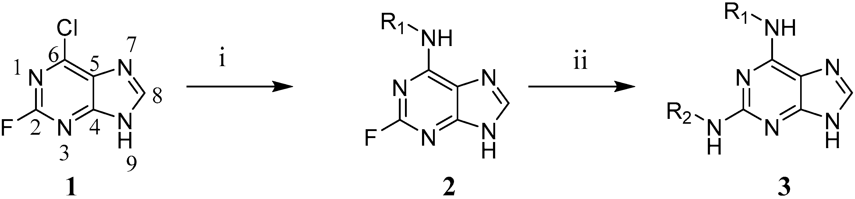
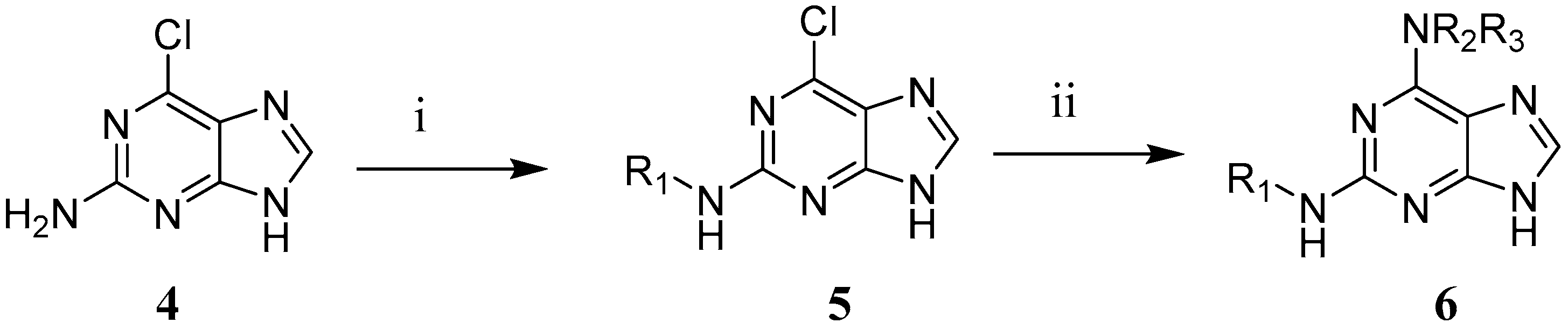
2.8.2. General Procedure I for the First Nucleophilic Aromatic Substitution in the 6-Position (2a and 2b)

To a solution of 6-chloro-2-fluoropurine (1 mmol, 1.00 equiv.) in n-butanol (4 mL), was added N,N-diisopropylethylamine (1.41 equiv.). The mixture was stirred at room temperature for 5 min. To the mixture, was added an amine (1.02 equiv.). The mixture was warmed to 65 °C and stirred for 4 h. Then, the reaction mixture was concentrated in vacuo and to the residue, was added cold water. The precipitate was filtered, washed with cold water and purified.

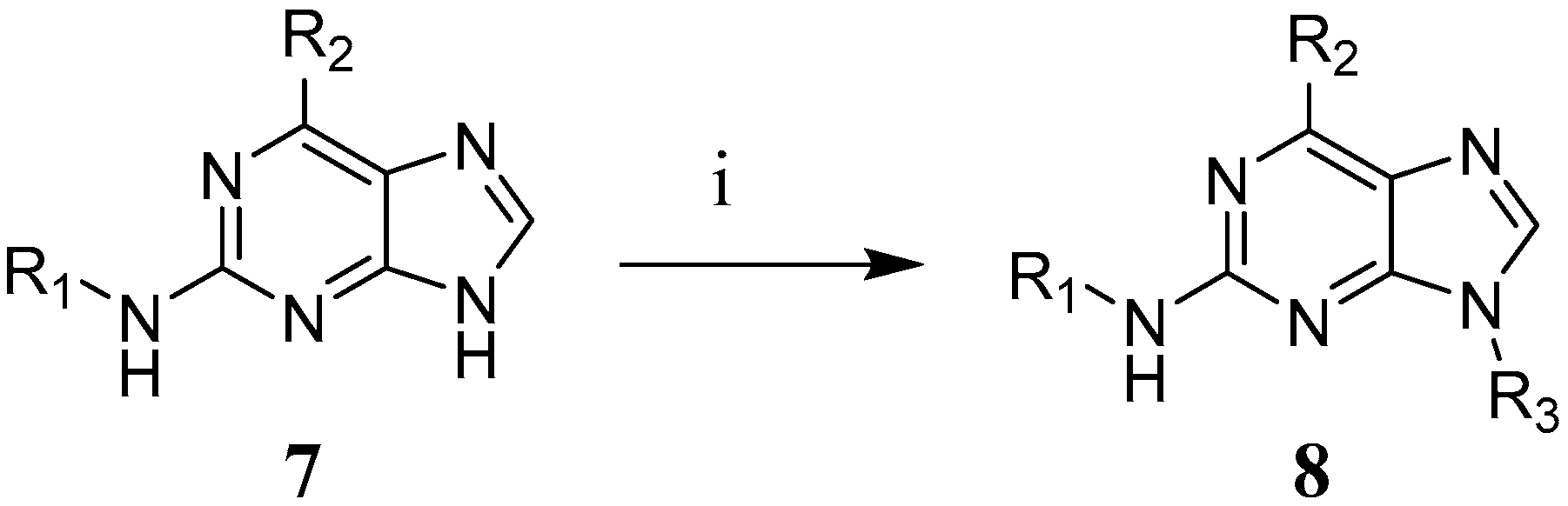
2.8.3. General Procedure II for the Second Nucleophilic Aromatic Substitution in the 2-Position (9 to 29 (Excluding 14 and 17), 32, 35, 38 and 39)

To a solution of a N6-substituted-2-fluoro-9H-purin-6-amine (1 mmol, 1.00 equiv.) in n-butanol (4 mL), was added N,N-diisopropylethylamine (2.20 equiv.). The mixture was stirred at room temperature for 5 min. To the mixture, was added an amine (2.00 equiv.). The mixture was heated to reflux and stirred overnight (16 h). Then, the reaction mixture was concentrated in vacuo and purified.

2.8.4. General Procedure III for the Reductive Amination in the 2-Position (5a to 5c)

To a solution of 2-amino-6-chloropurine (1 mmol, 1.00 equiv.) in ethyl acetate (4 mL), was added a benzaldehyde (1.20 equiv.). The mixture was cooled to 0 °C and was added trifluoroacetic acid (2.24 equiv.) and sodium triacetoxyborohydride (1.50 equiv.). The reaction was warmed to room temperature and stirred overnight (16 h). The mixture was quenched with 10% aq. NaOH solution (20 mL) to pH ~8–9 and extracted with ethyl acetate (3 × 20 mL). The organic extracts were combined, washed with brine (50 mL) and dried over MgSO4. Then, it was concentrated in vacuo and purified.

2.8.5. General Procedure IV for the Nucleophilic Aromatic Substitution in the 6-Position (14, 30, 31, 36 and 41 to 44)

To a solution of N2-substituted-6-chloro-9H-purin-2-amine (1 mmol, 1.00 equiv.) in n-butanol (4 mL), was added an amine (1.50 equiv.) and triethylamine (1.00 equiv.). The reaction was heated to reflux and stirred for 5 h. Then, the reaction mixture was concentrated in vacuo and purified.
Additional synthetic methods and spectroscopic data for individual compounds is provided in the Supplementary Materials S1.

3. Results

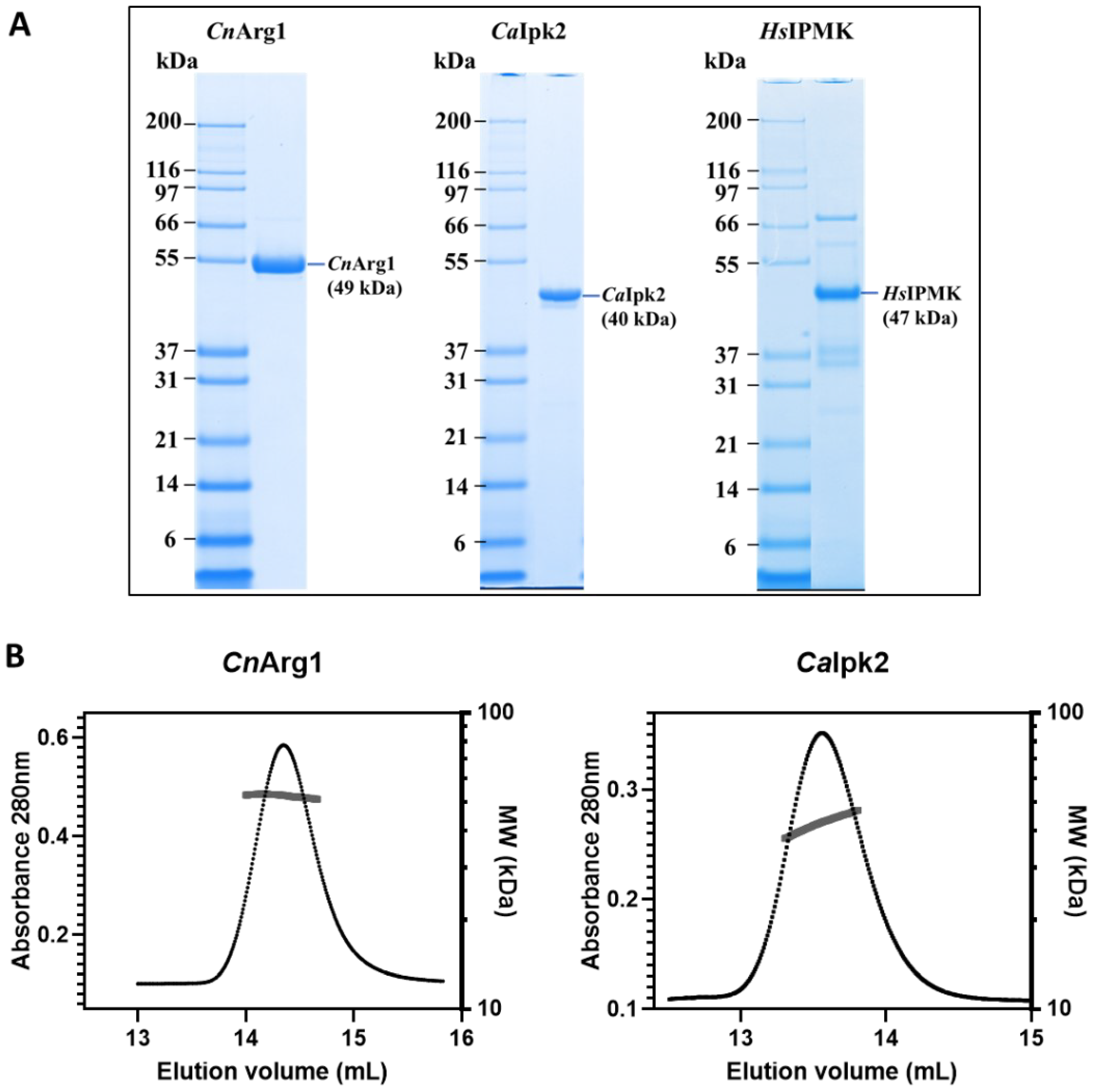
3.1. Purification of IP3-4K from C. neoformans, C. albicans and Human

IP3-4K from C. neoformans (CnArg1) and C. albicans (CaIpk2) and HsIPMK were expressed from pGEX-6P in E. coli as GST fusion proteins and purified in a two-step process involving glutathione-affinity chromatography, followed by GST cleavage by HRV 3C protease and size exclusion chromatography. SDS-PAGE indicated CnArg1 and CaIpk2 have molecular weights close to the predicted values of 49 kDa and 40 kDa, respectively, and a purity of >95% (Figure 1A). SEC-MALLS was used to assess the oligomeric state and was consistent with both IP3-4K enzymes being monomeric. The solution molecular weights of CnArg1 and CaIpk2 were calculated as 52 kDa and 43 kDa, respectively (i.e., within 10% of the predicted monomer molecular weight) (Figure 1B). From SDS-PAGE, the molecular weight of HsIPMK was ~47 kDa (Figure 1A), in agreement with the predicted molecular weight of 47.2 kDa [40]. HsIPMK purity was ~60%. Reduced HsIPMK purity could be due to limited expression levels and contamination with bacterial heat shock proteins. HsIPMK was previously shown to be monomeric [21,22,40,41]. The purification yielded ~3.6 mg per litre of CnArg1, ~8.8 mg per litre of CaIpk2 and ~0.3 mg per litre of HsIPMK proteins.

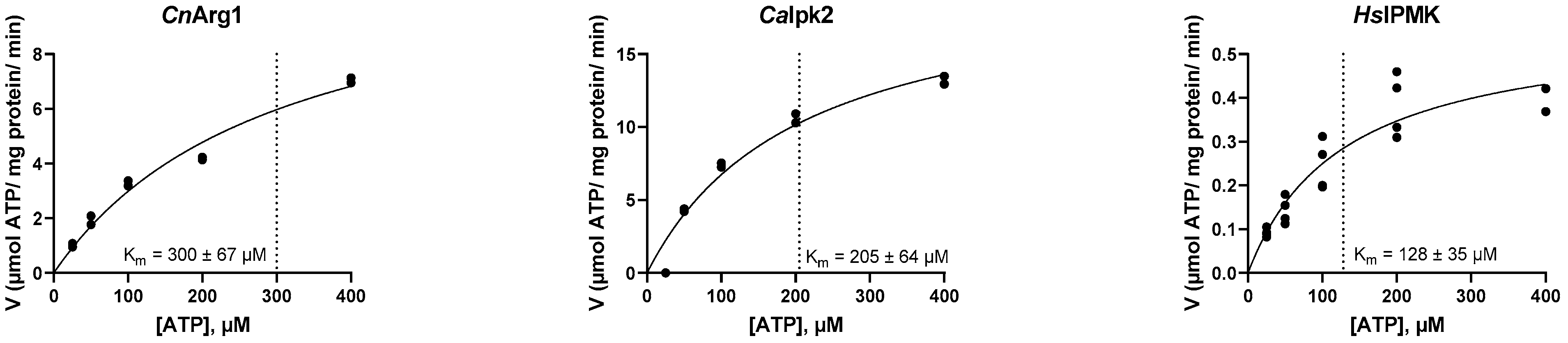
3.2. Establishing the Kinetic Properties of IP3-4K

A luminescence assay was optimised from [14] to determine the kinetic properties of CnArg1 and CaIpk2 using IP3 as the substrate. For CnArg1 and CaIpk2, the Km was 300 ± 67 µM and 213 ± 49 µM, and the Vmax was 12 ± 1.5 and 21 ± 3 µmol ATP/mg protein/min, respectively (Figure 2). Using the same assay conditions, HsIPMK had a lower Km (128 ± 35 µM) compared to the fungal IP3-4K enzymes and a lower Vmax (0.6 ± 0.07 µmol ATP/mg protein/min) (Figure 2), consistent with a higher affinity for ATP. This compares to a previously determined Km for ATP of 61 ± 6 µM when PIP2 was used as the substrate [22] and 10 µM when IP4 was used as the substrate [31]. The Km of rat IPMK for ATP was previously determined to be 64 µM when IP3 was used as the substrate [36].

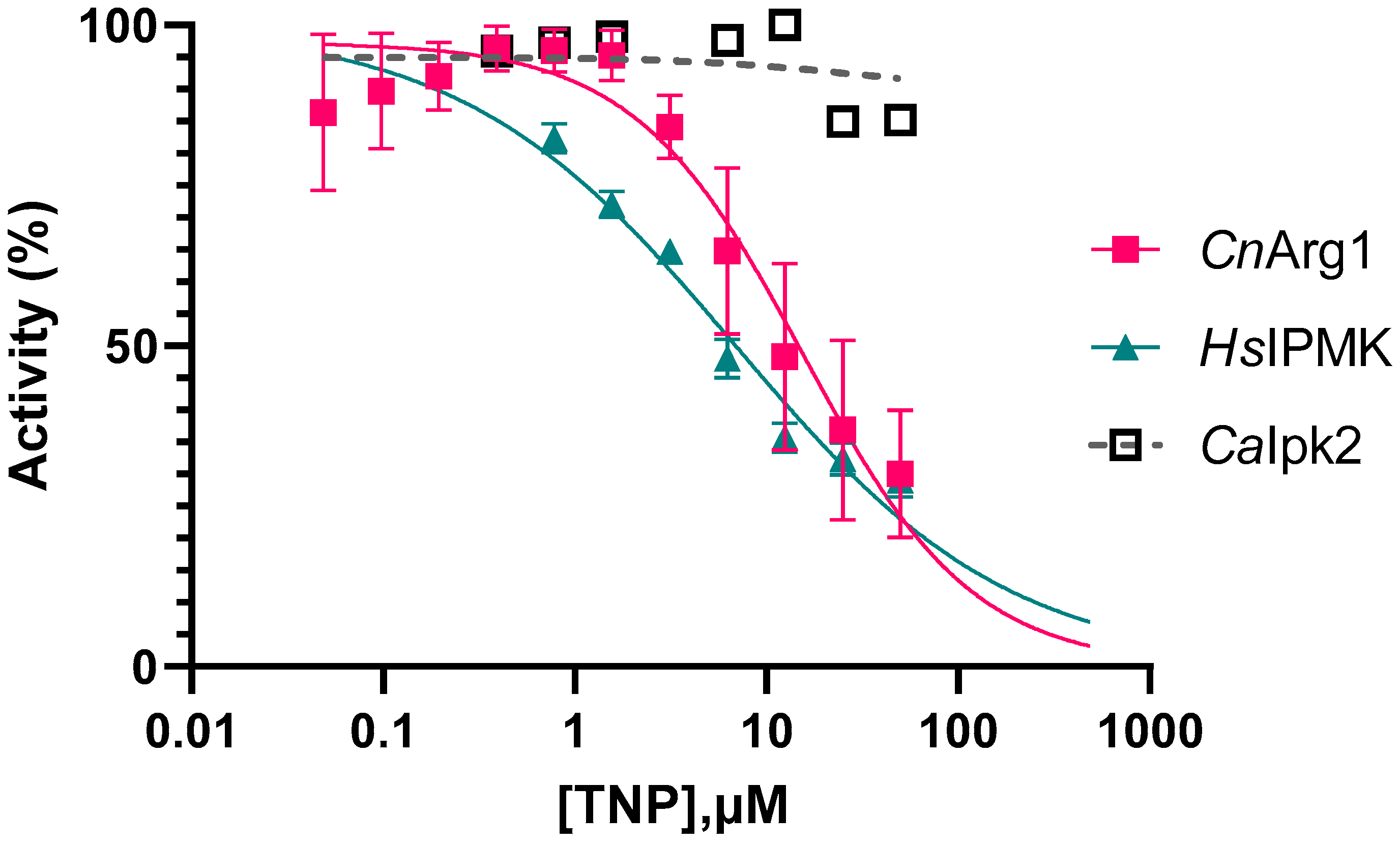
3.3. Optimizing the Enzyme Assay and Determining the IC50 of TNP

Next, we determined the inhibitory properties of TNP. TNP is an ATP-competitive inhibitor [34]. To bias the assay toward ATP-competitive inhibitors, the ATP concentration was reduced from 500 µM to 10 µM, which is a value lower than the Km of all IP3-4K enzymes (Figure 2). IP3 and enzyme concentrations were also optimised following the manufacturer’s instructions. Briefly, this involved measuring ATP consumption using a fixed saturated amount of kinase and varying the concentration of IP3 from 0 to 200 µM. The IP3 concentration that resulted in the largest change in luminescence was chosen as the optimal IP3 concentration. This optimal amount of IP3 was then used to test varying concentrations of the kinase ranging from 0 to 20 ng/µL. The amount of kinase that produced luminescence values in the linear range of the kinase titration curve was deemed to be the optimal kinase amount. The optimised concentrations of enzyme assay components are summarised in Table 2.
Using the optimized assay conditions (Table 2), TNP was found to inhibit CnArg1 and HsIPMK with an IC50 of 21 ± 6 µM and 7 ± 0.5 µM, respectively (Figure 3). To our knowledge, this is the first report that TNP inhibits HsIPMK. Interestingly, TNP did not inhibit CaIpk2.

3.4. Synthesis of 2,6-Disubstituted Purine Analogues

TNP analogues were synthesized using multiple strategies that expedited the inclusion of substituents at the N2- and N6-positions of the purine scaffold (Table 3, Table 4 and Table 5). In addition, modification at N9 was included in examples 45 and 46 (Table 6). Note that several of these compounds were previously reported in studies of human IPKs as well as other enzyme targets [34,35,37,42,43].
For fixed N6-amine substitutions, 2-fluoro-6-chloro-9H-purine, 1 was subjected to consecutive nucleophilic aromatic substitutions (Scheme 1) [37,44,45]. While substitution of 1 occurs in the 6-position preferentially, giving 2 [46], bis-substitution was a common observation initially. Monitoring the temperature and length of the first nucleophilic aromatic substitution reaction gave improved outcomes. The second substitution at the 2-position yielded the target compounds, 3 but also proved to be challenging, with reactions requiring high reagent concentrations to proceed.
In order to diversify the 6-position conveniently, an alternate synthetic route was adopted, where the group on the 2-position was introduced first. This has been achieved previously through the use of protecting groups or modification of the 6-chloro group to bypass the more reactive groups [28,37]. However, we pursued a more direct route by a reductive alkylation of 2-amino-6-chloro-9H-purine (Scheme 2). This was then followed by the nucleophilic aromatic substitution by various amines at the 6-chloro position to give 5. Reductive alkylation in the 2-position has previously been demonstrated [47,48], but to our knowledge, reductive alkylation in the 6-position has not been reported before. Substitution of 5 with various amines gave target compounds 6.
Lastly, the impact of an alkyl group in the 9-position was explored. This was accomplished through alkylation of 7 using an alkyl halide to give 8 (Scheme 3).

3.5. Assessment of the Inhibitory Properties of 2,6-Disubstituted Purine Analogues

Using the assay conditions established for TNP, all analogues were assessed for inhibition of CnArg1, HsIPMK and CaIpk2. We found that most analogues inhibited CnArg1 with IC50 values ranging from 10–100 μM (summarised in Table 3, Table 4, Table 5 and Table 6), but none inhibited CaIpk2 (see supplementary Figure S1). Compound structures and their IC50 values against CnArg1 and HsIPMK are summarised in Table 3, Table 4, Table 5 and Table 6. Only a selected number of analogues were tested for inhibitory activity against HsIPMK, with a focus on those with similar or better potencies than TNP (i.e., those with an IC50 less than 40 µM). Note that the reduced potencies of some of these compounds could be due to poor solubility in water although this was not investigated further. Enzyme inhibition assays could not be performed on 11, 18 and 19 as they were poorly soluble in 5% DMSO.
We first identified that 9, which lacked the nitro group of TNP, had a similar potency to TNP against CnArg1, but slightly reduced potency against HsIPMK. 9 had previously been described by [34,37] during their studies around IP3K and IP6K, respectively. They showed that the removal of the nitro group resulted in similar inhibition as TNP. Note that some structure activity relationship (SAR) against other human IPKs, IP3K and IP6K has been described before [34].
21 TNP analogues were created with modifications at the N2-position (see Table 3) encompassing a range of functional groups such as alternately substituted benzylamines (1024), alternate aromatic moieties (25, 26), alkyl ethers and alcohols (2729). In some cases, activity was abolished but overall, the inhibitory activity versus CnArg1 was only modestly affected compared to 9 (<2-fold difference). However, the selectivity of compounds for CnArg1 over HsIPMK was influenced by substitution. For example, 13 is a comparable inhibitor of CnArg1 to 9 but a much weaker inhibitor of HsIPMK.
Midway through this study, a research team at Johns Hopkins University published their strategy of first improving TNP solubility by replacing the N6-nitrobenzyl group with a methoxyethyl chain, before making further chemical modifications [37]. We employed the same strategy to explore the N2-position of the purine core, synthesizing a total of 11 analogues with the methoxyethyl chain at the N6-position (Table 4). It was reported that 30 had improved IP6K inhibition compared to TNP [37] but this was not observed with either CnArg1 or HsIPMK. Swapping benzylamine substitution at N2 gave a range of outcomes. Arylamine substitution as 37 and 40 was tolerated (IC50 of 17 ± 2 µM and 14 ± 0.5 µM, respectively).
Further variations at the N6-position were also considered (Table 5). One of these, 42, which had a 4-methyltetrahydropyran group, produced the most potent inhibition against CnArg1 with an IC50 of 10 ± 0.5 µM. Substitution in the 9-position was not tolerated in the two analogues, 45 and 46, which did not have any inhibitory activity for CnArg1 (Table 6).

3.6. Assessing Binding Affinities of TNP and Its Analogues

To complement the enzyme inhibition data, the binding affinities (KD) of TNP analogues were determined for CnArg1 using surface plasmon resonance (SPR) and the results are summarised in Table 7 (see Figure S2 for representative SPR sensorgrams and dose–response curves). A KD could not be determined for TNP and its closest analogue 9, due to poor behaviour on the SPR chip, which is most likely attributable to poor aqueous solubility. Chemical modification of TNP and 9 improved compound properties, allowing the binding affinities of 16 analogues to be determined. Apart from 37, these analogues bound to CnArg1, confirming a correlation between binding and inhibition. With the exception of 39 and 40, the analogues generally had comparable IC50 and KD values.

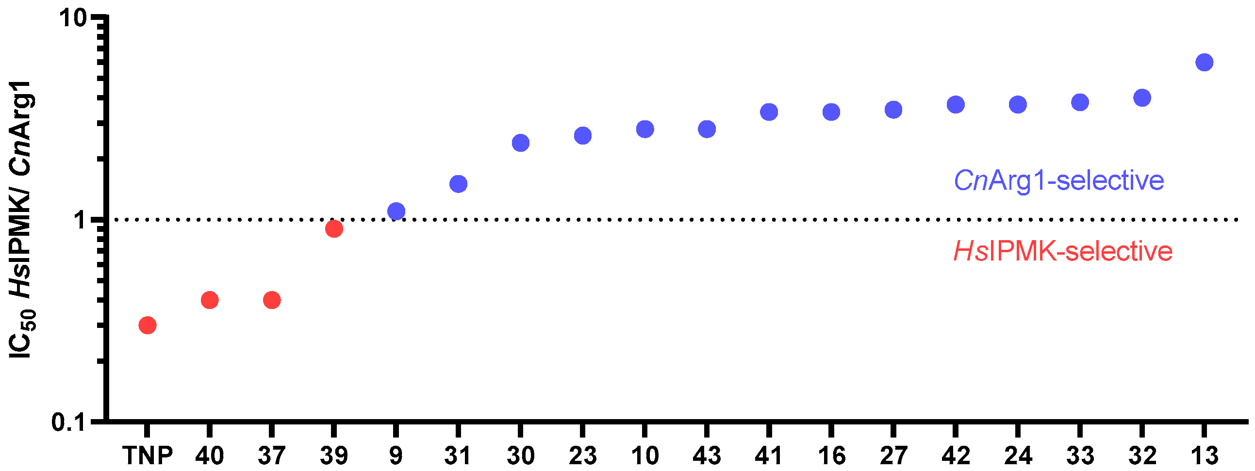
3.7. Comparing the Selectivity of TNP and Its Analogues for CnArg1

Using the IC50 data for TNP and the seventeen TNP analogues with the lowest IC50 against CnArg1, the HsIPMK/CnArg1 IC50 ratios were calculated (see last column in Table 3, Table 4, Table 5 and Table 6) and plotted (Figure 4). Fourteen TNP analogues were found to be more selective for CnArg1 (ratio higher than 1). While unable to identify significantly more potent analogues of CnArg1 than TNP, the collected assessment of enzyme inhibitory activities did show that the relative selectivity over the human orthologue could be influenced by altered substitutions, with the selectivity ratios changing 20-fold in going from TNP (3-CF3 substitution) to 13 (3-OCH3 substitution) (Table 3).

4. Discussion

IP3-4K is critical for the virulence of C. neoformans and C. albicans. An IP3-4K (CnArg1) deletion mutant failed to either grow at 37 °C or establish an infection in a mouse model [14]. The mutant was also defective in producing a plethora of virulence-related phenotypes including capsule, melanin and phospholipase B, was more readily phagocytosed by blood-derived monocytes, had a cell wall defect and reduced metabolic flexibility and could not upregulate phosphate acquisition machinery in response to phosphate deprivation [13,14,15,16,49]. Attempts to delete both alleles of IP3-4 kinase (CaIpk2) in the diploid yeast, C. albicans, were unsuccessful, suggesting that IP3-4K is an essential protein. Using a knockdown approach, CaIpk2 was shown to be essential for hyphal development, secretion of degradative enzymes and survival inside macrophages [17]. Targeting fungal IP3-4K is therefore a promising strategy for developing a novel class of antifungal drug. Fungal IP3-4K is also an attractive drug target due to the redundancy that exists in the analogous IP3 to IP5 conversion step in the human pathway.
Our SAR studies revealed that most TNP analogues inhibited CnArg1, but not CaIpk2, and that many TNP analogues were more selective for CnArg1 over HsIPMK (summarised in Figure 4). This suggests that potential differences exist in the active site of each kinase. Like many IPK enzymes, the protein structures of CnArg1 and CaIpk2 have not been experimentally determined. To investigate possible structural differences among the three IP3-4K proteins that could explain our results, we utilized the AlphaFold-predicted structures for CnArg1 (UniProt: J9W3G0) and CaIpk2 (UniProt: Q59YE9) [50,51] and compared them to the published HsIPMK crystal structure consisting of amino acid residues 50 to 416 in a complex with ADP (PDB: 5W2H) [21,22]. The HsIPMK protein used to obtain the crystal structure had residues 263–377 deleted, which included a nuclear localisation signal.
Both the fungal IP3-4 kinases share only a ~20% sequence homology with HsIPMK. However, an overlay of the AlphaFold models of CnArg1 and CaIpk2 with HsIPMK (PDB: 5W2H) revealed a conserved catalytic core consisting of the three characteristic domains: an N-terminal α + β domain (N-lobe), a C-terminal α + β domain (C-lobe) and an inositol binding domain [21,22]. The N- and C-terminal domains each contain one anti-parallel beta sheet where ADP is sandwiched in between, making up the ATP binding site (Figure 5A,B). Seven out of twelve of the amino acids in the HsIPMK active site that interact with ATP are conserved in CnArg1 and CaIpk2 (Figure 5C,D) and represent Pro111, Leu254, Asp144, Ile384, Asp385 and Lys75 (Figure 5E). In HsIPMK, Leu254 and Ile384 form van der Waals interactions with each other and Asp144 forms a hydrogen bond with the ribose group of ADP. Asp385 interacts with the two magnesium ions, which in turn contact the two phosphate groups of ADP [21]. Ile384 and Asp385 are part of an Ile-Asp-Phe tripeptide, which is conserved in the IPK family and known to interact with a metal cofactor [11,21,52]. Lys75 forms a salt bridge with the alpha-phosphate of ADP [21].
Differences between the fungal IP3-4Ks and HsIPMK (Figure 5E, circled residues) are the replacement of the negatively charged Asp132 in HsIPMK with the polar asparagine in CnArg1 (Asn100) and CaIpk2 (Asn102) and replacement of Glu131 in HsIPMK with Ala99 in CnArg1 and Ser101 in CaIpk2. In HsIPMK, Glu131 and hydrophobic Val133 form hydrogen bonds with the N6 and N1 atoms of adenine, respectively. Residues within the ATP binding site that are unique to CaIpk2 are Phe28, which is replaced by valine in HsIPMK (Val73) and CnArg1 (Val32). In HsIPMK, Val73 makes a van der Waals interaction with the adenine group of ADP [21]. The aromatic group on Phe28 could potentially interfere with the binding of TNP and its analogues. Other differences are Ser103 in CaIpk2 replacing Val133 (in HsIPMK) and Leu101 (in CnArg1), and Cys21 in CaIpk2 replacing Ile65 (in HsIPMK) and Val23 (in CnArg1). Ile65 in HsIPMK makes van der Waals interaction with the adenine group of ADP.
Human IP6K2 was modelled using the Entamoeba histolytica (Eh) IP6KA crystal structure (PDB ID: 4O4D) and was subsequently used for SAR analysis with flavonoids [41] and TNP [35]. Similarly, differences in the fungal IP3-4K active site residues can be further explored through molecular docking and/or via site-directed mutagenesis to identify residues responsible for the lack of inhibition observed for CaIpk2 and to facilitate the design of a more potent inhibitor of CnArg1, which also inhibits CaIpk2. This, TNP derivatives may become relevant to CaIpk2 inhibition once the affinity of the compounds is increased to give IC50 in the nanomolar range.

5. Conclusions

In summary, we have demonstrated that TNP and a series of analogues derived from it, inhibit purified, tag-free IP3-4K produced by the human fungal pathogen, C. neoformans. We also show that the relative selectivity of TNP and its analogues over the human orthologue can be influenced 20-fold by making substitutions at the N2 position of the purine. Recently released AlphaFold protein models of fungal pathogen IP3-4Ks have also revealed amino acid differences in the human and fungal ATP binding site, which are being used in combination with the assays developed to guide ongoing SAR studies aimed at improving the potency and selectivity of the TNP analogues for the fungal enzyme targets, and testing new IPK inhibitor scaffolds.

Supplementary Materials

The following supporting information can be downloaded at: https://www.mdpi.com/article/10.3390/biom12101526/s1, Data S1: Synthetic methods and spectroscopic data for individual compounds. Figure S1: TNP and its analogues did not inhibit CaIpk2 when tested at concentrations up to 50 μM. Figure S2: Surface plasmon resonance (SPR) sensorgrams and dose-response curves of compounds 39 and 41 are shown as representative data. The KD values in Table 7 were obtained using steady-state affinity.

Author Contributions

J.T.D. and P.E.T. conceptualized the project. D.D., D.T., L.W.-W., S.L., M.T. and C.D. developed the methodology and performed the investigation, formal analysis and validation. D.D., D.T., J.T.D. and P.E.T. wrote and reviewed the original manuscript draft. All authors reviewed and edited subsequent manuscript drafts. J.T.D., P.E.T., L.W.-W. and C.D. supervised project members. J.T.D. and P.E.T. administered the project. J.T.D., P.E.T., J.P.M., T.C.S. and J.M.M. were involved in funding acquisition. All authors have read and agreed to the published version of the manuscript.

Funding

This project was supported by seed grants from the University of Sydney Drug Discovery Institute and the Sydney Institute for Infectious Diseases, an Industry PhD scholarship from NSW Health and the University of Sydney and project grants to J.T. Djordjevic from the National Health and Medical Research Council (NHMRC) of Australia (APP1183939, APP1058779). Daniel Truong is a recipient of an RTP scholarship from the Australian Government.

Institutional Review Board Statement

Not applicable.

Informed Consent Statement

Not applicable.

Data Availability Statement

Not applicable.

Acknowledgments

We thank the Westmead Scientific Platforms, which is supported by the Westmead Institute for Medical Research, the Cancer Institute New South Wales and the NHMRC, for providing access to instrumentation to perform the enzyme assays. This research was facilitated by access to Sydney Analytical, a core research facility at the University of Sydney.

Conflicts of Interest

The authors declare no conflict of interest.

References

  1. Bongomin, F.; Gago, S.; Oladele, R.O.; Denning, D.W. Global and Multi-National Prevalence of Fungal Diseases-Estimate Precision. J. Fungi 2017, 3, 57. [Google Scholar] [CrossRef] [PubMed]
  2. Fisher, M.C.; Gurr, S.J.; Cuomo, C.A.; Blehert, D.S.; Jin, H.; Stukenbrock, E.H.; Stajich, J.E.; Kahmann, R.; Boone, C.; Denning, D.W.; et al. Threats Posed by the Fungal Kingdom to Humans, Wildlife, and Agriculture. mBio 2020, 11, e00449-20. [Google Scholar] [CrossRef] [PubMed]
  3. Wang, J.-L.; Chang, C.-H.; Young-Xu, Y.; Chan, K.A. Systematic Review and Meta-Analysis of the Tolerability and Hepatotoxicity of Antifungals in Empirical and Definitive Therapy for Invasive Fungal Infection. Antimicrob. Agents Chemother. 2010, 54, 2409–2419. [Google Scholar] [CrossRef] [PubMed] [Green Version]
  4. Perfect, J.R. The antifungal pipeline: A reality check. Nat. Rev. Drug Discov. 2017, 16, 603–616. [Google Scholar] [CrossRef] [Green Version]
  5. Cabañes, F.J. Aspergillosis, poultry farming and antifungal resistance. Rev. Iberoam. Micol. 2021, 38, 109–110. [Google Scholar] [CrossRef]
  6. Maligie, M.A.; Selitrennikoff, C.P. Cryptococcus neoformans Resistance to Echinocandins: (1,3)β-Glucan Synthase Activity Is Sensitive to Echinocandins. Antimicrob. Ag. Chemother. 2005, 49, 2851–2856. [Google Scholar] [CrossRef] [Green Version]
  7. Mroczyńska, M.; Brillowska-Dąbrowska, A. Review on Current Status of Echinocandins Use. Antibiotics 2020, 9, 227. [Google Scholar] [CrossRef]
  8. Chowdhary, A.; Prakash, A.; Sharma, C.; Kordalewska, M.; Kumar, A.; Sarma, S.; Tarai, B.; Singh, A.; Upadhyaya, G.; Upadhyay, S.; et al. A multicentre study of antifungal susceptibility patterns among 350 Candida auris isolates (2009–17) in India: Role of the ERG11 and FKS1 genes in azole and echinocandin resistance. J. Antimicrob. Chemother. 2018, 73, 891–899. [Google Scholar] [CrossRef]
  9. Li, C.; Lev, S.; Saiardi, A.; Desmarini, D.; Sorrell, T.C.; Djordjevic, J.T. Inositol Polyphosphate Kinases, Fungal Virulence and Drug Discovery. J. Fungi 2016, 2, 24. [Google Scholar] [CrossRef] [Green Version]
  10. Lev, S.; Bowring, B.; Desmarini, D.; Djordjevic, J.T. Inositol polyphosphate-protein interactions: Implications for microbial pathogenicity. Cell Microbiol. 2021, 23, e13325. [Google Scholar] [CrossRef]
  11. Saiardi, A.; Azevedo, C.; Desfougères, Y.; Portela-Torres, P.; Wilson, M.S.C. Microbial inositol polyphosphate metabolic pathway as drug development target. Adv. Biol. Regul. 2018, 67, 74–83. [Google Scholar] [CrossRef] [PubMed]
  12. Lev, S.; Li, C.; Desmarini, D.; Sorrell, T.C.; Saiardi, A.; Djordjevic, J.T. Fungal Kinases with a Sweet Tooth: Pleiotropic Roles of Their Phosphorylated Inositol Sugar Products in the Pathogenicity of Cryptococcus neoformans Present Novel Drug Targeting Opportunities. Front Cell Infect. Microbiol. 2019, 9, 248. [Google Scholar] [CrossRef] [PubMed] [Green Version]
  13. Lev, S.; Desmarini, D.; Li, C.; Chayakulkeeree, M.; Traven, A.; Sorrell, T.C.; Djordjevic, J.T. Phospholipase C of Cryptococcus neoformans regulates homeostasis and virulence by providing inositol trisphosphate as a substrate for Arg1 kinase. Infect. Immun. 2013, 81, 1245–1255. [Google Scholar] [CrossRef] [PubMed] [Green Version]
  14. Li, C.; Lev, S.; Desmarini, D.; Kaufman-Francis, K.; Saiardi, A.; Silva, A.P.G.; Mackay, J.P.; Thompson, P.E.; Sorrell, T.C.; Djordjevic, J.T. IP3-4 kinase Arg1 regulates cell wall homeostasis and surface architecture to promote Cryptococcus neoformans infection in a mouse model. Virulence 2017, 8, 1833–1848. [Google Scholar] [CrossRef] [Green Version]
  15. Lev, S.; Li, C.; Desmarini, D.; Saiardi, A.; Fewings, N.L.; Schibeci, S.D.; Sharma, R.; Sorrell, T.C.; Djordjevic, J.T. Fungal Inositol Pyrophosphate IP7 Is Crucial for Metabolic Adaptation to the Host Environment and Pathogenicity. mBio 2015, 6, e00531-15. [Google Scholar] [CrossRef] [Green Version]
  16. Li, C.; Lev, S.; Saiardi, A.; Desmarini, D.; Sorrell, T.C.; Djordjevic, J.T. Identification of a major IP5 kinase in Cryptococcus neoformans confirms that PP-IP5/IP7, not IP6, is essential for virulence. Sci. Rep. 2016, 6, 23927. [Google Scholar] [CrossRef]
  17. Li, J.; Zhang, B.; Ma, T.; Wang, H.; Zhang, B.; Yu, Q.; Li, M. Role of the Inositol Polyphosphate Multikinase Ipk2 in Regulation of Hyphal Development, Calcium Signaling and Secretion in Candida albicans. Mycopathologia 2017, 182, 609–623. [Google Scholar] [CrossRef]
  18. Peng, X.; Yu, Q.; Liu, Y.; Ma, T.; Li, M. Study on the Function of the Inositol Polyphosphate Kinases Kcs1 and Vip1 of Candida albicans in Energy Metabolism. Front Microbiol. 2020, 11, 566069. [Google Scholar] [CrossRef]
  19. Ma, T.; Yu, Q.; Ma, C.; Mao, X.; Liu, Y.; Peng, X.; Li, M. Role of the inositol polyphosphate kinase Vip1 in autophagy and pathogenesis in Candida albicans. Future Microbiol. 2020, 15, 1363–1377. [Google Scholar] [CrossRef]
  20. Resnick, A.C.; Snowman, A.M.; Kang, B.N.; Hurt, K.J.; Snyder, S.H.; Saiardi, A. Inositol polyphosphate multikinase is a nuclear PI3-kinase with transcriptional regulatory activity. Proc. Natl. Acad. Sci. USA 2005, 102, 12783–12788. [Google Scholar] [CrossRef]
  21. Wang, H.; Shears, S.B. Structural features of human inositol phosphate multikinase rationalize its inositol phosphate kinase and phosphoinositide 3-kinase activities. J. Biol. Chem. 2017, 292, 18192–18202. [Google Scholar] [CrossRef] [Green Version]
  22. Seacrist, C.D.; Blind, R.D. Crystallographic and kinetic analyses of human IPMK reveal disordered domains modulate ATP binding and kinase activity. Sci. Rep. 2018, 8, 16672. [Google Scholar] [CrossRef] [PubMed] [Green Version]
  23. Kim, S.; Kim, S.F.; Maag, D.; Maxwell, M.J.; Resnick, A.C.; Juluri, K.R.; Chakraborty, A.; Koldobskiy, M.A.; Cha, S.H.; Barrow, R.; et al. Amino Acid Signaling to mTOR Mediated by Inositol Polyphosphate Multikinase. Cell Metab. 2011, 13, 215–221. [Google Scholar] [CrossRef] [PubMed] [Green Version]
  24. Lee, B.; Park, S.J.; Hong, S.; Kim, K.; Kim, S. Inositol polyphosphate multikinase signaling: Multifaceted functions in health and disease. Mol. Cells 2021, 44, 187–194. [Google Scholar] [CrossRef] [PubMed]
  25. Morrison, B.H.; Haney, R.; Lamarre, E.; Drazba, J.; Prestwich, G.D.; Lindner, D.J. Gene deletion of inositol hexakisphosphate kinase 2 predisposes to aerodigestive tract carcinoma. Oncogene 2009, 28, 2383–2392. [Google Scholar] [CrossRef] [PubMed] [Green Version]
  26. Rao, F.; Cha, J.; Xu, J.; Xu, R.; Vandiver, M.S.; Tyagi, R.; Tokhunts, R.; Koldobskiy, M.A.; Fu, C.; Barrow, R.; et al. Inositol Pyrophosphates Mediate the DNA-PK/ATM-p53 Cell Death Pathway by Regulating CK2 Phosphorylation of Tti1/Tel2. Mol. Cell 2020, 79, 702. [Google Scholar] [CrossRef]
  27. Moritoh, Y.; Oka, M.; Yasuhara, Y.; Hozumi, H.; Iwachidow, K.; Fuse, H.; Tozawa, R. Inositol Hexakisphosphate Kinase 3 Regulates Metabolism and Lifespan in Mice. Sci. Rep. 2016, 6, 32072. [Google Scholar] [CrossRef] [Green Version]
  28. Ghoshal, S.; Zhu, Q.; Asteian, A.; Lin, H.; Xu, H.; Ernst, G.; Barrow, J.C.; Xu, B.; Cameron, M.D.; Kamenecka, T.M.; et al. TNP [N2-(m-Trifluorobenzyl), N6-(p-nitrobenzyl)purine] ameliorates diet induced obesity and insulin resistance via inhibition of the IP6K1 pathway. Mol. Metab. 2016, 5, 903–917. [Google Scholar] [CrossRef]
  29. Kröber, T.; Bartsch, S.M.; Fiedler, D. Pharmacological tools to investigate inositol polyphosphate kinases—Enzymes of increasing therapeutic relevance. Adv. Biol. Regul. 2021, 83, 100836. [Google Scholar] [CrossRef]
  30. Moritoh, Y.; Abe, S.I.; Akiyama, H.; Kobayashi, A.; Koyama, R.; Hara, R.; Kasai, S.; Watanabe, M. The enzymatic activity of inositol hexakisphosphate kinase controls circulating phosphate in mammals. Nat. Commun. 2021, 12, 4847. [Google Scholar] [CrossRef]
  31. Liao, G.; Ye, W.; Heitmann, T.; Ernst, G.; DePasquale, M.; Xu, L.; Wormald, M.; Hu, X.; Ferrer, M.; Harmel, R.K.; et al. Identification of Small-Molecule Inhibitors of Human Inositol Hexakisphosphate Kinases by High-Throughput Screening. ACS Pharmacol. Transl. Sci. 2021, 4, 780–789. [Google Scholar] [CrossRef] [PubMed]
  32. Zhou, Y.; Mukherjee, S.; Huang, D.; Chakraborty, M.; Gu, C.; Zong, G.; Stashko, M.A.; Pearce, K.H.; Shears, S.B.; Chakraborty, A.; et al. Development of Novel IP6K Inhibitors for the Treatment of Obesity and Obesity-Induced Metabolic Dysfunctions. J. Med. Chem. 2022, 65, 6869–6887. [Google Scholar] [CrossRef] [PubMed]
  33. Padmanabhan, U.; Dollins, D.E.; Fridy, P.C.; York, J.D.; Downes, C.P. Characterization of a selective inhibitor of inositol hexakisphosphate kinases: Use in defining biological roles and metabolic relationships of inositol pyrophosphates. J. Biol. Chem. 2009, 284, 10571–10582. [Google Scholar] [CrossRef] [PubMed] [Green Version]
  34. Chang, Y.T.; Choi, G.; Bae, Y.S.; Burdett, M.; Moon, H.S.; Lee, J.W.; Gray, N.S.; Schultz, P.G.; Meijer, L.; Chung, S.K.; et al. Purine-based inhibitors of inositol-1,4,5-trisphosphate-3-kinase. Chembiochem 2002, 3, 897–901. [Google Scholar] [CrossRef]
  35. Lee, S.; Park, B.B.; Kwon, H.; Kim, V.; Jeon, J.S.; Lee, R.; Subedi, M.; Lim, T.; Ha, H.; An, D.; et al. TNP and its analogs: Modulation of IP6K and CYP3A4 inhibition. J. Enzym. Inhib. Med. Chem. 2022, 37, 269–279. [Google Scholar] [CrossRef]
  36. Kolozsvari, B.; Parisi, F.; Saiardi, A. Inositol phosphates induce DAPI fluorescence shift. Biochem. J. 2014, 460, 377–385. [Google Scholar] [CrossRef]
  37. Wormald, M.M.; Ernst, G.; Wei, H.; Barrow, J.C. Synthesis and characterization of novel isoform-selective IP6K1 inhibitors. Bioorganic Med. Chem. Lett. 2019, 29, 126628. [Google Scholar] [CrossRef]
  38. Dubois, E.; Scherens, B.; Vierendeels, F.; Ho, M.M.; Messenguy, F.; Shears, S.B. In Saccharomyces cerevisiae, the inositol polyphosphate kinase activity of Kcs1p is required for resistance to salt stress, cell wall integrity, and vacuolar morphogenesis. J. Biol. Chem. 2002, 277, 23755–23763. [Google Scholar] [CrossRef] [Green Version]
  39. Lev, S.; Kaufman-Francis, K.; Desmarini, D.; Juillard, P.G.; Li, C.; Stifter, S.A.; Feng, C.G.; Sorrell, T.C.; Grau, G.E.; Bahn, Y.S.; et al. Pho4 Is Essential for Dissemination of Cryptococcus neoformans to the Host Brain by Promoting Phosphate Uptake and Growth at Alkaline pH. mSphere 2017, 2, e00381-16. [Google Scholar] [CrossRef]
  40. Nalaskowski, M.M.; Deschermeier, C.; Fanick, W.; Mayr, G.W. The human homologue of yeast ArgRIII protein is an inositol phosphate multikinase with predominantly nuclear localization. Biochem. J. 2002, 366, 549–556. [Google Scholar] [CrossRef]
  41. Gu, C.; Stashko, M.A.; Puhl-Rubio, A.C.; Chakraborty, M.; Chakraborty, A.; Frye, S.V.; Pearce, K.H.; Wang, X.; Shears, S.B.; Wang, H. Inhibition of Inositol Polyphosphate Kinases by Quercetin and Related Flavonoids: A Structure–Activity Analysis. J. Med. Chem. 2019, 62, 1443–1454. [Google Scholar] [CrossRef] [PubMed]
  42. Wang, X.; He, Q.; Wu, K.; Guo, T.; Du, X.; Zhang, H.; Fang, L.; Zheng, N.; Zhang, Q.; Ye, F. Design, synthesis and activity of novel 2,6-disubstituted purine derivatives, potential small molecule inhibitors of signal transducer and activator of transcription 3. Eur. J. Med. Chem. 2019, 179, 218–232. [Google Scholar] [CrossRef] [PubMed]
  43. Havlíček, L.; Hanuš, J.; Veselý, J.; Leclerc, S.; Meijer, L.; Shaw, G.; Strnad, M. Cytokinin-Derived Cyclin-Dependent Kinase Inhibitors:  Synthesis and cdc2 Inhibitory Activity of Olomoucine and Related Compounds. J. Med. Chem. 1997, 40, 408–412. [Google Scholar] [CrossRef]
  44. Savelieva, E.M.; Oslovsky, V.E.; Karlov, D.S.; Kurochkin, N.N.; Getman, I.A.; Lomin, S.N.; Sidorov, G.V.; Mikhailov, S.N.; Osolodkin, D.I.; Romanov, G.A. Cytokinin activity of N6-benzyladenine derivatives assayed by interaction with the receptors in planta, in vitro, and in silico. Phytochemistry 2018, 149, 161–177. [Google Scholar] [CrossRef] [PubMed]
  45. Gray, N.S.; Wodicka, L.; Thunnissen, A.-M.W.H.; Norman, T.C.; Kwon, S.; Espinoza, F.H.; Morgan, D.O.; Barnes, G.; LeClerc, S.; Meijer, L.; et al. Exploiting Chemical Libraries, Structure, and Genomics in the Search for Kinase Inhibitors. Science 1998, 281, 533–538. [Google Scholar] [CrossRef] [PubMed] [Green Version]
  46. Eicher, T.; Hauptmann, S.; Speicher, A. The Chemistry of Heterocycles: Structure, Reactions, Synthesis, and Applications; Wiley-VCH: Wernheim, Germany, 2013. [Google Scholar]
  47. Huang, L.; Wang, Z.; Geng, L.; Chen, R.; Xing, W.; Wang, Y.; Huang, J. Selective and recyclable rhodium nanocatalysts for the reductive N-alkylation of nitrobenzenes and amines with aldehydes. RSC Adv. 2015, 5, 56936–56941. [Google Scholar] [CrossRef]
  48. Adamska, E.; Barciszewski, J.; Markiewicz, W.T. Convenient and Efficient Syntheses of N6- and N4- Substituted Adenines and Cytosines and their 2′-Deoxyribosides. Nucleosides Nucleotides Nucleic Acids 2012, 31, 861–871. [Google Scholar] [CrossRef]
  49. Desmarini, D.; Lev, S.; Furkert, D.; Crossett, B.; Saiardi, A.; Kaufman-Francis, K.; Li, C.; Sorrell, T.C.; Wilkinson-White, L.; Matthews, J.; et al. IP7-SPX Domain Interaction Controls Fungal Virulence by Stabilizing Phosphate Signaling Machinery. mBio 2020, 11, e01920-20. [Google Scholar] [CrossRef]
  50. Sparkes, M. DeepMind’s Protein-Folding AI Cracks Biology’s Biggest Problem. 2022. Available online: https://www.newscientist.com/article/2330866-deepminds-protein-folding-ai-cracks-biologys-biggest-problem/ (accessed on 29 July 2022).
  51. Jumper, J.; Evans, R.; Pritzel, A.; Green, T.; Figurnov, M.; Ronneberger, O.; Tunyasuvunakool, K.; Bates, R.; Žídek, A.; Potapenko, A.; et al. Highly accurate protein structure prediction with AlphaFold. Nature 2021, 596, 583–589. [Google Scholar] [CrossRef]
  52. González, B.; Schell, M.J.; Letcher, A.J.; Veprintsev, D.B.; Irvine, R.F.; Williams, R.L. Structure of a Human Inositol 1,4,5-Trisphosphate 3-Kinase: Substrate Binding Reveals Why It Is Not a Phosphoinositide 3-Kinase. Mol. Cell 2004, 15, 689–701. [Google Scholar] [CrossRef]
Figure 1. Assessment of purity, size and multimerization state of recombinant fungal IP3-4K enzymes. IP3-4K cDNA from C. neoformans (CnArg1), C. albicans (CaIpk2) and human (HsIPMK) were cloned into the pGEX-6P expression vector and purified using a two-step procedure described in the methods. (A) SDS-PAGE was used to assess the size and purity of the recombinant fungal IP3-4K enzymes. The theoretical molecular weights (MW) of the fungal enzymes, calculated from their amino acid sequences, are indicated next to the bands. (B) SEC-MALLS analysis suggested that CnArg1 and CaIpk2 are monomeric with calculated molecular weights of 52 kDa and 43 kDa, respectively, in agreement with predicted values of 49 kDa and 40 kDa. The dotted lines represent the UV-profile of the eluted proteins, and the thick grey lines represent the SEC-MALLS-processed MW measurements.
Biomolecules 12 01526 g001
Figure 2. Kinetic properties of recombinant fungal IP3-4K enzymes and HsIPMK. Each enzyme was incubated with ATP (25–400 µM) and 200 µM of IP3 in a 50 µL reaction volume at room temperature for up to 10 min. Each data point was repeated at least twice. Km and Vmax values were obtained by fitting all data points to the Michaelis-Menten equation using GraphPad Prism 9 and are presented as the mean ± SEM. V: reaction velocity.
Biomolecules 12 01526 g002
Figure 3. TNP inhibits CnArg1 and HsIPMK but not CaIpk2. Reactions were carried out at room temperature for 10 min using 10 µM ATP and the optimal amount of enzyme and IP3 as summarized in Table 2. ATP consumption was calculated from the luminescence reading and expressed as percentage enzyme activity according to the methods. The IC50 curves were generated by plotting % enzyme activity against the concentration of TNP. For CnArg1, the IC50 of 21 ± 6 µM (SEM) was calculated from 2 biological replicates, each performed in technical triplicate, error bar = SD. For HsIPMK, the IC50 of 7 ± 0.5 µM (SEM) was calculated from 3 technical replicates, error bar = SD. For CaIpk2, no inhibitory activity was found up to 50 µM (n = 1).
Biomolecules 12 01526 g003
Scheme 1. General synthesis of 3 from 2 via 1. i, R1NH2, N,N-diisopropylethylamine, n-BuOH, room temp. 65 °C, 4 h, 66–75%; ii, R2NH2, N,N-diisopropylethylamine, n-BuOH, reflux, 16 h, 6–70%.
Biomolecules 12 01526 sch001
Scheme 2. General synthesis of 6 from 4 via 5. i, Aldehyde-R1, NaBH(OAc)3, TFA, EtOAc, room temp., 16 h, 50–64%; ii, NHR2R3, triethylamine, n-BuOH, reflux, 5 h, 46–97%.
Biomolecules 12 01526 sch002
Scheme 3. Synthesis of 8 via 7. i, R3X, 68–85%.
Biomolecules 12 01526 sch003
Figure 4. Selectivity of TNP and its analogues. The selectivity of TNP and the seventeen analogues with the lowest IC50 against CnArg1, for CnArg1 (blue dots) and HsIPMK (red dots), was determined by plotting the HsIPMK/CnArg1 IC50 ratios.
Biomolecules 12 01526 g004
Figure 5. Overlay of the ATP binding site of HsIPMK and the fungal IP3-4Ks to determine conservation of active site residues. ADP-bound HsIPMK structure (PDB:5W2H) showing the N-lobe in orange, C-lobe in yellow, hinge region in green and inositol binding domain (IP-domain) in blue is superimposed with the AlphaFold models of CnArg1 (cyan) (A) and CaIpk2 (pink) (B). Surface representation (in grey) of the ATP-binding pocket of CnArg1 (C) and CaIpk2 (D), with residues identical to those of HsIPMK in blue and residues non-identical to those of HsIPMK in yellow. Stick models depicting all residues involved in ATP binding (E) are depicted with CnArg1 in cyan, CaIpk2 in pink and HsIPMK coloured green for carbon, blue for nitrogen and red for oxygen. Residues that differ among the three IP3-4K proteins being circled. For all panels, ADP is shown as stick representation with green for carbon, blue for nitrogen, red for oxygen and orange for phosphorus, and magnesium ions are depicted as violet spheres.
Biomolecules 12 01526 g005
Table 1. Primers used to construct and verify expression plasmids.
Primer NamePrimer Sequence (5′ to 3′)Description
ARG1-BglII-s TACGagatctGACCTGCCCCTCACCCTCGSense primer to PCR-amplify CnArg1 cDNA with BglII recognition sequence and adapter sequence
ARG-EcoRI-a CAAGgaattcTCAAACACAACCCCGTTCAACCAntisense primer to PCR-amplify CnArg1 cDNA with EcoRI recognition sequence and adapter sequence
CaIPK2-BamHI-sACTCggatccATTCCCACTTTAAATTCACTCACTCCTSense primer to PCR-amplify CaIpk2 cDNA with BamHI recognition sequence and adapter sequence
CaIPK2-XhoI-aTCTActcgagGTACAACCATTGCCATCGGAntisense primer to PCR-amplify CaIpk2 cDNA with XhoI recognition sequence and adapter sequence
pGEX-6T-5’ TTTTGCGCCGACATCATAACGSequencing primer 1 to confirm successful cloning of IP3-4K sequences
pGEX-Seq-sGTGGCGACCATCCTCCAAASequencing primer 2 to confirm successful cloning of IP3-4K sequences
pGEX-Seq-aCAAGCTGTGACCGTCTCCGSequencing primer 3 to confirm successful cloning of IP3-4K sequences
Table 2. Optimized concentrations of IP3 and enzyme used in the inhibition assay.
ComponentCnArg1CaIpk2HsIPMK
ATP (µM)101010
Enzyme (ng/µL)2.52.56
IP3 (µM)502525
Table 3. Summary of inhibitory properties resulting from chemical exploration at the N2 position of the TNP purine core following removal of the NO2 group at the N6 position (21 analogues). Only potent and soluble analogues with an IC50 < 40 µM for CnArg1 were tested for inhibitory properties against HsIPMK. ND = not determined.
Biomolecules 12 01526 i001
CompoundR1CnArg1 IC50
(µM)
HsIPMK IC50 (µM)HsIPMK/CnArg1
TNPSee above for structure21 ± 67 ± 0.50.3
9Biomolecules 12 01526 i00228 ± 732 ± 31.1
10Biomolecules 12 01526 i00320 ± 155 ± 22.8
11Biomolecules 12 01526 i004Low solubility—NDNDND
12Biomolecules 12 01526 i00577 ± 14NDND
13Biomolecules 12 01526 i00625 ± 1150 ± 306.0
14Biomolecules 12 01526 i00744 ± 2NDND
15Biomolecules 12 01526 i00846 ± 22NDND
16Biomolecules 12 01526 i00914 ± 148 ± 23.4
17Biomolecules 12 01526 i01045 ± 3NDND
18Biomolecules 12 01526 i011Low solubility—NDNDND
19Biomolecules 12 01526 i012Low solubility—NDNDND
20Biomolecules 12 01526 i01371 ± 12NDND
21
(20 HCl salt)
Biomolecules 12 01526 i01457 ± 6NDND
22Biomolecules 12 01526 i015Not inhibitoryNDND
23Biomolecules 12 01526 i01621 ± 155 ± 42.6
24Biomolecules 12 01526 i01724 ± 189 ± 93.7
25Biomolecules 12 01526 i018Not inhibitoryNDND
26Biomolecules 12 01526 i019Not inhibitoryNDND
27Biomolecules 12 01526 i02035 ± 2123 ± 803.5
28Biomolecules 12 01526 i021127 ± 32NDND
29Biomolecules 12 01526 i02275 ± 9NDND
Table 4. Summary of inhibitory properties resulting from chemical exploration at the N2 position of N6-methoxyethyl purine (11 analogues). Only potent and soluble analogues with an IC50 < 40 µM for CnArg1 were tested for inhibitory properties against HsIPMK. ND = not determined.
Biomolecules 12 01526 i023
CompoundR1CnArg1 IC50
(µM)
HsIPMK IC50 (µM)HsIPMK/CnArg1
TNP 21 ± 67 ± 0.50.3
30Biomolecules 12 01526 i02428 ± 167 ± 62.4
31Biomolecules 12 01526 i02529 ± 343 ± 41.5
32Biomolecules 12 01526 i02612 ± 148 ± 34.0
33Biomolecules 12 01526 i02738 ± 5144 ± 553.8
34Biomolecules 12 01526 i028Not inhibitoryNDND
35Biomolecules 12 01526 i02987 ± 9NDND
36Biomolecules 12 01526 i03055 ± 6NDND
37Biomolecules 12 01526 i03117 ± 27 ± 10.4
38Biomolecules 12 01526 i03244 ± 6NDND
39Biomolecules 12 01526 i03322 ± 319 ± 10.9
40Biomolecules 12 01526 i03414 ± 15 ± 10.4
Table 5. Summary of inhibitory properties resulting from chemical exploration at both the R1 and R2 positions on the TNP purine core. Only potent and soluble analogues with an IC50 < 40 µM for CnArg1 were tested for inhibitory properties against HsIPMK. ND = not determined.
Biomolecules 12 01526 i035
CompoundR1R2CnArg1 IC50
(µM)
HsIPMK IC50 (µM)HsIPMK/CnArg1
TNP 21 ± 67 ± 0.50.3
41Biomolecules 12 01526 i036Biomolecules 12 01526 i03724 ± 182 ± 93.4
42Biomolecules 12 01526 i038Biomolecules 12 01526 i03910 ± 0.537 ± 23.7
43Biomolecules 12 01526 i040Biomolecules 12 01526 i04127 ± 275 ± 82.8
44Biomolecules 12 01526 i042Biomolecules 12 01526 i04382 ± 13NDND
Table 6. Exploration at the N9 position on the TNP purine core abolished inhibition against CnArg1. ND = not determined.
Biomolecules 12 01526 i044
CompoundR1R2R3CnArg1 IC50 (µM)HsIPMK IC50 (µM)HsIPMK/CnArg1
TNP 21 ± 67 ± 0.50.3
45Biomolecules 12 01526 i045Biomolecules 12 01526 i046Biomolecules 12 01526 i047Not inhibitoryNDND
46Biomolecules 12 01526 i048Biomolecules 12 01526 i049CH3Not inhibitoryNDND
Table 7. Summary to the binding affinities (KD) of TNP and its analogues to CnArg1 as determined by surface plasmon resonance (SPR). Analogue binding to N-terminally biotinylated CnArg1 immobilised onto a streptavidin chip was assessed between 6.25–200 µM at 10 °C in 20 mM HEPES, 150 mM NaCl, pH 7.5. The IC50 and KD values are presented as mean ± SEM (IC50n ≥ 3 technical replicates; KDn ≥ 1). ND = not determined.
CompoundIC50 (µM)KD (µM)
TNP21 ± 6Poor solubility—ND
928 ± 7Poor solubility—ND
1020 ± 117 ± 5
1325 ± 113 ± 3
2321 ± 122 ± 4
2424 ± 116 ± 9
28127 ± 32321 ± 10
2975 ± 9155 ± 27
3028 ± 170 ± 13
3129 ± 335 ± 9
3212 ± 0.538 ± 11
3655 ± 674 ± 10
3717 ± 2No binding
3922 ± 3188 ± 9
4013 ± 0.5316 ± 31
4124 ± 196 ± 10
4210 ± 0.548 ± 7
4327 ± 221 ± 9
Publisher’s Note: MDPI stays neutral with regard to jurisdictional claims in published maps and institutional affiliations.

