Article

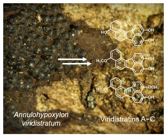
Viridistratins A−C, Antimicrobial and Cytotoxic Benzo[j]fluoranthenes from Stromata of Annulohypoxylon viridistratum (Hypoxylaceae, Ascomycota)

by
Kevin Becker
1,2,
Anna-Charleen Wessel
1,
J. Jennifer Luangsa-ard
3 and
Marc Stadler
1,2,*
1
Department of Microbial Drugs, Helmholtz Centre for Infection Research GmbH (HZI), Inhoffenstraße 7, 38124 Braunschweig, Germany
2
German Centre for Infection Research Association (DZIF), Partner site Hannover-Braunschweig, Inhoffenstraße 7, 38124 Braunschweig, Germany
3
National Center for Genetic Engineering and Biotechnology (BIOTEC), 113 Thailand Science Park, Phahonyothin Rd., Khlong Nueng, Khlong Luang, Pathum Thani 12120, Thailand
*
Author to whom correspondence should be addressed.
Biomolecules 2020, 10(5), 805; https://doi.org/10.3390/biom10050805
Submission received: 17 February 2020 / Revised: 17 May 2020 / Accepted: 21 May 2020 / Published: 23 May 2020
(This article belongs to the Special Issue 2020 Feature Papers by Biomolecules’ Editorial Board Members)

Abstract

:
During the course of our search for novel biologically active metabolites from tropical fungi, we are using chemotaxonomic and taxonomic methodology for the preselection of interesting materials. Recently, three previously undescribed benzo[j]fluoranthenes (13) together with the known derivatives truncatones A and C (4, 5) were isolated from the stromata of the recently described species Annulohypoxylon viridistratum collected in Thailand. Their chemical structures were elucidated by means of spectral methods, including nuclear magnetic resonance (NMR) spectroscopy and high-resolution mass spectrometry (HR-MS). The new compounds, for which we propose the trivial names viridistratins A−C, exhibited weak-to-moderate antimicrobial and cytotoxic activities in cell-based assays.

Graphical Abstract

1. Introduction

The genus Annulohypoxylon belongs to the fungal family Hypoxylaceae (order Xylariales), which was recently resurrected [1] to accommodate Hypoxylon and allied genera and is known for a remarkably diverse secondary metabolism [2]. It was segregated from Hypoxylon by Hsieh et al. in 2005 [3], and its type species, Annulohypoxylon truncatum, was previously known as Hypoxylon truncatum. In the past 15 years, these fungi have been the subject of intensive studies of their secondary metabolites, revealing an enormous diversity of bioactive compounds [4]. In particular, their stromatal pigments are of chemotaxonomic value [2,5]. Several studies of old types of material of Hypoxylon and allied genera using high-performance liquid chromatography coupled to diode array detection and mass spectrometry (HPLC-DAD/MS) have revealed that those pigments can remain stable for centuries [5,6,7]. A recent study on Hypoxylon fragiforme showed that these pigments can even endure many centuries and remain intact in fossil samples [7]. Two classes of secondary metabolites are predominant in the stromata of the Hypoxylaceae, i.e., azaphilones and naphthalene derivatives [8]. Recent major phylogenetic studies [1,9], which were now even confirmed by phylogenomics [10] have led to the segregation of the new genus Jackrogersella (with J. multiformis, J. cohaerens and J. minutella being examples of species that contain azaphilones as predominant stromatal pigments), which was segregated from Annulohypoxylon based on a combination of morphological and chemotaxonomic traits with the results of a multilocus molecular phylogeny. These azaphilones of Jackrogersella are of the cohaerin type, which also includes the multiformins and minutellins that are derived from the same unique polyketide scaffold [7,10,11]. Examples of common naphthalene derivatives in Annulohypoxylon spp. are the rather ubiquitous 1,1’-binaphthalene-4,4’,5,5’-tetrol (BNT) [2], as well as oxidized derivatives thereof, such as truncatones [2,12], hypoxylonols [2,13,14] and daldinones [2,11], with the three latter ones belonging to the subclass of benzo[j]fluoranthenes. The latter compound class has been exclusively found in Annulohypoxylon species and was so far not detected in Jackrogersella and Hypoxylon [2,4], even though binaphthalenes also occur in other, more distantly related Hypoxylaceae such as Daldinia [5].
Among the species that were only recently described in 2017 [2], Annulohypoxylon viridistratum was reported to have a unique secondary metabolite profile. Several yet unknown compounds were detected in the holotype specimen by database-aided HPLC-DAD/MS detection (see [2] and Figure S3). While the valuable holotype specimen was not suitable for preparative work, also because the stromatal material was rather scarce, we were recently able to collect another specimen in the field, which turned out to represent the same species and provided us with sufficient amounts of material to envisage the isolation of these unknown compounds. The current paper is dedicated to the description of their isolation, structure elucidation and biological activities.

2. Materials and Methods

2.1. General

Electrospray mass (ESI-MS) spectra were recorded with an UltiMate® 3000 Series uHPLC (Thermo Fisher Scientific, Waltman, MA, USA) utilizing a C18 Acquity® UPLC BEH column (2.1 × 50 mm, 1.7 µm; Waters, Milford, USA) connected to an amaZon speed® ESI-Iontrap-MS (Bruker, Billerica, MA, USA). HPLC parameters were set as follows: solvent A: H2O+0.1 % (v/v) formic acid, solvent B: acetonitrile (ACN)+0.1 % (v/v) formic acid, gradient 5 % B for 0.5 min, increasing to 100 % B in 19.5 min, keeping 100% B for further 5 min, flowrate 0.6 mL/min, and DAD detection 190–600 nm.
High-resolution electrospray mass (HR-ESI-MS) spectra were obtained with an Agilent 1200 Infinity Series HPLC (Agilent Technologies, Santa Clara, CA, USA) connected to a maXis® electrospray time-of-flight mass spectrometer (ESI-TOF-MS; Bruker; HPLC conditions same as for ESI-MS spectra).
Nuclear magnetic resonance (NMR) spectra were recorded with an Avance III 500 spectrometer (Bruker, 1H NMR: 500 MHz, and 13C NMR: 125 MHz). UV/Vis spectra were taken with a UV/Vis spectrophotometer UV-2450 (Shimadzu, Kyoto, Japan), and electronic circular dichroism (ECD) spectra (Figure S2) were recorded on a J-815 spectropolarimeter (JASCO, Pfungstadt, Germany).

2.2. Fungal Material

Stromata of Annulohypoxylon viridistratum were collected by L. Wendt from unidentified dead wood in a tropical rainforest in Thailand, Nan Bo Kluea, Khun Nan National Park (19.18301 N, 101.17801 E) in August of 2015 during the rainy season. A voucher specimen, which showed the characteristics of the species, was identified by E. Kuhnert and L. Wendt by a comparison of morphological characteristics, as well as molecular phylogenetic studies and HPLC profiling [2]. It is deposited in the herbarium of the National Center for Genetic Engineering and Biotechnology (BIOTEC), 113 Thailand Science Park, Phahonyothin Rd., Khlong Nueng, Khlong Luang, Pathum Thani 12120, Thailand (acc. No. BBH40533). This specimen is only the second record of this recently described species.

2.3. Extraction and Isolation

The dried stromata (1.45 g) were extracted a total of three times: (1) The stromata were carefully scraped off the dead wood and extracted with 100 mL of acetone in an ultrasonic bath (40°C, 60 min). The extract was centrifuged (4000 rpm, 5 min), and the supernatant transferred to a round-bottom flask. (2) The precipitate was extracted again with 100 mL of acetone, centrifuged and the supernatant transferred as described before. (3) Finally, the precipitate was crushed utilizing a mortar and pestle before being treated as in step (2) again. The supernatants of all three extractions were combined and dried in vacuo at 40°C, which yielded the crude extract (227.6 mg). The stromatal remnants were discarded.
The crude extract was prefractionated with a Strata® C18-E cartridge (10 g/60 mL, 55 µm, 70 Å; P/N: 8B-S001-MFF, Phenomenex, Torrance, CA, USA). At first, the sample was dispersed in water and added to the cartridge. Applying a vacuum of ca. 600 mbar removed the water while leaving the sample on the cartridge. Then, a step gradient of the solvent waters (solvent A), acetonitrile (ACN; solvent B) and acetone (solvent C) was applied with the following steps: (I) 80:20:0 (A:B:C, % v/v/v), (II) 50:50:0, (III) 0:100:0, (IV) 0:50:50 and (V) 0:0:100. For each step, 40 mL of solvent was used, and elution was aided by applying a vacuum (600 mbar), which yielded fractions I−V.
Fraction III (83.3 mg) was further processed using a PLC 2250 system (Gilson, Middleton, WI, USA). The sample was dissolved in 3 mL acetone:H2O:ACN 4:1:1 (v/v/v). A Nucleodur® C18ec column (125×40 mm, 7 µm, 100 Å; Machery-Nagel, Düren, Germany) was utilized using H2O+0.1 % (v/v) formic acid (solvent A) and ACN+0.1 % (v/v) formic acid (solvent B) as eluents. Using a flow rate of 50 mL/min, a gradient was applied from 35 % to 80 % B within 50 min, followed by an increase of 80 % to 100 % B in 10 min, followed by another 10 min of 100 % B. Fractions were taken every 20 mL.
This yielded the pure compounds 15: viridistratin A (1, fractions #36−41, 3.2 mg), truncatone C (5, #42−49, 9.6 mg), truncatone A (4, #59−61, 3.4 mg), viridistratin B (2, #70−80, 17.5 mg) and viridistratin C (3, #99−106, 8.0 mg).

2.4. Antimicrobial Activity Assay

Compounds 1, 2, 4 and 5 were dissolved in MeOH (1 mg/mL) for the antimicrobial activity assay, while compound 3 was dissolved in MeOH: DMSO 9+1 (1 mg/mL). The solvents were also used as negative controls.
Minimum inhibitory concentrations (MIC) were determined in a serial dilution assay, as described previously [15]; a detailed protocol can be found in the Supporting Information. The compounds (1 mg/mL) were diluted to a range of 66.7 to 0.52 µg/mL and incubated with the test organisms overnight. Inhibition of growth was visually evaluated the next day: the MIC is defined as the lowest concentration of the test compound where no growth of the test organism was observed. Various test organisms of fungal and bacterial origin were tested to cover a broad range of microorganisms. This selection is also being used as a standard test panel in our attempts to discover new anti-infectives, as it represents a broad spectrum of pathogens of clinical interest, as well as sensitive indicator strains (bacteria: Bacillus subtilis, Staphylococcus aureus, Micrococcus luteus, Chromobacterium violaceum, Escherichia coli and Pseudomonas aeruginosa; mycobacteria: Mycolicibacterium smegmatis and fungi: Candida albicans, Schizosaccharomyces pombe, Mucor hiemalis, Pichia anomala and Rhodotorula glutinis).

2.5. Cytotoxicity Assay

Compounds 15 were dissolved as described in the previous section. The cytotoxicity assay was initially performed against the cell lines L929 (mouse fibroblasts), as well as KB 3.1 (human papillomavirus-related endocervical adenocarcinoma), as described previously [16]. A detailed protocol, as well as sources of the cell lines, is given in the Supporting Information.
After incubating the cell lines with a serial dilution of the test compounds (final range: 37 to 0.6 × 10⁻³ µg/mL) for five days, the cells were dyed using 3-(4,5-dimethyl-2-thiazolyl)-2,5-diphenyl-2H-tetrazolium bromide (MTT), which is only converted to its purple formazan derivative by living cells. Then, the intensity of the purple derivative in relation to cells without additive (set to 100% viability) for each concentration of a test compound was quantified. For this, the absorption at 595 nm was measured using a microplate reader to calculate the percentage of cell viability. From this, the half-maximum inhibitory concentration (IC50, in µM) was calculated.
If an inhibition of cell viability with an IC50 < 50 µM was observed, further cell lines were subjected to the test compounds: PC-3 (human prostate adenocarcinoma), SK-OV-3 (human ovary adenocarcinoma), MCF-7 (human breast adenocarcinoma), A431 (human squamous carcinoma) and A549 (human lung carcinoma).

2.6. Spectral Data

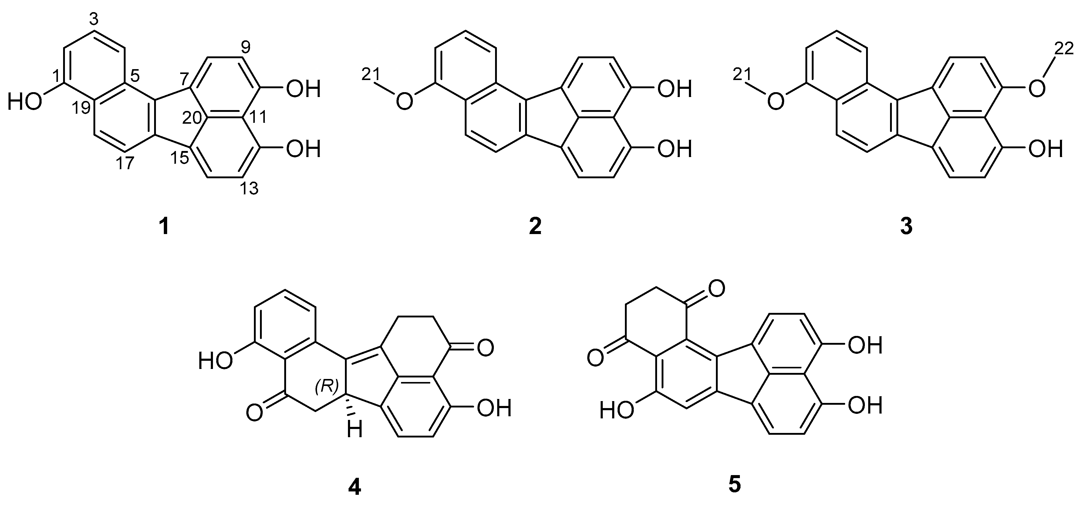
2.6.1. Viridistratin A (1)

Yellow powder. NMR (acetone-d6, 1H NMR: 500 MHz, 13C NMR: 125 MHz): see Table 1; UV/Vis (c = 0.01 mg/mL, ACN): λmax (ε) = 251 (4.45), 342 (4.14), 350 (4.12), 406 (3.94) and 428 (3.91) nm; ESI-MS: m/z 301.06 (M+H)+ and 298.94 (M−H); HR-ESI-MS: m/z 301.0855 (M+H)+ (calculated for C20H13O3, 301.0859); tR= 9.3 min.

2.6.2. Viridistratin B (2)

Yellow powder. NMR (acetone-d6, 1H NMR: 500 MHz, 13C NMR: 125 MHz): see Table 1; UV/Vis (c = 0.01 mg/mL, ACN): λmax (ε) = 248 (4.64), 327 (4.23), 341 (4.38), 384 (4.01) and 403 (4.07) nm; ESI-MS: m/z 315.10 (M+H)+ and 312.96 (M−H); HR-ESI-MS: m/z 315.1010 (M+H)+ (calculated for C21H15O3, 315.1016); tR= 12.2 min.

2.6.3. Viridistratin C (3)

Yellow powder. NMR (DMSO-d6, 1H NMR: 500 MHz and 13C NMR: 125 MHz): see Table 1; UV/Vis (c = 0.01 mg/mL, CHCl3): λmax (ε) = 326 (4.18), 341 (4.46), 384 (3.91) and 403 (3.97) nm; ESI-MS: m/z 329.12 (M+H)+ and 327.01 (M−H); HR ESI-MS: m/z 329.1168 (M+H)+ (calculated for C22H17O3, 329.1172); tR= 13.9 min.

3. Results

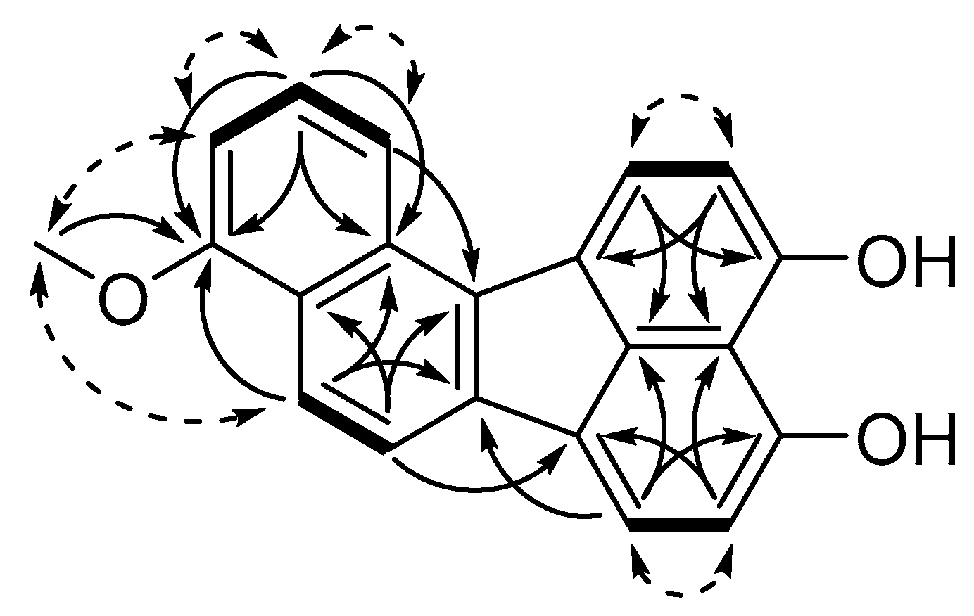
3.1. Structure Elucidation of Viridistratins A−C (13)

In total, five compounds were isolated from the stromatal extract of A. viridistratum in substantial amounts. Three of them represent novel secondary metabolites for which we propose the trivial names viridistratins A−C (13) (see Figure 1), while the remaining two compounds were identical to the previously reported truncatones A (4) and C (5) [12].
Viridistratin A (1) was isolated as a yellow powder and shown to possess a molecular formula of C20H12O3 by HR-ESI-MS, which corresponds to 15 double-bond equivalents. In combination with the UV/Vis maxima (see Figure S1), a large, highly conjugated aromatic system was indicated. In the 1H NMR and 1H/13C heteronuclear single quantum coherence spectroscopy (1H/13C-HSQC) spectra, nine aromatic methines were observed. Additionally, the 13C NMR and 1H/13C heteronuclear multiple-bond correlation spectroscopy (1H/13C HMBC) spectra showed the presence of eleven sp2-hybridized carbons. Three of those eleven carbons had chemical shifts indicating a linkage to a hydroxy group. Analysis of the 1H/1H correlation spectroscopy (1H/1H COSY) spectra revealed the presence of four separate spin systems: (1) 1-H, 2-H and 3-H; (2) 8-H and 9-H and (3) 13-H and 14-H, as well as (4) 17-H and 18-H, which was supported by their splitting patterns in the 1H NMR spectrum. Analysis of the 1H/13C HMBC correlations revealed two naphthalene moieties linked to each other by two C-C bonds to form a five-membered ring in between (i.e., a benzo[j]fluoranthene backbone). Three hydroxy groups were connected to the core structure by analysis of the proton and carbon shifts of the neighboring atoms.
Viridistratin B (2) was isolated as a yellow powder and showed a molecular formula of C21H14O3, indicating a formal addition of CH2 compared to 1. An additional singlet in the 1H and 1H/13C HSQC NMR spectra (21-H3, δH = 4.04), as well as a 1H/13C HMBC correlation of 21-H3 to C-1, revealed 2 to be the 1-methoxy derivative of 1. Key 1H/1H COSY, 1H/13C HMBC and rotating frame nuclear Overhauser effect spectroscopy (ROESY) correlations of 2 are depicted in Figure 2 as an example for the new compounds isolated in this work.
Viridistratin C (3) was isolated as a yellow powder and shown to have a molecular formula of C22H16O3, indicating yet another addition of a methylene in comparison to 2. The 1H/13C HMBC correlations of the singlet 22-H3H = 4.09) to C-10 showed their linkage via the hydroxy group OH-10. Eventually, 3 is the 1,10-dimethoxy derivative of 1.
Structures of truncatones A and C (4, 5) were elucidated by NMR spectroscopy and verified with data provided in the original publication by Sudarman et al. [12]. The stereochemistry of truncatone A was confirmed by the comparison of taken electronic circular dichroism (ECD) spectra (see Figure S2) with data published therein.

3.2. Antibacterial, Antifungal and Cytotoxic Activities of Compounds 15

The minimum inhibitory concentrations (MIC) of 15 were assessed as described in the Methods Section, and the results are summarized in Table 2. For simplified evaluation, measured MIC values were assigned to three descriptors whose thresholds were derived from the measured MIC of the references: strong (MICcompound < MICReference), moderate (MICcompound ≈ MICReference) and weak activity (MICcompound > MICReference).
Viridistratin A (1) exhibited weak antibacterial activities against Gram-positive bacteria, with the strongest activity against Micrococcus luteus (16.7 µg/mL). In addition, growth of the Gram-negative Chromobacterium violaceum was inhibited at 66.7 µg/mL. Furthermore, moderate antifungal activity against all tested fungi except Candida albicans was observed. Viridistratin B (2) showed a similar antimicrobial activity spectrum, additionally being able to inhibit the growth of Mycolicibacterium smegmatis (33.3 µg/mL). In comparison to 1, compound 2 was generally more active with the lowest MIC of 8.3 µg/mL against Micrococcus luteus, as well as a strong inhibition of Mucor hiemalis (4.2 µg/mL). Viridistratin C (3) and truncatone A (4) only exhibited weak antibacterial activity against Micrococcus luteus (33.3 and 16.7 µg/mL, respectively), while, for truncatone C (5), weak activities against Bacillus subtilis and Micrococcus luteus with MIC of 66.7 and 16.7 µg/mL were observed. Truncatone C additionally exhibited weak antifungal activity against Mucor hiemalis and moderate activity against Rhodotorula glutinis (66.7 and 16.7 µg/mL)
Concerning the cytotoxicity of 15, an effect of all compounds against the chosen cell lines was measured and is summarized in Table 3. Against the mouse fibroblast cell line L929, truncatone A (4) and viridistratin A (1) showed half-maximum inhibitory concentration (IC50) values of 10.4 µM (3.3 µg/mL) and 12.7 µM (8.5 µg/mL), respectively. Against human cell lines, viridistratin B (2) induced a decrease of cell viability at IC50 values of 1.1 µM (0.34 µg/mL) and 1.4 µM (0.45 µg/mL) against A431 and A549. Compounds 1, 4 and 5 exhibited a reduced cytotoxicity (i.e., a higher IC50) as compared to 2 and 3. Viridistratin C (3) showed a low decrease in cell viability (IC50 > 50 µM) against L929 and KB 3.1 and was excluded from tests against further cell lines.
To conclude, it can be summarized that the strongest antimicrobial activities (lowest MIC) among the tested compounds, as well as the strongest cytotoxicities (lowest IC50), were both exhibited by viridistratin B (2). In general, viridistratin C (3) showed the highest MIC values, as well as the highest IC50. The other compounds tested (1, 4 and 5) lied in between.

4. Discussion

The three secondary metabolites viridistratins A−C (13) described in this study constitute a new subclass of benzo[j]fluoranthenes from stromata of Annulohypoxylon, whose backbone only consists of aromatic carbons. Due to their aromaticity, no stereochemistry is present in those metabolites.
Peaks corresponding to the viridistratins were already detected by Kuhnert et al. from the holotype specimen and designated UCV1−3 [2]. Occurrence of those compounds has not been observed in any other Annulohypoxylon species yet, even though over two-hundred specimens, including almost all holotypes, have already been examined [2]. Thus, 13 can serve as chemotaxonomic markers for the distinction of A. viridistratum from other Annulohypoxylon spp. Along with the viridistratins, the truncatones A (4) and C (5) were isolated, which are common metabolites in Annulohypoxylon spp. and found in many representatives [2]. Interestingly, truncatone A (4) was not detected in crude extracts of the stromata and, thus, may be a conversion product from the isolation process (see Figure S3). In general, it should be mentioned that all benzo[j]fluoranthenes hitherto isolated from Xylariales were exclusively obtained from members of the genus Annulohypoxylon in the current sense. Previous reports on the isolation of these compounds from “Hypoxylontruncatum (which is now the type species of Annulohypoxylon [3]) actually may go back to this species or its morphologically similar relatives. The genus Hypoxylon sensu Wendt et al. [1], as well as the species of the recently erected genus Jackrogersella, are characterized by the lack of this compound class as stromatal metabolites, and this chemotaxonomic evidence has strongly supported the recent rearrangement of the genera of the Hypoxylaceae.
Sudarman et al. [12] postulated the benzo[j]fluoranthenes to be derived from the 1,8-DHN (dihydroxy naphthalene) pathway [17] of the melanin biosynthesis, with BNT as the precursor for further oxidations. Following this hypothesis, the complete absence of BNT in stromatal extracts of A. viridistratum means that BNT was fully converted to its biosynthetical successors. The origin of these aromatic compounds from the 1,8-DHN pathway has been established in other Ascomycota, and the corresponding polyketide biosynthetic gene clusters are thought to be derived from the 1,8-DHN melanin pathway [18]. However, so far, the biosynthetic genes for these secondary metabolites have not been identified for any species of Annulohypoxylon. This can only now be made possible, because high-quality genomes of several Hypoxylaceae, including the type species of Annulohypoxylon, have recently become available for the first time [19].
Viridistratins 13 exhibited antimicrobial activity against bacteria and fungi but with moderate-to-weak effects. Especially Micrococcus luteus was sensitive to the tested benzo[j]fluoranthenes. Among the tested compounds, vidiristratin B (2) showed the highest activities. Notably, growth of Mucor hiemalis was inhibited by 2 at comparably low concentrations of 4.2 µg/mL. For related benzo[j]fluoranthenes like truncatones [10], hypoxyonols [13,14] or daldinols [20,21], no antimicrobial assessments have been reported in the literature. Furthermore, viridistratin B (2) showed the lowest half-maximum inhibitory concentrations (IC50) against all human cell lines, followed by 1, 4 and 5, while viridistratin C (3) showed only a weak decrease of mammalian cell viability. Other related benzo[j]fluoranthenes like truncatones A, C and D were previously shown to be cytotoxic against the KB 3.1 and L929 cell lines, even though truncatones were reported to exhibit lower IC50 values [12]. Hypoxylonol C was shown to have cell-protective effects against stereptozotocin-induced damage in INS-1 cells [22], while hypoxylonol F was observed to improve insulin secretion [23]. These two reports of rather beneficial bioactivities, although apparently contradicting the cytotoxic activities of hypoxylonols A−F published before [13,14], can be explained by the usage of different doses, as well as target systems. Daldinols C and D were also shown to be cytotoxic against human colon adenocarcinoma cells [21]. Hence, it can be assumed that cytotoxic activities of the aforementioned compounds, as well as the new viridistratins, arise from the benzo[j]fluoranthene backbone, which is well-known for its cytotoxic and cancerogenic properties [24]. These properties arise from the hydrophobicity of the compounds, which reduce (essential) structural interactions between cellular macromolecules such as lipid layers or enzymes by inducing water stress [25,26]. These unspecific targets of such hydrophobic molecules consequently explain the measured bioactivity of substances such as viridistratins A−C (13) against diverse mammalian, bacterial, and fungal cells.

5. Conclusions

The novel compounds viridistratins A−C (13) described herein constitute a novel subclass of hydroxylated benzo[j]fluoranthenes with varying methoxylation levels. So far, they have exclusively been detected in A. viridistratum and may thus serve as chemotaxonomic markers for the species. Chemotaxonomic markers are the basis for HPLC fingerprinting [2], which can help to aid in the discrimination of morphologically similar species.
Several other benzo[j]fluoranthenes have been described from Annulohypoxylon spp. already [12,13,14,21], all of which possess cytotoxic activity against various mammalian cell lines. Viridistratins A−C additionally exhibited weak antibacterial and antifungal activities, with the lowest minimum inhibitory concentrations (MIC) against Micrococcus luteus and Mucor hiemalis. The broad range of activity suggests a nonselective mode of action against the tested microorganisms and mammalian cell lines.
Therefore, and because of the scarce availability and the instability observed in some derivatives, the viridistratins and related metabolites do not appear attractive for further developments as drugs, but they may have a protective function for the producer organisms in nature.

Supplementary Materials

The following are available online at https://www.mdpi.com/2218-273X/10/5/805/s1: Protocol: Antimicrobial Activity Assay. Protocol: Cytotoxicity Assay. Figure S1. HPLC-UV/Vis chromatograms at 210 nm and DAD and HR-ESI-MS(+) traces of viridistratins A−C (13) and truncatones A+C (4, 5). Figure S2. ECD spectrum of truncatone A (4). Figure S3. HPLC-UV/Vis chromatogram at 210 nm of the crude extract of A. viridistratum. Figures S4−S8. 1D and 2D NMR spectra of viridistratin A (1). Figures S9−S14. 1D and 2D NMR spectra of viridistratin B (2). Figures S15−S19. 1D and 2D NMR spectra of viridistratin C (3).

Author Contributions

K.B.: resources, investigation, conception and design, analysis, interpretation of data, drafting of the paper and critical review of the draft. A.-C.W.: resources, investigation, analysis and interpretation of data. J.J.L.-a. and M.S.: conception and design, supervision and critical review of the draft. All authors have read and agreed to the published version of the manuscript.

Funding

This project received funding from the European Union’s Horizon 2020 Research and Innovation Programme (RISE) under the Marie Skłodowska-Curie grant agreement No. 645701, project acronym “GoMyTri” (beneficiaries K.B., M.S. and J.J.L.). This research was also funded by Deutsche Forschungsgemeinschaft (DFG) in the priority program “Taxon-Omics: New Approaches for Discovering and Naming Biodiversity (SPP 1991)” (K.B. and M.S.).

Acknowledgments

The authors wish to thank Lucile Wendt for providing the fungal material and Eric Kuhnert for providing guidance with the extraction and isolation. Silke Reinecke and Esther Surges are thanked for expert assistance in the lab and Christel Kakoschke for recording the NMR spectra. We are grateful towards Wera Collisi for conducting the bioassays.

Conflicts of Interest

The authors declare no conflicts of interest.

References

  1. Wendt, L.; Sir, E.B.; Kuhnert, E.; Heitkämper, S.; Lambert, C.; Hladki, A.I.; Romero, A.I.; Luangsa-ard, J.J.; Srikitikulchai, P.; Peršoh, D.; et al. Resurrection and emendation of the Hypoxylaceae, recognised from a multigene phylogeny of the Xylariales. Mycol. Prog. 2018, 17, 115–154. [Google Scholar] [CrossRef] [Green Version]
  2. Kuhnert, E.; Sir, E.B.; Lambert, C.; Hyde, K.D.; Hladki, A.I.; Romero, A.I.; Rohde, M.; Stadler, M. Phylogenetic and chemotaxonomic resolution of the genus Annulohypoxylon (Xylariaceae) including four new species. Fungal Divers. 2017, 85, 1–43. [Google Scholar] [CrossRef]
  3. Hsieh, H.-M.; Ju, Y.-M.; Rogers, J.D. Molecular phylogeny of Hypoxylon and closely related genera. Mycologia 2017, 97, 844–865. [Google Scholar] [CrossRef]
  4. Helaly, S.E.; Thongbai, B.; Stadler, M. Diversity of biologically active secondary metabolites from endophytic and saprotrophic fungi of the ascomycete order Xylariales. Nat. Prod. Rep. 2018, 35, 992–1014. [Google Scholar] [CrossRef] [PubMed]
  5. Stadler, M.; Læssøe, T.; Fournier, J.; Decock, C.; Schmieschek, B.; Tichy, H.-V.; Peršoh, D. A polyphasic taxonomy of Daldinia (Xylariaceae). Stud. Mycol. 2014, 77, 1–143. [Google Scholar] [CrossRef] [PubMed] [Green Version]
  6. Stadler, M.; Fournier, J.; Granmo, A.; Beltrán-Tejera, E. The “red Hypoxylons” of the temperate and subtropical Northern hemisphere. N. Am. Fungi 2008, 3, 73–125. [Google Scholar] [CrossRef] [Green Version]
  7. Quang, D.N.; Hashimoto, T.; Nomura, Y.; Wollweber, H.; Hellwig, V.; Fournier, J.; Stadler, M.; Asakawa, Y. Cohaerins A and B, azaphilones from the fungus Hypoxylon cohaerens, and comparison of HPLC-based metabolite profiles in Hypoxylon sect. Annulata. Phytochemistry 2005, 66, 797–809. [Google Scholar] [CrossRef]
  8. Surup, F.; Narmani, A.; Wendt, L.; Pfütze, S.; Kretz, R.; Becker, K.; Menbrivès, C.; Giosa, A.; Elliott, M.; Petit, C.; et al. Identification of fungal fossils and novel azaphilone pigments in ancient carbonised specimens of Hypoxylon fragiforme from forest soils of Châtillon-sur-Seine (Burgundy). Fungal Divers. 2018, 92, 345–356. [Google Scholar] [CrossRef]
  9. Daranagama, D.A.; Hyde, K.D.; Sir, E.B.; Thambugala, K.M.; Tian, Q.; Samarakoon, M.C.; McKenzie, E.H.C.; Jayasiri, S.C.; Tibpromma, S.; Bhat, J.D.; et al. Towards a natural classification and backbone tree for Graphostromataceae, Hypoxylaceae, Lopadostomataceae and Xylariaceae. Fungal Divers. 2018, 88, 1–165. [Google Scholar] [CrossRef]
  10. Wibberg, D.; Stadler, M.; Lambert, C.; Bunk, B.; Spröer, C.; Rückert, C.; Kalinowski, J.; Cox, R.J.; Kuhnert, E. High quality genome sequences of thirteen Hypoxylaceae (Ascomycota) strengthen the phylogenetic family backbone and enable the discovery of new taxa. Fungal Divers. 2020, in press. [Google Scholar] [CrossRef]
  11. Kuhnert, E.; Surup, F.; Halecker, S.; Stadler, M. Minutellins A–D, azaphilones from the stromata of Annulohypoxylon minutellum (Xylariaceae). Phytochemistry 2017, 137, 66–71. [Google Scholar] [CrossRef] [PubMed]
  12. Sudarman, E.; Kuhnert, E.; Hyde, K.; Sir, E.; Surup, F.; Stadler, M. Truncatones A–D, benzo[j]fluoranthenes from Annulohypoxylon species (Xylariaceae, Ascomycota). Tetrahedron 2016, 72, 6450–6454. [Google Scholar] [CrossRef]
  13. Koyama, K.; Kuramochi, D.; Kinoshita, K.; Takahashi, K. Hypoxylonols A and B, novel reduced benzo[j]fluoranthene derivatives from the mushroom Hypoxylon truncatum. J. Nat. Prod. 2002, 65, 1489–1490. [Google Scholar] [CrossRef] [PubMed]
  14. Fukai, M.; Tsukada, M.; Miki, K.; Suzuki, T.; Sugita, T.; Kinoshita, K.; Takahashi, K.; Shiro, M.; Koyama, K. Hypoxylonols C-F, benzo[j]fluoranthenes from Hypoxylon truncatum. J. Nat. Prod. 2011, 75, 22–25. [Google Scholar] [CrossRef] [PubMed]
  15. Helaly, S.E.; Ashrafi, S.; Teponno, R.B.; Bernecker, S.; Dababat, A.A.; Maier, W.; Stadler, M. Nematicidal cyclic lipodepsipeptides and a xanthocillin derivative from a phaeosphariaceous fungus parasitizing eggs of the plant parasitic nematode Heterodera filipjevi. J. Nat. Prod. 2018, 81, 2228–2234. [Google Scholar] [CrossRef]
  16. Sandargo, B.; Michehl, M.; Praditya, D.; Steinmann, E.; Stadler, M.; Surup, F. Antiviral meroterpenoid rhodatin and sesquiterpenoids rhodocoranes A-E from the Wrinkled Peach Mushroom, Rhodotus palmatus. Org. Lett. 2019, 21, 3286–3289. [Google Scholar] [CrossRef]
  17. Eisenman, H.C.; Casadevall, A. Synthesis and assembly of fungal melanin. Appl. Microbiol. Biotechnol. 2012, 93, 931–940. [Google Scholar] [CrossRef] [Green Version]
  18. Cox, R.J.; Simpson, T.J. Chapter 3 Fungal Type I Polyketide Synthases. In Methods in Enzymology; Elsevier Inc.: Amsterdam, The Netherlands, 2009; Volume 459, pp. 49–78. ISBN 9780123745910. [Google Scholar]
  19. Stadler, M.; Lambert, C.; Wibberg, D.; Kalinowski, J.; Cox, R.J.; Kolařík, M.; Kuhnert, E. Intragenomic polymorphisms in the ITS region of high-quality genomes of the Hypoxylaceae (Xylariales, Ascomycota). Mycol. Prog. 2020, 19, 235–245. [Google Scholar] [CrossRef] [Green Version]
  20. Quang, D.N.; Hashimoto, T.; Tanaka, M.; Baumgartner, M.; Stadler, M.; Asakawa, Y. Chemical constituents of the ascomycete Daldinia concentrica. J. Nat. Prod. 2002, 65, 1869–1874. [Google Scholar] [CrossRef]
  21. Gu, W.; Ge, H.M.; Song, Y.C.; Ding, H.; Zhu, H.L.; Zhao, X.A.; Tan, R.X. Cytotoxic benzo[j]fluoranthene metabolites from Hypoxylon truncatum IFB-18, an endophyte of Artemisia annua. J. Nat. Prod. 2007, 70, 114–117. [Google Scholar] [CrossRef]
  22. Lee, D.; Choi, P.; Hwang, B.S.; Kim, T.; Kim, Y.; Kim, J.-C.; Song, J.H.; Park, J.S.; Hwang, G.S.; Yamabe, N.; et al. Protective effect of hypoxylonol C and 4,5,4′,5′-tetrahydroxy-1,1′-binaphthyl isolated from Annulohypoxylon annulatum against streptozotocin-induced damage in INS-1 cells. Bioorg. Chem. 2019, 90, 103053. [Google Scholar] [CrossRef] [PubMed]
  23. Lee, D.; Hwang, B.S.; Choi, P.; Kim, T.; Kim, Y.; Song, B.G.; Yamabe, N.; Hwang, G.S.; Kang, K.S.; Ham, J. Hypoxylonol F Isolated from Annulohypoxylon annulatum improves insulin secretion by regulating pancreatic β-cell metabolism. Biomolecules 2019, 9, 335. [Google Scholar] [CrossRef] [PubMed] [Green Version]
  24. U.S. Department of Health & Human Services, Public Health Service Agency for Toxic Substances and Disease Registry. Toxicological Profile for Polycyclic Aromatic Hydrocarbons, U.S. Department of Health & Human Services, Public Health Service, Agency for Toxic Substances and Disease Registry, Washington, D.C., August, 1985. J. Toxicol. Cutan. Ocul. Toxicol. 1999, 18, 141–147. [Google Scholar]
  25. Bhaganna, P.; Volkers, R.J.M.; Bell, A.N.W.; Kluge, K.; Timson, D.J.; McGrath, J.W.; Ruijssenaars, H.J.; Hallsworth, J.E. Hydrophobic substances induce water stress in microbial cells. Microb. Biotechnol. 2010, 3, 701–716. [Google Scholar] [CrossRef] [Green Version]
  26. Cray, J.A.; Stevenson, A.; Ball, P.; Bankar, S.B.; Eleutherio, E.C.A.; Ezeji, T.C.; Singhal, R.S.; Thevelein, J.M.; Timson, D.J.; Hallsworth, J.E. Chaotropicity: A key factor in product tolerance of biofuel-producing microorganisms. Curr. Opin. Biotechnol. 2015, 33, 228–259. [Google Scholar] [CrossRef]
Figure 1. Structures of secondary metabolites isolated from stromata of Annulohypoxylon viridistratum. 13: viridistratins A−C, 4: truncatone A and 5: truncatone C.
Biomolecules 10 00805 g001
Figure 2. Key nuclear magnetic resonance (NMR) correlations of viridistratin B (2). Bold bonds: 1H/1H correlation spectroscopy (COSY) correlations, plain arrows: 1H/13C (heteronuclear multiple bond correlation (HMBC) correlations and dashed arrows: rotating frame nuclear Overhauser effect spectroscopy (ROESY) correlations.
Biomolecules 10 00805 g002
Table 1. 1D nuclear magnetic resonance (NMR) data of 13 (1, 2: acetone-d6 and 3: DMSO-d6; 1H NMR: 500 MHz and 13C NMR: 125 MHz).
pos 11 2 3
δC, mult 2δH, mult 2 δC, multδH, mult δC, multδH, mult
1155.1, C 156.8, C 156.2, C
2108.2, CH6.90, d (7.53) 103.6, CH6.92, d (7.63) 104.0, CH6.97, m
3128.1, CH7.42, t (2 × 7.96) 127.5, CH7.52, t (2 × 8.09) 127.9, CH7.56, t (2 × 7.96)
4116.4, CH8.22, d (8.39) 116.8, CH8.29, d (8.39) 116.6, CH8.30, d (8.39)
5132.5, C 131.5, C 130.9, C
6133.5, C 133.0, C 131.6, C
7129.9, C 129.3, C 129.2, C
8127.4, CH8.45, m 126.9, CH8.45, d (7.78) 126.8, CH8.56, d (8.17)
9111.2, CH7.06, d (7.53) 110.8, CH7.06, d (7.78) 106.7, CH7.13, d (7.96)
10156.2, C 155.7, C 158.1, C
11112.6, C 112.1, C 112.7, C
12156.8, C 156.3, C 156.8, C
13110.9, CH7.01, d (7.53) 110.5, CH7.01, d (7.63) 112.0, CH7.00, m
14124.3, CH8.06, br s 123.8, CH8.05, m 124.6, CH8.09, m
15129.1, C 128.5, C 126.7, C
16137.9, C 137.5, C 137.3, C
17119.0, CH8.06, m 118.9, CH8.06, m 119.2, CH8.09, m
18121.8, CH8.26, d (8.60) 120.8, CH8.22, m 120.7, CH8.17, d (8.60)
19125.4, C 125.4, C 124.5, C
20135.2, C 134.7, C 134.1, C
21 55.5, CH34.04, s 56.0, CH34.02, s
22 56.8, CH34.09, s
1 pos: atom position (see Figure 1); 2 δCH: chemical shift [ppm]; mult: multiplicity, (br) s: (broad) singlet, d: doublet, t: triplet, and m: multiplet.
Table 2. Minimum inhibitory concentrations (MIC) of 15 against bacterial and fungal test organisms.
Test OrganismMinimum Inhibitory Concentration (MIC) (µg/mL)
12345Reference
Bacillus subtilis33.316.7>66.7>66.766.78.3 1
Staphylococcus aureus66.716.7>66.7>66.7>66.70.4 2
Micrococcus luteus16.78.366.733.316.70.8 2
Chromobacterium violaceum66.766.7>66.7>66.7>66.70.1 2
Escherichia coli>66.7>66.7>66.7>66.7>66.71.7 2
Pseudomonas aeruginosa>66.7>66.7>66.7>66.7>66.70.4 3
Mycolicibacterium smegmatis>66.733.3>66.7>66.7>66.73.3 4
Candida albicans>66.7>66.7>66.7>66.7>66.766.7 5
Schizosaccharomyces pombe66.733.3>66.7>66.7>66.733.3 5
Mucor hiemalis66.74.2>66.7>66.766.766.7 5
Pichia anomala66.733.3>66.7>66.7>66.766.7 5
Rhodotorula glutinis33.333.3>66.7>66.766.716.7 5
1 oxytetracycline 1 mg/mL, 2 oxytetracycline 0.1 mg/mL, 3 gentamicin 0.1 mg/mL, 4 kanamycin 0.1 mg/mL and 5 nystatin 1 mg/mL.
Table 3. Cytotoxicity of 15 against mammalian cell lines as half-maximum inhibitory concentrations (IC50). n.d.: not determined.
Cell LineCytotoxicity (IC50) (µM)
12345Reference 1
L929mouse fibroblasts12.717.261.010.416.30.00006
KB 3.1human endocervical adenocarcinoma (AC)28.317.285.444.030.10.00079
PC-3human prostate AC23.79.9n.d.44.025.60.00008
SK-OV-3human ovary AC56.77.3n.d.66.033.10.00034
MCF-7human breast AC9.75.1n.d.8.87.80.00012
A431human squamous AC8.71.1n.d.5.716.30.00005
A549human lung carcinoma20.01.4n.d.17.027.10.00008
1 epothilone B (1mg/mL).

