Screening of Molecular Targets of Action of Atractylodin in Cholangiocarcinoma by Applying Proteomic and Metabolomic Approaches

 ,
,
Abstract
:1. Introduction
2. Results
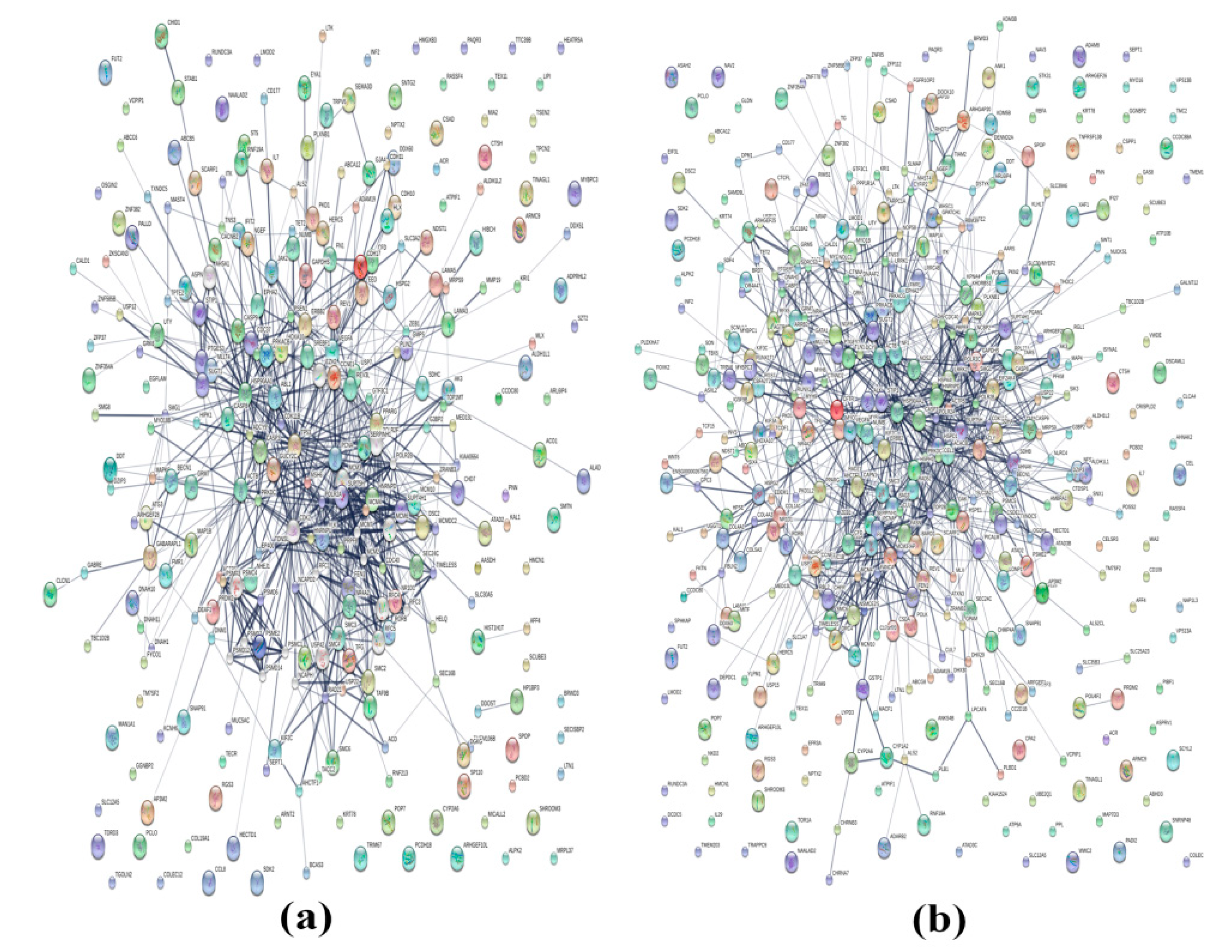
2.1. Proteomics
2.2. Metabolomics
2.3. Co-Analysis of Proteomics and Metabolomics
3. Discussion
4. Materials and Methods
4.1. Cell Culture
4.2. Sample Preparation
4.3. Proteomics
4.3.1. Protein Extraction
4.3.2. SDS-PAGE Analysis and In-Gel Digestion by Trypsin
4.3.3. LC-MS/MS Analysis
4.3.4. Protein Identification and Analysis
4.4. Metabolomics
4.4.1. Metabolite Extraction
4.4.2. Metabolite Identification
4.4.3. Metabolomics Analysis
5. Conclusions
Author Contributions
Funding
Acknowledgments
Conflicts of Interest
References
- Khan, S.A.; Davidson, B.R.; Goldin, R.; Pereira, S.P.; Rosenberg, W.M.; Taylor-Robinson, S.D.; Thillainayagam, A.V.; Thomas, H.C.; Thursz, M.R.; Wasan, H.; et al. Guidelines for the diagnosis and treatment of cholangiocarcinoma: Consensus document. Gut 2002, 51, VI1–VI9. [Google Scholar] [CrossRef] [PubMed]
- Nathan, H.; Aloia, T.A.; Vauthey, J.N.; Abdalla, E.K.; Zhu, A.X.; Schulick, R.D.; Choti, M.A.; Pawlik, T.M. A proposed staging system for intrahepatic cholangiocarcinoma. Ann. Surg. Oncol. 2009, 16, 14–22. [Google Scholar] [CrossRef] [PubMed]
- Mahavorasirikul, W.; Viyanant, V.; Chaijaroenkul, W.; Itharat, A.; Na-Bangchang, K. Cytotoxic activity of Thai medicinal plants against human cholangiocarcinoma, laryngeal and hepatocarcinoma cells in vitro. BMC Complement. Altern. Med. 2010, 10, 55. [Google Scholar] [CrossRef] [PubMed]
- Na-Bangchang, K.; Karbwang, J. Traditional herbal medicine for the control of tropical diseases. Trop. Med. Health 2014, 42, 3–13. [Google Scholar] [CrossRef] [PubMed]
- Chuang, C.H.; Cheng, Y.C.; Lin, S.C.; Lehman, C.W.; Wang, S.P.; Chen, D.Y.; Tsai, S.W.; Lin, C.C. Atractylodin Suppresses Dendritic Cell Maturation and Ameliorates Collagen-Induced Arthritis in a Mouse Model. J. Agric. Food Chem. 2019, 67, 6773–6784. [Google Scholar] [CrossRef] [PubMed]
- Nakai, Y.; Kido, T.; Hashimoto, K.; Kase, Y.; Sakakibara, I.; Higuchi, M.; Sasaki, H. Effect of the rhizomes of Atractylodes lancea and its constituents on the delay of gastric emptying. J. Ethnopharmacol. 2003, 84, 51–55. [Google Scholar] [CrossRef]
- Chae, H.S.; Kim, Y.M.; Chin, Y.W. Atractylodin Inhibits Interleukin-6 by Blocking NPM-ALK Activation and MAPKs in HMC-1. Molecules 2016, 21, 1169. [Google Scholar] [CrossRef]
- Liang, Y.; Li, S.; Chen, L. The physiological role of drug transporters. Protein Cell 2015, 6, 334–350. [Google Scholar] [CrossRef] [Green Version]
- Hlavac, V.; Brynychova, V.; Vaclavikova, R.; Ehrlichova, M.; Vrana, D.; Pecha, V.; Kozevnikovova, R.; Trnkova, M.; Gatek, J.; Kopperova, D.; et al. The expression profile of ATP-binding cassette transporter genes in breast carcinoma. Pharmacogenomics 2013, 14, 515–529. [Google Scholar] [CrossRef]
- Elsnerova, K.; Mohelnikova-Duchonova, B.; Cerovska, E.; Ehrlichova, M.; Gut, I.; Rob, L.; Skapa, P.; Hruda, M.; Bartakova, A.; Bouda, J.; et al. Gene expression of membrane transporters: Importance for prognosis and progression of ovarian carcinoma. Oncol. Rep. 2016, 35, 2159–2170. [Google Scholar] [CrossRef]
- Hlavata, I.; Mohelnikova-Duchonova, B.; Vaclavikova, R.; Liska, V.; Pitule, P.; Novak, P.; Bruha, J.; Vycital, O.; Holubec, L.; Treska, V.; et al. The role of ABC transporters in progression and clinical outcome of colorectal cancer. Mutagenesis 2012, 27, 187–196. [Google Scholar] [CrossRef] [PubMed] [Green Version]
- Stokes, E.; Shuang, T.; Zhang, Y.; Pei, Y.; Fu, M.; Guo, B.; Parissenti, A.; Wu, L.; Wang, R.; Yang, G. Efflux inhibition by H2S confers sensitivity to doxorubicin-induced cell death in liver cancer cells. Life Sci. 2018, 213, 116–125. [Google Scholar] [CrossRef] [PubMed]
- Bassi, M.T.; Manzoni, M.; Bresciani, R.; Pizzo, M.T.; Della Monica, A.; Barlati, S.; Monti, E.; Borsani, G. Cellular expression and alternative splicing of SLC25A23, a member of the mitochondrial Ca2+-dependent solute carrier gene family. Gene 2005, 345, 173–182. [Google Scholar] [CrossRef] [PubMed]
- Hoffman, N.E.; Chandramoorthy, H.C.; Shanmughapriya, S.; Zhang, X.Q.; Vallem, S.; Doonan, P.J.; Malliankaraman, K.; Guo, S.; Rajan, S.; Elrod, J.W.; et al. SLC25A23 augments mitochondrial Ca(2)(+) uptake, interacts with MCU, and induces oxidative stress-mediated cell death. Mol. Biol. Cell 2014, 25, 936–947. [Google Scholar] [CrossRef]
- Wissenbach, U.; Niemeyer, B.A.; Fixemer, T.; Schneidewind, A.; Trost, C.; Cavalie, A.; Reus, K.; Meese, E.; Bonkhoff, H.; Flockerzi, V. Expression of CaT-like, a novel calcium-selective channel, correlates with the malignancy of prostate cancer. J. Biol. Chem. 2001, 276, 19461–19468. [Google Scholar] [CrossRef]
- Suzuki, Y.; Chitayat, D.; Sawada, H.; Deardorff, M.A.; McLaughlin, H.M.; Begtrup, A.; Millar, K.; Harrington, J.; Chong, K.; Roifman, M.; et al. TRPV6 Variants Interfere with Maternal-Fetal Calcium Transport through the Placenta and Cause Transient Neonatal Hyperparathyroidism. Am. J. Hum. Genet. 2018, 102, 1104–1114. [Google Scholar] [CrossRef] [Green Version]
- Chow, J.; Norng, M.; Zhang, J.; Chai, J. TRPV6 mediates capsaicin-induced apoptosis in gastric cancer cells—Mechanisms behind a possible new “hot” cancer treatment. Biochim. Biophys. Acta (BBA)-Mol. Cell Res. 2007, 1773, 565–576. [Google Scholar] [CrossRef]
- Strzalka, W.; Ziemienowicz, A. Proliferating cell nuclear antigen (PCNA): A key factor in DNA replication and cell cycle regulation. Ann. Bot. 2011, 107, 1127–1140. [Google Scholar] [CrossRef]
- Paunesku, T.; Mittal, S.; Protic, M.; Oryhon, J.; Korolev, S.V.; Joachimiak, A.; Woloschak, G.E. Proliferating cell nuclear antigen (PCNA): Ringmaster of the genome. Int. J. Radiat. Biol. 2001, 77, 1007–1021. [Google Scholar] [CrossRef]
- Feki, A.; Jefford, C.E.; Berardi, P.; Wu, J.-Y.; Cartier, L.; Krause, K.-H.; Irminger-Finger, I. BARD1 induces apoptosis by catalysing phosphorylation of p53 by DNA-damage response kinase. Oncogene 2005, 24, 3726–3736. [Google Scholar] [CrossRef]
- Woods, D.; Turchi, J.J. Chemotherapy induced DNA damage response: Convergence of drugs and pathways. Cancer Biol. Ther. 2013, 14, 379–389. [Google Scholar] [CrossRef] [PubMed]
- Kotawong, K.; Chaijaroenkul, W.; Muhamad, P.; Na-Bangchang, K. Cytotoxic activities and effects of atractylodin and beta-eudesmol on the cell cycle arrest and apoptosis on cholangiocarcinoma cell line. J. Pharmacol. Sci. 2018, 136, 51–56. [Google Scholar] [CrossRef] [PubMed]
- Liu, Y.; Hyde, A.S.; Simpson, M.A.; Barycki, J.J. Emerging regulatory paradigms in glutathione metabolism. Adv. Cancer Res. 2014, 122, 69–101. [Google Scholar] [PubMed]
- Gumireddy, K.; Li, A.; Cao, L.; Yan, J.; Liu, L.; Xu, X.; Pazoles, C.; Huang, Q. NOV-002, A Glutathione Disulfide Mimetic, Suppresses Tumor Cell Invasion and Metastasis. J. Carcinog. Mutagen. 2013, 2013, s7-002. [Google Scholar] [PubMed]
- Muhlbauer, M.; Allard, B.; Bosserhoff, A.K.; Kiessling, S.; Herfarth, H.; Rogler, G.; Scholmerich, J.; Jobin, C.; Hellerbrand, C. Differential effects of deoxycholic acid and taurodeoxycholic acid on NF-kappa B signal transduction and IL-8 gene expression in colonic epithelial cells. Am. J. Physiol. Gastrointest. Liver Physiol. 2004, 286, G1000–G1008. [Google Scholar] [CrossRef]
- Xie, Q.; Li, G.M.; Zhou, X.Q.; Liao, D.; Yu, H.; Guo, Q. Effect of Tauroursodeoxycholic acid on cytochrome C-mediated apoptosis in HepG2 cells. Zhonghua Gan Zang Bing Za Zhi 2003, 11, 298–301. [Google Scholar]
- Rybinski, B.; Franco-Barraza, J.; Cukierman, E. The wound healing, chronic fibrosis, and cancer progression triad. Physiol. Genom. 2014, 46, 223–244. [Google Scholar] [CrossRef]
- Lima, L.G.; Monteiro, R.Q. Activation of blood coagulation in cancer: Implications for tumour progression. Biosci. Rep. 2013, 33, e00064. [Google Scholar] [CrossRef]
- Scappaticci, F.A.; Fehrenbacher, L.; Cartwright, T.; Hainsworth, J.D.; Heim, W.; Berlin, J.; Kabbinavar, F.; Novotny, W.; Sarkar, S.; Hurwitz, H. Surgical wound healing complications in metastatic colorectal cancer patients treated with bevacizumab. J. Surg. Oncol. 2005, 91, 173–180. [Google Scholar] [CrossRef]
- Mathema, V.B.; Chaijaroenkul, W.; Na-Bangchang, K. Cytotoxic activity and molecular targets of atractylodin in cholangiocarcinoma cells. J. Pharm. Pharmacol. 2019, 71, 185–195. [Google Scholar] [CrossRef]
- Palta, S.; Saroa, R.; Palta, A. Overview of the coagulation system. Indian J. Anaesth. 2014, 58, 515–523. [Google Scholar] [CrossRef] [PubMed]
- Castaman, G.; Matino, D. Hemophilia A and B: Molecular and clinical similarities and differences. Haematologica 2019, 104, 1702–1709. [Google Scholar] [CrossRef] [PubMed]
- Lowry, O.H.; Rosebrough, N.J.; Farr, A.L.; Randall, R.J. Protein measurement with the Folin phenol reagent. J. Biol. Chem. 1951, 193, 265–275. [Google Scholar] [PubMed]
- Kotawong, K.; Thitapakorn, V.; Roytrakul, S.; Phaonakrop, N.; Viyanant, V.; Na-Bangchang, K. Plasma phosphoproteome and differential plasma phosphoproteins with opisthorchis viverrini-related cholangiocarcinoma. Asian Pac. J. Cancer Prev. 2015, 16, 1011–1018. [Google Scholar] [CrossRef] [PubMed]
- Yuan, M.; Breitkopf, S.B.; Yang, X.; Asara, J.M. A positive/negative ion-switching, targeted mass spectrometry-based metabolomics platform for bodily fluids, cells, and fresh and fixed tissue. Nat. Protoc. 2012, 7, 872–881. [Google Scholar] [CrossRef]


| Transporter Proteins Identified at 24 h Incubation | ||
| Protein Names | Protein Names | Protein Names |
| ATP-binding cassette sub-family A member 12 | Polycystic kidney disease protein 1-like 2 | Retinal-specific ATP-binding cassette transporter |
| ATP-binding cassette sub-family G member 8 | Polycystic kidney disease protein 1-like 3 | Sodium/hydrogen exchanger 9B2 |
| CSC1-like protein 1 | Potassium voltage-gated channel subfamily A member 4 | Transient receptor potential cation channel subfamily V member 4 |
| Gamma-aminobutyric acid receptor subunit epsilon | Potassium voltage-gated channel subfamily H member 4 | Transient receptor potential cation channel subfamily V member 6 |
| Melanoma inhibitory activity protein 2 | Potassium voltage-gated channel subfamily H member 8 | Voltage-dependent L-type calcium channel subunit beta-2 |
| Mitochondrial inner membrane protein OXA1L | Potassium voltage-gated channel subfamily KQT member 3 | V-type proton ATPase 116 kDa subunit α isoform 2 |
| Mitochondrial pyruvate carrier 1 | Potassium voltage-gated channel subfamily S member 1 | Protein shisa-7 |
| Neuronal acetylcholine receptor subunit alpha-10 | Potassium-transporting ATPase subunit beta | |
| NTF2-related export protein 1 | Probable phospholipid-transporting ATPase VD | |
| Transporter Proteins Identified at 48 h Incubation | ||
| Protein Names | Protein Names | Protein Names |
| Amiloride-sensitive sodium channel subunit gamma | Mitochondrial pyruvate carrier 1 | Protein shisa-7 |
| ATP-binding cassette sub-family A member 12 | Neuronal acetylcholine receptor subunit alpha-10 | Sodium/bile acid cotransporter |
| ATP-binding cassette sub-family G member 8 | Neuronal acetylcholine receptor subunit alpha-7 | Sodium/hydrogen exchanger 9B2 |
| Calcium permeable stress-gated cation channel 1 | Neuronal acetylcholine receptor subunit beta-3 | Synaptic vesicular amine transporter |
| Calcium-activated chloride channel regulator 4 | NTF2-related export protein 1 | Transient receptor potential cation channel subfamily V member 6 |
| Calcium-binding mitochondrial carrier protein SCaMC-3 | Phospholipid-transporting ATPase IB | Transmembrane channel-like protein 2 |
| Ceramide-1-phosphate transfer protein | Polycystic kidney disease protein 1-like 2 | Vacuolar protein sorting-associated protein 26B |
| Excitatory amino acid transporter 5 | Polycystic kidney disease protein 1-like 3 | Vesicular acetylcholine transporter |
| Importin subunit alpha-4 | Potassium voltage-gated channel subfamily H member 4 | Zinc transporter ZIP6 |
| Melanoma inhibitory activity protein 2 | Potassium voltage-gated channel subfamily S member 1 | |
| Mitochondrial glutamate carrier 2 | Potassium-transporting ATPase subunit beta | |
| Up-Regulation of Intracellular Metabolites at 24 h of Exposure | |||||
| Name | Fold Change | Name | Fold Change | Name | Fold Change |
| Inosine | 8.05 | Threonine | 3.37 | Taurodeoxycholic acid | 2.24 |
| Phenylpyruvate | 6.25 | S-ribosyl-L-homocysteine | 3.31 | Phenylalanine | 2.22 |
| 6-phospho-D-glucono-1,5-lactone | 5.86 | Indole | 2.81 | Kynurenine | 2.16 |
| Cytidine | 5.66 | Aconitate | 2.73 | Folate | 2.14 |
| Choline | 5.60 | Creatine | 2.70 | Tyrosine | 2.13 |
| UDP-D-glucose | 5.32 | L-arginino-succinate | 2.52 | 2-oxo-4-methylthiobutanoate | 2.12 |
| Serine | 3.85 | D-gluconate | 2.24 | Pyridoxine | 2.01 |
| Down-Regulation of Intracellular Metabolites at 24 h of Exposure | |||||
| Name | Fold Change | Name | Fold Change | Name | Fold Change |
| Glutathione disulfide | 6.84 | AMP | 2.71 | Cholesteryl sulfate | 2.16 |
| Lactate | 5.11 | Thiamine | 2.61 | 6-phospho-D-gluconate | 2.08 |
| Methionine sulfoxide | 4.43 | Cystine | 2.53 | Glucosamine | 2.06 |
| Glutamine | 4.21 | Aspartate | 2.43 | Glucose-1-phosphate | 2.04 |
| Citrulline | 3.68 | Cellobiose | 2.41 | UTP | 2.01 |
| 4-aminobutyrate | 2.86 | Asparagine | 2.34 | ||
| N-acetyl-glutamate | 2.83 | dAMP | 2.17 | ||
| Up-Regulation of Extracellular Metabolites at 24 h of Exposure | |||||
| Name | Fold Change | Name | Fold Change | Name | Fold Change |
| Methylnicotinamide | 8.57 | Glycerate | 3.00 | Orotidine-5-phosphate | 2.12 |
| Adenine | 5.81 | 2,3-dihydroxybenzoic acid | 2.73 | Serine | 2.07 |
| Pyroglutamic acid | 3.87 | Hydroxyisocaproic acid | 2.25 | Adenosine | 2.04 |
| Cytidine | 3.61 | Dihydroorotate | 2.16 | ||
| Down-Regulation of Extracellular Metabolites at 24 h of Exposure | |||||
| Name | Fold Change | Name | Fold Change | Name | Fold Change |
| CDP | 4.83 | N-carbamoyl-L-aspartate | 2.79 | Acetylphosphate | 2.44 |
| Glutathione disulfide | 3.96 | Orotate | 2.71 | Citraconic acid | 2.20 |
| N6-Acetyl-L-lysine | 3.39 | 2-ketohaxanoic acid | 2.66 | Glucose-1-phosphate | 2.12 |
| Citrate | 3.11 | 2-dehydro-D-gluconate | 2.61 | Taurodeoxycholic acid | 2.11 |
| Phenylpyruvate | 3.05 | Shikimate-3-phosphate | 2.60 | ||
| 2-oxo-4-methylthiobutanoate | 2.84 | D-gluconate | 2.48 | ||
| Up-Regulation of Intracellular Metabolites at 48 h of Exposure | |||||
| Name | Fold Change | Name | Fold Change | Name | Fold Change |
| Taurodeoxycholic acid | 22.41 | Glucosamine | 3.09 | Taurine | 2.25 |
| 2-Aminooctanoic acid | 21.35 | 2-oxo-4-methylthiobutanoate | 2.97 | Citrate | 2.23 |
| Cystine | 17.21 | Allantoate | 2.96 | Methylnicotinamide | 2.20 |
| Orotidine-5-phosphate | 6.65 | Ribose-phosphate | 2.94 | N-acetyl-glutamate | 2.19 |
| 2-dehydro-D-gluconate | 5.77 | Asparagine | 2.89 | Ornithine | 2.19 |
| Shikimate | 5.29 | Methylcysteine | 2.82 | Hexose-phosphate | 2.17 |
| Acetylphosphate | 4.87 | Glycerate | 2.67 | 2,3-dihydroxybenzoic acid | 2.14 |
| 4-Pyridoxic acid | 4.76 | N-Acetyl-L-ornithine | 2.66 | Citrate-isocitrate | 2.11 |
| 2-ketohaxanoic acid | 4.57 | Pantothenate | 2.63 | Acetyllysine | 2.11 |
| Shikimate-3-phosphate | 4.43 | Kynurenic acid | 2.56 | Thymidine | 2.08 |
| Cytidine | 4.14 | Folate | 2.56 | Allantoin | 2.06 |
| Threonine | 4.10 | Succinate | 2.54 | Tryptophan | 2.06 |
| Aconitate | 4.06 | 6-phospho-D-gluconate | 2.54 | Histidine | 2.04 |
| N6-Acetyl-L-lysine | 3.92 | Nicotinamide | 2.49 | Cholesteryl sulfate | 2.04 |
| Cellobiose | 3.71 | Hydroxyisocaproic acid | 2.47 | Thymine | 2.03 |
| Citrulline | 3.45 | Glucono-D-lactone | 2.41 | Orotate | 2.02 |
| N-carbamoyl-L-aspartate | 3.27 | N-acetyl-glutamine | 2.40 | Deoxyribose-phosphate | 2.01 |
| Serine | 3.19 | CDP | 2.31 | L-arginino-succinate | 2.01 |
| Xanthine | 3.14 | Lysine | 2.30 | ||
| Indole | 3.10 | Phenylpropiolic acid | 2.26 | ||
| Down-Regulation of Intracellular Metabolites at 48 h of Exposure | |||||
| Name | Fold Change | Name | Fold Change | Name | Fold Change |
| Glutathione disulfide | 61.87 | ATP | 4.17 | IMP | 2.78 |
| AMP | 6.01 | NAD+ | 4.07 | 1-Methyladenosine | 2.35 |
| Glycerophosphocholine | 5.30 | GMP | 3.62 | Kynurenine | 2.27 |
| UDP-D-glucose | 4.51 | UMP | 3.25 | ||
| Up-Regulation of Extracellular Metabolites at 48 h of Exposure | |||||
| Name | Fold Change | Name | Fold Change | Name | Fold Change |
| GMP | 2.24 | Serine | 2.94 | Adenine | 5.51 |
| Glycerate | 2.73 | UMP | 4.12 | Cytidine | 6.18 |
| S-ribosyl-L-homocysteine | 2.77 | Methylnicotinamide | 4.79 | ||
| Down-Regulation of Extracellular Metabolites at 48 h of Exposure | |||||
| Name | Fold Change | Name | Fold Change | Name | Fold Change |
| Glutathione disulfide | 65.53 | UDP-D-glucose | 3.63 | Allantoin | 2.47 |
| CDP | 17.44 | Phenylpropiolic acid | 3.58 | Betaine | 2.42 |
| 6-phospho-D-glucono-1,5-lactone | 10.16 | Cholesteryl sulfate | 3.57 | AMP | 2.40 |
| 2-ketohaxanoic acid | 7.30 | Uric acid | 3.51 | Thymine | 2.37 |
| Phenylpyruvate | 6.62 | Methylmalonic acid | 3.42 | Ng,NG-dimethyl-L-arginine | 2.36 |
| Shikimate-3-phosphate | 6.57 | Deoxyribose-phosphate | 3.28 | p-hydroxybenzoate | 2.33 |
| Citrate | 6.33 | Kynurenic acid | 3.21 | Ribose-phosphate | 2.29 |
| Citraconic acid | 5.77 | Hydroxyphenylacetic acid | 3.17 | Acadesine | 2.26 |
| Citrate-isocitrate | 5.23 | Acetylphosphate | 3.14 | Hydroxyisocaproic acid | 2.26 |
| N-carbamoyl-L-aspartate | 5.08 | 2-oxo-4-methylthiobutanoate | 3.01 | Biotin | 2.24 |
| Octulose-1,8-bisphosphate (OBP) | 5.07 | Nicotinamide | 2.95 | SN-glycerol-3-phosphate | 2.24 |
| 2-dehydro-D-gluconate | 5.01 | NAD+ | 2.91 | Sarcosine | 2.21 |
| D-gluconate | 4.79 | Shikimate | 2.84 | Imidazoleacetic acid | 2.18 |
| Xanthine | 4.11 | Purine | 2.83 | Acetylcarnitine DL | 2.11 |
| UTP | 4.11 | Lactate | 2.66 | 2-Hydroxy-2-methylthiobutanoate | 2.10 |
| Allantoate | 4.00 | Thiamine | 2.64 | Malate | 2.05 |
| L-arginino-succinate | 3.96 | Acetyllysine | 2.58 | Pantothenate | 2.00 |
| A-ketoglutarate | 3.89 | 1,3-diphopshateglycerate | 2.51 | ||
| 4-Pyridoxic acid | 3.86 | Thymidine | 2.50 | ||
| N-Acetyl-L-alanine | 3.83 | Hexose-phosphate | 2.48 | ||
© 2019 by the authors. Licensee MDPI, Basel, Switzerland. This article is an open access article distributed under the terms and conditions of the Creative Commons Attribution (CC BY) license (http://creativecommons.org/licenses/by/4.0/).
Share and Cite
Kotawong, K.; Chaijaroenkul, W.; Roytrakul, S.; Phaonakrop, N.; Na-Bangchang, K. Screening of Molecular Targets of Action of Atractylodin in Cholangiocarcinoma by Applying Proteomic and Metabolomic Approaches. Metabolites 2019, 9, 260. https://doi.org/10.3390/metabo9110260
Kotawong K, Chaijaroenkul W, Roytrakul S, Phaonakrop N, Na-Bangchang K. Screening of Molecular Targets of Action of Atractylodin in Cholangiocarcinoma by Applying Proteomic and Metabolomic Approaches. Metabolites. 2019; 9(11):260. https://doi.org/10.3390/metabo9110260
Chicago/Turabian StyleKotawong, Kanawut, Wanna Chaijaroenkul, Sittiruk Roytrakul, Narumon Phaonakrop, and Kesara Na-Bangchang. 2019. "Screening of Molecular Targets of Action of Atractylodin in Cholangiocarcinoma by Applying Proteomic and Metabolomic Approaches" Metabolites 9, no. 11: 260. https://doi.org/10.3390/metabo9110260