
Diagnostic Value of Salivary Amino Acid Levels in Cancer


Abstract
:
1. Introduction
2. Materials and Methods
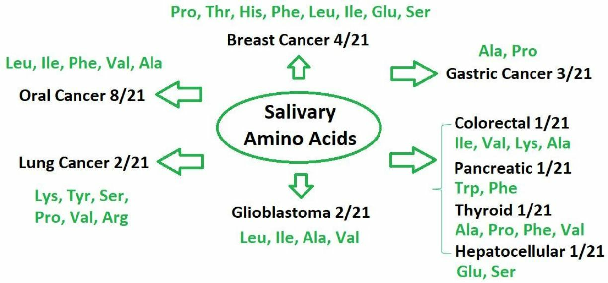
3. Results
4. Discussion
5. Conclusions
Author Contributions
Funding
Conflicts of Interest
Abbreviations
References
- Safrhansova, L.; Hlozkova, K.; Starkova, J. Targeting amino acid metabolism in cancer. In International Review of Cell and Molecular Biology; Buqué, A., Galluzzi, L., Eds.; Academic Press: Cambridge, MA, USA, 2022; Volume 373, pp. 37–79. [Google Scholar]
- Lieu, E.L.; Nguyen, T.; Rhyne, S.; Kim, J. Amino Acids in Cancer. Exp. Mol. Med. 2020, 52, 15–30. [Google Scholar] [CrossRef]
- Wei, Z.; Liu, X.; Cheng, C.; Yu, W.; Yi, P. Metabolism of Amino Acids in Cancer. Front. Cell Dev. Biol. 2021, 8, 603837. [Google Scholar] [CrossRef]
- Ragni, M.; Fornelli, C.; Nisoli, E.; Penna, F. Amino Acids in Cancer and Cachexia: An Integrated View. Cancers 2022, 14, 5691. [Google Scholar] [CrossRef] [PubMed]
- Jaune-Pons, E.; Vasseur, S. Role of amino acids in regulation of ROS balance in cancer. Arch. Biochem. Biophys. 2020, 689, 108438. [Google Scholar] [CrossRef] [PubMed]
- Zhao, Y.; Pu, C.; Liu, Z. Essential amino acids deprivation is a potential strategy for breast cancer treatment. Breast 2022, 62, 152–161. [Google Scholar] [CrossRef]
- Lukey, M.J.; Katt, W.P.; Cerione, R.A. Targeting amino acid metabolism for cancer therapy. Drug Discov. Today 2017, 22, 796–804. [Google Scholar] [CrossRef] [PubMed] [Green Version]
- Fu, S.; Xu, S.; Zhang, S. The role of amino acid metabolism alterations in pancreatic cancer: From mechanism to application. BBA-Rev. Cancer 2023, 1878, 188893. [Google Scholar] [CrossRef]
- Han, X.; Li, D.; Wang, S.; Lin, Y.; Liu, Y.; Lin, L.; Qiao, L. Serum amino acids quantification by plasmonic colloidosome-coupled MALDI-TOF MS for triple-negative breast cancer diagnosis. Mater. Today Bio. 2022, 17, 100486. [Google Scholar] [CrossRef]
- Lu, H.; Li, Y.; Zhang, H.; Chingin, K.; Wei, Y.; Huang, K.; Feng, S. Direct quantitative profiling of amino acids in tissues for the assessment of lung cancer. Talanta 2021, 233, 122544. [Google Scholar] [CrossRef]
- Bel’skaya, L.V. Possibilities of using saliva for the diagnosis of cancer. Klin. Lab. Diagn. Russ. Clin. Lab. Diagn. 2019, 64, 333–336. [Google Scholar] [CrossRef]
- Kaczor-Urbanowicz, K.E.; Wei, F.; Rao, S.L.; Kim, J.; Shin, H.; Cheng, J.; Tu, M.; Wong, D.T.W.; Kim, Y. Clinical validity of saliva and novel technology for cancer detection. BBA-Rev. Cancer 2019, 1872, 49–59. [Google Scholar] [CrossRef] [PubMed]
- Roblegg, E.; Coughran, A.; Sirjani, D. Saliva: An all-rounder of our body. Eur. J. Pharm. Biopharm. 2019, 142, 133–141. [Google Scholar] [CrossRef] [PubMed]
- Huang, Z.; Yang, X.; Huang, Y.; Tang, Z.; Chen, Y.; Liu, H.; Huang, M.; Qing, L.; Li, L.; Wang, Q.; et al. Saliva—A new opportunity for fluid biopsy. Clin. Chem. Lab. Med. 2022, 61, 4–32. [Google Scholar] [CrossRef]
- Khurshid, Z.; Warsi, I.; Moin, S.F.; Slowey, P.D.; Latif, M.; Zohaib, S.; Zafar, M.S. Biochemical analysis of oral fluids for disease detection. Adv. Clin. Chem. 2021, 100, 205–253. [Google Scholar]
- Cui, Y.; Yang, M.; Zhu, J.; Zhang, H.; Duan, Z.; Wang, S.; Liao, Z.; Liu, W. Developments in diagnostic applications of saliva in human organ diseases. Med. Nov. Technol. Devices 2022, 13, 100115. [Google Scholar] [CrossRef]
- Song, M.; Bai, H.; Zhang, P.; Zhou, X.; Ying, B. Promising applications of human-derived saliva biomarker testing in clinical diagnostics. Int. J. Oral. Sci. 2023, 15, 2. [Google Scholar] [CrossRef] [PubMed]
- Boroumand, M.; Olianas, A.; Cabras, T.; Manconi, B.; Fanni, D.; Faa, G.; Desiderio, C.; Messana, I.; Castagnola, M. Saliva, a bodily fluid with recognized and potential diagnostic applications. J. Sep. Sci. 2021, 44, 3677–3690. [Google Scholar] [CrossRef]
- Nijakowski, K.; Zdrojewski, J.; Nowak, M.; Gruszczyński, D.; Knoll, F.; Surdacka, A. Salivary Metabolomics for Systemic Cancer Diagnosis: A Systematic Review. Metabolites 2023, 13, 28. [Google Scholar] [CrossRef]
- Melguizo-Rodríguez, L.; Costela-Ruiz, V.J.; Manzano-Moreno, F.J.; Ruiz, C.; Illescas-Montes, R. Salivary Biomarkers and Their Application in the Diagnosis and Monitoring of the Most Common Oral Pathologies. Int. J. Mol. Sci. 2020, 21, 5173. [Google Scholar] [CrossRef]
- Poboẑy, E.; Czarkowska, W.; Trojanowicz, M. Determination of amino acids in saliva using capillary electrophoresis with fluorimetric detection. J. Biochem. Biophys. Methods 2006, 67, 37–47. [Google Scholar] [CrossRef]
- Martín Santos, P.; del Nogal Sánchez, M.; Pérez Pavón, J.L.; Moreno Cordero, B. Non-separative method based on a single quadrupole mass spectrometer for the semi-quantitative determination of amino acids in saliva samples. A preliminary study. Talanta 2019, 208, 120381. [Google Scholar] [CrossRef]
- Qu, C.; Jian, C.; Ge, K.; Zheng, D.; Bao, Y.; Jia, W.; Zhao, A. A rapid UHPLC-QDa method for quantification of human salivary amino acid profiles. J. Chromatogr. B 2022, 1211, 123485. [Google Scholar] [CrossRef]
- Sugimoto, M.; Wong, D.T.; Hirayama, A.; Soga, T.; Tomita, M. Capillary electrophoresis mass spectrometry-based saliva metabolomics identified oral, breast and pancreatic cancer-specific profiles. Metabolomics 2010, 6, 78–95. [Google Scholar] [CrossRef] [PubMed] [Green Version]
- Wei, J.; Xie, G.; Zhou, Z.; Shi, P.; Qiu, Y.; Zheng, X.; Chen, T.; Su, M.; Zhao, A.; Jia, W. Salivary metabolite signatures of oral cancer and leukoplakia. Int. J. Cancer 2011, 129, 2207–2217. [Google Scholar] [CrossRef] [PubMed]
- Reddy, I.; Sherlin, H.J.; Ramani, P.; Premkumar, P.; Natesan, A.; Chandrasekar, T. Amino acid profile of saliva from patients with oral squamous cell carcinoma using high performance liquid chromatography. J. Oral. Sci. 2012, 54, 279–283. [Google Scholar] [CrossRef] [Green Version]
- Wang, Q.; Gao, P.; Cheng, F.; Wang, X.; Duan, Y. Measurement of salivary metabolite biomarkers for early monitoring of oral cancer with ultra performance liquid chromatography–mass spectrometry. Talanta 2014, 119, 299–305. [Google Scholar] [CrossRef]
- Wang, Q.; Gao, P.; Wang, X.; Duan, Y. The early diagnosis and monitoring of squamous cell carcinoma via saliva metabolomics. Sci. Rep. 2014, 4, 6802. [Google Scholar] [CrossRef] [Green Version]
- Wang, Q.; Gao, P.; Wang, X.; Duan, Y. Investigation and identification of potential biomarkers in human saliva for the early diagnosis of oral squamous cell carcinoma. Clin. Chim. Acta 2014, 427, 79–85. [Google Scholar] [CrossRef]
- Ohshima, M.; Sugahara, K.; Kasahara, K.; Katakura, A. Metabolomic analysis of the saliva of Japanese patients with oral squamous cell carcinoma. Oncol. Rep. 2017, 37, 2727–2734. [Google Scholar] [CrossRef] [PubMed] [Green Version]
- Lohavanichbutr, P.; Zhang, Y.; Wang, P.; Gu, H.; Nagana Gowda, G.A.; Djukovic, D.; Buas, M.F.; Raftery, D.; Chen, C. Salivary metabolite profiling distinguishes patients with oral cavity squamous cell carcinoma from normal controls. PLoS ONE 2018, 13, e0204249. [Google Scholar] [CrossRef] [PubMed] [Green Version]
- Yatsuoka, W.; Ueno, T.; Miyano, K.; Enomoto, A.; Ota, S.; Sugimoto, M.; Uezono, Y. Time-Course of Salivary Metabolomic Profiles during Radiation Therapy for Head and Neck Cancer. J. Clin. Med. 2021, 10, 2631. [Google Scholar] [CrossRef]
- de Sá Alves, M.; de Sá Rodrigues, N.; Bandeira, C.M.; Chagas, J.F.S.; Pascoal, M.B.N.; Nepomuceno, G.L.J.T.; da Silva Martinho, H.; Alves, M.G.O.; Mendes, M.A.; Dias, M.; et al. Identification of Possible Salivary Metabolic Biomarkers and Altered Metabolic Pathways in South American Patients Diagnosed with Oral Squamous Cell Carcinoma. Metabolites 2021, 11, 650. [Google Scholar] [CrossRef]
- Cheng, F.; Wang, Z.; Huang, Y.; Duan, Y.; Wang, X. Investigation of salivary free amino acid profile for early diagnosis of breast cancer with ultra performance liquid chromatography-mass spectrometry. Clin. Chim. Acta 2015, 447, 23–31. [Google Scholar] [CrossRef]
- Zhong, L.; Cheng, F.; Lu, X.; Duan, Y.; Wang, X. Untargeted saliva metabonomics study of breast cancer based on ultra performance liquid chromatography coupled to mass spectrometry with HILIC and RPLC separations. Talanta 2016, 158, 351–360. [Google Scholar] [CrossRef] [PubMed]
- Murata, T.; Yanagisawa, T.; Kurihara, T.; Kaneko, M.; Ota, S.; Enomoto, A.; Tomita, M.; Sugimoto, M.; Sunamura, M.; Hayashida, T.; et al. Salivary metabolomics with alternative decision tree-based machine learning methods for breast cancer discrimination. Breast Cancer Res. Treat 2019, 177, 591–601. [Google Scholar] [CrossRef] [PubMed]
- Zhang, Z.; Liu, Y.; Liu, P.; Yang, L.; Jiang, X.; Luo, D.; Yang, D. Non-invasive detection of gastric cancer relevant d-amino acids with luminescent DNA/silver nanoclusters. Nanoscale 2017, 9, 19367–19373. [Google Scholar] [CrossRef] [PubMed]
- Chen, Y.; Cheng, S.; Zhang, A.; Song, J.; Chang, J.; Wang, K.; Zhang, Y.; Li, S.; Liu, H.; Alfranca, G.; et al. Salivary Analysis Based on Surface Enhanced Raman Scattering Sensors Distinguishes Early and Advanced Gastric Cancer Patients from Healthy Persons. J. Biomed. Nanotechnol. 2018, 14, 1773–1784. [Google Scholar] [CrossRef] [PubMed]
- Li, Z.; Liu, W.; Ni, P.; Zhang, C.; Wang, B.; Duan, G.; Chen, C.; Jiang, Y.; Lu, Y. Carbon dots confined in N-doped carbon as peroxidase-like nanozyme for detection of gastric cancer relevant D-amino acids. Chem. Eng. J. 2022, 428, 131396. [Google Scholar] [CrossRef]
- Jiang, X.; Chen, X.; Chen, Z.; Yu, J.; Lou, H.; Wu, J. High-Throughput Salivary Metabolite Profiling on an Ultralow Noise Tip-Enhanced Laser Desorption Ionization Mass Spectrometry Platform for Noninvasive Diagnosis of Early Lung Cancer. J. Proteome Res. 2021, 20, 4346–4356. [Google Scholar] [CrossRef] [PubMed]
- Takamori, S.; Ishikawa, S.; Suzuki, J.; Oizumi, H.; Uchida, T.; Ueda, S.; Edamatsu, K.; Iino, M.; Sugimoto, M. Differential diagnosis of lung cancer and benign lung lesion using salivary metabolites: A preliminary study. Thorac. Cancer 2022, 13, 460–465. [Google Scholar] [CrossRef]
- García-Villaescusa, A.; Morales-Tatay, J.M.; Monleón-Salvadó, D.; González-Darder, J.M.; Bellot-Arcis, C.; Montiel-Company, J.M.; Almerich-Silla, J.M. Using NMR in saliva to identify possible biomarkers of glioblastoma and chronic periodontitis. PLoS ONE 2018, 13, e0188710. [Google Scholar] [CrossRef] [PubMed]
- Muller Bark, J.; Karpe, A.V.; Doecke, J.D.; Leo, P.; Jeffree, R.L.; Chua, B.; Day, B.W.; Beale, D.J.; Punyadeera, C. A pilot study: Metabolic profiling of plasma and saliva samples from newly diagnosed glioblastoma patients. Cancer Med. 2023, 12, 11427–11437. [Google Scholar] [CrossRef] [PubMed]
- Zhang, J.; Wen, X.; Li, Y.; Zhang, J.; Li, X.; Qian, C.; Tian, Y.; Ling, R.; Duan, Y. Diagnostic approach to thyroid cancer based on amino acid metabolomics in saliva by ultra-performance liquid chromatography with high resolution mass spectrometry. Talanta 2021, 235, 122729. [Google Scholar] [CrossRef]
- Hershberger, C.E.; Rodarte, A.I.; Siddiqi, S.; Moro, A.; Acevedo-Moreno, L.A.; Brown, J.M.; Allende, D.S.; Aucejo, F.; Rotroff, D.M. Salivary Metabolites are Promising Non-Invasive Biomarkers of Hepatocellular Carcinoma and Chronic Liver Disease. Liver Cancer Int. 2021, 2, 33–44. [Google Scholar] [CrossRef]
- Kuwabara, H.; Katsumata, K.; Iwabuchi, A.; Udo, R.; Tago, T.; Kasahara, K.; Mazaki, J.; Enomoto, M.; Ishizaki, T.; Soya, R.; et al. Salivary metabolomics with machine learning for colorectal cancer detection. Cancer Sci. 2022, 113, 3234–3243. [Google Scholar] [CrossRef] [PubMed]
- Hirayama, A.; Kami, K.; Sugimoto, M.; Sugawara, M.; Toki, N.; Onozuka, H.; Kinoshita, T.; Saito, N.; Ochiai, A.; Tomita, M. Quantitative metabolome profiling of colon and stomach cancer microenvironment by capillary electrophoresis time-of-flight mass spectrometry. Cancer Res. 2009, 69, 4918–4925. [Google Scholar] [CrossRef] [Green Version]
- Miyagi, Y.; Higashiyama, M.; Gochi, A.; Akaike, M.; Ishikawa, T.; Miura, T.; Saruki, N.; Bando, E.; Kimura, H.; Imamura, F. Plasma free amino acid profiling of five types of cancer patients and its application for early detection. PLoS ONE 2011, 6, e24143. [Google Scholar] [CrossRef] [Green Version]
- Jain, M.; Nilsson, R.; Sharma, S.; Madhusudhan, N.; Kitami, T.; Souza, A.L.; Kafri, R.; Kirschner, M.W.; Clish, C.B.; Mootha, V.K. Metabolite profiling identifies a key role for glycine in rapid cancer cell proliferation. Science 2012, 336, 1040–1044. [Google Scholar] [CrossRef] [Green Version]
- Löb, S.; Königsrainer, A.; Zieker, D.; Brücher, B.L.D.M.; Rammensee, H.G.; Opelz, G.; Terness, P. IDO1 and IDO2 are expressed in human tumors: Levobut not dextro-1-methyl tryptophan inhibits tryptophan catabolism. Cancer Immunol. Immunother. 2009, 58, 153–157. [Google Scholar] [CrossRef]
- Son, J.; Lyssiotis, C.A.; Ying, H.; Wang, X.; Hua, S.; Ligorio, M.; Perera, R.M.; Ferrone, C.R.; Mullarky, E.; Shyh-Chang, N.; et al. Glutamine supports pancreatic cancer growth through a KRAS-regulated metabolic pathway. Nature 2013, 496, 101–105. [Google Scholar] [CrossRef] [Green Version]
- Yoo, H.C.; Yu, Y.C.; Sung, Y.; Han, J.M. Glutamine reliance in cell metabolism. Exp. Mol. Med. 2020, 52, 1496–1516. [Google Scholar] [CrossRef] [PubMed]
- Tiziani, S.; Lopes, V.; Gunther, U.L. Early stage diagnosis of oral cancer using H-1 NMR-based metabolomics. Neoplasia 2009, 11, 269–276. [Google Scholar] [CrossRef] [PubMed] [Green Version]
- Mu, Y.; Zhou, Y.; Wang, Y.; Li, W.; Zhou, L.; Lu, X.; Gao, P.; Gao, M.; Zhao, Y.; Wang, Q.; et al. Serum Metabolomics Study of Nonsmoking Female Patients with Non-Small Cell Lung Cancer Using Gas Chromatography-Mass Spectrometry. J. Proteome Res. 2019, 18, 2175–2184. [Google Scholar] [CrossRef] [PubMed]
- Kim, H.J.; Jang, S.H.; Ryu, J.-S.; Lee, J.E.; Kim, Y.C.; Lee, M.K.; Jang, T.W.; Lee, S.-Y.; Nakamura, H.; Nishikata, N.; et al. The performance of a novel amino acid multivariate index for detecting lung cancer: A case control study in Korea. Lung Cancer 2015, 90, 522–527. [Google Scholar] [CrossRef] [Green Version]
- Callejón-Leblic, B.; García-Barrera, T.; Grávalos-Guzmán, J.; Pereira-Vega, A.; Gómez-Ariza, J.L. Metabolic profiling of potential lung cancer biomarkers using bronchoalveolar lavage fluid and the integrated direct infusion/gas chromatography mass spectrometry platform. J. Proteomics 2016, 145, 197–206. [Google Scholar] [CrossRef] [Green Version]
- Lee, K.B.; Ang, L.; Yau, W.P.; Seow, W.J. Association between metabolites and the risk of lung cancer: A systematic literature review and meta-analysis of observational studies. Metabolites 2020, 10, 362. [Google Scholar] [CrossRef]
- Bulakbasi, N.; Kocaoglu, M.; Ors, F.; Tayfun, C.; Ucoz, T. Combination of single-voxel proton MR spectroscopy and apparent diffusion coefficient calculation in the evaluation of common brain tumors. AJNR Am. J. Neuroradiol. 2003, 24, 225–233. [Google Scholar]
- Tanaka, S.; Nakada, M.; Nobusawa, S.; Suzuki, S.O.; Sabit, H.; Miyashita, K.; Hayashi, Y. Epithelioid glioblastoma arising from pleomorphic xanthoastrocytoma with the BRAF V600E mutation. Brain Tumor Pathol. 2014, 31, 172–176. [Google Scholar] [CrossRef]
- Abooshahab, R.; Hooshmand, K.; Razavi, S.A.; Gholami, M.; Sanoie, M.; Hedayati, M. Plasma metabolic profiling of human thyroid nodules by gas chromatography-mass spectrometry (GC-MS)-Based untargeted metabolomics. Front. Cell Dev. Biol. 2020, 8, 385–398. [Google Scholar] [CrossRef]
- Huang, F.Q.; Li, J.; Jiang, L.; Wang, F.X.; Alolga, R.N.; Wang, M.J.; Min, W.J.; Ma, G.; Zhao, Y.J.; Wang, S.L.; et al. Serum-plasma matched metabolomics for comprehensive characterization of benign thyroid nodule and papillary thyroid carcinoma. Int. J. Cancer 2019, 144, 868–876. [Google Scholar] [CrossRef] [Green Version]
- Gong, Z.-G.; Zhao, W.; Zhang, J.; Wu, X.; Hu, J.; Yin, G.-C.; Xu, Y.J. Metabolomics and eicosanoid analysis identified serum biomarkers for distinguishing hepatocellular carcinoma from hepatitis B virus related cirrhosis. Oncotarget 2017, 8, 63890–63900. [Google Scholar] [CrossRef] [Green Version]
- Chen, T.; Xie, G.; Wang, X.; Fan, J.; Qiu, Y.; Zheng, X.; Qi, X.; Cao, Y.; Su, M.; Wang, X.; et al. Serum and urine metabolite profiling reveals potential biomarkers of human hepatocellular carcinoma. Mol. Cell Proteomics. 2011, 10, M110.004945. [Google Scholar]
- Gao, H.; Lu, Q.; Liu, X.; Cong, H.; Zhao, L.; Wang, H.; Lin, D. Application of 1H NMR-based metabonomics in the study of metabolic profiling of human hepatocellular carcinoma and liver cirrhosis. Cancer Sci. Engl. 2009, 100, 782–785. [Google Scholar] [CrossRef]
- Gao, R.; Cheng, J.; Fan, C.; Shi, X.; Cao, Y.; Sun, B.; Ding, H.; Hu, C.; Dong, F.; Yan, X. Serum Metabolomics to Identify the Liver Disease-Specific Biomarkers for the Progression of Hepatitis to Hepatocellular Carcinoma. Sci. Rep. 2015, 5, 18175. [Google Scholar] [CrossRef] [PubMed] [Green Version]
- Liu, P.; Lu, D.; Al-Ameri, A.; Wei, X.; Ling, S.; Li, J.; Zhu, H.; Xie, H.; Zhu, L.; Zheng, S.; et al. Glutamine synthetase promotes tumor invasion in hepatocellular carcinoma through mediating epithelial-mesenchymal transition. Hepatol. Res. Off. J. Jpn. Soc. Hepatol. Neth. 2020, 50, 246–257. [Google Scholar] [CrossRef] [PubMed]
- Fang, F.; He, X.; Deng, H.; Chen, Q.; Lu, J.; Spraul, M.; Yu, Y. Discrimination of metabolic profiles of pancreatic cancer from chronic pancreatitis by highresolution magic angle spinning 1H nuclear magnetic resonance and principal components analysis. Cancer Sci. 2007, 98, 1678–1682. [Google Scholar] [CrossRef]

| № | Type of Cancer | Author | Method of Analysis | Study Group | Amino Acids (AAs) * |
|---|---|---|---|---|---|
| 1 | OSCC | Sugimoto M. et al., 2010 [24] | CE-TOF-MS | OSCC—69, HC—87 | Ala, Leu + Ile, Tyr, Glu, Phe, Ser, His, Pro, Lys, Gly, Asp, Gln, Val, Trp, Thr |
| 2 | OSCC | Wei J. et al., 2011 [25] | UPLC-QTOF-MS | OSCC—37, leukoplakia (OLK)—32, HC—34 | Val, Phe |
| 3 | OSCC | Reddy I. et al., 2012 [26] | HPLC | OSCC—16 (well-differentiated—8, moderately differentiated—8), HC—8 | Asp, Glu, Ser, His, Gly, Thr, Ala, Arg, Tyr, Val, Met, Phe, Ile, Leu, Lys |
| 4 | OSCC | Wang Q. et al., 2014 [27,28,29] | UPLC–ESI–MS | OSCC—60, HC—30 | Phe, Leu |
| 5 | OSCC | Ohshima M. et al., 2017 [30] | CE-TOF–MS | OSCC—22, HC—21 | Val, Leu, Ile, Trp, Ala |
| 6 | OSCC | Lohavanichbutr P. et al., 2018 [31] | HILIC–UPLC–MS | OSCC—101, OPC—58, HC—35 | Gly, Pro |
| 7 | OSCC | Yatsuoka W. et al., 2021 [32] | CE-TOF-MS | Head and neck cancer—9 (underwent radiation therapy) | His, Tyr, Gly, Glu, Asp, Trp, Lys, Met |
| 8 | OSCC | de Sá Alves M. et al., 2021 [33] | GC-MS | OSCC—27, HC—41 | Met, Leu |
| 9 | Breast cancer | Sugimoto M. et al., 2010 [24] | CE-TOF-MS | Breast cancer—30, HC—87 | Ala, Leu + Ile, Tyr, Glu, Phe, Ser, His, Pro, Lys, Gly, Asp, Gln, Val, Trp, Thr |
| 10 | Breast cancer | Cheng F. et al., 2015 [34] | HILIC–UPLC–MS | Breast cancer—27 (Stage I—5, II—12, III—10) | Leu, Phe, Trp, Met, Val, Pro, Ala, Thr, Glu, Gln, Ser, Asp, Arg, Lys, His |
| 11 | Breast cancer | Zhong L. et al., 2016 [35] | RPLC-ESI-MS HILIC-ESI-MS | Breast cancer—30 (Stage I—7, II—14, III—8, IV—1), HC—25 | Phe, His |
| 12 | Breast cancer | Murata T. et al., 2019 [36] | CE-TOF–MS | Invasive breast carcinoma—101, Ductal carcinoma in situ—23, HC—42 | Leu, Gln, Ile, Ser |
| 13 | Gastric cancer | Zhang Z. et al., 2017 [37] | DNA/Ag NCs based biosensing system | - | DAA index (D-Ala, D-Pro) |
| 14 | Gastric cancer | Chen Y. et al., 2018 [38] | SERS sensors | Gastric Cancer (earlier—20, advanced—84), HC—116 | Gly, Gln, His, Ala, Glu, Pro, Tyr |
| 15 | Gastric cancer | Li Z. et al., 2022 [39] | UV–vis absorption spectra | Gastric cancer—5, HC—5 | D-Pro and D-Ala |
| 16 | Lung Cancer | Jiang X. et al., 2021 [40] | MALDI-TOF-MS | Lung cancer—100 (early—89 and advanced—11), HC—50 | Ser, Pro, Val, Arg |
| 17 | Lung Cancer | Takamori S. et al., 2022 [41] | CE-TOF-MS | Lung Cancer—41, benign lung lesion (BLL)—21 | Ile, Leu, Lys, Phe, Tyr, Trp |
| 18 | Glioblastoma | García-Villaescusa A. et al., 2018 [42] | NMR spectroscopy | Glioblastoma—10, HC—120 | Leu, Val, Ile, Ala |
| 19 | Glioblastoma | Bark J.M. et al., 2023 [43] | UPLC-QTOF-MS | Glioblastoma—21 | dl-Val |
| 20 | Pancreatic cancer | Sugimoto M. et al., 2010 [24] | CE-TOF-MS | Pancreatic cancer—18, HC—87 | Ala, Leu + Ile, Tyr, Glu, Phe, Ser, His, Pro, Lys, Gly, Asp, Gln, Val, Trp, Thr |
| 21 | Thyroid cancer | Zhang J. et al., 2021 [44] | HILIC–UPLC–MS | Papillary thyroid carcinoma—61, HC—61 | Gly, Ala, Pro, Val, Thr, Leu, Ile, Met, Phe, Trp |
| 22 | Hepatocellular carcinoma | Hershberger C.E. et al., 2021 [45] | GC-TOF-MS | Hepatocellular carcinoma—37, cirrhosis—30, HC—43 | Gln, Ser |
| 23 | Colorectal cancer | Kuwabara H. et al., 2022 [46] | CE-TOF-MS | Colorectal cancer (CRC)—235, adenoma (AD)—50, HC—2317 | Ile, Val, Lys, Ala |
| AA | OSCC | BC | GC | LC | GBM | PC | TC | HCC | CRC | ∑ | ||||||||||||||
|---|---|---|---|---|---|---|---|---|---|---|---|---|---|---|---|---|---|---|---|---|---|---|---|---|
| 24 * | 25 | 26 | 27 | 30 | 31 | 32 | 33 | 24 | 34 | 35 | 36 | 37 | 38 | 39 | 40 | 41 | 42 | 43 | 24 | 44 | 45 | 46 | ||
| Ala | ↑ | ↓ | ↑ | ↑ | ↓ | ↑ | ↑ | ↑ | ↑ | ↑ | ↑ | ↑ | ↓ | ↑ | 14 | |||||||||
| Arg | ↑ | ↑ | ↓ | 3 | ||||||||||||||||||||
| Asn | 0 | |||||||||||||||||||||||
| Asp | ↑ | ↑ | ↓ | ↑ | ↑ | ↑ | ↑ | 7 | ||||||||||||||||
| Cys | 0 | |||||||||||||||||||||||
| Gln | ↑ | ↓ | ↑ | ↑ | ↑ | ↑ | ↑ | ↑ | 8 | |||||||||||||||
| Glu | ↑ | ↑ | ↓ | ↑ | ↑ | ↑ | ↑ | 7 | ||||||||||||||||
| Gly | ↑ | ↑ | ↓ | ↑ | ↑ | ↑ | ↑ | ↓ | 8 | |||||||||||||||
| His | ↑ | ↑ | ↓ | ↑ | ↑ | ↑ | ↑ | ↑ | ↑ | 9 | ||||||||||||||
| Ile | ↑ | ↓ | ↑ | ↑ | ↓ | ↑ | ↑ | ↓ | ↑ | ↑ | ↓ | ↑ | 12 | |||||||||||
| Leu | ↑ | ↓ | ↑ | ↓ | ↑ | ↓ | ↓ | ↑ | ↑ | ↑ | ↓ | ↑ | ↑ | ↓ | 14 | |||||||||
| Lys | ↑ | ↑ | ↓ | ↑ | ↑ | ↑ | ↓ | ↑ | ↑ | 9 | ||||||||||||||
| Met | ↑ | ↑ | ↑ | ↑ | ↓ | 5 | ||||||||||||||||||
| Phe | ↑ | ↓ | ↑ | ↓ | ↓ | ↑ | ↑ | ↑ | ↑ | ↓ | ↑ | ↓ | 12 | |||||||||||
| Pro | ↑ | ↓ | ↓ | ↑ | ↑ | ↑ | ↑ | ↑ | ↓ | ↑ | ↓ | 11 | ||||||||||||
| Ser | ↑ | ↑ | ↓ | ↑ | ↑ | ↑ | ↓ | ↑ | ↑ | 9 | ||||||||||||||
| Thr | ↑ | ↓ | ↑ | ↓ | ↑ | ↑ | ↑ | ↓ | 8 | |||||||||||||||
| Trp | ↑ | ↑ | ↑ | ↑ | ↑ | ↓ | ↑ | ↓ | 8 | |||||||||||||||
| Tyr | ↑ | ↑ | ↓ | ↑ | ↑ | ↑ | ↓ | ↑ | 8 | |||||||||||||||
| Val | ↑ | ↓ | ↑ | ↑ | ↓ | ↑ | ↑ | ↓ | ↑ | ↑ | ↑ | ↓ | ↑ | 13 | ||||||||||
| AA | Oral Cancer (OSCC) | Breast Cancer | Pancreatic Cancer | |||||
|---|---|---|---|---|---|---|---|---|
| [24] | [25] | [26] * | [30] | [24] | [34] * | [36] **** | [24] | |
| Ala | 3.91 | 1.85/5.91 ** | 1.3 | 1.94 | 1.68/1.99 *** | ~1.5 | 3.67 | |
| Arg | 4.68/12.6 | 1.29/1.26 | ~1.6 | |||||
| Asn | ||||||||
| Asp | 1.63 | 6.89/17.2 | 1.70 | 2.12/2.09 | 4.10 | |||
| Cys | ||||||||
| Gln | 2.35 | 1.59 | 2.24/2.55 | ~2.5 | 4.96 | |||
| Glu | 2.87 | 0.76/2.01 | 2.12 | 4.80 | ||||
| Gly | 1.38 | 4.43/8.49 | 2.32 | 3.10 | ||||
| His | 1.70 | 1.33/2.34 | 1.35 | 1.35/1.24 | 2.02 | |||
| Ile | 4.65 | 7.15/13.4 | 2.7 | 3.05 | ~2.0 | 7.71 | ||
| Leu | 16.1/33.4 | 2.5 | 1.81/2.10 | ~2.5 | ||||
| Lys | 1.84 | 1.63/0.56 | 2.96 | 1.90/1.97 | 3.97 | |||
| Met | 13.5/104.4 | 4.93/2.17 | ||||||
| Phe | 2.25 | 0.74 | 9.54/33.5 | 1.78 | 1.67/1.45 | 3.54 | ||
| Pro | 1.63 | 2.48 | 3.25/3.97 | 3.99 | ||||
| Ser | 1.74 | 3.74/10.3 | 1.66 | 2.62/2.96 | ~2.2 | 4.34 | ||
| Thr | 2.15 | 2.77/4.62 | 1.71 | 2.21/2.39 | ~1.6 | 4.75 | ||
| Trp | 4.26 | 1.9 | 1.59 | 2.07/1.56 | 6.47 | |||
| Tyr | 1.84 | 3.06/5.38 | 1.99 | 2.90 | ||||
| Val | 4.53 | 0.56 | 4.34/8.42 | 2.6 | 2.64 | 2.82/6.64 | ~1.5 | 5.92 |
| N | Type of Cancer | AAs | AUC | Sensitivity, % | Specificity, % | Cutoff Point (ng/mL) |
|---|---|---|---|---|---|---|
| 1 | OSCC [25] | Val | 0.81 (0.706–0.911) | 82.4 | 75.7 | - |
| Phe | 0.64 (0.508–0.765) | 52.9 | 56.8 | - | ||
| 2 | OSCC [27] | Phe | 0.695/0.767 * | 84.6/47.1 | 61.7/95.0 | - |
| Leu | 0.863/0.852 * | 84.6/82.4 | 81.7/80.0 | - | ||
| 3 | OSCC [33] | Met | 0.925 | - | - | - |
| Leu | 0.923 | - | - | - | ||
| 4 | Breast cancer [34] | Phe | 0.748/0.739 * | 64.7/70.0 | 82.1/82.1 | 599.3/570.3 |
| Trp | 0.763/0.786 | 82.4/90.0 | 71.4/71.4 | 46.1/45.1 | ||
| Met | 0.786/0.786 | 82.4/90.0 | 71.4/71.4 | 6.8/5.9 | ||
| Pro | 0.866/0.857 | 70.6/80.0 | 92.8/92.9 | 11,119.7/10,959.1 | ||
| Thr | 0.830/0.886 | 76.5/90.0 | 85.7/85.7 | 408.3/412.3 | ||
| Asp | 0.792/0.696 | 82.4/80.0 | 67.9/67.9 | 362.1/360.2 | ||
| Ser | 0.750/0.832 | 76.5/90.0 | 67.9/71.4 | 931.3/1010.5 | ||
| His | 0.695/0.646 | 52.9/50.0 | 82.1/82.1 | 1317.9/1317.0 | ||
| Gln | 0.769/0.832 | 58.8/90.0 | 82.1/64.3 | 852.6/531.6 | ||
| Leu | 0.748/0.857 | 76.5/100.0 | 75.0/71.4 | 1011.6/959.9 | ||
| Val | 0.727/0.961 | 70.6/90.0 | 71.4/92.8 | 280.1/532.6 | ||
| Glu | 0.798/0.861 | 58.8/90.0 | 89.3/89.3 | 1977.4/1925.8 | ||
| Lys | 0.706/0.821 | 76.5/80.0 | 60.7/51.4 | 3210.8/3807.8 | ||
| 5 | Breast cancer [35] | Phe | 0.739 (0.597–0.881) | 77.8 | 66.7 | - |
| His | 0.847 (0.736–0.958) | 96.3 | 62.5 | - | ||
| 6 | Thyroid cancer [44] | Gly | 0.743 (0.650–0.837) | 100.0 | 51.0 | 879.6 |
| Ala | 0.814 (0.736–0.891) | 72.1 | 76.5 | 388.2 | ||
| Pro | 0.754 (0.665–0.843) | 50.8 | 92.2 | 1241.7 | ||
| Val | 0.833 (0.758–0.907) | 80.3 | 78.4 | 2806.7 | ||
| Thr | 0.755 (0.663–0.848) | 63.9 | 92.2 | 198.3 | ||
| Leu | 0.746 (0.657–0.835) | 62.3 | 76.5 | 332.5 | ||
| Ile | 0.689 (0.589–0.789) | 86.9 | 47.1 | 96.6 | ||
| Met | 0.678 (0.576–0.779) | 90.2 | 45.1 | 36.3 | ||
| Phe | 0.749 (0.658–0.839) | 98.4 | 43.1 | 592.0 | ||
| Trp | 0.732 (0.641–0.824) | 63.9 | 76.5 | 53.7 | ||
| 7 | Lung cancer [41] | Ile | 0.620 | - | - | - |
| Leu | 0.621 | - | - | - | ||
| Lys | 0.620 | - | - | - | ||
| Phe | 0.634 | - | - | - | ||
| Tyr | 0.618 | - | - | - | ||
| Trp | 0.663 | - | - | - | ||
| Meanvalue | 0.748 ± 0.026 | 75.1 ± 4.9 | 71.8 ± 5.1 | - | ||
| N | Statistical Methods | Type of Cancer | Variables in the Model | Characteristics |
|---|---|---|---|---|
| 1 | Principal component analysis (PCA); Multiple logistic regression (MLR) | OSCC [24] | Alanine, Choline, Leucine + Isoleucine, Glutamic acid, 120.0801 m/z, Phenylalanine, alpha-Aminobutyric acid, Serine | AUC—0.865 (0.810) |
| 2 | Principal component analysis (PCA); Orthogonal partial least squares-discriminant analysis (OPLS-DA); Logistic regression (LR) | OSCC [25] | OSCC vs. HC: Lactic acid and Valine | AUC—0.890 (0.813–0.972) Sensitivity—86.5% Specificity—82.4% |
| OSCC vs. leukoplakia: Lactic acid, Phenylalanine, Valine | AUC—0.970 (0.932–1.000) Sensitivity—94.6% Specificity—84.4% | |||
| 3 | Logistic regression (LR) | OSCC [27,28,29] | HC vs. T1–2: Phenylalanine, Leucine | AUC—0.871 (0.767–0.974) Sensitivity—92.3% Specificity—81.7% |
| HC vs. T3–4: Phenylalanine, Leucine | AUC—0.899 (0.827–0.971) Sensitivity—94.1% Specificity—75.0% | |||
| 4 | Partial least squares regression-discriminant analysis (PLS-DA) | OSCC [32] | Histidine, Tyrosine | AUC—0.94 (0.79−1.0) |
| 5 | Principal component analysis (PCA); Multiple logistic regression (MLR) | Breast cancer [24] | 173.0285 m/z, Lysine, 409.2312 m/z, Threonine, Leucine + Isoleucine, Putrescine, 131.1174 m/z, Glutamic acid, Tyrosine, Piperideine, Valine, Glycine, 437.7442 m/z | AUC—0.973 (0.881) |
| 6 | Multiple logistic regression (MLR) | Breast cancer [34] | SFAA index: Proline, Threonine, Histidine | HC vs. T1–2: AUC—0.916 (0.834–0.998) Sensitivity—88.2% Specificity—85.7% |
| 7 | Multiple logistic regression (MLR); Alternative decision tree (ADTree + Bagging) | Breast cancer [36] | Spermine, ribulose 5-phosphate, 1,3-Diaminopropane, Butanoate, Threonine, DHAP, Leucine, Cadaverine, GABA, Propionate, N-acetylneuraminate, N1-acetylspermine, Arginine, Carnitine, N1-acetylspermidine, Lactate, Ile, Spermidine, Serine, Succinate, Alanine, gamma-Butyrobetaine, 5-Aminovalerate, Choline, Glutamine, Valine | AUC—0.912 (0.838–0.961) |
| 8 | Principal component analysis (PCA); Cluster analysis; Orthogonal partial least squares-discriminant analysis (OPLS-DA); ANN model | Lung cancer [40] | Gamma aminobutyric acid (GABA), Cytosine, Uracil, Creatinine, Pyroglutamic acid, Ketoleucine, Adenine, Imidazolepropionic acid, Allysine, Guanine, 3-hydroxyanthranilic acid, gentisic acid, N-acetylproline, N-acetylhistidine, Serine, Proline, Valine, Phenylglyoxylic acid, Xanthine, Arginine, N-acetyl-L-glutamic acid, N-acetyltaurine, Glycylphenylalanine | AUC—0.986 Sensitivity—97.2% Specificity—92.0% |
| 9 | Multiple logistic regression (MLR) | Lung cancer [41] | Diethanolamine, Cytosine, Lysine, Tyrosine | AUC—0.663 (0.516–0.810) |
| Principal component analysis (PCA); Logistic regression (LR) | Gastric cancer [38] | Taurine, Glycine, Glutamine, Ethanolamine, Histidine, Alanine, Glutamic acid, Hydroxylysine, Proline, Tyrosine | Sensitivity > 80% Specificity > 87.7% | |
| 10 | Principal component analysis (PCA); Partial least-squares discriminant analysis (PLS-DA); Binary logistic regression (BLR) | Thyroid cancer [44] | Alanine, Proline, Phenylalanine, Valine | AUC—0.936 (0.894–0.977) Sensitivity—91.2% Specificity—85.2% |
| 11 | Principal component analysis (PCA); Multiple logistic regression (MLR) | Pancreatic cancer [24] | Phenylalanine, Tryptophan, Ethanolamine, Carnitine, 173.0919 m/z | AUC—0.993 (0.944) |
| 12 | Principal component analysis (PCA); Random Forest model with a leave-one-out cross-validation (LOOCV); classification and regression tree method (CART) | Hepatocellular cancer [45] | 125 metabolites (RF125) | AUC—0.845 Sensitivity—81.8% Specificity—87.2% |
| 12 metabolites (iRF12): Octadecanol, Acetophenone, Lauric acid, 1-monopalmitin, Dodecanol, Salicylaldehyde, Glycyl-proline, 1-monostearin, Creatinine, Glutamine, Serine, 4-hydroxybutyric acid | AUC—0.886 Sensitivity—84.8% Specificity—92.4% | |||
| 4 metabolites (iRF4): Octadecanol, Acetophenone, 1-monopalmitin, 1-monostearin | AUC—0.917 Sensitivity—87.9% Specificity—95.5% | |||
| CART: Octadecanol, Acetophenone, 1-monopalmitin, 1-monostearin | AUC—0.907 Sensitivity—87.9% Specificity—93.5% | |||
| 13 | Partial least squares-discriminant analysis (PLS-DA); Multiple logistic regression (MLR); ADTree algorithm | Colorectal cancer [46] | N-acetylputrescine, N1-acetylspermine, N1,N8 -diacetylspermidine, N8-acetylspermidine, N1 -acetylspermidine, N1,N12-diacetylspermine, pyruvate, lactate, succinate, malate, 4-methyl-2-oxopentanoate, 5-oxoproline, Isoleucine, Valine, Lysine, Alanine, 3-Aminoisobutyrate, alpha-Aminoadipate, 2AB, Cadaverine, 2-Hydroxy-4-methylpentanoate, gamma-Butyrobetaine, Creatine | CRC + AD vs. HC AUC—0.870 (0.837–0.903) |
Disclaimer/Publisher’s Note: The statements, opinions and data contained in all publications are solely those of the individual author(s) and contributor(s) and not of MDPI and/or the editor(s). MDPI and/or the editor(s) disclaim responsibility for any injury to people or property resulting from any ideas, methods, instructions or products referred to in the content. |
© 2023 by the authors. Licensee MDPI, Basel, Switzerland. This article is an open access article distributed under the terms and conditions of the Creative Commons Attribution (CC BY) license (https://creativecommons.org/licenses/by/4.0/).
Share and Cite
Bel’skaya, L.V.; Sarf, E.A.; Loginova, A.I. Diagnostic Value of Salivary Amino Acid Levels in Cancer. Metabolites 2023, 13, 950. https://doi.org/10.3390/metabo13080950
Bel’skaya LV, Sarf EA, Loginova AI. Diagnostic Value of Salivary Amino Acid Levels in Cancer. Metabolites. 2023; 13(8):950. https://doi.org/10.3390/metabo13080950
Chicago/Turabian StyleBel’skaya, Lyudmila V., Elena A. Sarf, and Alexandra I. Loginova. 2023. "Diagnostic Value of Salivary Amino Acid Levels in Cancer" Metabolites 13, no. 8: 950. https://doi.org/10.3390/metabo13080950