Review

Iron Metabolism in the Disorders of Heme Biosynthesis

1
Regional Reference Centre for Diagnosing and Management of Porphyrias, Internal Medicine Unit and Centre for Hemochromatosis and Hereditary Liver Diseases, ERN-EuroBloodNet Centre for Iron Disorders, Azienda Ospedaliero-Universitaria Policlinico di Modena, 41124 Modena, Italy
2
Department of Medical and Surgical Science for Children and Adults, University of Modena and Reggio Emilia, 41124 Modena, Italy
*
Author to whom correspondence should be addressed.
Metabolites 2022, 12(9), 819; https://doi.org/10.3390/metabo12090819
Submission received: 3 August 2022 / Revised: 26 August 2022 / Accepted: 29 August 2022 / Published: 31 August 2022

Abstract

:
Given its remarkable property to easily switch between different oxidative states, iron is essential in countless cellular functions which involve redox reactions. At the same time, uncontrolled interactions between iron and its surrounding milieu may be damaging to cells and tissues. Heme—the iron-chelated form of protoporphyrin IX—is a macrocyclic tetrapyrrole and a coordination complex for diatomic gases, accurately engineered by evolution to exploit the catalytic, oxygen-binding, and oxidoreductive properties of iron while minimizing its damaging effects on tissues. The majority of the body production of heme is ultimately incorporated into hemoglobin within mature erythrocytes; thus, regulation of heme biosynthesis by iron is central in erythropoiesis. Additionally, heme is a cofactor in several metabolic pathways, which can be modulated by iron-dependent signals as well. Impairment in some steps of the pathway of heme biosynthesis is the main pathogenetic mechanism of two groups of diseases collectively known as porphyrias and congenital sideroblastic anemias. In porphyrias, according to the specific enzyme involved, heme precursors accumulate up to the enzyme stop in disease-specific patterns and organs. Therefore, different porphyrias manifest themselves under strikingly different clinical pictures. In congenital sideroblastic anemias, instead, an altered utilization of mitochondrial iron by erythroid precursors leads to mitochondrial iron overload and an accumulation of ring sideroblasts in the bone marrow. In line with the complexity of the processes involved, the role of iron in these conditions is then multifarious. This review aims to summarise the most important lines of evidence concerning the interplay between iron and heme metabolism, as well as the clinical and experimental aspects of the role of iron in inherited conditions of altered heme biosynthesis.

1. Introduction

Iron (Fe) is fundamental to life in mammals. Given its remarkable property to easily switch between different oxidative states, this element is an essential cofactor for countless cellular functions which involve redox reactions. At the same time, uncontrolled interactions between iron and its surrounding milieu may be damaging to cells and tissues, for instance, through the production of reactive oxygen species (ROS) [1].
Heme—the iron-chelated form of protoporphyrin IX—is a complex, macrocyclic molecule by which our body manages to exploit the catalytic, gas-binding, and oxidoreductive properties of iron in the most diverse biochemical settings. In fact, most of the iron reserves in mammalian cells are routed to the pathway of heme biosynthesis—the remainder being employed in other prosthetic groups, i.e., iron–sulfur [Fe-S] clusters. The majority of the body production of heme, in turn, is incorporated into hemoglobin, the major oxygen-carrying protein in mammals—thus, regulation of heme biosynthesis by iron is central in erythropoiesis. Additionally, heme is a cofactor in several essential metabolic pathways (i.e., nitric oxide biosynthesis and signal transduction, tryptophan, and homocysteine metabolism), which can be modulated by iron-dependent signals as well.
Two disease groups, collectively known as porphyrias and congenital sideroblastic anemias, are both caused by an impairment in some steps of heme biosynthesis. In porphyrias, according to the specific enzyme involved, heme precursors accumulate, up to the enzyme stop, in disease-specific patterns and organs [2]. Therefore, these diseases manifest themselves under strikingly different clinical pictures [2]. In sideroblastic anemias, several distinct dysfunctions in different enzymes—more or less strictly involved with heme biosynthesis—cause an abnormal utilization of mitochondrial iron by erythroid precursors [3].
In line with the complexity of the processes involved, the role of iron in porphyrias and congenital sideroblastic anemias is multifarious [4,5,6]. This review aims at summarising the most important lines of evidence concerning the interplay between iron and heme metabolism, as well as the clinical and experimental aspects of the role of iron in inherited conditions of altered heme biosynthesis.

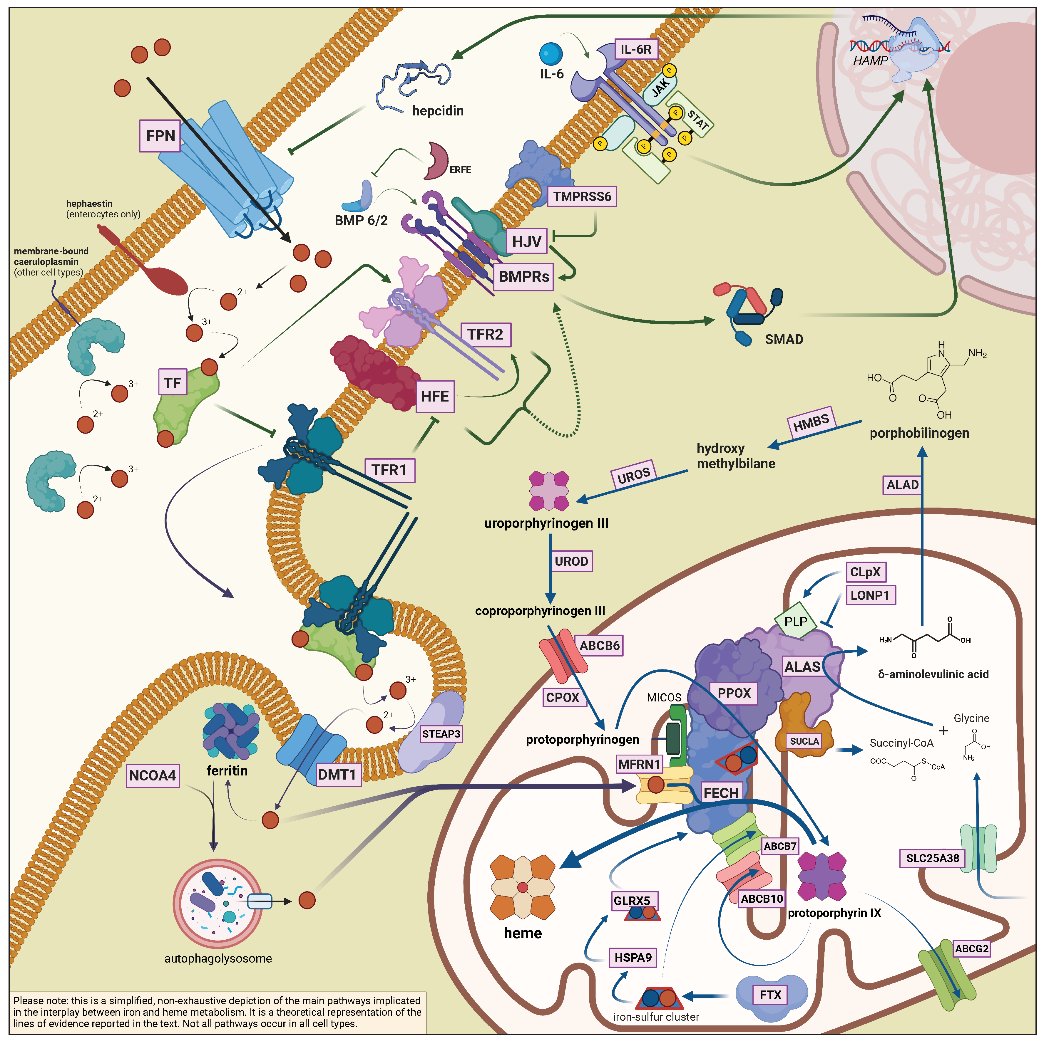
2. Role of Iron in the Biosynthesis of Heme

The main actors of iron and heme metabolism, which are mentioned in this section, are summarized in Table 1 and Table 2, with a non-exhaustive list of the known associated diseases. Figure 1 recapitulates the main metabolic pathways in the interplay between iron and heme biosynthesis (not all pathways occur in all cell types).

2.1. General Aspects of Iron Metabolism in Mammals

Under physiologic conditions, the total content of iron in the human body is about 3–4 g—less in females than males. While iron entry and mobilization are tightly regulated by several proteins, iron excretion is possible only through the turnover of the intestinal epithelium, skin cell or hair shedding, menstrual cycles, or blood losses. Crucially, the recycling of heme-bound iron represents one of the main sources for maintaining adequate supplies (together with mobilization of iron reserves from the liver), whereas the intestinal absorption of dietary iron, under physiologic conditions, only compensates for daily losses. In macrophages, heme recycling is realized through heme oxygenase (HO), which extracts iron in its ferrous form—releasing carbon monoxide in the process—and decomposes the protoporphyrin ring into carbon dioxide and biliverdin IXa—the latter is eventually reduced to bilirubin. With regards to dietary intakes, instead, non-heme iron is a substrate of divalent metal transporter 1 (DMT1) after being reduced to its ferrous form (Fe2+) by duodenal cytochrome B (DCYTB) at the apical membrane of duodenal and proximal jejunal enterocytes [7].
Whether from reticuloendothelial cells or enterocytes, iron is exported into the bloodstream by ferroportin (FPN1, coded by the gene SLC40A1)—the only known iron exporter [8]. This is a crucial step in the entire cycle of iron homeostasis, and the only which can be endogenously tuned in the exchange of iron with the environment: in fact, by regulating iron release from enterocytes and the reticuloendothelial system, ferroportin ultimately determines iron circulating levels. Ferroportin membrane expression can be chiefly modulated through vesicle internalization: in particular, hepcidin (HAMP) triggers ferroportin polyubiquitination by direct binding and thereby leads to its degradation in lysosomes [9]. More recently, it has been reported that hepcidin can occlude the central cavity of ferroportin, thus blocking iron export also by means of an endocytosis-independent mechanism [10].
To adequately regulate systemic iron homeostasis in physiological processes, hepatic HAMP expression is influenced by several factors, such as plasma and tissue iron levels, various erythropoietic stimuli (e.g., hypoxia or anemia), and infections or other causes of inflammation [11,12]. At the intracellular level, HAMP expression is chiefly regulated by the bone morphogenetic protein (BMP)/suppressor of mothers against the decapentaplegic (SMAD) signal transduction pathway [13,14].
It has been proposed that when tissue iron stores increase, liver sinusoidal endothelial cells (LSECs) enhance the expression of BMP6 and BMP2, which are thereby secreted and likely form heterodimers [15]. On the hepatocyte cell membrane, BMP6 and BMP2, most likely in the form of heterodimers, bind to a tetramer composed of BMP type I (activin receptor-like kinase—ALK-2 and 3) and type II (activin receptor—ACTR- 2A and BMP receptor 2) receptors and complexed with the co-receptor hemojuvelin (HJV). As a result, the intracellular signal transductors SMAD1, SMAD5, and SMAD8 are phosphorylated and bind SMAD4 to enter the nucleus and trigger HAMP expression. Transmembrane protease serine 6 (TMPRSS6), instead, has a negative effect on HAMP expression: when tissue iron levels decrease, TMPRSS6 cleaves HJV, decreasing hepcidin levels and rebalancing the iron stores [11,12,15]. When plasma iron levels increase, iron-loaded transferrin binds transferrin receptor 1 (TFR1) and transferrin receptor 2 (TFR2) on the hepatocyte membrane, inducing an increase in TFR2 protein stability and likely weakening the interaction between TFR1 and the human homeostatic iron regulator protein (HFE) [15]. As a consequence, HFE dissociates from TFR1 and forms a complex with TFR2 to start a signal transduction cascade to the HAMP gene, possibly interacting with HJV and the BMP/SMAD pathway and ultimately increasing hepcidin levels [11,12].
Moreover, hypoxia and anemia stimulate erythropoiesis and act as negative regulators of HAMP expression. In hypoxic or anemic conditions, the kidney produces higher amounts of erythropoietin (EPO), which in turn promotes erythroferrone (ERFE) production by red blood cell precursors. Then, ERFE binds BMPs, interfering with BMP-BMP receptor interaction and the following signaling cascade, thus resulting in decreased hepcidin levels [11,12]. Both infections and inflammation result in the secretion of interleukin 6 (IL-6) by macrophages. IL-6 induces the activation of the Janus kinase (JAK)-signal transducer and activator of transcription 3 (STAT3) signaling pathway in hepatocytes, leading to the upregulation of HAMP expression [11,12].
In the bloodstream, iron is taken up by TF after being oxidized to its ferric form (Fe3+) by membrane-bound hephaestin (HEPH) or the copper-carrying enzyme ceruloplasmin (CP). Transferrin is capable of carrying two atoms of ferric iron, which can be released to cells through endocytosis by binding to TFR1; during this process, iron is eventually reduced to its ferrous form by the metalloreductase six-transmembrane epithelial antigen of prostate 3 (STEAP3) and transported through DMT1. At the intracellular level, up to 4500 ferrous iron atoms can be stored in ferritin, which is composed of 24 subunits of light (FTL) and heavy chains (FTH1), differently combined. Iron uptake is dependent on FTL, whereas FTH1 displays ferroxidase properties [16]. The stored iron is then released and made available to the cell through lysosomal degradation of ferritin (ferritinophagy), in a process mediated by nuclear receptor coactivator 4 (NCOA4) [17].
Iron delivery to mitochondria could be accomplished through either a ferritin-dependent or a ferritin-independent pathway: in the former, a role in regulating the iron flux has been reported for NCOA4 and poly rC–binding protein 1 (PCBP1), a cytosolic iron chaperone which mediates iron delivery to ferritin [18]; in the ferritin-independent pathway, iron could be directly transferred to the mitochondrion from the outside through transferrin-containing endosomes [19,20]. Both pathways seem to play a role in erythropoiesis and heme biosynthesis [18,19,20].
Concerning the regulation of several proteins involved in iron homeostasis, a very versatile, post-transcriptional mechanism is represented by the interaction between iron regulatory proteins (IRP1 and IRP2) and the iron response element (IRE) in the 5 - or 3 -untranslated regions (UTRs) of mRNAs [21]. In conditions of iron deprivation, IRPs bind avidly to IREs. When intracellular iron is high, instead, IRP1 incorporates a [4Fe-4S] cubane cluster and gains aconitase activity, releasing the IRE, whereas IRP2 undergoes ubiquitination and proteosomal degradation. IREs, in turn, are highly conserved RNA stem-loops and differentially regulate the translation of their transcript according to where they are located. For instance, in the condition of iron deficiency, ferritin and ferroportin are negatively regulated by an IRP-IRE interaction in the 5 -UTR, while TFR1 and DMT1 translation is enhanced by IRP binding to the IREs in their 3 -UTRs. As explained in the following Section (see Section 3.3), this regulatory mechanism may be bypassed in the final steps of erythropoiesis to allow for the highly increased demand for iron required for hemoglobinization [22].
Among the several known disorders of iron metabolism, hemochromatosis (HC) is the most common inherited cause of primary iron overload [23]. A homozygous pathogenic variant (p.C282Y/p.C282Y) in HFE is the cause of the classic form of HC [24,25], whereas other rarer forms of HC are caused by pathogenic variants in other genes involved in iron metabolism, namely TFR2, HAMP or HJV; additionally, uncommon gain-of-function mutations in SLC40A1 which cause ferroportin to be resistant to hepcidin activity are associated to a phenotype indistinguishable from HC. Loss-of-function variants in SLC40A1, instead, give rise to a distinct form of iron overload (ferroportin disease). Furthermore, several other inherited conditions are associated with alterations in the metabolism of iron and iron overloads, such as iron deficiency–iron refractory anemia (IRIDA, caused by pathogenic variants in TMPRSS6), aceruloplasminemia or hypoceruloplasminemia, and iron-loading anemias [3,24,26,27].

2.2. The Interplay between Heme Biosynthesis and Iron

From a molecular standpoint, heme, as iron protoporphyrin-IX, is a cyclic tetrapyrrole and a coordination complex for diatomic gases (e.g., oxygen, carbon monoxide, nitric oxide), accurately engineered by evolution to exploit the redox properties of iron while minimizing its damaging effects on tissues. When incorporated into hemoglobin—or myoglobin—heme serves the irreplaceable function of the oxygen carrier, but this molecule is also fundamental, as a prosthetic group, to the correct functioning of several other hemeprotein, such as cytochromes, catalases, nitric oxide synthases, soluble guanylate cyclases or enzymes involved in the metabolism of some amino acids (tryptophan/serotonin, methionine/homocysteine, and others) [28]. In general, every cell in the body needs heme to carry out some vital metabolic processes.
At the same time, an excess of free heme can be toxic to tissues: several mechanisms of damage have been described [29,30], many of which culminate with the release of ROS through iron-catalyzed Fenton reactions. It should also be remarked that heme, as a lipophilic molecule, intercalates into lipid membranes, either reacting within the hydrophobic phospholipid bilayer and yielding lipoperoxides, or quickly entering cells, thus precipitating cytolysis or causing intracellular damage to proteins or DNA [29,30]. From a clinical standpoint, heme toxicity becomes a threatening issue in all those conditions which are burdened by hemolysis, such as sickle cell disease, β -thalassemia, malaria, ischemia-reperfusion, hemolytic-uremic syndrome, paroxysmal nocturnal hemoglobinuria or even severe sepsis [31], as well as rhabdomyolysis with myoglobinuria. As a detoxifying countermeasure, free heme can be efficiently scavenged by hemopexin, an acute phase plasma glycoprotein, and secondarily albumin [32]. The kidney is particularly susceptible to heme toxicity; in this regard, fundamental protection against heme-induced tubular damage is provided by the inducible form of heme oxygenase (HO-1), whose expression is enhanced, among others, by free heme itself, cytokines, and several oxidant species [33,34]. Anecdotally, a transient decrease in kidney function has been reported in a patient suffering from acute intermittent porphyria after therapy with heme arginate was vigorously implemented to treat a porphyric attack [35]. Other than the inducible HO-1, two other HO isoenzymes have been identified: in particular, HO-2 is a constitutive isoform that plays a role in the modulation of signaling to tissues through the synthesis of carbon monoxide, a diatomic gas with vasodilatory and neurotransmitting properties similar to nitric oxide [36,37].
The major heme-synthesizing organ in humans is, understandably, the bone marrow, which accounts for ∼80% of total heme production and requires around 20 mg of iron supply daily [22]; the liver, in turn, produces ∼15% of total heme in the body, followed, in terms of overall contribution, by the kidney [38,39].
Heme biosynthesis is realized through an eight-step metabolic pathway [28,40]: in animals, it starts with the rate-limiting enzyme aminolevulinate synthase (ALAS), which produces δ -aminolevulinic acid (ALA) from glycine and succinyl-CoA, in the so-called Shemin pathway. Two isoforms of ALAS exists: ALAS1 is constitutively expressed by every cell in the body, whereas ALAS2 is specific to erythroid tissues and is expressed at ∼30 fold higher levels than its counterpart in the liver [2]. Notably, the ALAS2 gene is on the X chromosome (Xp11.21). Both forms need pyridoxal phosphate (PLP) as a cofactor.
Consistent with their different evolutionary purposes, the two isoforms are regulated by different stimuli: ALAS1 is under a negative feedback by heme, whereas ALAS2 is repressed by iron deficiency. The translation of ALAS2 depends on iron availability through the presence of an IRE in the gene 5 -untranslated region (5 -UTR): in a setting of iron deficiency, IRPs bind to the IRE, inhibiting ALAS2 translation [41,42], even though this mechanism may be by-passed in the terminal steps of erythropoiesis (see Section 3.3) [22]. Recently, a new mechanism of post-translational regulation of ALAS1 has been proposed in human cells, involving the ATP-dependent proteases lon peptidase 1, mitochondrial (LONP1), and caseinolytic mitochondrial matrix peptidase chaperone subunit X (ClpX): both proteases seem to be involved in the negative feedback regulation of heme biosynthesis through heme-dependent degradation of ALAS1 [43,44]. Additionally, a significant increase in ALA levels has been observed in mouse erythroleukemia cells knock-out for the ClpX gene, together with decreased PPOX and FECH activities, resulting in anemia and PPIX accumulation—a phenotype reminiscent of erythroid protoporphyria. The authors of this study suggested that CLPX could also regulate ALAS2 turnover, increasing its proteolytic degradation, and concomitantly optimize PPOX and FECH activities with molecular mechanisms which still remain unknown [45]. Intriguingly, ClpX also promotes ALAS activity by catalyzing the insertion of PLP [46]. Thus, opposite regulatory mechanisms operate within the same molecular machinery, possibly allowing a better fine-tuning of the enzyme activity.
In the second step of heme biosynthesis, two molecules of ALA are condensed by ALA dehydratase (ALAD) to form porphobilinogen (PBG), which undergoes polymerization into hydroxymethylbilane (HMB) by HMB synthase (HMBS). Afterward, HMB undergoes cyclization by uroporphyrinogen III synthase (UROD) to yield uroporphyrinogen III, which is further modified to coproporphyrinogen III by uroporphyrinogen III decarboxylase (UROD), then to protoporphyrinogen by coproporphyrinogen oxidase (CPOX) and protoporphyrin IX by protoporphyrinogen oxidase (PPOX). Finally, in the last step of heme biosynthesis, an atom of ferrous iron is chelated into the protoporphyrin IX ring by the enzyme ferrochelatase (FECH). The first and last three steps of the pathway happen in the mitochondrion, the middle ones in the cytoplasm—in fact, coproporphyrinogen III is transported through the mitochondrial transported ABCB6, whereas the exact mechanisms underlying ALA translocation are not entirely clear. Noteworthy, ALAD, HMBS, and uroporphyrinogen III synthase (UROS) have both a “house-keeping” and an erythroid-specific promoter in their transcript, although only HMBS displays two different isoforms –erythroid and non-erythroid [2].
Ferrochelatase, a homodimeric enzyme located on the inner surface of the inner mitochondrial matrix [47], represents a fundamental crossroad between porphyrin and iron metabolism [48]. Notably, FECH activity is enhanced through a signaling pathway that culminates with FECH phosphorylation and is activated by erythropoietin (EPO) [49].
FECH possesses a [Fe-S] cluster, which confers structural stability to the enzyme [50] and possibly endows it with redox sensing functions [51]. Apparently, FECH receives iron from mitoferrin, a mitochondrial solute carrier that is essential for the proper assembling of heme [52]. Additionally, a role in the delivery of iron to FECH could be played by frataxin [53], which binds to FECH [54] but also contributes to the assembling of [Fe-S] clusters [55]. It also appears that the topological integrity of the mitochondrial cristae is essential to the correct functioning of FECH [56]. Even though FECH can accept several divalent metal ions as substrates, it is deemed that in vivo, only iron and zinc have access to it through mitochondrial transporters. Interestingly, some divalent heavy metals (e.g., Hg and Pb) can be accepted as a substrate, but the product of the chelation is poorly released from the enzyme [57].
Intriguingly, it has been reported that mice lacking one or two functional copies of the mitoferrin 1 (Mfrn1) gene in hepatocytes, when fed ALA, show an increase in protoporphyrin IX production, with histological liver alterations (chronic cholestasis, early-to-advanced fibrosis) reminiscent of erythropoietic protoporphyria (EPP) [58]. The authors of the study suggested that the observed protoporphyrin IX build-up could be chiefly caused by an insufficient iron supply to the mitochondria—due to a decreased/absent function of mitoferrin 1, which could not be replaced by mitoferrin 2. In mice not treated with ALA, alterations in Mfrn1 were not associated with signs of protoporphyria, possibly because of the Irp1-mediated suppression of Alas2—in fact, Mfrn1-deficient mice start to develop protoporphyrin accumulation when the interaction between Alas2 and Irp1 is impaired [59]. Furthermore, it has been shown that MFRN1 mRNA levels strongly correlate with FECH activity, both in conditions of normal and impaired FECH expression [60].
In the last few decades, many different proteins have been involved in the assembly and transport of [Fe-S] clusters: adenosine triphosphate binding cassette subfamily B member 7 (ABCB7) is the main mitochondrial [Fe-S] exporter to the cytosol; heat-shock protein family A member 9 (HSPA9) is a mitochondrial chaperone essential to the transfer of newly formed [2Fe-2S] clusters to glutaredoxin 5 (GLRX5); GLRX5, in turn, represents the immediate donor of [2Fe-2S] clusters to target apoproteins, such as FECH, respiratory complex proteins, or IRP1 cytosolic aconitase. Defects in the production or activity of each of these proteins are responsible for an impairment in the functioning of [Fe-S] clusters, which causes an increased iron uptake and reduced iron utilization, ultimately leading to the clinical picture of sideroblastic anemias (see Section 4.1.1 and Section 4.2.2) [61].
It has been recently reported that at least ferrochelatase, protoporphyrinogen oxidase, and aminolevulinic acid synthase-2 form a mitochondrial complex (metabolon) [62], which may possibly include additional factors involved in the transport of iron (mitoferrin) and porphyrin (ABCB10), [Fe-S] cluster biogenesis (ABCB7), and ALAS2 substrate synthesis (succinyl-CoA synthetase) [62,63,64,65]. Topological proximity is certainly convenient for all these factors which handle iron within the mitochondrial matrix in that it may constitute a safeguard mechanism against the excessive spillover of such potentially toxic element in a milieu very sensitive to oxidative damage.
Table 1. Main actors of iron metabolism in mammals.
ProteinGeneFunctionAssociated Diseases
Bone morphogenetic protein 2BMP2Ligand of the BMP-SMAD signaling pathway regulating hepcidin expression in response to iron
Bone morphogenetic protein 6BMP6Ligand of the BMP-SMAD signaling pathway regulating hepcidin expression in response to ironBMP6-associated iron-overload
CaeruloplasminCPSoluble/membrane-bound copper-carrying ferroxidaseAceruloplasminemia
Divalent metal transporter 1DMT1 or SLC11A2Ferrous iron importerDMT1 deficiency
Duodenal cytochrome BDCYTBReduces dietary ferric iron to ferrous form at the apical border of enterocytes
ErythroferroneERFEHepcidin inhibitor, produced by the bone marrow in response to erythropoietin
Ferritin heavy chainFTH1Subunit of ferritin, with ferroxidase activityFTH-related iron-overload
Ferritin light chainFTLSubunit of ferritin, with iron storage propertiesHyperferritinemia-cataract syndrome Hereditary benign hyperferritinemia Neuroferritinopathy L-ferritin deficiency [66]
FerroportinSLC40A1Ferrous iron exporterFerroportin disease (loss-of-function)SLC40A1-related hemochromatosis (gain-of-function)
FrataxinFXNIron carrier, participates in iron-sulfur cluster biogenesisFriedreich’s ataxia
HaephastinHEPHMembrane-bound ferroxidase
HemojuvelinHJV or HFE2BMP co-receptor, involved in the iron-sensing pathway which regulates hepcidinHJV-related hemochromatosis
HepcidinHAMPIron regulating hormone; internalises ferroportinHAMP-related hemochromatosis
Human homeostatic iron regulator proteinHFEProtein involved in the iron-sensing pathway which regulates hepcidinHFE-related HH
IRE binding protein 1IREB1 or IRP1 or ACO1Iron-sensing regulator of translation; aconitase activity in the presence of iron
IRE binding protein 2IREB2 or IRP2Iron-sensing regulator of translationIRP2-related protoporphyria
MatriptaseTMPRSS6Cleaves membrane-bound HJV; negative regulator of hepcidin in response to iron deficiencyIron-deficient iron refractory anaemia (IRIDA)
Mitoferrin-1MFRN1Intramitochondrial iron carrier
Nuclear receptor coactivator 4NCOA4Delivers ferritin to autophagolysosomes (ferritinophagy)
Six-transmembrane epithelial antigen of prostate 3STEAP3Membrane-bound metalloreductaseSideroblastic anaemia with primary hypogonadism
TransferrinTFFerric iron carrierHypo/Atransferrinemia
Transferrin receptorTFRCReceptor for endocytosis-mediated iron uptake; one of the plasma iron sensorsTFRC-related combined immunodeficiency [67]
Transferrin receptor 2TFR2Protein involved in the iron-sensing pathway which regulates hepcidinTFR2-related hemochromatosis
Table 2. Main actors of heme metabolism in mammals.
ProteinGeneFunctionAssociated Diseases
δ -aminolaevulinate dehydrataseALADDehydrates ALA to yield PBGALAD-deficiency (Doss) porphyria
δ -aminolaevulinate synthase 1ALAS1Condenses glycine and succinyl-CoA to yield ALA
δ -aminolaevulinate synthase 2ALAS2Condenses glycine and succinyl-CoA to yield ALA (erythroid-specific isoform)X-linked congenital sideroblastic anaemia (loss-of-function) X-linked erythropoietic protoporphyria (gain-of-function)
ATP-binding cassette super-family B member 6ABCB6Imports porphyrins into mithocondriaPhenotype modifier in porphyrias [68]
ATP-binding cassette super-family B member 7ABCB7Mitochondrial [Fe-S] cluster exporterX-linked sideroblastic anaemia with ataxia
ATP-binding cassette super-family B member 10ABCB10Mitochondrial exporter with putative roles in porphyrin or iron transport; complexes with MFRN1 and FECH to enhance heme biosynthesis
ATP-binding cassette super-family G member 2ABCG2Cytosolic and mitochondrial exporter of protoporphyrin IX; also involved in the export of heme
caseinolytic mitochondrial matrix peptidase chaperone subunit XCLpXMitochondrial protein with ATP-dependent protease and unfoldase activity; regulates ALAS turnover; activates ALAS catalyzing PLP insertionPhenotype modifier in protoporphyria (see Section 3.3)
coproporphyrinogen III oxidaseCPOXEliminates two carboxyl groups from coproporphyrinogen III side chains to yield protoporphyrinogen IXHereditary coproporphyria (autosomal dominant) Harderoporphyria (autosomal recessive)
ferrochelataseFECHChelates iron into protoporphyrin IX to yield hemeErythropoietic protoporphyria
glutaredoxin 5GLRX5Mitochondrial protein with thiol reductase activity; involved in [Fe-S] cluster assemblyAutosomal recessive sideroblastic anaemia
hydroxymethylbilane synthaseHMBSCondensates four PBG molecules into HMBAcute intermittent porphyria (AIP)Autosomal recessive AIP
heme oxygenase 1HO-1Cleaves heme into biliverdin IX α , releasing CO and ferrous iron (inducible isoform)HO-1 deficiency
heme oxygenase 2HO-2Cleaves heme into biliverdin IX α , releasing CO and ferrous iron (constitutive isoform); involved in the CO signalling pathway
hemopexinHPXHeme scavenger in the plasma
heat shock protein family A member 9HSPA9Mitochondrial protein with chaperone activity for [Fe-S] clustersAutosomal recessive sideroblastic anaemia
lon peptidase 1, mitochondrialLONP1ATP-dependent protease involved in the turnover of mitochondrial matrix proteinCODAS (Cerebral, Ocular, Dental, Auricular and Skeletal) syndrome [69]
protoporphyrinogen oxidasePPOXDehydrogenates protoporphyrinogen IX to yield protoporphyrin IXVariegate porphyria (VP) Autosomal recessive VP
mitochondrial solute carrier family 25 member A38SLC25A38Mitochondrial glycine transporterAutosomal recessive sideroblastic anaemia
succinyl-CoA synthaseSUCLAControls the flux of heme precursors catalyzing a reversible conversion from succinate + coenzime A to succynil-CoA (precursor of ALA)
uroporphyrinogen III decarboxylaseURODEliminates four carboxyl groups from uroporphyrinogen III side chains to yield coproporphyrinogen IIIPorphyria cutanea tarda (sporadic or familial)Hepatoerythropoietic porphyria
uroporphyrinogen III synthaseUROSConverts linear PBG to cyclic uroporphyrinogen IIICongenital erythropoietic porphyria (Günther disease)

3. Clinical and Experimental Aspects of the Role of Iron in Porphyrias

3.1. Porphyria Cutanea Tarda

Porphyria Cutanea Tarda (PCT) exists both as an inherited disorder (familial PCT, F-PCT), caused by a heterozygous mutation of uroporphyrinogen III decarboxylase (UROD), and as an acquired entity (sporadic PCT, S-PCT) [70]. More than two-thirds of patients with PCT belong to the latter group, and even those with a pathogenic variant of UROD do not usually develop F-PCT unless in the presence of other factors [71]. Namely, PCT is strongly associated with conditions of iron overload [72,73,74,75], such as hemochromatosis, hepatitis C virus infection, and alcoholism. PCT is an almost exclusively cutaneous disorder, presenting with sunlight-sensitive, painful, blistering lesions (bullae, erosions) which heal slowly and leave scars, most typically in the back of hands or around the mouth; hyperpigmentation or hypertrichosis can occur as well.
Patients with PCT almost universally benefit from iron depletion through phlebotomy, whereas iron supplementation therapy unleashes or exacerbates the symptoms [75,76,77]. Beneficial effects were also noticed after implementing some dietary changes, i.e., with a vegetable-fruit, low-calorie (∼500 Kcal/day) diet, which would reduce the total iron content of the body and improve symptoms and biochemical signs of PCT [78].
Compared to non-porphyric controls, significant differences have been found in PCT patients regarding the presence of variants associated with HFE-related HC [74,79,80,81,82,83,84]. One study found that ∼20% of PCT patients were homozygous for the pathogenic variant p.C282Y∖p.C282Y in the HFE gene (the typical genotype associated with the risk of developing HFE-HC [25]), ∼7% were compound heterozygous p.C282Y∖p.H63D (which can predispose, in the presence of other acquired factors, to a phenotype reminiscent of an “intermediate”, milder form of HC). Consistent with this, the frequency of heterozygous p.C282Y, p.H63D, or homozygous p.H63D∖p.H63D genotypes (which are not usually associated per se to pathologic iron accumulation) were not significantly different between PCT patients and controls [74]. All patients with PCT had clear biochemical or histological signs of iron overload, with homozygous p.C282Y∖p.C282Y patients with PCT displaying the worst phenotype. Additionally, the allelic frequency of the p.H63D variant was significantly higher in chromosomes from F-PCT compared to S-PCT patients [74].
Interestingly, a useful animal model for studying PCT is represented by Urod + , Hfe mice, which are also carriers of a homozygous null mutation in Hfe, other than a heterozygous null mutation in Urod [85].
Even though hepatic UROD is produced at half the normal levels in F-PCT patients, both forms of PCT present a reduction in hepatic UROD activity to ∼25% [86,87,88], leading to suspect the presence of an inhibiting compound. In fact, it has been demonstrated that uroporphyrinogen undergoes iron-dependent partial oxidation of one bridge carbon of the tetrapyrrolic macrocycle to yield uroporphomethene, a competitive inhibitor of UROD [88], in a reaction likely catalyzed by cytochrome 1A2 of the P450 family (CYP1A2) [88,89].

3.2. Congenital Erythropoietic Porphyria

Congenital erythropoietic porphyria (CEP)—also known as Günther’s disease—arises from a biallelic impairment of uroporphyrinogen III synthase (UROS)—or, less frequently, X-linked hematopoietic transcription factor GATA-binding factor 1 (GATA1) [90,91]. In CEP, UROS deficiency reroutes the metabolic pathway of hydroxymethylbilane towards non-enzymatic cyclisation, which yields porphyrin isomer I metabolites - uroporphyrinogen I and coproporphyrinogen I. The latter cannot be further processed by coproporphyrinogen III oxidase -CPOX, an enzyme stereospecific for the III isomer- and both metabolites are non-enzymatically oxidized to uroporphyrin I and coproporphyrin I, respectively [91,92].
In CEP, the severity of the clinical manifestations varies widely, ranging from mild photosensitivity, with cutaneous symptoms reminiscent of PCT, to devastating blistering lesions leading to deformities by photomutilation (i.e., loss of fingers, eyelids, ears, as well as other sunlight-exposed areas). Anemia is present as the result of both intravascular hemolysis, which can be very severe, and ineffective erythropoiesis. Of note, a few cases have been described of patients with hematological disorders in whom the replication of clonal myeloid subsets with somatic mutations resulted in acquired phenotypes similar to CEP [93,94]. Liver involvement is unusual, although hepatopathy caused by fibrosis and signs of diffuse intrasinusoidal extramedullary hematopoiesis (characterized by the presence of erythroid precursors within markedly congested and dilated sinusoids), possibly a consequence of moderate to severe chronic anemia, has been described [95]. In patients with CEP, bone marrow transplantation is potentially curative and should be considered, in the presence of a suitable donor, according to the phenotype, genotype, and biochemical profile of the disease [96].
Until recently, most of the available therapeutic options were based on a pioneering report of a patient whose symptoms of CEP could be satisfactorily managed with hypertransfusion therapy (blood hematocrit above 39%), aiming at nearly complete suppression of endogenous erythropoiesis, coupled with iron chelation by deferoxamine to manage transfusion-related iron overload [97]. In this patient, the urinary iron excretion was maximal and the urinary porphyrin content was minimal after 6 days since blood transfusion (under deferoxamine): this lag would account for the time required for the bone marrow to adapt and suppress endogenous erythropoiesis after transfusions [97]. Similar to patients with transfusion-dependent thalassemias, liver and heart iron overload with high levels of serum iron and ferritin can be burdensome complications of chronic hypertransfusion, notwithstanding iron chelation therapy [96]. Furthermore, at least one case has been described concerning a pediatric CEP patient with transfusion-related kidney iron overload and nephrotic syndrome [98].
Quite recently, it has been first reported the case of a CEP patient who underwent a remarkable improvement in her symptoms of porphyria in the setting of iron depletion caused by gastrointestinal bleeding [95]. Following this anecdotal finding, the patient started an off-label iron-chelating therapy with deferasirox, which effectively reduced uroporphyrin and coproporphyrin urinary levels and alleviated the disease phenotype [95]. In vitro studies on the bone marrow from the patient (or her sibling) showed that partial iron restriction, obtained by an intermediate ratio of holo- to apo-transferrin in the culture medium, was the most effective in allowing the maturation of UROS-deficient erythroid cells [95]. Furthermore, a decrease in ALAS2 activity and porphyrin levels could be observed, in the same cell population, after iron deprivation in vitro [95].
These findings have proved instrumental in disclosing the possibility of inducing iron deficiency in patients with CEP to alleviate the disease phenotype. Given the higher risk of adverse events associated with iron chelation, iterative phlebotomies have been proposed as a safer means to achieve iron depletion -understandably, this approach is only feasible in patients without severe anemia [6,99,100]. To avoid an abrupt compensatory induction of erythropoiesis, gradual increases in the volume of blood withdrawn during phlebotomies has sometimes been attempted, starting from small quantities [99].
Concerning the physiopathology of the disease, interesting results have been disclosed in vivo by comparing the phenotype of iron overload, with signs of severe hemolytic anaemia, in a CEP mouse model [101] with models of hemojuvelin (Hjv )-related hemochromatosis [102]. While Hjv mice displayed severe iron overload in hepatocytes, with no iron deposits in the spleen, in animals with CEP the iron accumulation was observed predominantly in Kupffer cells, with spleen enlargement as a manifestation of compensatory extramedullary erythropoiesis, and high levels of total iron content in the spleen. Although high levels of ferritin could be measured in both models, both serum iron and transferrin were elevated in CEP mice, with normal transferrin saturation, whereas Hjv mice showed high transferrin saturation. Importantly, CEP mice displayed early signs of iron deficiency in the course of erythropoietic differentiation, such as low reticulocyte hemoglobin content, as well as signs of enhanced compensatory erythropoiesis, such as an increase in erythropoietin levels and bone morphogenic protein 4, and suppression of hepcidin. Notably, in CEP models, ferroportin was increased in enterocytes and liver, and an accumulation of iron in the kidney cortex, most pronounced in proximal tubules, was highlighted by Perl’s staining. In sum, an efficient erythroid response to intravascular hemolysis could be observed in this particular CEP mouse strain, albeit a following study of the same group showed that different patterns of iron overload and alterations of iron metabolism can be observed in different CEP mouse strains, according to their specific pathogenic variant [103].
It has been suggested that ALAS2 expression, which is dependent on iron status through the IRP/IRE system, can act as a disease modifier in CEP [104,105], thus suggesting a rationale for therapeutic iron depletion. Interestingly, delivery of short hairpin RNAs through lentiviral vectors has been attempted to silence ALAS2 or HMBS in UROS-deficient CD34+ cells derived from a CEP patient: this RNA interference approach yielded a significant reduction in porphyrin overload and disclosed a correlation between porphyrin accumulation and ALAS2 expression [106]. Similar results were also obtained with the oral iron chelator deferiprone, which could also reverse the skin symptoms, hematological abnormalities, and liver/spleen lesions (decrease in erythroid clusters from extramedullary erythropoiesis) in CEP mouse models [106].

3.3. Erythropoietic and X-Linked Protoporphyria

In the final step of heme biosynthesis, iron is complexed into a protoporphyrin IX ring by the enzyme ferrochelatase (FECH) [107].
Patients with erythropoietic protoporphyria (EPP) carry an inactive copy of the FECH gene in one allele, together with an in trans intronic single-nucleotide variant (c.315-48T>C, previously known as IVS3-48T/C), which corresponds to a common hypomorphic haplotype [108]. Therefore, EPP displays an autosomal pseudo dominant pattern of inheritance, with patients being usually compound heterozygous even though homozygous genotypes have been described [108,109]. Patients with EPP have a FECH activity which is ∼10–30% of the normal [108]. A gain-of-function mutation of ALAS2, instead, gives rise to X-linked protoporphyria (XLP), a rarer condition which has been discovered only in 2008 and presents with almost identical clinical findings. In both EPP and XLP, protoporphyrins accumulate in multiple sites, most notably the bone marrow, the liver, and the skin. Consistent with this, patients with EPP and XLP present with mild microcytic hypochromic anaemia and thrombocytopenia, which were found to be strongly correlated with higher erythrocyte protoporphyrin IX content [110]; liver disease, which is caused by accumulation in the biliary system of hydrophobic protoporphyrins may precipitate a life-threatening acute cholestatic hepatitis in a small percentage of cases and cutaneous photosensitivity symptoms, perhaps the most burdensome for patients, may suffer from a range of painful, burning skin reactions when exposed to sunlight or artificial light of a particular wavelength (e.g., emitted by surgical lamps which are not filtered on purpose) [107,110,111,112,113].
Several modifier genes have been identified, which may contribute to the severity of the phenotype of protoporphyrias: in particular, an abnormal transcript of mitoferrin-1 has been identified in patients with a known mutation leading to EPP or XLP, as well as some patients with a phenotype consistent with protoporphyria but no other identified mutation [114].
Differently from other conditions, such as lead intoxication, primary iron deficiency, or other red blood cells disorders, the protoporphyrins which accumulate are metal-free, rather than zinc-chelated; in XLP, zinc-protoporphyrins usually exceed 15% of the total, whereas in EPP they are less than 15%. This implies that the levels of zinc-protoporphyrins cannot be fully relied upon to grade the severity of iron deficiency in these patients.
Both EPP and XLP patients often present with signs of iron deficiency, such as low serum ferritin levels (often lower than 20 ng/mL) which may be refractory to iron supplementation [111,112,115,116,117]. An early study estimated that EPP patients could not have more than 250 mg of iron stores which could be readily mobilized after phlebotomy [72]. Several studies reported a strong association between serum ferritin and hemoglobin levels, together with the absence of significant signs of chronic disease anemia [110,116,118]. At least in humans, ringed sideroblasts and mitochondrial iron deposition have been reported [119].
Interestingly, it has been highlighted that soluble transferrin receptor and serum iron levels are often unaltered in patients with EPP [110,115,116,118], which led some authors to conclude that iron deficiency would not be a strong limiting factor in the synthesis of hemoglobin and erythropoiesis [110]. Noteworthy, it has been shown that hepcidin levels in EPP patients are decreased as an appropriate measure to iron deficiency [115,116], and gastrointestinal absorption of ferrous sulfate is normal [115], which would exclude an impairment in the intestinal absorption of iron [115]. In this regard, a mouse model of EPP failed to show any improvement in the biochemical and histological signs of the disease after being treated with an RNA interference agent targeting TMPRSS6 to mimic a condition of IRIDA, in which hepcidin is up-regulated [120].
Intriguingly, it has been hypothesized that, at least in FECH-deficient mice, a compensatory enhancement of extramedullary erythropoiesis causes a redistribution of iron reserves from peripheral tissues to the spleen while maintaining intact the total iron content of the body [121].
It has also been shown that serum transferrin levels positively correlate with erythrocyte protoporphyrin IX content [110], which would corroborate the hypothesis, first proposed in FECH-deficient mice [121], that protoporphyrin IX may induce the hepatic synthesis of transferrin, functioning as a sensor of intracellular iron shortage to the erythroid lineage. Increased protoporphyrin IX levels have also been significantly associated with male gender and growth/differentiation factor 15 (GDF15) levels [116].
Of note, phlebotomy has been attempted in patients with EPP and severe liver damage, in an approach similar to CEP, with promising results in terms of reduction in serum protoporphyrin levels and markers of liver injury: understandably, these findings were paralleled by a decrease in hemoglobin and serum ferritin [122].
The effects of iron supplementation therapy in EPP and XLP are controversial [4,5,6]. Early reports mention a beneficial effect of iron supplementation on EPP symptoms, as well as protoporphyrin and transaminase levels [123,124,125], whereas other authors warned against a worsening of cutaneous and hepatic symptoms [126,127,128]. Some beneficial effects of iron supplementation have been observed in patients with XLP [117] and it has been claimed that the clinical and biochemical improvement previously reported in patients with EPP was due to them having XLP instead [117,129]. In the latter condition, iron supplementation would enhance the activity of a normally functioning FECH, coupling with the substrate excess of protoporphyrin IX and converting it to heme [117,129]. On the other hand, benefits from iron supplementation in EPP patients could also derive from chelation of protoporphyrins in the guts in the context of their enterohepatic circulation, non enzymatic intracellular chelation, or iron-induced enhancement of FECH activity [117,125].
From a biochemical perspective, iron would enhance the activity of both ALAS2—through the IRP/IRE regulatory system—and FECH—which requires iron both as a substrate and for its [Fe-S] clusters. Therefore, iron supplementation may be beneficial or detrimental according to the ratio of activation between the two ends of the heme biosynthetic pathway. In human erythroleukemic cells, it has been demonstrated that iron chelation increases the mRNA levels of ALAS2, but not FECH, while decreasing the protein levels of both. Thus it has been suggested that ALAS2 transcription may be induced—through as-yet-unknown mechanisms—in conditions of impaired FECH activity, but ALAS2 translation would still be stopped by means of the IRP/IRE interaction. In EPP, iron depletion would then result in a safeguard mechanism, as it would impede ALAS2 overexpression with a subsequent increase in protoporphyrin IX levels [130].
Interestingly, it has been reported that in the terminal steps of erythroid maturation, the IRP/IRE regulatory mechanisms may be bypassed to allow high expression of both ALAS2 and TFR1 while maintaining low ferritin levels [22]. In this study, the authors used primary mouse erythroid cells, which allowed them to closely follow the terminal steps of erythroid differentiation, including full hemoglobinization and enucleation [22]. In particular, a difference could be noticed between self-renewing erythroid progenitors, which maintain the precursor cell pool and are under the classic iron-dependent IRP/IRE regulation, and maturing erythroblast, which instead show an uncoupling between ALAS2, ferritin, and TFR1 expression. From a physiologic perspective, this would permit us to reach the saturating levels of iron needed for massive hemoglobinization in the terminal stages of erythropoiesis while not wasting resources in iron storage, which would be futile at this stage [22]. It may be speculated whether an exacerbation of such mechanisms may intervene in the context of EPP, in which very low ferritin levels may be an expression of enhanced, but poorly effective, erythropoiesis. In fact, it has been reported an enhanced expression of ALAS2 in young erythrocytes and higher levels of ALAS2 mRNA in the peripheral blood cells of EPP patients, which would be unexpected given their putative status of iron deficiency [129]. Moreover, FECH inhibition through N-methyl-protoporphyrin led to an increase in ALAS2 mRNA in an in vitro model of erythroleukemic cells [129], a finding which was also reported in mice fed with griseofulvin (which is converted to N-methyl-protoporphyrin in the liver) [131]. As mentioned in Section 2.2, a putative uncoupling mechanism could involve CLPX-dependent regulation of ALAS2 [132]. In this regard, a clinical and biochemical phenotype consistent with protoporphyria (including sideropenic anemia) has been recently reported in a female patient who harbored two different mutations: one in the IRE of ALAS2, inherited from the maternal lineage (whose members were asymptomatic), and another in the CLPX gene, which was shared with the patient’s father and uncle (who presented both with high protoporphyrin levels, but only mild photosensitivity) [133,134]. Functional studies on this particular mutation in CLPX suggested that the mutated protein may form heterohexamers with the wild-type and act as a dominant-negative to impair ALAS turnover, thus increasing protoporphyrin IX levels as well as ALAS protein levels and activity [133].

IRP2-Related Protoporphyria

It is worth mentioning that mice which lack IRP2 expression (IRP2 ) display a peculiar phenotype, featuring Alas2 overexpression, markedly elevated levels of protoporphyrin IX (metal-free > zinc), and an increase in levels of Hmbs mRNA in the spleen, with also skin and eye lesions [135]. Additionally, IRP2 mice show microcytic anaemia with severe iron depletion in the bone marrow and markedly reduced levels of transferrin receptor in erythroid precursors, albeit with hyperferritinemia and percentages of transferrin saturation comparable to wild-type controls. Several of these manifestations are more severe when also one IRP1 allele is non-functional (IRP1 + IRP2 ) [135]. The most notable symptom in IRP2-deficient mice, though, is adult-onset neurodegeneration, likely caused by an abnormal regulation of iron metabolism, with axonal degeneration, neuronal loss, and signs of iron overload in axonal structures and oligodendrocytes [136,137]. Intriguingly, the total content of iron in the brain and spleen is unchanged relative to controls, whereas the total content of iron in the liver is increased [135].
In 2019, the first case of a human with bi-allelic loss of IREB2 (coding for IRP2) was described, recapitulating the phenotype observed in animals: the patient was a 16-year old with neurodegeneration, microcytic hypochromic anemia refractory to iron supplementation, mildly elevated ferritin levels with normal serum transferrin and iron levels, low transferrin receptor expression, and a marked increase in zinc-protoporphyrin IX levels. Strikingly, the clinical picture could be reversed by lentiviral gene therapy, which restored IRP2 expression. No photosensitivity has been reported in this patient [138].

3.4. Acute Hepatic Porphyrias

Acute hepatic porphyrias (AHPs) are caused by heterozygous pathogenic variants of HMBS (acute intermittent porphyria—AIP– which is the most common), CPOX (hereditary coproporphyria—HCP), PPOX (variegate porphyria—VP), or, in the case of the exceedingly rare ALAD-deficiency porphyria, by pathogenic variants of ALAD in homozygosis [139]. Of note, lead intoxication acts through the steric hindrance of ALAD, thus recapitulating the corresponding inherited condition [140].
Patients with AHPs are at risk of acute neurovisceral attacks (acute porphyric attacks—APAs), which can pose a serious threat in terms of survival or long-term impairment. APAs can be unleashed by exogenous or endogenous stimuli that supposedly increase the metabolic demand for heme, thus enhancing ALAS expression. As a consequence, given the enzymatic deficiency down the biosynthetic pathway, heme precursor levels increase manifold. Consistent with the enzymes involved, in AIP only ALA and PBG accumulate, whereas in HCP and VP there is also an increase in cyclic porphyrin levels, which gives rise to phototoxic cutaneous manifestations reminiscent of PCT. Patients with AHPs are also burdened by long-term complications, such as arterial hypertension, chronic neuropathy, porphyria-associated kidney disease, or non-cirrhotic hepatocellular carcinoma, which can be further debilitating [141].
The build-up of non-porphyrin heme precursors (ALA and PBG) is deemed to be a major mechanism of damage in AHPs [140], albeit relative heme deficiency may also play a role [142]. In the presence of iron and oxygen, ALA undergoes a phosphate-catalyzed auto-enolization to yield the oxidizing species superoxide anion (O2·), HO· radical, and ALA enoyl radical (ALA·) [143,144]. In the following step, ALA· is oxidized by iron to 4,5-dioxovaleric acid (DOVA), a DNA alkylating agent [145] to which guanine moieties are particularly susceptible in the presence of ferritin [146]. Crucially, it has been shown that the ALA enoyl radical causes ferritin to release Fe2+ ions [144,147]. In fact, ALA-driven oxidation may selectively damage the tryptophan and cysteine moieties of apoferritin, thus altering its secondary and tertiary structure and impairing FTL-dependent iron uptake (but not FTH1-dependent ferroxidase activity) [148].
Additionally, in the brain of AIP animal models (either rats treated with succinyl acetone methyl ester or intraperitoneally injected with ALA), increases in total non-heme iron, ferritin, and lipid peroxidation have been reported, together with enhanced antioxidant activity of the copper-zinc superoxide dismutase [149,150]. Higher non-heme iron levels have also been observed in the liver of AIP animal models [147].
The oxidative damage exerted by ALA may have more general effects on iron homeostasis as well as incubation with ALA increases IRP1 activity: in this regard, a cell-permeable antioxidant as N-acetylcysteine was able to revert this condition, in contrast with extracellular-acting catalases or superoxide dismutases [151].
Additionally, ALAS1 activity is enhanced by fasting through the key action of a transcriptional coactivator, peroxisome proliferator-activated receptor-gamma coactivator 1 (PGC-1 α ) [152]; at the same time, signalling through PGC-1 α enhances hepcidin expression during starvation, with subsequent ferroportin degradation, low serum iron levels, and iron accumulation in the liver [153].
In AHPs, the negative feedback exerted by heme on the expression of ALAS1 also has a clinical significance: acute porphyric attacks can be managed with heme arginate (hemin) infusions, which downregulate ALAS1 and reduce circulating ALA levels. Until recently, off-label prophylactic hemin infusions have been the only therapeutic option for patients with several or markedly severe APAs. As a foreseeable complication of this approach, patients under hemin maintenance therapy are prone to develop (liver) iron overload with high serum ferritin levels.
In recent years, a novel siRNA-based agent, givosiran, has been approved for the treatment of AHPs [154]. Givosiran exploits the RNA-induced silencing complex (RISC) machinery to inhibit ALAS1 mRNA translation. Treatment with givosiran has been very effective in reducing the annualized rate of APAs and improving the overall quality of life of patients with AHPs [154,155,156], although some adverse effect must be taken into consideration and caution should be warranted as to the occurrence of unexpected others. Of interest, at least some adverse events may be possibly related to drug-induced relative heme deficiency in its non-hematopoietic routes of utilization: for instance, an impairment in heme biosynthesis induced by givosiran may play a role in the observed alterations of homocysteine catabolism [157,158,159,160,161,162,163], cytochrome P450 function [164], or nitric oxide synthase-regulated kidney microcirculation [141,165].

4. Clinical and Experimental Aspects of the Role of Iron in Congenital Sideroblastic Anaemias

Congenital sideroblastic anemias (CSAs) are a group of rare anemias characterized by both an altered use of mitochondrial iron by erythroid precursors and an accumulation of ring sideroblasts in the bone marrow [3].
CSAs are caused by pathogenic variants in several genes involved in the biosynthesis of heme (ALAS2, SLC25A38, and FECH), [Fe-S] cluster assembly (ABCB7, GLRX5, HSPA9, HSBC), as well as the translation of other mitochondrial proteins (TRNT1, PUS1, YARS2, SLC19A2, NDUFB11). The latter group is mainly responsible for syndromic sideroblastic anemias, which are usually associated with milder anemia without systemic iron overload, and whose description goes beyond the aim of this review [166,167]. Instead, an overview of CSAs caused by defects of [Fe-S] cluster assembly is provided, as increasing lines of evidence have supported the direct role of [Fe-S] cluster biogenesis in the heme biosynthetic pathway, e.g., by affecting the supply of succinyl-CoA substrate, the IRP-mediated regulation of ALAS2 translation, and the post-translational regulation of FECH activity [50,168].
The most common form of CSA is ALAS2-related, X-linked sideroblastic anemia (40% percent of all CSA cases), followed by autosomal recessive sideroblastic anemias (ARCSA), which account for 35–40% of cases. Pathogenic variants in SLC25A38 cause <10% of all CSA cases [169].
Mitochondrial iron overload occurs in all sideroblastic anemias: in sideroblasts (which are erythroblasts containing iron inclusions) iron deposits in perinuclear mitochondria and is visible as a “ring” when cells are stained with Perl’s blue. Ringed sideroblasts may also be present in other conditions characterised by alterations in iron metabolism, such as myeloproliferative disorders or copper deficiency [170].
It has been demonstrated that in sideroblastic anemias, most of the iron in excess is contained in mitochondrial ferritin (MtF)—a mechanism deemed to protect organelles from oxidative damage [171,172]. Even though the translation of cytosolic ferritin is regulated by means of an IRP1/IRE interaction, a recognizable IRE has not been identified in the gene for Mtf. In analogy with frataxin down-regulation in Friedreich ataxia, feedback inhibition of the MtF gene (FTMT) by protoporphyrin IX synthesis has been proposed [173].
A common pathophysiologic feature of CSAs is the iron-driven increase of ROS in mitochondria, leading to cell damage and impaired erythropoiesis. Although deposition of non-heme iron in the mitochondria of erythroid precursors is present in all cases of CSA, systemic iron overload does not always develop in untransfused patients but occurs mainly when the inherited defect is restricted to the erythroid cell line (XLSA, GLRX5, and SLC25A38 mutations), as a consequence of inefficient erythropoiesis and increased iron absorption [3]. The main site of iron accumulation is the liver, which in untreated patients is at risk of chronic damage—progressing to fibrosis and cirrhosis—whereas cardiac iron overload is much rarer [170].

4.1. ALAS2-Related X-Linked Sideroblastic Anaemia

First described by Cooley in 1945 [174], the X-linked sideroblastic anemia (XLSA) related to pathogenic variants in the erythroid-specific form of aminolevulinate synthase (ALAS2) is the most common form of CSA [169]. The prevalence of XLSA is unknown: about 200 cases and fewer than 100 unrelated probands have been described in the literature [166].
In the last few decades, more than 100 different pathogenic variants have been reported, most of which are missense, loss-of-function mutations affecting the ALAS2 catalytic domain, the interaction site between ALAS2 and succinyl-CoA synthetase, or the binding domain for pyridoxal phosphate [169,175]. The presence of the latter mutation site accounts for pyridoxin-responsiveness in about two-thirds of patients with XLSA [176]. Additionally, pathogenic variants in the ALAS2 promoter have been reported in about 5% of congenital sideroblastic anemias [177]: recently, two variants in a new enhancer region containing a GATA binding site have been identified as responsible for a decrease in ALAS2 transcription and sideroblastic anemia in male patients [178].
Due to an X-linked pattern of inheritance, XLSA presents mainly in hemizygous males, during childhood or adolescence, with a microcytic, hypochromic anaemia and signs of iron overload [3].
Female carriers of ALAS2 mutations are usually asymptomatic, but occasionally they may have a late presentation of CSA as a result of familial-skewed inactivation of the normal X-chromosome, also referred to as “unfortunate skewing” [167]. The phenotype of women heterozygous for an ALAS2 mutation depend on the percentage of cells in which the normally functioning X chromosome is inactivated, as well as on the impact of the respective mutation. Elderly-onset XLSA has also been described—in fact, age has been identified as an additional cause of acquired skewed lyonization in hematopoietic cells [179]. Interestingly, most females with XLSA exhibit a macrocytic phenotype, possibly related to an accelerated, EPO-driven release of premature erythrocytes from the bone marrow in conditions of anemic hypoxia. The severity of the mutations leading to XLSA in females is responsible for the complete absence of mutant erythrocytes in peripheral blood. Moreover, these mutations would be lethal in affected males, which is why no males with XLSA are present in the families of female probands [180].
As previously anticipated, in about two-thirds of XLSA cases, pyridoxine (vitamin B6) supplementation induces a gradual increase in hemoglobin levels up to normalization. Severe anemia, unresponsive to pyridoxine, requires chronic transfusion therapy, which inevitably leads to secondary iron overload and a need for iron chelation therapy [176].

4.1.1. X-Linked Sideroblastic Anaemia with Ataxia

A rare form of XLSA has been associated with pathogenic variants of ABCB7: cerebellar ataxia, incoordination, and diminished deep tendon reflex may be evident by the first year of age but are not progressive. The degree of anemia is milder than in ALAS2-related XLSA and is associated with elevated levels of erythrocyte protoporphyrin [170].
Recently, ABCB7 has been identified as an actor in the mitochondrial complex formation (see Section 2.2): a connection with the carboxy-terminal region of ferrochelatase has been demonstrated, leading to speculation about a regulating function in the export of [Fe-S] clusters from the mitochondrial to the cytoplasmic space. Iron, trapped in mitochondria, is responsible for the peculiar erythrocyte modifications; the increase in erythrocyte protoporphyrin levels would suggest a normal ALAS2 activity associated with some degree of iron unavailability for FECH, i.e., for its [Fe-S] clusters [181].
XLSA with ataxia (XLSA/A) is not associated with systemic iron overload: in general, anemia is a secondary component in syndromic forms of CSA, and iron overload is usually not reported [3].

4.2. Autosomal Recessive Sideroblastic Anaemias

4.2.1. SLC25A38 Mutations

Pathogenic variants of the erythroid-specific mitochondrial transporter SLC25A38, located on chromosome 3 (3p 22.1), have been reported in a group of patients with severe microcytic anemia and systemic iron overload in early childhood [169].
SLC25A38 is a mitochondrial glycine transporter, preferentially expressed in transferrin receptor (CD71) positive cells: its absence has been associated with decreased ALA levels, despite normal ALAS2 enzymatic activity, decreased heme production, consistent with the essential role of glycine as a substrate in the first step of heme synthesis (see Section 2.2).
A recent case series reported that CSA associated with SLC25A38 mutations usually presents at birth or infancy (<12 months) with severe reticulocytopenic anemia requiring chronic transfusion. Iron overload developed in all patients surviving childhood and required chelation therapy. Allogenic hematopoietic stem cell transplantation has been successfully performed in some of the most severe patients [182].
Guernsey et al. first described loss-of-function mutations in the SLC25A38 gene, causing a non-syndromic autosomal recessive form of CSA in 12 patients with unexplained CSA. They previously demonstrated, with mRNA knockdown experiments in zebrafish embryos, that simultaneous injection of morpholinos directed to the two zebrafish genes scl25a38 orthologs lead to an anemic phenotype [183]. Furthermore, the authors reported that yeast deletion strain could not reduce sodium nitroprusside (in a heme-dependent reaction used as a surrogate of heme biosynthesis) unless supplemented with glycine or ALA [183,184].

4.2.2. GLRX5, HSPA9, HSCB Mutations

[Fe-S] clusters are an essential component in many mitochondrial and extra-mitochondrial processes (see Section 2.2). Alterations in this complex have been associated with neurologic and metabolic impairment, e.g., in Friedreich ataxia [55] and mitochondrial myopathies [185].
Other than ABCB7 mutations causing XLSA/A (see Section 4.2), pathogenic variants in three additional proteins involved in the transfer of [Fe-S] clusters have been associated with non-syndromic congenital sideroblastic anemia.
So far, only three cases of CSA caused by mutations in glutaredoxin-5 (GLRX5) have been described [186,187,188]: two men that, at the age of 29 and 44 years, respectively, presented with severe microcytic hypochromic anemia and clinical signs of iron overload (bronze diabetes, hypogonadism, hepatosplenomegaly, and cirrhosis), along with a 14-year old girl with anemia, elevated serum ferritin level and hepatic iron overload (LIC 200 micromol/g), without clinical signs of organ damage. In all three cases, a significant clinical improvement has been reported after chelation therapy with deferoxamine (DFO) was started: serum ferritin level and liver iron content gradually decreased, whereas hemoglobin levels increased until transfusions and iron chelation were postponed or stopped.
Glutaredoxins (GRX) are small proteins with thiol reductase activity involved in the reduction of oxidized glutathione and other disulfide bonds; however, their function in [Fe-S] cluster biogenesis has not been completely assessed yet. Ye and colleagues proposed that GLRX5 could act as a scaffold, delivering [2Fe-2S] cluster to different mitochondrial proteins, mainly FECH [189].
GLRX5 knockdown experiments, conducted in HeLa S3 cells, revealed a significant decrease in mitochondrial aconitase enzymatic activity without reduction of aconitase levels. A relevant increase in total non-heme iron was observed and related to mitochondrial iron overload, whereas cytosolic iron levels were normal. In line with the cytosolic iron-depleted state and [Fe-S] cluster deficiency, a marked increase in IRP levels was observed, associated with ALAS2 and ferritin repression and TFR1 RNA stabilization [189].
In erythroid K562 cells, GLRX5 RNA-interference induced a decreased expression of ALAS2, FECH, and ferritin: this observation supported the specific role of GLRX5 in the heme biosynthetic pathway in hematopoietic cells.
Interesting data emerged from gene profiling in GLRX5-deficient K562 cells: ferroportin transcript levels were markedly increased, likely in response to stress caused by the alteration in mitochondrial iron homeostasis. The same transcriptional upregulation was not observed in HeLa S3 cells and has been attributed to a newly identified FPN1 mRNA variant, FPN1b, which does not contain an IRE and therefore evades IRP-mediated repression [189]. The specificity of FPN1b and ALAS2 for erythroid cells may account for the erythroid-specific phenotype of GLRX5 mutations.
In conclusion, GLRX5 mutations impair the assembly of [Fe-S] clusters and are associated with reduced ferrochelatase and mitochondrial aconitase activities. Reduction in heme biosynthesis and mitochondrial iron overload, with sideroblast formation, may be considered as expected consequences; however, total porphyrin and PPIX levels are maintained unexpectedly low despite the significant reduction of FECH activity, probably because of a reduction in ALAS2 activity [188].
Loss-of-function mutations of HSPA9 inherited as an autosomal recessive trait and, in some cases, with a pseudodominant pattern, have been described in 11 families with CSA [190]. A single patient with CSA and mild pancytopenia due to a frameshift variant on the heat-shock cognate B (HSCB) promoter has been described [191]. HSPA9 is a heat-shock protein family member (also known as mortalin), which seems to act as a chaperone for [2Fe-2S] clusters from the mitochondrial scaffold proteins to GLRX5. HSBC, instead, functions as a co-chaperone, stimulating the ATPase activity of HSPA9 and facilitating the release of [Fe-S] clusters [61,166].
In shiraz zebrafish mutants for glrx5, the overexpression of ALAS2 RNA without the IRE in the 5 -UTR rescued the production of hemoglobin [192]; the same benefit has been observed in hscb knockdown experiments in zebrafish embryos [61]. It may be speculated whether targeting the IRE in ALAS2, as well as limiting IRP-mediated repression on the translation of proteins which are crucial to heme biosynthesis and iron regulation, could be some successful strategies to prevent anemia in CSA due to mitochondrial [Fe-S] cluster defects.

4.2.3. STEAP3-Related Sideroblastic Anaemia with Primary Hypogonadism and DMT1 Deficiency

To complete this review, an outline is provided of two rare forms of iron-loading microcytic anaemia associated with defects in the TF-TFR1 endosomal cycle (only STEAP3-related anemia is associated with ringed sideroblasts) [193].
A defect in STEAP3, which encodes a ferroreductase involved in iron uptake in erythroblasts, has been associated with a severe form of hypochromic microcytic anemia with ringed sideroblasts. In 2005, Ohgami et al. demonstrated that mouse mutant nm1054 was associated with a severe form of hypochromic microcytic anemia caused by an impairment in ferroreductase activity, which could be corrected by the induction of Steap3 overexpression [194]. A few years later, the first and so far only human STEAP3 mutation has been described in three siblings, two males and one female, presenting since infancy or adolescence with a severe form of transfusion-dependent hypochromic microcytic anemia, with iron overload and primary hypogonadism. Blood marrow smears showed some ringed sideroblasts, although the mechanism leading to mitochondrial iron deposition remains unexplained. Since both parents were healthy, a recessive pattern of transmission has been suggested. In fact, a heterozygous nonsense (p.Cys100Stop) mutation in STEAP3 was found in the probands as well as their father, whereas only the probands displayed low levels of STEAP3 mRNA in the blood. Instead, no clearly pathogenic variant was identified in the mother, so the authors suggested that the coinheritance of a paternal mutated allele and a maternal hypomorphic or weakly expressed allele could be responsible for the clinical manifestations in the probands [195]. All the siblings required regular blood transfusions, EPO administrations to lengthen the transfusion intervals and chronic iron chelation therapy [195].
Ten patients suffering from hypochromic microcytic anemia, sometimes already present at birth, have been described as harboring mutations in SLC11A2. All individuals but two presented with liver iron overload (in the two more recent cases, liver iron levels have not been assessed yet) [196]. SLC11A2 encodes for divalent metal transporter 1 (DMT1), a ferrous iron importer in erythroblasts, duodenal cells, and macrophages. It was first identified in the mk mouse model, in which a missense mutation of the transmembrane domain of the protein was associated with severe iron-deficient anemia [197]. To date, at least four patients with DMT1 deficiency have been treated with EPO administration, obtaining a significant amelioration of anemia and stopping blood transfusions in a few of them [196]. However, a comparable decrease in liver iron overload has not been obtained as iron chelation therapy has been ineffective or led to a sharp decrease in hemoglobin levels [193,198,199].

5. Conclusions

From a biochemical standpoint, proper handling of iron is essential to the health of eukaryotes. To perform this task effectively, heme has been perfected by evolution as a versatile iron-containing coordination complex and an irreplaceable prosthetic group for countless reactions which rely on the properties of iron. Understandably, heme and iron reciprocally regulate each other’s metabolism through sophisticated molecular machinery which intervenes at all levels of both biochemical pathways.
In conditions of altered heme biosynthesis, iron plays a pivotal role in several mechanisms of damage. Alterations in iron metabolism are a feature of several kinds of porphyrias as well as congenital sideroblastic anemias, and pharmacological alterations of iron metabolism can have wide-ranging clinical consequences, either in positive (as in CEP), negative (as in hemin treatment in AHPs), or still undefined directions.
Deepening our understanding of the interplay between iron and heme metabolism can help to shed light not only on some as-yet-unknown details of their biochemical roles in living beings but also on the pathogenic mechanisms of the disorders of heme biosynthesis in humans and ultimately pave the way to finding more effective therapeutics for the treatment of this heterogeneous group of diseases.

Author Contributions

Conceptualization, A.R., G.D.B., E.B. (Elisa Bergamini), E.C., E.B. (Elena Buzzetti) and P.V.; writing—original draft preparation, A.R., G.D.B. and E.B. (Elisa Bergamini); writing—review and editing, E.C., E.B. (Elena Buzzetti) and P.V.; supervision, P.V. All authors have read and agreed to the published version of the manuscript.

Funding

This research received no external funding.

Conflicts of Interest

The authors declare no conflict of interest.

Abbreviations

5 -UTR5 untranslated region
ABCBadenosine triphosphate-binding cassette subfamily B
ACTRactivin receptor
AIPacute intermittent porphyria
AHPsacute hepatic porphyrias
ALA δ -aminolevulinic acid
ALA·enoyl (ALA) radical
ALADALA dehydratase
ALAS1ALA synthase 1
ALAS2ALA synthase 2
ALKactivin receptor-like kinase
APAacute porphyric attack
ARCSAautosomal recessive sideroblastic anaemia
BMPbone morphogenetic protein
CEPcongenital erythropoietic porphyria
ClpXcaseinolytic mitochondrial matrix peptidase chaperone subunit X
CPceruloplasmin
CPOXcoproporphyrinogen III oxidase
CSAcongenital sideroblastic anaemias
CYP1A2cytochrome 1A2 of the P450 family
DCYTBduodenal cytochrome B
DFOdeferoxamine
DMT1divalent metal transporter 1
DOVA4,5-dioxovaleric acid
EPPerythropoietic protoporphyria
EPOerythropoietin
ERFEerythroferrone
Feiron
FECHferrochelatase
Fe-S clusteriron-sulfur cluster
F-PCTfamilial porphyria cutanea tarda
FPN1ferroportin (protein)
FTH1ferritin heavy chain
FTLferritin light chain
FXNfrataxin
GATA1GATA-binding factor 1
GLRX5glutaredoxin 5
GDF15growth/differentation factor 15
GRXglutaredoxin
HAMPhepcidin gene
HCPhereditary coproporphyria
HChemochromatosis
HEPHhephaestin
HJV or HFE2hemojuvelin gene
HFEhuman homeostatic iron regulator protein
HMBhydroxymethylbilane
HMBSHMB synthase
HOheme oxygenase
HSCBheat-shock cognate B
HSPA9heat-shock protein family A member 9
IL-6interleukin 6
IREiron responsive element
IRIDAiron deficiency-iron refractory anaemia
IRP1IRE-binding protein 1
IRP2IRE-binding protein 2
LICliver iron content
LONP1lon peptidase 1, mitochondrial
LSECliver sinusoidal endothelial cell
Mfrn1mitoferrin 1
MtFmitochondrial ferritin
NCOA4nuclear receptor coactivator 4
PBGporphobilinogen
PCBP1poly rC–binding protein 1
PCTporphyria cutanea tarda
PLPpyridoxal phosphate
PPOXprotoporphyrinogen oxidase
RISCRNA-induced silencing complex
ROSreactive oxygen species
S-PCTsporadic porphyria cutanea tarda
SLC40A1ferroportin (gene)
SMADsuppressor of mothers against decapentaplegic
STAT3signal transducer and activator of transcription 3
STEAP3six-transmembrane epithelial antigen of prostate 3
TFtransferrin
TFR1transferrin receptor 1 (protein)
TFR2transferrin receptor 2
TFRCtransferrin receptor (gene)
TMPRSS6transmembrane protease serine 6
URODuroporphyrinogen decarboxylase
UROSuroporphyrinogen III synthase
UTRuntranslated region
VPvariegate porphyria
XLPX-linked protoporphyria
XLSAX-linked sideroblastic anaemia
XLSA/AXLSA with ataxia

References

  1. Pietrangelo, A. Mechanisms of iron hepatotoxicity. J. Hepatol. 2016, 65, 226–227. [Google Scholar] [CrossRef] [PubMed]
  2. Balwani, M.; Desnick, R.J. The porphyrias: Advances in diagnosis and treatment. Blood J. Am. Soc. Hematol. 2012, 120, 4496–4504. [Google Scholar]
  3. Camaschella, C. Hereditary sideroblastic anemias: Pathophysiology, diagnosis, and treatment. Semin. Hematol. 2009, 46, 371–377. [Google Scholar] [CrossRef] [PubMed]
  4. Di Pierro, E.; Granata, F. Nutrients and Porphyria: An Intriguing Crosstalk. Int. J. Mol. Sci. 2020, 21, 3462. [Google Scholar] [CrossRef] [PubMed]
  5. Buzzetti, E.; Ventura, P.; Corradini, E. Iron in Porphyrias: Friend or Foe? Diagnostics 2022, 12, 272. [Google Scholar] [CrossRef] [PubMed]
  6. Poli, A.; Schmitt, C.; Moulouel, B.; Mirmiran, A.; Puy, H.; Lefèbvre, T.; Gouya, L. Iron, Heme Synthesis and Erythropoietic Porphyrias: A Complex Interplay. Metabolites 2021, 11, 798. [Google Scholar] [CrossRef]
  7. West, A.R.; Oates, P.S. Mechanisms of heme iron absorption: Current questions and controversies. World J. Gastroenterol. WJG 2008, 14, 4101. [Google Scholar] [CrossRef]
  8. Donovan, A.; Lima, C.A.; Pinkus, J.L.; Pinkus, G.S.; Zon, L.I.; Robine, S.; Andrews, N.C. The iron exporter ferroportin/Slc40a1 is essential for iron homeostasis. Cell Metab. 2005, 1, 191–200. [Google Scholar] [CrossRef]
  9. Nemeth, E.; Tuttle, M.S.; Powelson, J.; Vaughn, M.B.; Donovan, A.; Ward, D.M.; Ganz, T.; Kaplan, J. Hepcidin regulates cellular iron efflux by binding to ferroportin and inducing its internalization. Science 2004, 306, 2090–2093. [Google Scholar] [CrossRef]
  10. Aschemeyer, S.; Qiao, B.; Stefanova, D.; Valore, E.V.; Sek, A.C.; Ruwe, T.A.; Vieth, K.R.; Jung, G.; Casu, C.; Rivella, S.; et al. Structure-function analysis of ferroportin defines the binding site and an alternative mechanism of action of hepcidin. Blood 2018, 131, 899–910. [Google Scholar] [CrossRef]
  11. Katsarou, A.; Pantopoulos, K. Basics and principles of cellular and systemic iron homeostasis. Mol. Asp. Med. 2020, 75, 100866. [Google Scholar] [CrossRef] [PubMed]
  12. Xu, Y.; Alfaro-Magallanes, V.M.; Babitt, J.L. Physiological and pathophysiological mechanisms of hepcidin regulation: Clinical implications for iron disorders. Br. J. Haematol. 2021, 193, 882–893. [Google Scholar] [CrossRef]
  13. Andriopoulos Jr, B.; Corradini, E.; Xia, Y.; Faasse, S.A.; Chen, S.; Grgurevic, L.; Knutson, M.D.; Pietrangelo, A.; Vukicevic, S.; Lin, H.Y.; et al. BMP6 is a key endogenous regulator of hepcidin expression and iron metabolism. Nat. Genet. 2009, 41, 482–487. [Google Scholar] [CrossRef] [PubMed]
  14. Sangkhae, V.; Nemeth, E. Regulation of the Iron Homeostatic Hormone Hepcidin. Adv. Nutr. 2017, 8, 126–136. [Google Scholar] [CrossRef]
  15. Fisher, A.L.; Babitt, J.L. Coordination of iron homeostasis by bone morphogenetic proteins: Current understanding and unanswered questions. Dev. Dyn. 2022, 251, 26–46. [Google Scholar] [CrossRef]
  16. Harrison, P.M.; Arosio, P. The ferritins: Molecular properties, iron storage function and cellular regulation. Biochim. Biophys. Acta BBA Bioenerg. 1996, 1275, 161–203. [Google Scholar] [CrossRef]
  17. Mancias, J.D.; Wang, X.; Gygi, S.P.; Harper, J.W.; Kimmelman, A.C. Quantitative proteomics identifies NCOA4 as the cargo receptor mediating ferritinophagy. Nature 2014, 509, 105–109. [Google Scholar] [CrossRef]
  18. Ryu, M.S.; Zhang, D.; Protchenko, O.; Shakoury-Elizeh, M.; Philpott, C.C. PCBP1 and NCOA4 regulate erythroid iron storage and heme biosynthesis. J. Clin. Investig. 2017, 127, 1786–1797. [Google Scholar] [CrossRef]
  19. Zhang, A.S.; Sheftel, A.D.; Ponka, P. Intracellular kinetics of iron in reticulocytes: Evidence for endosome involvement in iron targeting to mitochondria. Blood 2005, 105, 368–375. [Google Scholar] [CrossRef]
  20. Sheftel, A.D.; Zhang, A.S.; Brown, C.; Shirihai, O.S.; Ponka, P. Direct interorganellar transfer of iron from endosome to mitochondrion. Blood J. Am. Soc. Hematol. 2007, 110, 125–132. [Google Scholar] [CrossRef]
  21. Pantopoulos, K. Iron Metabolism and the IRE/IRP Regulatory System: An Update. Ann. N. Y. Acad. Sci. 2004, 1012, 1–13. [Google Scholar] [CrossRef] [PubMed]
  22. Schranzhofer, M.; Schifrer, M.; Cabrera, J.A.; Kopp, S.; Chiba, P.; Beug, H.; Müllner, E.W. Remodeling the regulation of iron metabolism during erythroid differentiation to ensure efficient heme biosynthesis. Blood 2006, 107, 4159–4167. [Google Scholar] [CrossRef]
  23. Pietrangelo, A. Hereditary hemochromatosis: Pathogenesis, diagnosis, and treatment. Gastroenterology 2010, 139, 393–408. [Google Scholar] [CrossRef] [PubMed]
  24. Corradini, E.; Buzzetti, E.; Pietrangelo, A. Genetic iron overload disorders. Mol. Asp. Med. 2020, 75, 100896. [Google Scholar] [CrossRef] [PubMed]
  25. Zoller, H.; Schaefer, B.; Vanclooster, A.; Griffiths, B.; Bardou-Jacquet, E.; Corradini, E.; Porto, G.; Ryan, J.; Cornberg, M. EASL Clinical Practice Guidelines on haemochromatosis. J. Hepatol. 2022, 77, 479–502. [Google Scholar] [CrossRef]
  26. Ramsay, A.J.; Quesada, V.; Sanchez, M.; Garabaya, C.; Sardà, M.P.; Baiget, M.; Remacha, A.; Velasco, G.; López-Otín, C. Matriptase-2 mutations in iron-refractory iron deficiency anemia patients provide new insights into protease activation mechanisms. Hum. Mol. Genet. 2009, 18, 3673–3683. [Google Scholar] [CrossRef]
  27. Gitlin, J.D. Aceruloplasminemia. Pediatr. Res. 1998, 44, 271–276. [Google Scholar] [CrossRef]
  28. Heinemann, I.U.; Jahn, M.; Jahn, D. The biochemistry of heme biosynthesis. Arch. Biochem. Biophys. 2008, 474, 238–251. [Google Scholar] [CrossRef]
  29. Chiabrando, D.; Vinchi, F.; Fiorito, V.; Mercurio, S.; Tolosano, E. Heme in pathophysiology: A matter of scavenging, metabolism and trafficking across cell membranes. Front. Pharmacol. 2014, 5, 61. [Google Scholar] [CrossRef]
  30. Kumar, S.; Bandyopadhyay, U. Free heme toxicity and its detoxification systems in human. Toxicol. Lett. 2005, 157, 175–188. [Google Scholar] [CrossRef]
  31. Larsen, R.; Gozzelino, R.; Jeney, V.; Tokaji, L.; Bozza, F.A.; Japiassú, A.M.; Bonaparte, D.; Cavalcante, M.M.; Ângelo, C.; Ferreira, A.; et al. A Central Role for Free Heme in the Pathogenesis of Severe Sepsis. Sci. Transl. Med. 2010, 2, 51ra71. [Google Scholar] [CrossRef] [PubMed] [Green Version]
  32. Smith, A.; McCulloh, R. Hemopexin and haptoglobin: Allies against heme toxicity from hemoglobin not contenders. Front. Physiol. 2015, 6, 187. [Google Scholar] [CrossRef] [PubMed]
  33. Nath, K.A.; Haggard, J.J.; Croatt, A.J.; Grande, J.P.; Poss, K.D.; Alam, J. The Indispensability of Heme Oxygenase-1 in Protecting against Acute Heme Protein-Induced Toxicity in Vivo. Am. J. Pathol. 2000, 156, 1527–1535. [Google Scholar] [CrossRef]
  34. Deuel, J.W.; Schaer, C.A.; Boretti, F.S.; Opitz, L.; García-Rubio, I.; Baek, J.; Spahn, D.R.; Buehler, P.W.; Schaer, D.J. Hemoglobinuria-related acute kidney injury is driven by intrarenal oxidative reactions triggering a heme toxicity response. Cell Death Dis. 2016, 7, e2064. [Google Scholar] [CrossRef] [PubMed]
  35. Dhar, G.J.; Bossenmaier, I.; Cardinal, R.; Petryka, Z.; Watson, C. Transitory renal failure following rapid administration of a relatively large amount of hematin in a patient with acute intermittent porphyria in clinical remission. Acta Medica Scand. 1978, 203, 437–443. [Google Scholar] [CrossRef] [PubMed]
  36. Christodoulides, N.; Durante, W.; Kroll, M.H.; Schafer, A.I. Vascular smooth muscle cell heme oxygenases generate guanylyl cyclase–stimulatory carbon monoxide. Circulation 1995, 91, 2306–2309. [Google Scholar] [CrossRef] [PubMed]
  37. Burnett, A.L.; Johns, D.G.; Kriegsfeld, L.J.; Klein, S.L.; Calvin, D.C.; Demas, G.E.; Schramm, L.P.; Tonegawa, S.; Nelson, R.J.; Snyder, S.H.; et al. Ejaculatory abnormalities in mice with targeted disruption of the gene for heme oxygenase-2. Nat. Med. 1998, 4, 84–87. [Google Scholar] [CrossRef]
  38. Szlendak, U.; Bykowska, K.; Lipniacka, A. Clinical, Biochemical and Molecular Characteristics of the Main Types of Porphyria. Adv. Clin. Exp. Med. 2016, 25, 361–368. [Google Scholar] [CrossRef]
  39. Woods, J. Regulation of porphyrin and heme metabolism in the kidney. Semin. Hematol. 1988, 25, 336–348. [Google Scholar]
  40. Phillips, J.D. Heme biosynthesis and the porphyrias. Mol. Genet. Metab. 2019, 128, 164–177. [Google Scholar] [CrossRef]
  41. Melefors, O.; Goossen, B.; Johansson, H.E.; Stripecke, R.; Gray, N.K.; Hentze, M. Translational control of 5-aminolevulinate synthase mRNA by iron-responsive elements in erythroid cells. J. Biol. Chem. 1993, 268, 5974–5978. [Google Scholar] [CrossRef]
  42. Sadlon, T.J.; Dell’Oso, T.; Surinya, K.H.; May, B.K. Regulation of erythroid 5-aminolevulinate synthase expression during erythropoiesis. Int. J. Biochem. Cell Biol. 1999, 31, 1153–1167. [Google Scholar] [CrossRef]
  43. Tian, Q.; Li, T.; Hou, W.; Zheng, J.; Schrum, L.W.; Bonkovsky, H.L. Lon peptidase 1 (LONP1)-dependent breakdown of mitochondrial 5-aminolevulinic acid synthase protein by heme in human liver cells. J. Biol. Chem. 2011, 286, 26424–26430. [Google Scholar] [CrossRef]
  44. Kubota, Y.; Nomura, K.; Katoh, Y.; Yamashita, R.; Kaneko, K.; Furuyama, K. Novel mechanisms for heme-dependent degradation of ALAS1 protein as a component of negative feedback regulation of heme biosynthesis. J. Biol. Chem. 2016, 291, 20516–20529. [Google Scholar] [CrossRef] [PubMed]
  45. Rondelli, C.M.; Perfetto, M.; Danoff, A.; Bergonia, H.; Gillis, S.; O’Neill, L.; Jackson, L.; Nicolas, G.; Puy, H.; West, R.; et al. The ubiquitous mitochondrial protein unfoldase CLPX regulates erythroid heme synthesis by control of iron utilization and heme synthesis enzyme activation and turnover. J. Biol. Chem. 2021, 297, 100972. [Google Scholar] [CrossRef]
  46. Kardon, J.R.; Yien, Y.Y.; Huston, N.C.; Branco, D.S.; Hildick-Smith, G.J.; Rhee, K.Y.; Paw, B.H.; Baker, T.A. Mitochondrial ClpX activates a key enzyme for heme biosynthesis and erythropoiesis. Cell 2015, 161, 858–867. [Google Scholar] [CrossRef]
  47. Harbin, B.M.; Dailey, H.A. Orientation of ferrochelatase in bovine liver mitochondria. Biochemistry 1985, 24, 366–370. [Google Scholar] [CrossRef]
  48. Obi, C.D.; Bhuiyan, T.; Dailey, H.A.; Medlock, A.E. Ferrochelatase: Mapping the intersection of iron and porphyrin metabolism in the mitochondria. Front. Cell Dev. Biol. 2022, 10, 894591. [Google Scholar] [CrossRef] [PubMed]
  49. Chung, J.; Wittig, J.G.; Ghamari, A.; Maeda, M.; Dailey, T.A.; Bergonia, H.; Kafina, M.D.; Coughlin, E.E.; Minogue, C.E.; Hebert, A.S.; et al. Erythropoietin signaling regulates heme biosynthesis. eLife 2017, 6, e24767. [Google Scholar] [CrossRef]
  50. Crooks, D.R.; Ghosh, M.C.; Haller, R.G.; Tong, W.H.; Rouault, T.A. Posttranslational stability of the heme biosynthetic enzyme ferrochelatase is dependent on iron availability and intact iron-sulfur cluster assembly machinery. Blood 2010, 115, 860–869. [Google Scholar] [CrossRef]
  51. Shah, D.I.; Takahashi-Makise, N.; Cooney, J.D.; Li, L.; Schultz, I.J.; Pierce, E.L.; Narla, A.; Seguin, A.; Hattangadi, S.M.; Medlock, A.E.; et al. Mitochondrial Atpif1 regulates haem synthesis in developing erythroblasts. Nature 2012, 491, 608–612. [Google Scholar] [CrossRef]
  52. Shaw, G.; Cope, J.; Li, L.; Corson, K.; Hersey, C.; Ackermann, G.; Gwynn, B.; Lambert, A.; Wingert, R.; Traver, D.; et al. Mitoferrin is essential for erythroid iron assimilation. Nature 2006, 440, 96–100. [Google Scholar] [CrossRef]
  53. Yoon, T.; Cowan, J. Frataxin-mediated iron delivery to ferrochelatase in the final step of heme biosynthesis. J. Biol. Chem. 2004, 279, 25943–25946. [Google Scholar] [CrossRef]
  54. Bencze, K.Z.; Yoon, T.; Millán-Pacheco, C.; Bradley, P.B.; Pastor, N.; Cowan, J.; Stemmler, T.L. Human frataxin: Iron and ferrochelatase binding surface. Chem. Commun. 2007, 1798–1800. [Google Scholar] [CrossRef]
  55. Schmucker, S.; Puccio, H. Understanding the molecular mechanisms of Friedreich’s ataxia to develop therapeutic approaches. Hum. Mol. Genet. 2010, 19, R103–R110. [Google Scholar] [CrossRef]
  56. Dietz, J.V.; Willoughby, M.M.; Piel, R.B.; Ross, T.A.; Bohovych, I.; Addis, H.G.; Fox, J.L.; Lanzilotta, W.N.; Dailey, H.A.; Wohlschlegel, J.A.; et al. Mitochondrial contact site and cristae organizing system (MICOS) machinery supports heme biosynthesis by enabling optimal performance of ferrochelatase. Redox Biol. 2021, 46, 102125. [Google Scholar] [CrossRef]
  57. Medlock, A.E.; Carter, M.; Dailey, T.A.; Dailey, H.A.; Lanzilotta, W.N. Product release rather than chelation determines metal specificity for ferrochelatase. J. Mol. Biol. 2009, 393, 308–319. [Google Scholar] [CrossRef]
  58. Troadec, M.B.; Warner, D.; Wallace, J.; Thomas, K.; Spangrude, G.J.; Phillips, J.; Khalimonchuk, O.; Paw, B.H.; Ward, D.M.; Kaplan, J. Targeted deletion of the mouse Mitoferrin1 gene: From anemia to protoporphyria. Blood 2011, 117, 5494–5502. [Google Scholar] [CrossRef]
  59. Chung, J.; Anderson, S.A.; Gwynn, B.; Deck, K.M.; Chen, M.J.; Langer, N.B.; Shaw, G.C.; Huston, N.C.; Boyer, L.F.; Datta, S.; et al. Iron regulatory protein-1 protects against mitoferrin-1-deficient porphyria. J. Biol. Chem. 2014, 289, 7835–7843. [Google Scholar] [CrossRef]
  60. Phillips, J.; Farrell, C.; Wang, Y.; Singal, A.K.; Anderson, K.; Balwani, M.; Bissell, M.; Bonkovsky, H.; Seay, T.; Paw, B.; et al. Strong correlation of ferrochelatase enzymatic activity with Mitoferrin-1 mRNA in lymphoblasts of patients with protoporphyria. Mol. Genet. Metab. 2019, 128, 391–395. [Google Scholar] [CrossRef]
  61. Crispin, A.; Guo, C.; Chen, C.; Campagna, D.R.; Schmidt, P.J.; Lichtenstein, D.; Cao, C.; Sendamarai, A.K.; Hildick-Smith, G.J.; Huston, N.C.; et al. Mutations in the iron-sulfur cluster biogenesis protein HSCB cause congenital sideroblastic anemia. J. Clin. Investig. 2020, 130, 5245–5256. [Google Scholar] [CrossRef]
  62. Medlock, A.E.; Shiferaw, M.T.; Marcero, J.R.; Vashisht, A.A.; Wohlschlegel, J.A.; Phillips, J.D.; Dailey, H.A. Identification of the mitochondrial heme metabolism complex. PLoS ONE 2015, 10, e0135896. [Google Scholar]
  63. Chen, W.; Dailey, H.A.; Paw, B.H. Ferrochelatase forms an oligomeric complex with mitoferrin-1 and Abcb10 for erythroid heme biosynthesis. Blood 2010, 116, 628–630. [Google Scholar] [CrossRef]
  64. Bishop, D.F.; Tchaikovskii, V.; Hoffbrand, A.V.; Fraser, M.E.; Margolis, S. X-linked sideroblastic anemia due to carboxyl-terminal ALAS2 mutations that cause loss of binding to the β-subunit of succinyl-CoA synthetase (SUCLA2). J. Biol. Chem. 2012, 287, 28943–28955. [Google Scholar] [CrossRef]
  65. Furuyama, K.; Sassa, S. Interaction between succinyl CoA synthetase and the heme-biosynthetic enzyme ALAS-E is disrupted in sideroblastic anemia. J. Clin. Investig. 2000, 105, 757–764. [Google Scholar] [CrossRef]
  66. Cadenas, B.; Fita-Torró, J.; Bermúdez-Cortés, M.; Hernandez-Rodriguez, I.; Fuster, J.L.; Llinares, M.E.; Galera, A.M.; Romero, J.L.; Pérez-Montero, S.; Tornador, C.; et al. L-Ferritin: One Gene, Five Diseases; from Hereditary Hyperferritinemia to Hypoferritinemia—Report of New Cases. Pharmaceuticals 2019, 12, 17. [Google Scholar] [CrossRef]
  67. Jabara, H.H.; Boyden, S.E.; Chou, J.; Ramesh, N.; Massaad, M.J.; Benson, H.; Bainter, W.; Fraulino, D.; Rahimov, F.; Sieff, C.; et al. A missense mutation in TFRC, encoding transferrin receptor 1, causes combined immunodeficiency. Nat. Genet. 2016, 48, 74–78. [Google Scholar] [CrossRef]
  68. Fukuda, Y.; Cheong, P.L.; Lynch, J.; Brighton, C.; Frase, S.; Kargas, V.; Rampersaud, E.; Wang, Y.; Sankaran, V.G.; Yu, B.; et al. The severity of hereditary porphyria is modulated by the porphyrin exporter and Lan antigen ABCB6. Nat. Commun. 2016, 7, 1–9. [Google Scholar]
  69. Dikoglu, E.; Alfaiz, A.; Gorna, M.; Bertola, D.; Chae, J.H.; Cho, T.J.; Derbent, M.; Alanay, Y.; Guran, T.; Kim, O.H.; et al. Mutations in LONP1, a mitochondrial matrix protease, cause CODAS syndrome. Am. J. Med. Genet. Part A 2015, 167, 1501–1509. [Google Scholar] [CrossRef]
  70. Elder, G.H. Porphyria cutanea tarda. Semin. Liver Dis. 1998, 18, 67–75. [Google Scholar] [CrossRef]
  71. Badenas, C.; To-Figueras, J.; Phillips, J.; Warby, C.; Munoz, C.; Herrero, C. Identification and characterization of novel uroporphyrinogen decarboxylase gene mutations in a large series of porphyria cutanea tarda patients and relatives. Clin. Genet. 2009, 75, 346–353. [Google Scholar] [CrossRef] [PubMed]
  72. Turnbull, A.; Baker, H.; Vernon-Roberts, B.; Magnus, I.A. Iron Metabolism in Porphyria Cutanea Tarda and in Erythropoietic Protoporphyria. QJM Int. J. Med. 1973, 42, 341–355. [Google Scholar] [CrossRef]
  73. Alla, V.; Bonkovsky, H.L. Iron in nonhemochromatotic liver disorders. Semin. Liver Dis. 2005, 25, 461–472. [Google Scholar] [CrossRef] [PubMed]
  74. Bulaj, Z.J.; Phillips, J.D.; Ajioka, R.S.; Franklin, M.R.; Griffen, L.M.; Guinee, D.J.; Edwards, C.Q.; Kushner, J.P. Hemochromatosis genes and other factors contributing to the pathogenesis of porphyria cutanea tarda. Blood 2000, 95, 1565–1571. [Google Scholar] [CrossRef]
  75. Ryan Caballes, F.; Sendi, H.; Bonkovsky, H.L. Hepatitis C, porphyria cutanea tarda and liver iron: An update. Liver Int. 2012, 32, 880–893. [Google Scholar] [CrossRef]
  76. Lundvall, O.; Weinfeld, A. Studies of the clinical and metabolic effects of phlebotomy treatment in porphyria cutanea tarda. Acta Med. Scand. 1968, 184, 191–199. [Google Scholar] [CrossRef]
  77. Felsher, B.F.; Jones, M.L.; Redeker, A.G. Iron and Hepatic Uroporphyrin Synthesis: Relation in Porphyria Cutanea Tarda. JAMA 1973, 226, 663–665. [Google Scholar] [CrossRef]
  78. Dabrowska, E.; Jabłońska-Kaszewska, I.; Falkiewicz, B. Effect of high fiber vegetable-fruit diet on the activity of liver damage and serum iron level in porphyria cutanea tarda (PCT). Med. Sci. Monit. Int. Med. J. Exp. Clin. Res. 2001, 7, 282–286. [Google Scholar]
  79. Roberts, A.G.; Whatley, S.D.; Nicklin, S.; Worwood, M.; Pointon, J.J.; Stone, C.; Elder, G.H. The frequency of hemochromatosis-associated alleles is increased in British patients with sporadic porphyria cutanea tarda. Hepatology 1997, 25, 159–161. [Google Scholar] [CrossRef]
  80. Stuart, K.A.; Busfield, F.; Jazwinska, E.C.; Gibson, P.; Butterworth, L.A.; Cooksley, W.G.; Powell, L.W.; Crawford, D.H. The C282Y mutation in the haemochromatosis gene (HFE) and hepatitis C virus infection are independent cofactors for porphyria cutanea tarda in Australian patients. J. Hepatol. 1998, 28, 404–409. [Google Scholar] [CrossRef]
  81. Bonkovsky, H.L.; Poh-Fitzpatrick, M.; Pimstone, N.; Obando, J.; Di Bisceglie, A.; Tattrie, C.; Tortorelli, K.; LeClair, P.; Mercurio, M.G.; Lambrecht, R.W. Porphyria cutanea tarda, hepatitis C, and HFE gene mutations in North America. Hepatology 1998, 27, 1661–1669. [Google Scholar] [CrossRef] [PubMed]
  82. Sampietro, M.; Fiorelli, G.; Fargion, S. Iron overload in porphyria cutanea tarda. Haematologica 1999, 84, 248–253. [Google Scholar] [PubMed]
  83. Tannapfel, A.; Stölzel, U.; Köstler, E.; Melz, S.; Richter, M.; Keim, V.; Schuppan, D.; Wittekind, C. C282Y and H63D mutation of the hemochromatosis gene in German porphyria cutanea tarda patients. Virchows Arch. 2001, 439, 1–5. [Google Scholar] [CrossRef]
  84. Egger, N.G.; Goeger, D.E.; Payne, D.A.; Miskovsky, E.P.; Weinman, S.A.; Anderson, K.E. Porphyria cutanea tarda: Multiplicity of risk factors including HFE mutations, hepatitis C, and inherited uroporphyrinogen decarboxylase deficiency. Dig. Dis. Sci. 2002, 47, 419–426. [Google Scholar] [CrossRef]
  85. Phillips, J.D.; Jackson, L.K.; Bunting, M.; Franklin, M.R.; Thomas, K.R.; Levy, J.E.; Andrews, N.C.; Kushner, J.P. A mouse model of familial porphyria cutanea tarda. Proc. Natl. Acad. Sci. USA 2001, 98, 259–264. [Google Scholar] [CrossRef] [PubMed]
  86. Kushner, J.P.; Barbuto, A.J.; Lee, G.R. An inherited enzymatic defect in porphyria cutanea tarda: Decreased uroporphyrinogen decarboxylase activity. J. Clin. Investig. 1976, 58, 1089–1097. [Google Scholar] [CrossRef]
  87. Elder, G.H.; Lee, G.B.; Tovey, J.A. Decreased Activity of Hepatic Uroporphyrinogen Decarboxylase in Sporadic Porphyria Cutanea Tarda. N. Eng. J. Med. 1978, 299, 274–278. [Google Scholar] [CrossRef]
  88. Phillips, J.D.; Bergonia, H.A.; Reilly, C.A.; Franklin, M.R.; Kushner, J.P. A porphomethene inhibitor of uroporphyrinogen decarboxylase causes porphyria cutanea tarda. Proc. Natl. Acad. Sci. USA 2007, 104, 5079–5084. [Google Scholar] [CrossRef]
  89. Sinclair, R.P.; Gorman, N.; Dalton, T.; Walton, S.H.; Bement, J.W.; Sinclair, F.J.; Smith, G.A.; Nebert, W.D. Uroporphyria produced in mice by iron and 5-aminolaevulinic acid does not occur in Cyp1a2 (-/-) null mutant mice. Biochem. J. 1998, 330, 149–153. [Google Scholar] [CrossRef]
  90. Phillips, J.D.; Steensma, D.P.; Pulsipher, M.A.; Spangrude, G.J.; Kushner, J.P. Congenital erythropoietic porphyria due to a mutation in GATA1: The first trans-acting mutation causative for a human porphyria. Blood 2007, 109, 2618–2621. [Google Scholar] [CrossRef]
  91. Di Pierro, E.; Brancaleoni, V.; Granata, F. Advances in understanding the pathogenesis of congenital erythropoietic porphyria. Br. J. Haematol. 2016, 173, 365–379. [Google Scholar] [CrossRef] [PubMed]
  92. Erwin, A.L.; Desnick, R.J. Congenital erythropoietic porphyria: Recent advances. Mol. Genet. Metab. 2019, 128, 288–297. [Google Scholar] [CrossRef] [PubMed]
  93. Podlipnik, S.; Guijarro, F.; Combalia, A.; To-Figueras, J.; Badenas, C.; Costa, D.; Rozman, M.; Jorge, S.; Aguilera, P.; Gaya, A. Acquired erythropoietic uroporphyria secondary to myelodysplastic syndrome with chromosome 3 alterations: A case report. Br. J. Dermatol. 2018, 179, 486–490. [Google Scholar] [CrossRef] [PubMed]
  94. Serra-García, L.; Morgado-Carrasco, D.; Pérez-Valencia, A.I.; Castaño-Díez, S.; Alamon-Reig, F.; Badenas, C.; To-Figueras, J.; Aguilera, P. Acquired Erythropoietic Uroporphyria secondary to Myeloid Malignancy: Case report and literature review. Photodermatol. Photoimmunol. Photomed. 2021, 38, 86–91. [Google Scholar] [CrossRef]
  95. Egan, D.N.; Yang, Z.; Phillips, J.; Abkowitz, J.L. Inducing iron deficiency improves erythropoiesis and photosensitivity in congenital erythropoietic porphyria. Blood J. Am. Soc. Hematol. 2015, 126, 257–261. [Google Scholar] [CrossRef] [Green Version]
  96. Katugampola, R.; Anstey, A.; Finlay, A.; Whatley, S.; Woolf, J.; Mason, N.; Deybach, J.; Puy, H.; Ged, C.; de Verneuil, H.; et al. A management algorithm for congenital erythropoietic porphyria derived from a study of 29 cases. Br. J. Dermatol. 2012, 167, 888–900. [Google Scholar] [CrossRef]
  97. Piomelli, S.; Poh-Fitzpatrick, M.B.; Seaman, C.; Skolnick, L.M.; Berdon, W.E. Complete Suppression of the Symptoms of Congenital Erythropoietic Porphyria by Long-Term Treatment with High-Level Transfusions. N. Eng. J. Med. 1986, 314, 1029–1031. [Google Scholar] [CrossRef]
  98. Lange, B.; Hofweber, K.; Waldherr, R.; Schärer, K. Congenital erythropoietic porphyria associated with nephrotic syndrome and renal siderosis. Acta Pædiatr. 1995, 84, 1325–1328. [Google Scholar] [CrossRef]
  99. Mirmiran, A.; Poli, A.; Ged, C.; Schmitt, C.; Lefebvre, T.; Manceau, H.; Daher, R.; Moulouel, B.; Peoc’h, K.; Simonin, S.; et al. Phlebotomy as an efficient long-term treatment of congenital erythropoietic porphyria. Haematologica 2021, 106, 913. [Google Scholar] [CrossRef]
  100. Blouin, J.M.; Ged, C.; Bernardo-Seisdedos, G.; Cabantous, T.; Pinson, B.; Poli, A.; Puy, H.; Millet, O.; Gouya, L.; Morice-Picard, F.; et al. Identification of novel UROS mutations in a patient with congenital erythropoietic porphyria and efficient treatment by phlebotomy. Mol. Genet. Metab. Rep. 2021, 27, 100722. [Google Scholar] [CrossRef]
  101. Ged, C.; Mendez, M.; Robert, E.; Lalanne, M.; Lamrissi-Garcia, I.; Costet, P.; Daniel, J.; Dubus, P.; Mazurier, F.; Moreau-Gaudry, F.; et al. A knock-in mouse model of congenital erythropoietic porphyria. Genomics 2006, 87, 84–92. [Google Scholar] [CrossRef]
  102. Millot, S.; Delaby, C.; Moulouel, B.; Lefebvre, T.; Pilard, N.; Ducrot, N.; Ged, C.; Lettéron, P.; De Franceschi, L.; Deybach, J.C.; et al. Hemolytic anemia repressed hepcidin level without hepatocyte iron overload: Lesson from Günther disease model. Haematologica 2017, 102, 260. [Google Scholar] [CrossRef] [PubMed]
  103. Lefebvre, T.; Millot, S.; Richard, E.; Blouin, J.M.; Lalanne, M.; Lamrissi-Garcia, I.; Costet, P.; Lyoumi, S.; Gouya, L.; Puy, H.; et al. Genetic background influences hepcidin response to iron imbalance in a mouse model of hemolytic anemia (Congenital erythropoietic porphyria). Biochem. Biophys. Res. Commun. 2019, 520, 297–303. [Google Scholar] [CrossRef] [PubMed]
  104. To-Figueras, J.; Ducamp, S.; Clayton, J.; Badenas, C.; Delaby, C.; Ged, C.; Lyoumi, S.; Gouya, L.; de Verneuil, H.; Beaumont, C.; et al. ALAS2 acts as a modifier gene in patients with congenital erythropoietic porphyria. Blood 2011, 118, 1443–1451. [Google Scholar] [CrossRef]
  105. Peoc’h, K.; Nicolas, G.; Schmitt, C.; Mirmiran, A.; Daher, R.; Lefebvre, T.; Gouya, L.; Karim, Z.; Puy, H. Regulation and tissue-specific expression of δ-aminolevulinic acid synthases in non-syndromic sideroblastic anemias and porphyrias. Mol. Genet. Metab. 2019, 128, 190–197. [Google Scholar] [CrossRef]
  106. Blouin, J.M.; Ged, C.; Lalanne, M.; Lamrissi-Garcia, I.; Morice-Picard, F.; Costet, P.; Daher, R.; Moreau-Gaudry, F.; Bedel, A.; Puy, H.; et al. Iron chelation rescues hemolytic anemia and skin photosensitivity in congenital erythropoietic porphyria. Blood 2020, 136, 2457–2468. [Google Scholar] [CrossRef]
  107. Di Pierro, E.; Granata, F.; De Canio, M.; Rossi, M.; Ricci, A.; Marcacci, M.; De Luca, G.; Sarno, L.; Barbieri, L.; Ventura, P.; et al. Recognized and Emerging Features of Erythropoietic and X-Linked Protoporphyria. Diagnostics 2022, 12, 151. [Google Scholar] [CrossRef]
  108. Gouya, L.; Puy, H.; Robreau, A.M.; Bourgeois, M.; Lamoril, J.; Da Silva, V.; Grandchamp, B.; Deybach, J.C. The penetrance of dominant erythropoietic protoporphyria is modulated by expression of wildtype FECH. Nat. Genet. 2002, 30, 27–28. [Google Scholar] [CrossRef]
  109. Balwani, M. Erythropoietic Protoporphyria and X-Linked Protoporphyria: Pathophysiology, genetics, clinical manifestations, and management. Mol. Genet. Metab. 2019, 128, 298–303. [Google Scholar] [CrossRef]
  110. Delaby, C.; Lyoumi, S.; Ducamp, S.; Martin-Schmitt, C.; Gouya, L.; Deybach, J.; Beaumont, C.; Puy, H. Excessive erythrocyte PPIX influences the hematologic status and iron metabolism in patients with dominant erythropoietic protoporphyria. Cell. Mol. Biol. 2009, 55, 45–52. [Google Scholar]
  111. Schneider-Yin, X.; Minder, E.I. Erythropoietic protoporphyria and X-linked dominant protoporphyria. In Handbook of Porphyrin Science (Volume 29) with Applications to Chemistry, Physics, Materials Science, Engineering, Biology and Medicine—Volume 29: Porphyrias and Sideroblastic Anemias; World Scientific: Singapore, 2014; pp. 299–328. [Google Scholar]
  112. Wahlin, S.; Floderus, Y.; Stål, P.; Harper, P. Erythropoietic protoporphyria in Sweden: Demographic, clinical, biochemical and genetic characteristics. J. Intern. Med. 2011, 269, 278–288. [Google Scholar] [CrossRef] [PubMed]
  113. Graziadei, G.; Duca, L.; Granata, F.; De Luca, G.; De Giovanni, A.; Brancaleoni, V.; Nava, I.; Di Pierro, E. Microcytosis in Erythropoietic Protoporphyria. Front. Physiol. 2022, 13, 321. [Google Scholar] [CrossRef] [PubMed]
  114. Wang, Y.; Langer, N.B.; Shaw, G.C.; Yang, G.; Li, L.; Kaplan, J.; Paw, B.H.; Bloomer, J.R. Abnormal mitoferrin-1 expression in patients with erythropoietic protoporphyria. Exp. Hematol. 2011, 39, 784–794. [Google Scholar] [CrossRef] [PubMed]
  115. Bossi, K.; Lee, J.; Schmeltzer, P.; Holburton, E.; Groseclose, G.; Besur, S.; Hwang, S.; Bonkovsky, H.L. Homeostasis of iron and hepcidin in erythropoietic protoporphyria. Eur. J. Clin. Investig. 2015, 45, 1032–1041. [Google Scholar] [CrossRef]
  116. Barman-Aksoezen, J.; Girelli, D.; Aurizi, C.; Schneider-Yin, X.; Campostrini, N.; Barbieri, L.; Minder, E.I.; Biolcati, G. Disturbed iron metabolism in erythropoietic protoporphyria and association of GDF15 and gender with disease severity. J. Inherit. Metab. Dis. 2017, 40, 433–441. [Google Scholar] [CrossRef]
  117. Landefeld, C.; Kentouche, K.; Gruhn, B.; Stauch, T.; Rößler, S.; Schuppan, D.; Whatley, S.D.; Beck, J.F.; Stölzel, U. X-linked protoporphyria: Iron supplementation improves protoporphyrin overload, liver damage and anaemia. Br. J. Haematol. 2016, 173, 482–484. [Google Scholar] [CrossRef]
  118. Holme, S.A.; Worwood, M.; Anstey, A.V.; Elder, G.H.; Badminton, M.N. Erythropoiesis and iron metabolism in dominant erythropoietic protoporphyria. Blood 2007, 110, 4108–4110. [Google Scholar] [CrossRef]
  119. Rademakers, L.H.P.M.; Koningsberger, J.C.; Sorber, C.W.J.; Faille, H.B.D.L.; Hattum, J.V.; Marx, J.J.M. Accumulation of iron in erythroblasts of patients with erythropoietic protoporphyria. Eur. J. Clin. Investig. 1993, 23, 130–138. [Google Scholar] [CrossRef]
  120. Schmidt, P.J.; Hollowell, M.L.; Fitzgerald, K.; Butler, J.S.; Fleming, M.D. Mild iron deficiency does not ameliorate the phenotype of a murine erythropoietic protoporphyria model. Am. J. Hematol. 2020, 95, 492–496. [Google Scholar] [CrossRef]
  121. Lyoumi, S.; Abitbol, M.; Andrieu, V.; Henin, D.; Robert, E.; Schmitt, C.; Gouya, L.; de Verneuil, H.; Deybach, J.C.; Montagutelli, X.; et al. Increased plasma transferrin, altered body iron distribution, and microcytic hypochromic anemia in ferrochelatase-deficient mice. Blood 2007, 109, 811–818. [Google Scholar] [CrossRef]
  122. Hagiwara, S.; Nishida, N.; Ida, H.; Ueshima, K.; Minami, Y.; Takita, M.; Aoki, T.; Morita, M.; Chishina, H.; Komeda, Y.; et al. Role of phlebotomy in the treatment of liver damage related to erythropoietic porphyria. Sci. Rep. 2022, 12, 6100. [Google Scholar] [CrossRef] [PubMed]
  123. Kniffen, J. Protoporphyrin removal in intrahepatic porphyrastasis. Gastroenterology 1970, 58, 1027. [Google Scholar]
  124. Gordeuk, V.R.; Brittenham, G.M.; Hawkins, C.W.; Mukhtar, H.; Bickers, D.R. Iron therapy for hepatic dysfunction in erythropoietic protoporphyria. Ann. Intern. Med. 1986, 105, 27–31. [Google Scholar] [CrossRef]
  125. Holme, S.A.; Thomas, C.L.; Whatley, S.D.; Bentley, D.P.; Anstey, A.V.; Badminton, M.N. Symptomatic response of erythropoietic protoporphyria to iron supplementation. J. Am. Acad. Dermatol. 2007, 56, 1070–1072. [Google Scholar] [CrossRef]
  126. Milligan, A.; Graham-Brown, R.; Sarkany, I.; Baker, H. Erythropoietic protoporphyria exacerbated by oral iron therapy. Br. J. Dermatol. 1988, 119, 63–66. [Google Scholar] [CrossRef] [PubMed]
  127. Baker, H. Erythropoietic protoporphyria provoked by iron therapy. Proc. Roy. Soc. Med. 1971, 64, 610–611. [Google Scholar] [CrossRef]
  128. McClements, B.; Bingham, A.; Callender, M.; Trimble, E.R. Erythropoietic protoporphyria and iron therapy. Br. J. Dermatol. 1990, 122, 423–424. [Google Scholar] [CrossRef]
  129. Barman-Aksözen, J.; Minder, E.I.; Schubiger, C.; Biolcati, G.; Schneider-Yin, X. In ferrochelatase-deficient protoporphyria patients, ALAS2 expression is enhanced and erythrocytic protoporphyrin concentration correlates with iron availability. Blood Cells Mol. Dis. 2015, 54, 71–77. [Google Scholar] [CrossRef]
  130. Barman-Aksözen, J.; Halloy, F.; Iyer, P.S.; Schümperli, D.; Minder, A.E.; Hall, J.; Minder, E.I.; Schneider-Yin, X. Delta-aminolevulinic acid synthase 2 expression in combination with iron as modifiers of disease severity in erythropoietic protoporphyria. Mol. Genet. Metab. 2019, 128, 304–308. [Google Scholar] [CrossRef]
  131. Inafuku, K.; Takamiyagi, A.; Oshiro, M.; Kinjo, T.; Nakashima, Y.; Nonaka, S. Alteration of mRNA levels of δ-aminolevulinic acid synthase, ferrochelatase and heme oxygenase-1 in griseofulvin induced protoporphyria mice. J. Dermatol. Sci. 1999, 19, 189–198. [Google Scholar] [CrossRef]
  132. Whitman, J.C.; Paw, B.H.; Chung, J. The role of ClpX in erythropoietic protoporphyria. Hematol. Transfus. Cell Ther. 2018, 40, 182–188. [Google Scholar] [CrossRef] [PubMed]
  133. Yien, Y.Y.; Ducamp, S.; van der Vorm, L.N.; Kardon, J.R.; Manceau, H.; Kannengiesser, C.; Bergonia, H.A.; Kafina, M.D.; Karim, Z.; Gouya, L.; et al. Mutation in human CLPX elevates levels of δ-aminolevulinate synthase and protoporphyrin IX to promote erythropoietic protoporphyria. Proc. Natl. Acad. Sci. USA 2017, 114, E8045–E8052. [Google Scholar] [CrossRef] [PubMed]
  134. Ducamp, S.; Luscieti, S.; Ferrer-Cortès, X.; Nicolas, G.; Manceau, H.; Peoc’h, K.; Yien, Y.Y.; Kannengiesser, C.; Gouya, L.; Puy, H.; et al. A mutation in the iron-responsive element of ALAS2 is a modifier of disease severity in a patient suffering from CLPX associated erythropoietic protoporphyria. Haematologica 2021, 106, 2030. [Google Scholar] [CrossRef]
  135. Cooperman, S.S.; Meyron-Holtz, E.G.; Olivierre-Wilson, H.; Ghosh, M.C.; McConnell, J.P.; Rouault, T.A. Microcytic anemia, erythropoietic protoporphyria, and neurodegeneration in mice with targeted deletion of iron-regulatory protein 2. Blood 2005, 106, 1084–1091. [Google Scholar] [CrossRef]
  136. LaVaute, T.; Smith, S.; Cooperman, S.; Iwai, K.; Land, W.; Meyron-Holtz, E.; Drake, S.K.; Miller, G.; Abu-Asab, M.; Tsokos, M.; et al. Targeted deletion of the gene encoding iron regulatory protein-2 causes misregulation of iron metabolism and neurodegenerative disease in mice. Nat. Genet. 2001, 27, 209–214. [Google Scholar] [CrossRef] [PubMed]
  137. Smith, S.R.; Cooperman, S.; Lavaute, T.; Tresser, N.; Ghosh, M.; Meyron-Holtz, E.; Land, W.; Ollivierre, H.; Jortner, B.; Switzer III, R.; et al. Severity of neurodegeneration correlates with compromise of iron metabolism in mice with iron regulatory protein deficiencies. Ann. N. Y. Acad. Sci. 2004, 1012, 65–83. [Google Scholar] [CrossRef]
  138. Costain, G.; Ghosh, M.C.; Maio, N.; Carnevale, A.; Si, Y.C.; Rouault, T.A.; Yoon, G. Absence of iron-responsive element-binding protein 2 causes a novel neurodegenerative syndrome. Brain 2019, 142, 1195–1202. [Google Scholar] [CrossRef]
  139. Marcacci, M.; Ricci, A.; Cuoghi, C.; Marchini, S.; Pietrangelo, A.; Ventura, P. Challenges in diagnosis and management of acute hepatic porphyrias: From an uncommon pediatric onset to innovative treatments and perspectives. Orphanet J. Rare Dis. 2022, 17, 1–10. [Google Scholar] [CrossRef]
  140. Ricci, A.; Di Pierro, E.; Marcacci, M.; Ventura, P. Mechanisms of Neuronal Damage in Acute Hepatic Porphyrias. Diagnostics 2021, 11, 2205. [Google Scholar] [CrossRef]
  141. Ricci, A.; Guida, C.C.; Manzini, P.; Cuoghi, C.; Ventura, P. Kidney Involvement in Acute Hepatic Porphyrias: Pathophysiology and Diagnostic Implications. Diagnostics 2021, 11, 2324. [Google Scholar] [CrossRef]
  142. Ricci, A.; Sandri, G.; Marcacci, M.; Di Pierro, E.; Granata, F.; Cuoghi, C.; Marchini, S.; Pietrangelo, A.; Ventura, P. Endothelial Dysfunction in Acute Hepatic Porphyrias. Diagnostics 2022, 12, 1303. [Google Scholar] [CrossRef] [PubMed]
  143. Oteiza, P.; Bechara, E. 5-Aminolevulinic Acid Induces Lipid Peroxidation in Cardiolipin-Rich Liposomes. Arch. Biochem. Biophys. 1993, 305, 282–287. [Google Scholar] [CrossRef] [PubMed]
  144. Rocha, M.E.; Ferreira, A.M.; Bechara, E.J. Roles of phosphate and an enoyl radical in ferritin iron mobilization by 5-aminolevulinic acid. Free. Radic. Biol. Med. 2000, 29, 1272–1279. [Google Scholar] [CrossRef]
  145. Douki, T.; Onuki, J.; Medeiros, M.H.; Bechara, E.J.; Cadet, J.; Di Mascio, P. DNA alkylation by 4, 5-dioxovaleric acid, the final oxidation product of 5-aminolevulinic acid. Chem. Res. Toxicol. 1998, 11, 150–157. [Google Scholar] [CrossRef]
  146. Di Mascio, P.; Teixeira, P.C.; Onuki, J.; Medeiros, M.H.; Dörnemann, D.; Douki, T.; Cadet, J. DNA damage by 5-aminolevulinic and 4, 5-dioxovaleric acids in the presence of ferritin. Arch. Biochem. Biophys. 2000, 373, 368–374. [Google Scholar] [CrossRef] [PubMed]
  147. Oteiza, P.; Kleinman, C.; Demasi, M.; Bechara, E. 5-Aminolevulinic Acid Induces Iron Release from Ferritin. Arch. Biochem. Biophys. 1995, 316, 607–611. [Google Scholar] [CrossRef]
  148. Rocha, M.E.M.; Dutra, F.; Bandy, B.; Baldini, R.L.; Gomes, S.L.; Faljoni-Alário, A.; Liria, C.W.; Miranda, M.T.M.; Bechara, E.J.H. Oxidative damage to ferritin by 5-aminolevulinic acid. Arch. Biochem. Biophys. 2003, 409, 349–356. [Google Scholar] [CrossRef]
  149. Rocha, M.E.M.; Bandy, B.; Costa, C.A.; de Barros, M.P.; Pinto, A.M.; Bechara, E.J. Iron mobilization by succinylacetone methyl ester in rats. A model study for hereditary tyrosinemia and porphyrias characterized by 5-Aminolevulinic acid overload. Free Radic. Res. 2000, 32, 343–353. [Google Scholar] [CrossRef]
  150. Demasi, M.; Penatti, C.A.; Delucia, R.; Bechara, E.J. The prooxidant effect of 5-aminolevulinic acid in the brain tissue of rats: Implications in neuropsychiatric manifestations in porphyrias. Free Radic. Biol. Med. 1996, 20, 291–299. [Google Scholar] [CrossRef]
  151. Carvalho, H.; Bechara, E.J.H.; Meneghini, R.; Demasi, M. Haem precursor δ-aminolaevulinic acid induces activation of the cytosolic iron regulatory protein 1. Biochem. J. 1997, 328, 827–832. [Google Scholar] [CrossRef]
  152. Handschin, C.; Lin, J.; Rhee, J.; Peyer, A.K.; Chin, S.; Wu, P.H.; Meyer, U.A.; Spiegelman, B.M. Nutritional Regulation of Hepatic Heme Biosynthesis and Porphyria through PGC-1α. Cell 2005, 122, 505–515. [Google Scholar] [CrossRef] [PubMed]
  153. Vecchi, C.; Montosi, G.; Garuti, C.; Corradini, E.; Sabelli, M.; Canali, S.; Pietrangelo, A. Gluconeogenic signals regulate iron homeostasis via hepcidin in mice. Gastroenterology 2014, 146, 1060–1069. [Google Scholar] [CrossRef] [PubMed]
  154. Balwani, M.; Sardh, E.; Ventura, P.; Peiró, P.A.; Rees, D.C.; Stölzel, U.; Bissell, D.M.; Bonkovsky, H.L.; Windyga, J.; Anderson, K.E.; et al. Phase 3 trial of RNAi therapeutic givosiran for acute intermittent porphyria. N. Engl. J. Med. 2020, 382, 2289–2301. [Google Scholar] [CrossRef]
  155. Ventura, P.; Bonkovsky, H.L.; Gouya, L.; Aguilera-Peiró, P.; Montgomery Bissell, D.; Stein, P.E.; Balwani, M.; Anderson, D.K.E.; Parker, C.; Kuter, D.J.; et al. Efficacy and safety of givosiran for acute hepatic porphyria: 24-month interim analysis of the randomized phase 3 ENVISION study. Liver Int. 2022, 42, 161–172. [Google Scholar] [CrossRef] [PubMed]
  156. Ricci, A.; Ventura, P. Givosiran for the treatment of acute hepatic porphyria. Expert Rev. Clin. Pharmacol. 2022, 15, 383–393. [Google Scholar] [CrossRef]
  157. To-Figueras, J.; Lopez, R.M.; Deulofeu, R.; Herrero, C. Preliminary report: Hyperhomocysteinemia in patients with acute intermittent porphyria. Metabolism 2010, 59, 1809–1810. [Google Scholar] [CrossRef]
  158. Ventura, P.; Corradini, E.; Di Pierro, E.; Marchini, S.; Marcacci, M.; Cuoghi, C.; Buzzetti, E.; Pietrangelo, A. Hyperhomocysteinemia in patients with acute porphyrias: A potentially dangerous metabolic crossroad? Eur. J. Intern. Med. 2020, 79, 101–107. [Google Scholar] [CrossRef]
  159. Fontanellas, A.; Ávila, M.A.; Arranz, E.; de Salamanca, R.E.; Morales-Conejo, M. Acute intermittent porphyria, givosiran, and homocysteine. J. Inherit. Metab. Dis. 2021, 44, 790. [Google Scholar] [CrossRef]
  160. To-Figueras, J.; Wijngaard, R.; García-Villoria, J.; Aarsand, A.K.; Aguilera, P.; Deulofeu, R.; Brunet, M.; Gómez-Gómez, À.; Pozo, O.J.; Sandberg, S. Dysregulation of homocysteine homeostasis in acute intermittent porphyria patients receiving heme arginate or givosiran. J. Inherit. Metab. Dis. 2021, 44, 961–971. [Google Scholar] [CrossRef]
  161. Petrides, P.E.; Klein, M.; Schuhmann, E.; Torkler, H.; Molitor, B.; Loehr, C.; Obermeier, Z.; Beykirch, M.K. Severe homocysteinemia in two givosiran-treated porphyria patients: Is free heme deficiency the culprit? Ann. Hematol. 2021, 100, 1685–1693. [Google Scholar] [CrossRef]
  162. Vassiliou, D.; Sardh, E. Homocysteine elevation in givosiran treatment: Suggested ALAS1 siRNA effect on cystathionine beta-synthase. J. Intern. Med. 2021, 290, 928–930. [Google Scholar] [CrossRef]
  163. Ricci, A.; Marcacci, M.; Cuoghi, C.; Pietrangelo, A.; Ventura, P. Hyperhomocysteinemia in patients with acute porphyrias: A possible effect of ALAS1 modulation by siRNAm therapy and its control by vitamin supplementation. Eur. J. Intern. Med. 2021, 92, 121–123. [Google Scholar] [CrossRef]
  164. Bins, S.; Sardh, E.; Langendonk, J.G. Givosiran Likely Inhibits Cytochrome P450 More Substantially Than Reported. Clin. Pharmacol. Ther. 2022, 112, 24. [Google Scholar] [CrossRef]
  165. Lazareth, H.; Poli, A.; Bignon, Y.; Mirmiran, A.; Rabant, M.; Schmitt, C.; Puy, H.; Karras, A.; Gouya, L.; Pallet, N.; et al. Renal function decline with small interfering RNA silencing ALAS1 for Acute Intermittent Porphyria. Kidney Int. Rep. 2021, 6, 1904–1911. [Google Scholar] [CrossRef]
  166. Ducamp, S.; Fleming, M.D. The molecular genetics of sideroblastic anemia. Blood J. Am. Soc. Hematol. 2019, 133, 59–69. [Google Scholar] [CrossRef]
  167. Abu-Zeinah, G.; DeSancho, M.T. Understanding sideroblastic anemia: An overview of genetics, epidemiology, pathophysiology and current therapeutic options. J. Blood Med. 2020, 11, 305. [Google Scholar] [CrossRef]
  168. Chiabrando, D.; Bertino, F.; Tolosano, E. Hereditary ataxia: A focus on heme metabolism and fe-s cluster biogenesis. Int. J. Mol. Sci. 2020, 21, 3760. [Google Scholar] [CrossRef]
  169. Bergmann, A.K.; Campagna, D.R.; McLoughlin, E.M.; Agarwal, S.; Fleming, M.D.; Bottomley, S.S.; Neufeld, E.J. Systematic molecular genetic analysis of congenital sideroblastic anemia: Evidence for genetic heterogeneity and identification of novel mutations. Pediatr. Blood Cancer 2010, 54, 273–278. [Google Scholar] [CrossRef]
  170. Camaschella, C. Recent advances in the understanding of inherited sideroblastic anaemia. Br. J. Haematol. 2008, 143, 27–38. [Google Scholar] [CrossRef]
  171. Furuyama, K.; Kaneko, K. Iron metabolism in erythroid cells and patients with congenital sideroblastic anemia. Int. J. Hematol. 2018, 107, 44–54. [Google Scholar] [CrossRef]
  172. Campanella, A.; Rovelli, E.; Santambrogio, P.; Cozzi, A.; Taroni, F.; Levi, S. Mitochondrial ferritin limits oxidative damage regulating mitochondrial iron availability: Hypothesis for a protective role in Friedreich ataxia. Hum. Mol. Genet. 2009, 18, 1–11. [Google Scholar] [CrossRef] [PubMed]
  173. Cazzola, M.; Invernizzi, R.; Bergamaschi, G.; Levi, S.; Corsi, B.; Travaglino, E.; Rolandi, V.; Biasiotto, G.; Drysdale, J.; Arosio, P. Mitochondrial ferritin expression in erythroid cells from patients with sideroblastic anemia. Blood J. Am. Soc. Hematol. 2003, 101, 1996–2000. [Google Scholar] [CrossRef] [PubMed]
  174. Cooley, T.B. A severe type of Hereditary Anemia with elliptocytosis-interisting sequence of splecectomy. Am. J. Med. Sci. 1945, 209, 561–568. [Google Scholar] [CrossRef]
  175. Ducamp, S.; Kannengiesser, C.; Touati, M.; Garçon, L.; Guerci-Bresler, A.; Guichard, J.F.; Vermylen, C.; Dochir, J.; Poirel, H.A.; Fouyssac, F.; et al. Sideroblastic anemia: Molecular analysis of the ALAS2 gene in a series of 29 probands and functional studies of 10 missense mutations. Hum. Mutat. 2011, 32, 590–597. [Google Scholar] [CrossRef]
  176. Bottomley, S.S.; Fleming, M.D. Sideroblastic anemia: Diagnosis and management. Hematol. Clin. 2014, 28, 653–670. [Google Scholar]
  177. Campagna, D.R.; de Bie, C.I.; Schmitz-Abe, K.; Sweeney, M.; Sendamarai, A.K.; Schmidt, P.J.; Heeney, M.M.; Yntema, H.G.; Kannengiesser, C.; Grandchamp, B.; et al. X-linked sideroblastic anemia due to ALAS2 intron 1 enhancer element GATA-binding site mutations. Am. J. Hematol. 2014, 89, 315–319. [Google Scholar] [CrossRef]
  178. Kaneko, K.; Furuyama, K.; Fujiwara, T.; Kobayashi, R.; Ishida, H.; Harigae, H.; Shibahara, S. Identification of a novel erythroid-specific enhancer for the ALAS2 gene and its loss-of-function mutation which is associated with congenital sideroblastic anemia. Haematologica 2014, 99, 252. [Google Scholar] [CrossRef]
  179. Cazzola, M.; May, A.; Bergamaschi, G.; Cerani, P.; Rosti, V.; Bishop, D.F. Familial-skewed X-chromosome inactivation as a predisposing factor for late-onset X-linked sideroblastic anemia in carrier females. Blood J. Am. Soc. Hematol. 2000, 96, 4363–4365. [Google Scholar]
  180. Aivado, M.; Gattermann, N.; Rong, A.; Giagounidis, A.A.; Prall, W.C.; Czibere, A.; Hildebrandt, B.; Haas, R.; Bottomley, S.S. X-linked sideroblastic anemia associated with a novel ALAS2 mutation and unfortunate skewed X-chromosome inactivation patterns. Blood Cells Mol. Dis. 2006, 37, 40–45. [Google Scholar] [CrossRef]
  181. Bekri, S.; Kispal, G.; Lange, H.; Fitzsimons, E.; Tolmie, J.; Lill, R.; Bishop, D.F. Human ABC7 transporter: Gene structure and mutation causing X-linked sideroblastic anemia with ataxia with disruption of cytosolic iron-sulfur protein maturation. Blood J. Am. Soc. Hematol. 2000, 96, 3256–3264. [Google Scholar]
  182. Heeney, M.M.; Berhe, S.; Campagna, D.R.; Oved, J.H.; Kurre, P.; Shaw, P.J.; Teo, J.; Shanap, M.A.; Hassab, H.M.; Glader, B.E.; et al. SLC25A38 congenital sideroblastic anemia: Phenotypes and genotypes of 31 individuals from 24 families, including 11 novel mutations, and a review of the literature. Hum. Mutat. 2021, 42, 1367–1383. [Google Scholar] [CrossRef] [PubMed]
  183. Guernsey, D.L.; Jiang, H.; Campagna, D.R.; Evans, S.C.; Ferguson, M.; Kellogg, M.D.; Lachance, M.; Matsuoka, M.; Nightingale, M.; Rideout, A.; et al. Mutations in mitochondrial carrier family gene SLC25A38 cause nonsyndromic autosomal recessive congenital sideroblastic anemia. Nat. Genet. 2009, 41, 651–653. [Google Scholar] [CrossRef] [PubMed]
  184. Liu, G.; Guo, S.; Kang, H.; Zhang, F.; Hu, Y.; Wang, L.; Li, M.; Ru, Y.; Camaschella, C.; Han, B.; et al. Mutation spectrum in Chinese patients affected by congenital sideroblastic anemia and a search for a genotype-phenotype relationship. Haematologica 2013, 98, e158. [Google Scholar] [CrossRef] [PubMed]
  185. Casas, K.A.; Fischel-Ghodsian, N. Mitochondrial myopathy and sideroblastic anemia. Am. J. Med. Genet. Part A 2004, 125, 201–204. [Google Scholar] [CrossRef]
  186. Camaschella, C.; Campanella, A.; De Falco, L.; Boschetto, L.; Merlini, R.; Silvestri, L.; Levi, S.; Iolascon, A. The human counterpart of zebrafish shiraz shows sideroblastic-like microcytic anemia and iron overload. Blood J. Am. Soc. Hematol. 2007, 110, 1353–1358. [Google Scholar] [CrossRef]
  187. Liu, G.; Guo, S.; Anderson, G.J.; Camaschella, C.; Han, B.; Nie, G. Heterozygous missense mutations in the GLRX5 gene cause sideroblastic anemia in a Chinese patient. Blood J. Am. Soc. Hematol. 2014, 124, 2750–2751. [Google Scholar] [CrossRef]
  188. Daher, R.; Mansouri, A.; Martelli, A.; Bayart, S.; Manceau, H.; Callebaut, I.; Moulouel, B.; Gouya, L.; Puy, H.; Kannengiesser, C.; et al. GLRX5 mutations impair heme biosynthetic enzymes ALA synthase 2 and ferrochelatase in Human congenital sideroblastic anemia. Mol. Genet. Metab. 2019, 128, 342–351. [Google Scholar] [CrossRef]
  189. Ye, H.; Jeong, S.Y.; Ghosh, M.C.; Kovtunovych, G.; Silvestri, L.; Ortillo, D.; Uchida, N.; Tisdale, J.; Camaschella, C.; Rouault, T.A.; et al. Glutaredoxin 5 deficiency causes sideroblastic anemia by specifically impairing heme biosynthesis and depleting cytosolic iron in human erythroblasts. J. Clin. Investig. 2010, 120, 1749–1761. [Google Scholar] [CrossRef]
  190. Schmitz-Abe, K.; Ciesielski, S.J.; Schmidt, P.J.; Campagna, D.R.; Rahimov, F.; Schilke, B.A.; Cuijpers, M.; Rieneck, K.; Lausen, B.; Linenberger, M.L.; et al. Congenital sideroblastic anemia due to mutations in the mitochondrial HSP70 homologue HSPA9. Blood J. Am. Soc. Hematol. 2015, 126, 2734–2738. [Google Scholar] [CrossRef]
  191. Crispin, A.; Schmidt, P.; Campagna, D.; Cao, C.; Lichtenstein, D.; Sendamarai, A.; Guo, C.; Chen, C.; Hildick-Smith, G.J.; Huston, N.C.; et al. Hscb, a mitochondrial iron-sulfur cluster assembly co-chaperone, is a novel candidate gene for congenital sideroblastic anemia. Blood 2017, 130, 79. [Google Scholar] [CrossRef]
  192. Wingert, R.A.; Galloway, J.L.; Barut, B.; Foott, H.; Fraenkel, P.; Axe, J.L.; Weber, G.J.; Dooley, K.; Davidson, A.J.; Schmidt, B.; et al. Deficiency of glutaredoxin 5 reveals Fe–S clusters are required for vertebrate haem synthesis. Nature 2005, 436, 1035–1039. [Google Scholar] [CrossRef] [PubMed]
  193. Donker, A.E.; Raymakers, R.A.; Vlasveld, L.T.; van Barneveld, T.; Terink, R.; Dors, N.; Brons, P.P.; Knoers, N.V.; Swinkels, D.W. Practice guidelines for the diagnosis and management of microcytic anemias due to genetic disorders of iron metabolism or heme synthesis. Blood J. Am. Soc. Hematol. 2014, 123, 3873–3886. [Google Scholar] [CrossRef] [PubMed]
  194. Ohgami, R.S.; Campagna, D.R.; Greer, E.L.; Antiochos, B.; McDonald, A.; Chen, J.; Sharp, J.J.; Fujiwara, Y.; Barker, J.E.; Fleming, M.D. Identification of a ferrireductase required for efficient transferrin-dependent iron uptake in erythroid cells. Nat. Genet. 2005, 37, 1264–1269. [Google Scholar] [CrossRef] [PubMed]
  195. Grandchamp, B.; Hetet, G.; Kannengiesser, C.; Oudin, C.; Beaumont, C.; Rodrigues-Ferreira, S.; Amson, R.; Telerman, A.; Nielsen, P.; Kohne, E.; et al. A novel type of congenital hypochromic anemia associated with a nonsense mutation in the STEAP3/TSAP6 gene. Blood J. Am. Soc. Hematol. 2011, 118, 6660–6666. [Google Scholar] [CrossRef]
  196. Romero-Cortadellas, L.; Hernández, G.; Ferrer-Cortès, X.; Zalba-Jadraque, L.; Fuster, J.L.; Bermúdez-Cortés, M.; Galera-Miñarro, A.M.; Pérez-Montero, S.; Tornador, C.; Sánchez, M. New Cases of Hypochromic Microcytic Anemia Due to Mutations in the SLC11A2 Gene and Functional Characterization of the G75R Mutation. Int. J. Mol. Sci. 2022, 23, 4406. [Google Scholar] [CrossRef] [PubMed]
  197. Fleming, M.D.; Trenor, C.C.; Su, M.A.; Foernzler, D.; Beier, D.R.; Dietrich, W.F.; Andrews, N.C. Microcytic anaemia mice have a mutation in Nramp2, a candidate iron transporter gene. Nat. Genet. 1997, 16, 383–386. [Google Scholar] [CrossRef]
  198. Iolascon, A.; d’Apolito, M.; Servedio, V.; Cimmino, F.; Piga, A.; Camaschella, C. Microcytic anemia and hepatic iron overload in a child with compound heterozygous mutations in DMT1 (SCL11A2). Blood 2006, 107, 349–354. [Google Scholar] [CrossRef]
  199. Iolascon, A.; De Falco, L.; Beaumont, C. Molecular basis of inherited microcytic anemia due to defects in iron acquisition or heme synthesis. Haematologica 2009, 94, 395. [Google Scholar] [CrossRef] [Green Version]
Figure 1. From iron to heme. Both iron metabolism and heme biosynthesis are complexly regulated to efficiently exploit the properties of iron in the most diverse biochemical settings. The mechanisms and factors depicted are described in detail in Section 2. ALAD, δ -aminolaevulinate dehydratase; ALAS, δ -aminolaevulinate-synthase; ABCB6, ATP-binding cassette super-family B member 6; ABCB7, ATP-binding cassette super-family B member 7; ABCB10, ATP-binding cassette super-family B member 10; ABCG2, ATP-binding cassette superfamily G member 2; BMP 6/2, bone morphogenetic protein 6 and 2 dimer; BMPRs, bone morphogenetic protein receptors; CLpX, caseinolytic mitochondrial matrix peptidase chaperone subunit X; CPOX, coproporphyrinogen III oxidase; DMT1, divalent metal transporter 1; ERFE, erythroferrone; FECH, ferrochelatase; FPN, ferroportin; FTX, frataxin; GLRX5, glutaredoxin 5; HAMP, hepcidin gene; HFE, human homeostatic iron regulator protein; HJV, hemojuvelin; HMBS, hydroxymethylbilane synthase; HSPA9, heat-shock protein family A member 9; IL-6, interleukin 6; IL-6R, interleukin 6 receptor; JAK, Janus kinase; LONP1, lon peptidase 1, mitochondrial; MICOS, mitochondrial contact site and cristae organizing system; MFRN1, mitoferrin 1; NCOA4, nuclear receptor coactivator 4; PLP, pyridoxal phosphate; PPOX, protoporphyrinogen oxidase; SLC25A38, mitochondrial solute carrier family member 25 A38 (glycine transporter); SMAD, small mother against decapentaplegic (protein family); STAT, signal transducer and activator of transcription protein; STEAP3, the six-transmembrane epithelial antigen of prostate 3 (metalloreductase); SUCLA, succinyl-CoA synthase; TF, transferrin; TFR1, transferrin receptor 1; TFR2, transferrin receptor 2; TMPRSS6, matriptase; UROD, uroporphyrinogen III decarboxylase; UROS, uroporphyrinogen III synthase. The red circles represent iron atoms; when needed, ferrous (2+) or ferric (3+) states are indicated. Created with Biorender.com (last accessed on 25 August 2022).
Metabolites 12 00819 g001
Publisher’s Note: MDPI stays neutral with regard to jurisdictional claims in published maps and institutional affiliations.

