
Metabolites of Moringa oleifera Activate Physio-Biochemical Pathways for an Accelerated Functional Recovery after Sciatic Nerve Crush Injury in Mice
 , , and
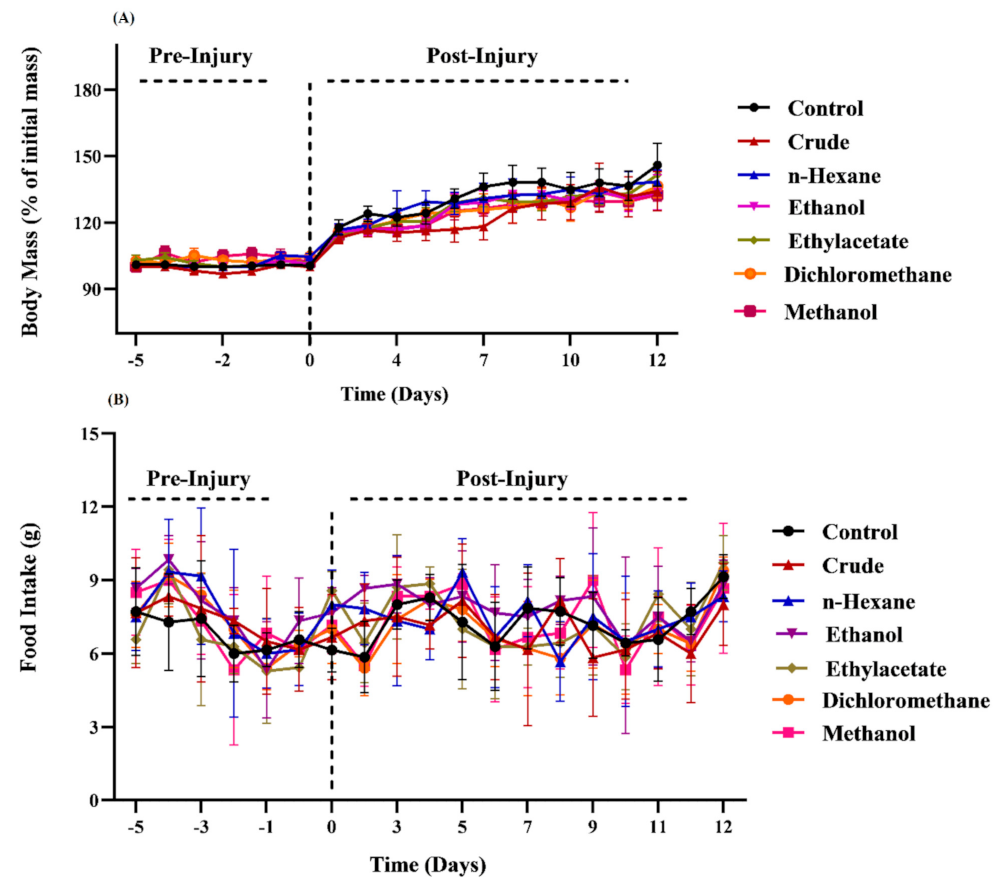
Abstract
:1. Introduction
2. Material and Methods
2.1. Animals
2.2. Extract Preparation and Supplementation
2.3. Induction of Sciatic Nerve Crush Injury
2.4. Behavioral Tests
2.4.1. Sciatic Functional Index
2.4.2. Hot Plate Test
2.4.3. Measurement of Grip Strength of Muscle
2.5. Biochemical Analysis of Systemic Indexes
2.5.1. Total Antioxidant Capacity
2.5.2. Total Oxidant Status
2.5.3. Random Blood Glucose
3. Results
3.1. Effects of M. oleifera Extracts on Food Consumption and Body Mass
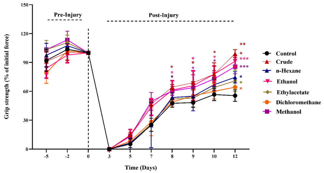
3.2. Effects of M. oleifera on Muscle Strength and Retrieval of Motor Functions
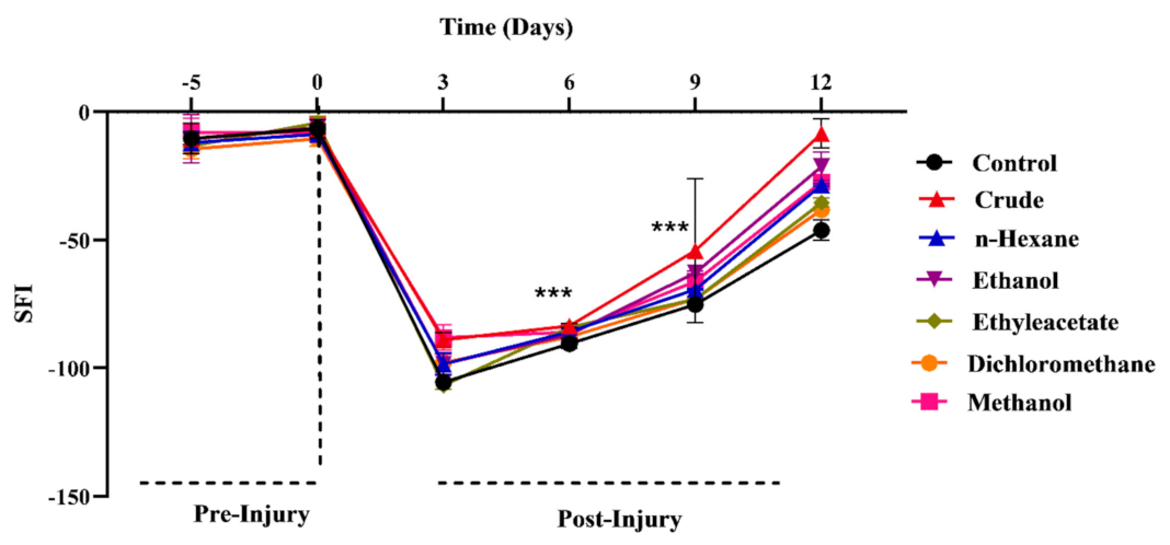
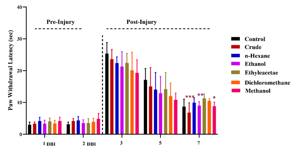
3.3. Effects of M. oleifera on Regain of Sensory Functions
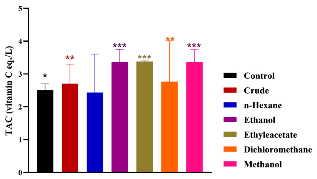
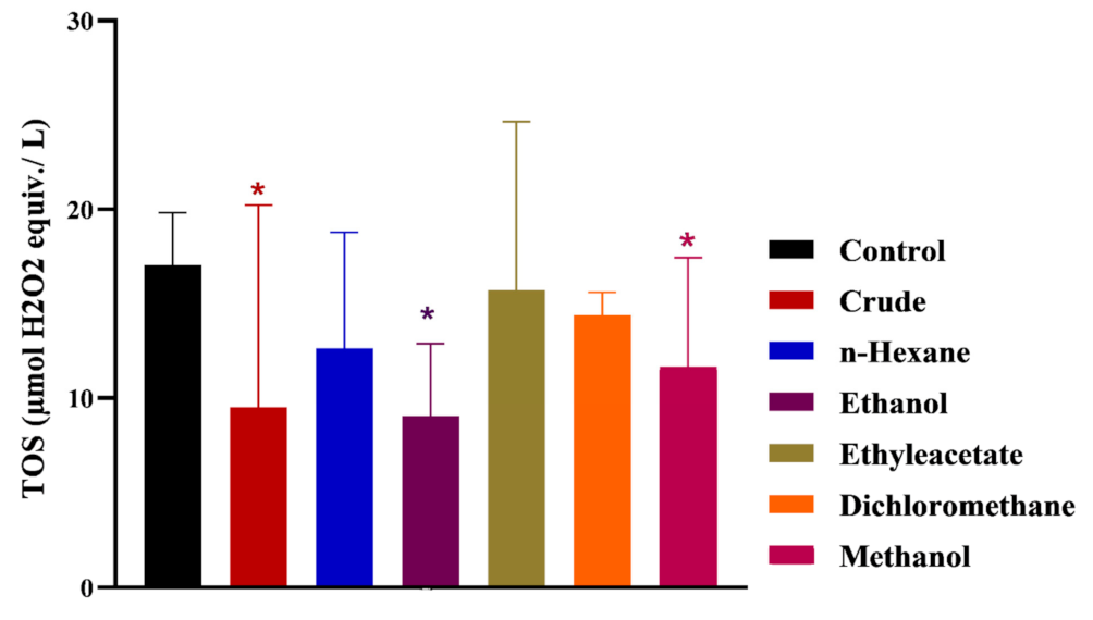
3.4. Effects of M. oleifera on Oxidative Stress and Blood Glucose Level
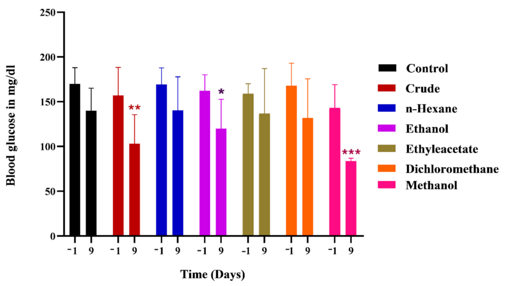
3.5. Effects of M. oleifera on Glycemic Level following Sciatic Nerve Lesion
4. Discussion
5. Conclusions
Author Contributions
Funding
Institutional Review Board Statement
Informed Consent Statement
Data Availability Statement
Acknowledgments
Conflicts of Interest
Abbreviations
References
- Ezzat, S.M.; Jeevanandam, J.; Egbuna, C.; Kumar, S.; Ifemeje, J.C. Phytochemicals as Sources of Drugs. In Phytochemistry: An In-silico and In-Vitro Update; Springer: Berlin/Heidelberg, Germany, 2019; pp. 3–22. [Google Scholar]
- Choudhari, A.S.; Mandave, P.C.; Deshpande, M.; Ranjekar, P.; Prakash, O. Phytochemicals in Cancer Treatment: From Preclinical Studies to Clinical Practice. Front. Pharmacol. 2020, 10, 1614. [Google Scholar] [CrossRef]
- Swain, S.S.; Hussain, T.; Pati, S. Drug-Lead Anti-Tuberculosis Phytochemicals: A Systematic Review. Curr. Top. Med. Chem. 2021, 21, 1832–1868. [Google Scholar] [CrossRef]
- Sharma, S.; Bhatia, V. Phytochemicals for Drug Discovery in Alzheimer’s Disease: In Silico Advances. Curr. Pharm. Des. 2021, 27, 2848–2860. [Google Scholar] [CrossRef]
- Liu, Y.; Chen, Z.; Li, A.; Liu, R.; Yang, H.; Xia, X. The Phytochemical Potential for Brain Disease Therapy and the Possible Nanodelivery Solutions for Brain Access. Front. Oncol. 2022, 12, 2748. [Google Scholar] [CrossRef]
- Li, X.; Gao, D.; Paudel, Y.N.; Li, X.; Zheng, M.; Liu, G.; Ma, Y.; Chu, L.; He, F.; Jin, M. Anti-Parkinson’s Disease Activity of Sanghuangprous Vaninii Extracts in the MPTP-Induced Zebrafish Model. ACS Chem. Neurosci. 2022, 13, 330–339. [Google Scholar] [CrossRef]
- Benga, A.; Zor, F.; Korkmaz, A.; Marinescu, B.; Gorantla, V. The Neurochemistry of Peripheral Nerve Regeneration. Indian J. Plast. Surg. 2017, 50, 5–15. [Google Scholar] [CrossRef] [PubMed]
- Carvalho, C.R.; Oliveira, J.M.; Reis, R.L. Modern Trends for Peripheral Nerve Repair and Regeneration: Beyond the Hollow Nerve Guidance Conduit. Front. Bioeng. Biotechnol. 2019, 7, 337. [Google Scholar] [CrossRef]
- Yow, Y.-Y.; Goh, T.-K.; Nyiew, K.-Y.; Lim, L.-W.; Phang, S.-M.; Lim, S.-H.; Ratnayeke, S.; Wong, K.-H. Therapeutic Potential of Complementary and Alternative Medicines in Peripheral Nerve Regeneration: A Systematic Review. Cells 2021, 10, 2194. [Google Scholar] [CrossRef]
- Lopes, B.; Sousa, P.; Alvites, R.; Branquinho, M.; Sousa, A.C.; Mendonça, C.; Atayde, L.M.; Luís, A.L.; Varejão, A.S.P.; Maurício, A.C. Peripheral Nerve Injury Treatments and Advances: One Health Perspective. Int. J. Mol. Sci. 2022, 23, 918. [Google Scholar] [CrossRef]
- Dhakad, A.K.; Ikram, M.; Sharma, S.; Khan, S.; Pandey, V.V.; Singh, A. Biological, Nutritional, and Therapeutic Significance of Moringa oleifera Lam. Phytother. Res. 2019, 33, 2870–2903. [Google Scholar] [CrossRef]
- Liu, R.; Liu, J.; Huang, Q.; Liu, S.; Jiang, Y. Moringa oleifera: A Systematic Review of Its Botany, Traditional Uses, Phytochemistry, Pharmacology and Toxicity. J. Pharm. Pharmacol. 2022, 74, 296–320. [Google Scholar] [CrossRef]
- Wang, F.; Long, S.; Zhang, J.; Yu, J.; Xiong, Y.; Zhou, W.; Qiu, J.; Jiang, H. Antioxidant Activities and Anti-Proliferative Effects of Moringa oleifera L. Extracts with Head and Neck Cancer. Food Biosci. 2020, 37, 100691. [Google Scholar] [CrossRef]
- Kirisattayakul, W.; Wattanathorn, J.; Tong-Un, T.; Muchimapura, S.; Wannanon, P.; Jittiwat, J. Cerebroprotective Effect of Moringa oleifera against Focal Ischemic Stroke Induced by Middle Cerebral Artery Occlusion. Oxidative Med. Cell. Longev. 2013, 2013, 951415. [Google Scholar] [CrossRef]
- Sutalangka, C.; Wattanathorn, J.; Muchimapura, S.; Thukham-mee, W. Moringa oleifera Mitigates Memory Impairment and Neurodegeneration in Animal Model of Age-Related Dementia. Oxidative Med. Cell. Longev. 2013, 2013, 695936. [Google Scholar] [CrossRef] [PubMed]
- Ghimire, S.; Subedi, L.; Acharya, N.; Gaire, B.P. Moringa oleifera: A Tree of Life as a Promising Medicinal Plant for Neurodegenerative Diseases. J. Agric. Food Chem. 2021, 69, 14358–14371. [Google Scholar] [CrossRef]
- González-Burgos, E.; Ureña-Vacas, I.; Sánchez, M.; Gómez-Serranillos, M.P. Nutritional Value of Moringa oleifera Lam. Leaf Powder Extracts and Their Neuroprotective Effects via Antioxidative and Mitochondrial Regulation. Nutrients 2021, 13, 2203. [Google Scholar] [CrossRef]
- Sunila, E.S.; Kuttan, G. A Preliminary Study on Antimetastatic Activity of Thuja Occidentalis, L. in Mice Model. Immunopharmacol. Immunotoxicol. 2006, 28, 269–280. [Google Scholar] [CrossRef]
- Cheng, X.-l.; Xiong, X.-b.; Xiang, M.-q. Effect of Arborvitae Seed on Cognitive Function and α7nAChR Protein Expression of Hippocampus in Model Rats with Alzheimer’s Disease. Cell Biochem. Biophys. 2013, 67, 181–184. [Google Scholar] [CrossRef] [PubMed]
- Hussain, G.; Schmitt, F.; Henriques, A.; Lequeu, T.; Rene, F.; Bindler, F.; Dirrig-Grosch, S.; Oudart, H.; Palamiuc, L.; Metz-Boutigue, M.-H.; et al. Systemic Down-Regulation of Delta-9 Desaturase Promotes Muscle Oxidative Metabolism and Accelerates Muscle Function Recovery following Nerve Injury. PLoS ONE 2013, 8, e64525. [Google Scholar] [CrossRef] [PubMed]
- Imran, A.; Xiao, L.; Ahmad, W.; Anwar, H.; Rasul, A.; Imran, M.; Aziz, N.; Razzaq, A.; Arshad, M.U.; Shabbir, A. Foeniculum Vulgare (Fennel) Promotes Functional Recovery and Ameliorates Oxidative Stress Following a Lesion to the Sciatic Nerve in Mouse Model. J. Food Biochem. 2019, 43, e12983. [Google Scholar] [CrossRef]
- Naz, S.; Farooq, T.; Hameed, A.; Anwar, H.; Aslam, R.; Malik, S.A.; Akram, R.; Sajid, F.; Saeed, Z.; Hussain, G. Thuja Occidentalis l. Expedites Functional Recovery after Sciatic Nerve Crush Injury in Mice. Pak. J. Med. Health Sci. 2022, 16, 396. [Google Scholar] [CrossRef]
- Maqbool, J.; Anwar, H.; Iqbal, J.; Rasul, A.; Imran, A.; Ahmad Malik, S.; Shabbir, A.; Ijaz, F.; Sajid, F.; Akram, R.; et al. Methanolic Extract of Fennel (Foeniculum vulgare) Escalates Functional Restoration Following a Compression Injury to the Sciatic Nerve in a Mouse Model. Food Sci. Nutr. 2021, 9, 701–710. [Google Scholar] [CrossRef] [PubMed]
- Erel, O. A New Automated Colorimetric Method for Measuring Total Oxidant Status. Clin. Biochem. 2005, 38, 1103–1111. [Google Scholar] [CrossRef] [PubMed]
- Rubio, C.P.; Hernández-Ruiz, J.; Martinez-Subiela, S.; Tvarijonaviciute, A.; Ceron, J.J. Spectrophotometric Assays for Total Antioxidant Capacity (TAC) in Dog Serum: An Update. BMC Vet. Res. 2016, 12, 166. [Google Scholar] [CrossRef] [PubMed]
- Wu, R.; Feng, J.; Yang, Y.; Dai, C.; Lu, A.; Li, J.; Liao, Y.; Xiang, M.; Huang, Q.; Wang, D. Significance of Serum Total Oxidant/Antioxidant Status in Patients with Colorectal Cancer. PLoS ONE 2017, 12, e0170003. [Google Scholar] [CrossRef] [PubMed]
- Bao, Y.; Xiao, J.; Weng, Z.; Lu, X.; Shen, X.; Wang, F. A Phenolic Glycoside from Moringa oleifera Lam. Improves the Carbohydrate and Lipid Metabolisms through AMPK in Db/Db Mice. Food Chem. 2020, 311, 125948. [Google Scholar] [CrossRef]
- Yang, Y.; Lin, L.; Zhao, M.; Yang, X. The Hypoglycemic and Hypolipemic Potentials of Moringa oleifera Leaf Polysaccharide and Polysaccharide-Flavonoid Complex. Int. J. Biol. Macromol. 2022, 210, 518–529. [Google Scholar] [CrossRef] [PubMed]
- Shah, K.H.; Oza, M.J. Comprehensive Review of Bioactive and Molecular Aspects of Moringa oleifera Lam. Food Rev. Int. 2022, 38, 1427–1460. [Google Scholar] [CrossRef]
- Natsir, H.; Wahab, A.; Budi, P.; Dali, S.; Arif, A. Amino Acid and Mineral Composition of Moringa Oleivera Leaves Extract and Its Bioactivity as Antioxidant. J. Phys. Conf. Ser. 2019, 1317, 012030. [Google Scholar] [CrossRef]
- Adewumi, O.O.; Felix-Minnaar, J.V.; Jideani, V.A. Functional Properties and Amino Acid Profile of Bambara Groundnut and Moringa oleifera Leaf Protein Complex. Processes 2022, 10, 205. [Google Scholar] [CrossRef]
- Su, B.; Chen, X. Current Status and Potential of Moringa oleifera Leaf as an Alternative Protein Source for Animal Feeds. Front. Vet. Sci. 2020, 7, 53. [Google Scholar] [CrossRef]
- Barodia, K.; Cheruku, S.P.; Kanwal, A.; Menon, A.; Rajeevan, R.; Rukade, A.; Kumar Shenoy, R.U.; Prabhu, C.; Sharma, V.; Divya, K.P.; et al. Effect of Moringa oleifera Leaf Extract on Exercise and Dexamethasone-Induced Functional Impairment in Skeletal Muscles. J. Ayurveda Integr. Med. 2022, 13, 100503. [Google Scholar] [CrossRef]
- Caillaud, M.; Aung Myo, Y.P.; McKiver, B.D.; Osinska Warncke, U.; Thompson, D.; Mann, J.; Del Fabbro, E.; Desmoulière, A.; Billet, F.; Damaj, M.I. Key Developments in the Potential of Curcumin for the Treatment of Peripheral Neuropathies. Antioxidants 2020, 9, 950. [Google Scholar] [CrossRef]
- Oliveira, M.A.; Heimfarth, L.; Passos, F.R.S.; Miguel-dos-Santos, R.; Mingori, M.R.; Moreira, J.C.F.; Lauton, S.S.; Barreto, R.S.; Araújo, A.A.; Oliveira, A.P. Naringenin Complexed with Hydroxypropyl-β-Cyclodextrin Improves the Sciatic Nerve Regeneration through Inhibition of p75NTR and JNK Pathway. Life Sci. 2020, 241, 117102. [Google Scholar] [CrossRef]
- Muratori, L.; Fregnan, F.; Maurina, M.; Haastert-Talini, K.; Ronchi, G. The Potential Benefits of Dietary Polyphenols for Peripheral Nerve Regeneration. Int. J. Mol. Sci. 2022, 23, 5177. [Google Scholar] [CrossRef] [PubMed]
- Gerhke, S.A.; Shibli, J.A.; Salles, M.B. Potential of the Use of an Antioxidant Compound to Promote Peripheral Nerve Regeneration after Injury. Neural Regenration Res. 2015, 10, 1063–1064. [Google Scholar] [CrossRef]
- Fakhri, S.; Abbaszadeh, F.; Moradi, S.Z.; Cao, H.; Khan, H.; Xiao, J. Effects of Polyphenols on Oxidative Stress, Inflammation, and Interconnected Pathways during Spinal Cord Injury. Oxidative Med. Cell. Longev. 2022, 2022, 8100195. [Google Scholar] [CrossRef]
- Bekiari, C.; Tekos, F.; Skaperda, Z.; Argyropoulou, A.; Skaltsounis, A.-L.; Kouretas, D.; Tsingotjidou, A. Antioxidant and Neuroprotective Effect of a Grape Pomace Extract on Oxaliplatin-Induced Peripheral Neuropathy in Rats: Biochemical, Behavioral and Histopathological Evaluation. Antioxidants 2022, 11, 1062. [Google Scholar] [CrossRef] [PubMed]
- Moradi, S.Z.; Jalili, F.; Farhadian, N.; Joshi, T.; Wang, M.; Zou, L.; Cao, H.; Farzaei, M.H.; Xiao, J. Polyphenols and Neurodegenerative Diseases: Focus on Neuronal Regeneration. Crit. Rev. Food Sci. Nutr. 2022, 62, 3421–3436. [Google Scholar] [CrossRef]
- Tian, H.; Liang, Y.; Liu, G.; Li, Y.; Deng, M.; Liu, D.; Guo, Y.; Sun, B. Moringa oleifera Polysaccharides Regulates Caecal Microbiota and Small Intestinal Metabolic Profile in C57BL/6 Mice. Int. J. Biol. Macromol. 2021, 182, 595–611. [Google Scholar] [CrossRef]
- Louisa, M.; Patintingan, C.G.H.; Wardhani, B.W.K. Moringa oleifera Lam. in Cardiometabolic Disorders: A Systematic Review of Recent Studies and Possible Mechanism of Actions. Front. Pharmacol. 2022, 13, 792794. [Google Scholar] [CrossRef]
- Monraz-Méndez, C.A.; Escutia-Gutiérrez, R.; Rodriguez-Sanabria, J.S.; Galicia-Moreno, M.; Monroy-Ramírez, H.C.; Sánchez-Orozco, L.; García-Bañuelos, J.; De la Rosa-Bibiano, R.; Santos, A.; Armendáriz-Borunda, J.; et al. Moringa oleifera Improves MAFLD by Inducing Epigenetic Modifications. Nutrients 2022, 14, 4225. [Google Scholar] [CrossRef]
- Mohamed, M.A.; Ahmed, M.A.; El Sayed, R.A. Molecular Effects of Moringa Leaf Extract on Insulin Resistance and Reproductive Function in Hyperinsulinemic Male Rats. J. Diabetes Metab. Disord. 2019, 18, 487–494. [Google Scholar] [CrossRef] [PubMed]
- Alejandra Sánchez-Muñoz, M.; Valdez-Solana, M.A.; Campos-Almazán, M.I.; Flores-Herrera, O.; Esparza-Perusquía, M.; Olvera-Sánchez, S.; García-Arenas, G.; Avitia-Domínguez, C.; Téllez-Valencia, A.; Sierra-Campos, E. Streptozotocin-Induced Adaptive Modification of Mitochondrial Supercomplexes in Liver of Wistar Rats and the Protective Effect of Moringa oleifera Lam. Biochem. Res. Int. 2018, 2018, 5681081. [Google Scholar] [CrossRef] [PubMed]
- Duranti, G.; Maldini, M.; Crognale, D.; Horner, K.; Dimauro, I.; Sabatini, S.; Ceci, R. Moringa oleifera Leaf Extract Upregulates Nrf2/HO-1 Expression and Ameliorates Redox Status in C2C12 Skeletal Muscle Cells. Molecules 2021, 26, 5041. [Google Scholar] [CrossRef]
- Ceci, R.; Maldini, M.; Olson, M.E.; Crognale, D.; Horner, K.; Dimauro, I.; Sabatini, S.; Duranti, G. Moringa oleifera Leaf Extract Protects C2C12 Myotubes against H2O2-Induced Oxidative Stress. Antioxidants 2022, 11, 1435. [Google Scholar] [CrossRef]
- Chigurupati, S.; Al-Murikhy, A.; Almahmoud, S.A.; Almoshari, Y.; Ahmed, A.S.; Vijayabalan, S.; Felemban, S.G.; Palanimuthu, V.R. Molecular Docking of Phenolic Compounds and Screening of Antioxidant and Antidiabetic Potential of Moringa oleifera Ethanolic Leaves Extract from Qassim Region, Saudi Arabia. Saudi J. Biol. Sci. 2022, 29, 854–859. [Google Scholar] [CrossRef]
- Sango, K.; Mizukami, H.; Horie, H.; Yagihashi, S. Impaired Axonal Regeneration in Diabetes. Perspective on the Underlying Mechanism from In Vivo and In Vitro Experimental Studies. Front. Endocrinol. 2017, 8, 12. [Google Scholar] [CrossRef] [PubMed]
- Kim, Y.J.; Kim, K.J.; Lee, J.H.; Park, S.-U.; Cho, S.-Y. Effect of Herbal Extracts on Peripheral Nerve Regeneration after Microsurgery of the Sciatic Nerve in Rats. BMC Complement. Med. Ther. 2021, 21, 162. [Google Scholar] [CrossRef]
- Wang, F.; Bao, Y.; Zhang, C.; Zhan, L.; Khan, W.; Siddiqua, S.; Ahmad, S.; Capanoglu, E.; Skalicka-Woźniak, K.; Zou, L. Bioactive Components and Anti-Diabetic Properties of Moringa oleifera Lam. Crit. Rev. Food Sci. Nutr. 2022, 62, 3873–3897. [Google Scholar] [CrossRef]


Publisher’s Note: MDPI stays neutral with regard to jurisdictional claims in published maps and institutional affiliations. |
© 2022 by the authors. Licensee MDPI, Basel, Switzerland. This article is an open access article distributed under the terms and conditions of the Creative Commons Attribution (CC BY) license (https://creativecommons.org/licenses/by/4.0/).
Share and Cite
Imran, M.; Hussain, G.; Hameed, A.; Iftikhar, I.; Ibrahim, M.; Asghar, R.; Nisar, I.; Farooq, T.; Khalid, T.; Rehman, K.; et al. Metabolites of Moringa oleifera Activate Physio-Biochemical Pathways for an Accelerated Functional Recovery after Sciatic Nerve Crush Injury in Mice. Metabolites 2022, 12, 1242. https://doi.org/10.3390/metabo12121242
Imran M, Hussain G, Hameed A, Iftikhar I, Ibrahim M, Asghar R, Nisar I, Farooq T, Khalid T, Rehman K, et al. Metabolites of Moringa oleifera Activate Physio-Biochemical Pathways for an Accelerated Functional Recovery after Sciatic Nerve Crush Injury in Mice. Metabolites. 2022; 12(12):1242. https://doi.org/10.3390/metabo12121242
Chicago/Turabian StyleImran, Muhammad, Ghulam Hussain, Arruje Hameed, Iqra Iftikhar, Muhammad Ibrahim, Rahat Asghar, Izzat Nisar, Tahir Farooq, Tanzila Khalid, Kanwal Rehman, and et al. 2022. "Metabolites of Moringa oleifera Activate Physio-Biochemical Pathways for an Accelerated Functional Recovery after Sciatic Nerve Crush Injury in Mice" Metabolites 12, no. 12: 1242. https://doi.org/10.3390/metabo12121242