Liquid Chromatography-High-Resolution Mass Spectrometry-Based In Vitro Toxicometabolomics of the Synthetic Cathinones 4-MPD and 4-MEAP in Pooled Human Liver Microsomes

 , ,
, ,
Abstract
:
1. Introduction
2. Results and Discussion
2.1. Infrared Spectroscopy
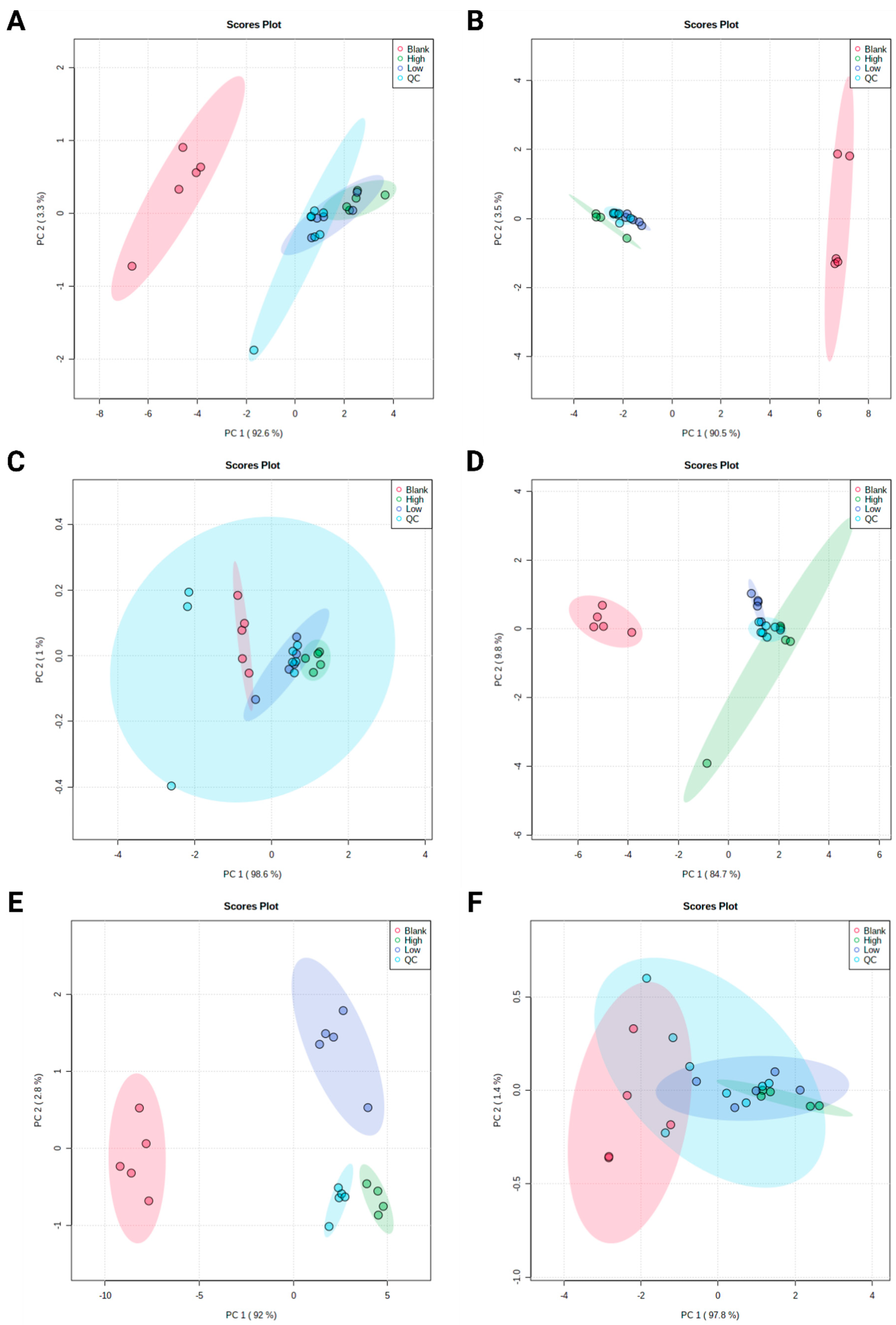
2.2. Untargeted Metabolomics
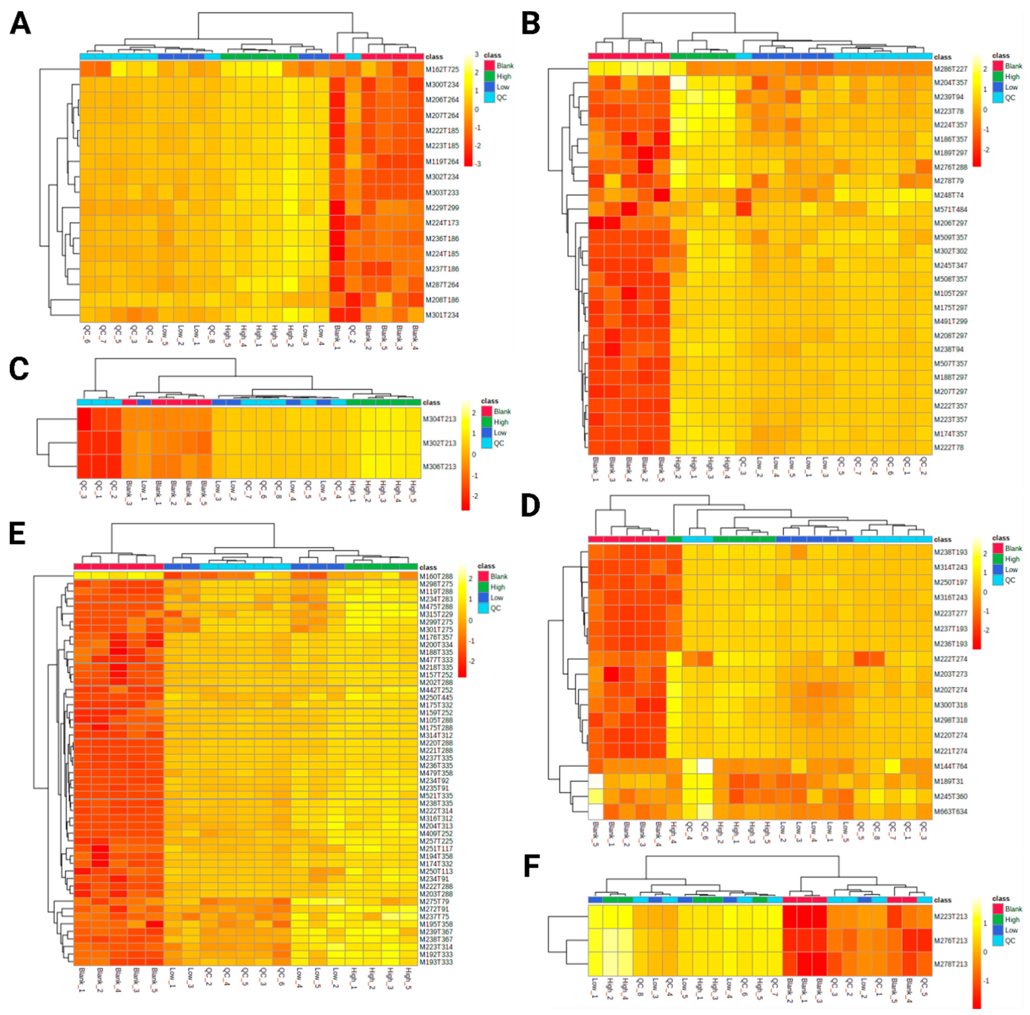
2.3. Identification of Significant Features
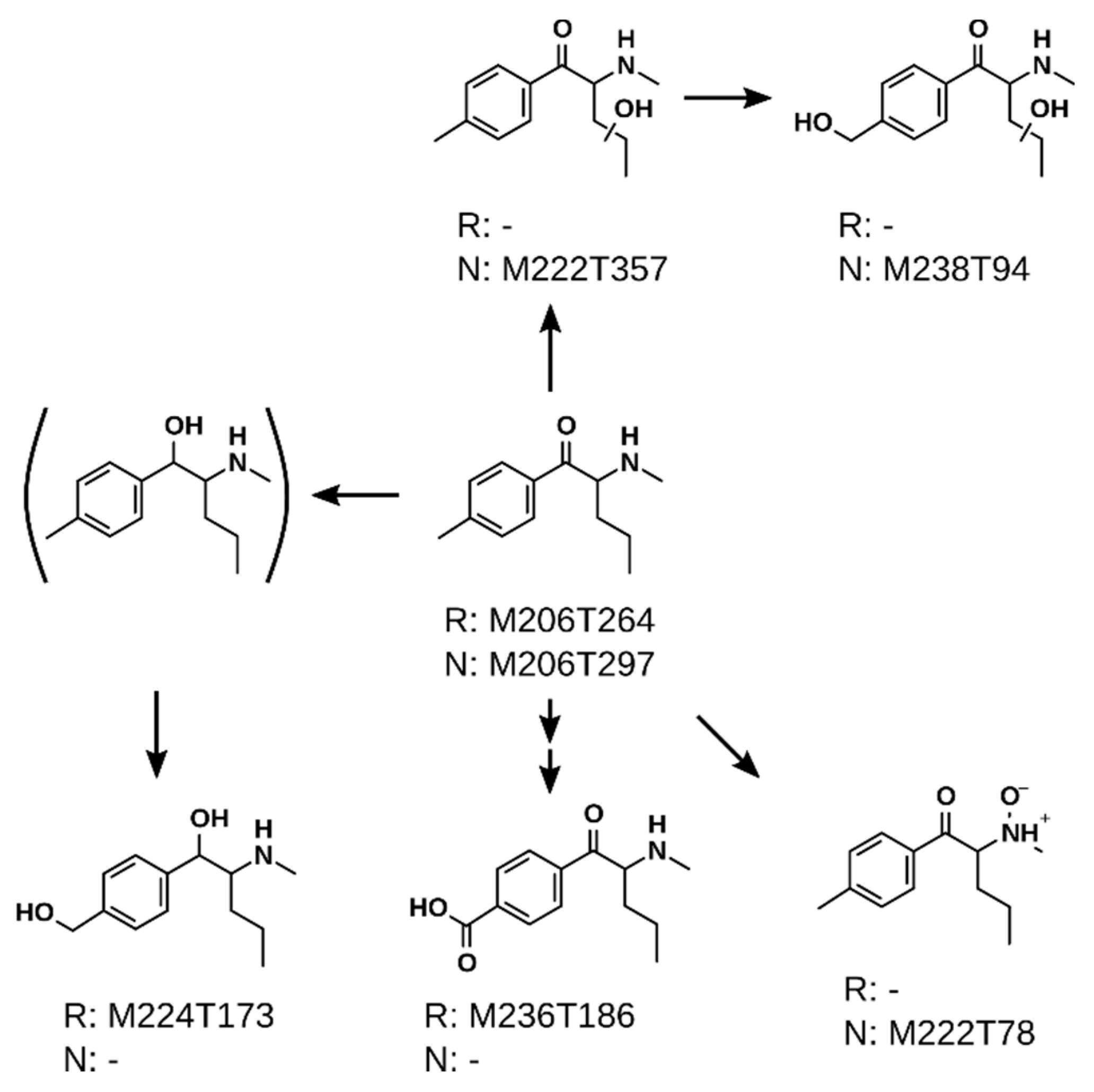
2.3.1. 4-MPD
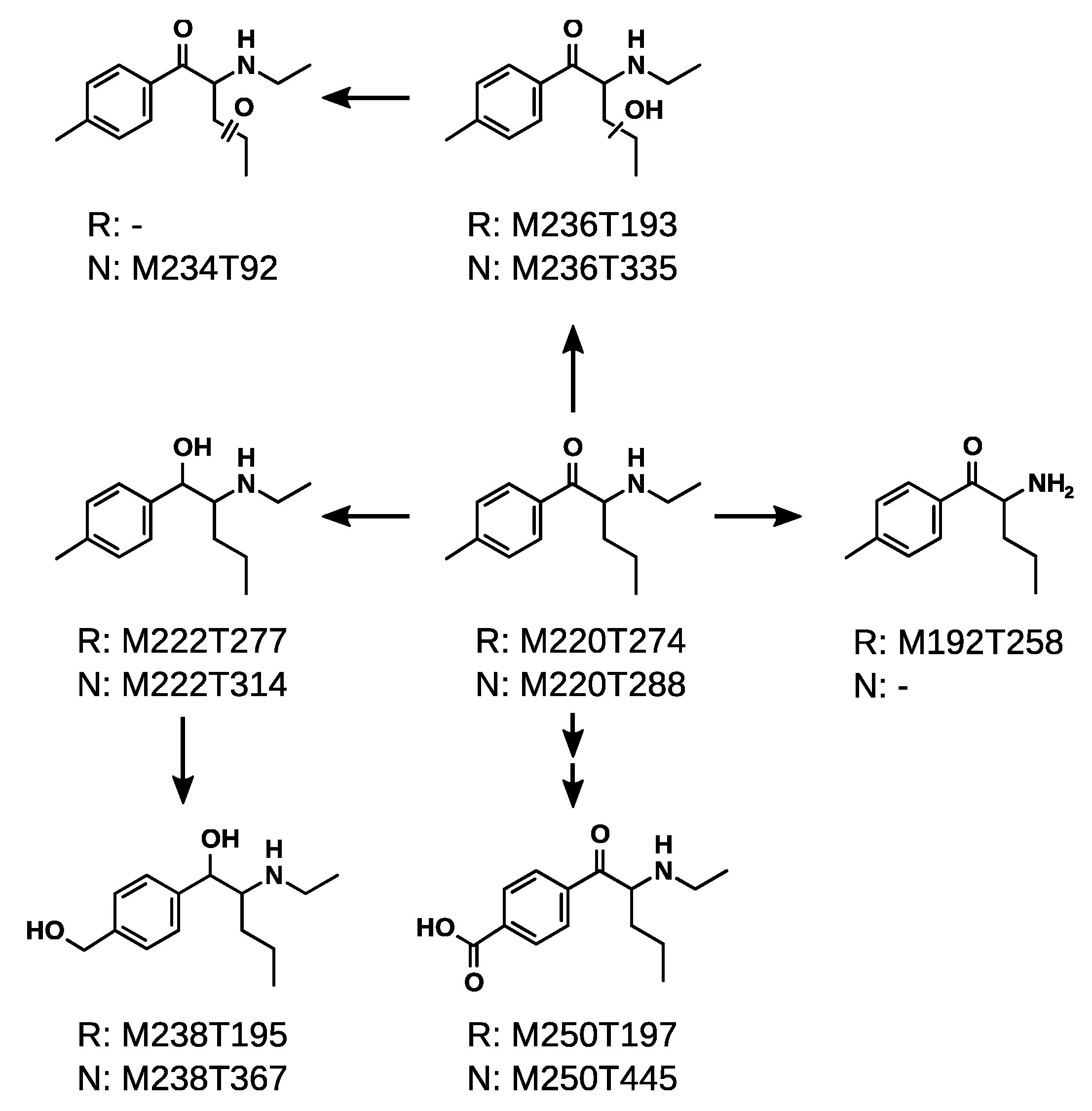
2.3.2. 4-MEAP
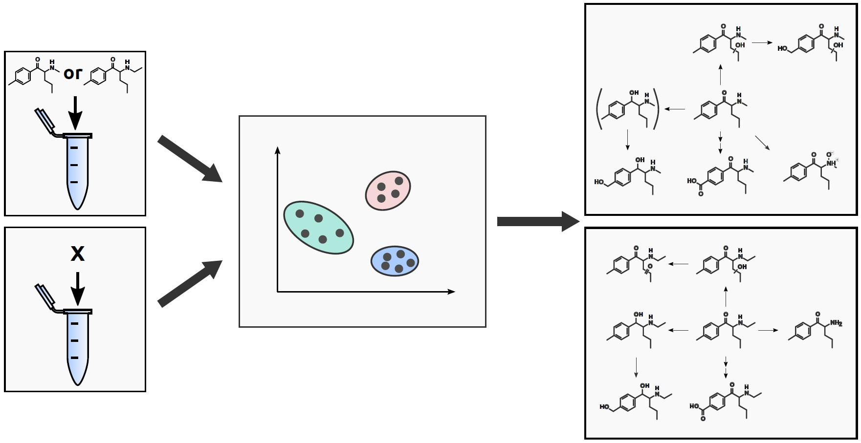
2.4. Proposed Metabolic Pathways
3. Materials and Methods
3.1. Chemicals and Reagents
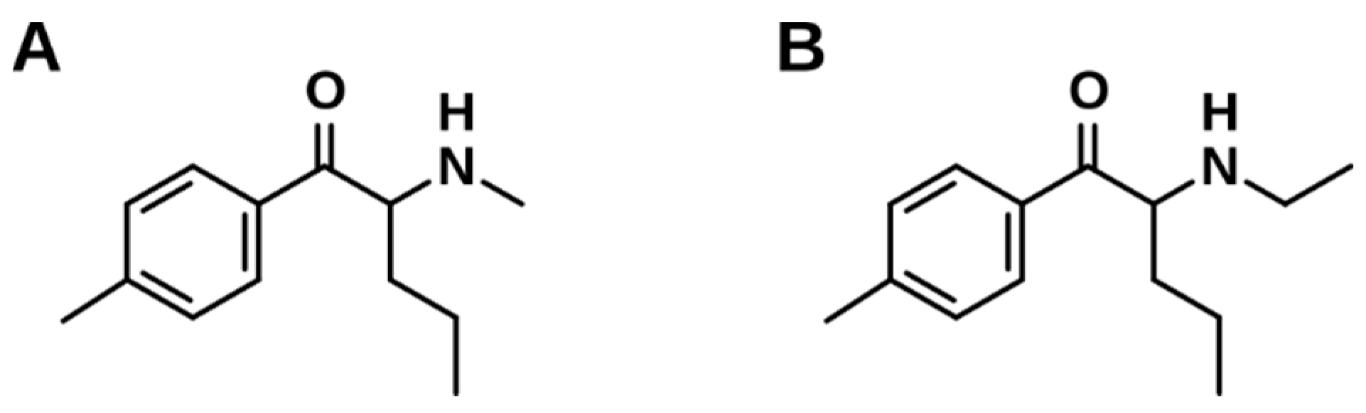
3.2. Synthesis of 2-(Methylamino)-1-(4-Methylphenyl)-1-Pentanone (4-MPD) and 2-(Ethylamino)-1-(4-Methylphenyl)-1-Pentanone (4-MEAP)
3.3. Infrared Spectroscopy Apparatus and Analysis
3.4. HPLC-HRMS/MS Apparatus
3.5. Microsomal Incubations Using pHLM
3.6. Data Processing for Untargeted Metabolomics
3.7. Identification of Significant Features
4. Conclusions
Supplementary Materials
Author Contributions
Funding
Institutional Review Board Statement
Informed Consent Statement
Data Availability Statement
Acknowledgments
Conflicts of Interest
References
- EMDDA–Europol. EU Drug Markets Report 2019; European Monitoring Centre for Drugs and Drug Addiction: Luxembourg, 2019. [Google Scholar]
- Simmler, L.D.; Buser, T.A.; Donzelli, M.; Schramm, Y.; Dieu, L.H.; Huwyler, J.; Chaboz, S.; Hoener, M.C.; Liechti, M.E. Pharmacological characterization of designer cathinones in vitro. Br. J. Pharmacol. 2013, 168, 458–470. [Google Scholar] [CrossRef] [PubMed] [Green Version]
- Hamby, D.; Burnett, A.; Jablonsky, M.; Twamley, B.; Kavanagh, P.V.; Gardner, E.A. Identification of 2-(ethylamino)-1-(4-methylphenyl)-1-pentanone (4-MEAP), a New “Legal High” Sold by an Internet Vendor as 4-Methyl Pentedrone. J. Forensic Sci. 2015, 60, 721–726. [Google Scholar] [CrossRef] [PubMed]
- EMDDA–Europol. 2014 Annual Report Implementation Council Decision 2005/387/JHA; Publications Office of the European Union: Luxembourg, 2015. [Google Scholar]
- Varma, A.; Patel, N.; Ford, L.; Jones, R.; Vale, J.A. Misuse of 2-(ethylamino)-1-(4-methylphenyl)-1-pentanone (4-MEAP), a synthetic cathinone. Clin. Toxicol. 2017, 55, 231–232. [Google Scholar] [CrossRef] [PubMed]
- Benedicte, L.; Camille, R.; Audrey, C.; Deborah, I.; Morgan, B.; Marie, D.; David, B.; Delphine, A.; Severine, F.; Guillaume, D.; et al. Case report on two-cathinones abuse: MPHP and N-ethyl-4′methylnorpentedrone, with a fatal outcome. Forensic Toxicol. 2020, 38, 243–254. [Google Scholar] [CrossRef]
- Sinz, M.A.; Lyubimov, A.V. In Vitro and In Vivo Models of Drug Metabolism. In Encyclopedia of Drug Metabolism and Interactions; John Wiley & Sons, Inc.: Hoboken, NJ, USA, 2011. [Google Scholar] [CrossRef]
- Manier, S.K.; Richter, L.H.J.; Schaper, J.; Maurer, H.H.; Meyer, M.R. Different in vitro and in vivo tools for elucidating the human metabolism of alpha-cathinone-derived drugs of abuse. Drug Test. Anal. 2018. [Google Scholar] [CrossRef]
- Helfer, A.G.; Turcant, A.; Boels, D.; Ferec, S.; Lelievre, B.; Welter, J.; Meyer, M.R.; Maurer, H.H. Elucidation of the metabolites of the novel psychoactive substance 4-methyl-N-ethyl-cathinone (4-MEC) in human urine and pooled liver microsomes by GC-MS and LC-HR-MS/MS techniques and of its detectability by GC-MS or LC-MS(n) standard screening approaches. Drug Test. Anal. 2015, 7, 368–375. [Google Scholar] [CrossRef] [Green Version]
- Barnes, S.; Benton, H.P.; Casazza, K.; Cooper, S.J.; Cui, X.; Du, X.; Engler, J.A.; Kabarowski, J.H.; Li, S.; Pathmasiri, W.; et al. Training in metabolomics research. I. Designing the experiment, collecting and extracting samples and generating metabolomics data. J. Mass Spectrom. 2016, 51, 461–475. [Google Scholar] [CrossRef] [Green Version]
- Lindon, J.C.; Nicholson, J.K.; Holmes, E.; Everett, J.R. Metabonomics: Metabolic processes studied by NMR spectroscopy of biofluids. Concept. Magn. Res. 2000, 12, 289–320. [Google Scholar] [CrossRef]
- Manier, S.K.; Keller, A.; Schaper, J.; Meyer, M.R. Untargeted metabolomics by high resolution mass spectrometry coupled to normal and reversed phase liquid chromatography as a tool to study the in vitro biotransformation of new psychoactive substances. Sci. Rep. 2019, 9, 2741. [Google Scholar] [CrossRef] [Green Version]
- Manier, S.K.; Wagmann, L.; Flockerzi, V.; Meyer, M.R. Toxicometabolomics of the new psychoactive substances alpha-PBP and alpha-PEP studied in HepaRG cell incubates by means of untargeted metabolomics revealed unexpected amino acid adducts. Arch. Toxicol. 2020, 94, 2047–2059. [Google Scholar] [CrossRef] [Green Version]
- Mortele, O.; Vervliet, P.; Gys, C.; Degreef, M.; Cuykx, M.; Maudens, K.; Covaci, A.; van Nuijs, A.L.N.; Lai, F.Y. In vitro Phase I and Phase II metabolism of the new designer benzodiazepine cloniprazepam using liquid chromatography coupled to quadrupole time-of-flight mass spectrometry. J Pharm. Biomed. Anal. 2018, 153, 158–167. [Google Scholar] [CrossRef] [PubMed]
- Vervliet, P.; Mortelé, O.; Gys, C.; Degreef, M.; Lanckmans, K.; Maudens, K.; Covaci, A.; van Nuijs, A.L.N.; Lai, F.Y. Suspect and non-target screening workflows to investigate the in vitro and in vivo metabolism of the synthetic cannabinoid 5Cl-THJ-018. Drug Test. Anal. 2019, 11, 479–491. [Google Scholar] [CrossRef] [PubMed]
- Wagmann, L.; Maurer, H.H. Bioanalytical Methods for New Psychoactive Substances. Handb. Exp. Pharm. 2018, 252, 413–439. [Google Scholar] [CrossRef]
- Meyer, M.R. Toxicokinetics of NPS: Update 2017. Handb. Exp. Pharm. 2018, 252, 441–459. [Google Scholar] [CrossRef]
- Heacock, R.A.; Marion, L. The infrared spectra of secondary amines and their salts. Can. J. Chem. 1956, 34, 1782–1795. [Google Scholar] [CrossRef]
- SI National Forensic Laboratory. Analytical Report 4-MPD. 2016. Available online: https://www.policija.si/apps/nfl_response_web/seznam.php (accessed on 27 November 2020).
- SWGDRUG. 4-Methyl Pentedrone Monograph; SWGDRUG: 2018. Available online: https://swgdrug.org/Monographs/4-Methylpentedrone.pdf (accessed on 27 November 2020).
- Wehrens, R.; Hageman, J.A.; van Eeuwijk, F.; Kooke, R.; Flood, P.J.; Wijnker, E.; Keurentjes, J.J.; Lommen, A.; van Eekelen, H.D.; Hall, R.D.; et al. Improved batch correction in untargeted MS-based metabolomics. Metabolomics 2016, 12, 88. [Google Scholar] [CrossRef] [Green Version]
- Remane, D.; Meyer, M.R.; Wissenbach, D.K.; Maurer, H.H. Ion suppression and enhancement effects of co-eluting analytes in multi-analyte approaches: Systematic investigation using ultra-high-performance liquid chromatography/mass spectrometry with atmospheric-pressure chemical ionization or electrospray ionization. Rapid. Commun. Mass Spectrom. 2010, 24, 3103–3108. [Google Scholar] [CrossRef]
- Sumner, L.W.; Amberg, A.; Barrett, D.; Beale, M.H.; Beger, R.; Daykin, C.A.; Fan, T.W.; Fiehn, O.; Goodacre, R.; Griffin, J.L.; et al. Proposed minimum reporting standards for chemical analysis Chemical Analysis Working Group (CAWG) Metabolomics Standards Initiative (MSI). Metabolomics 2007, 3, 211–221. [Google Scholar] [CrossRef] [Green Version]
- Apirakkan, O.; Frinculescu, A.; Shine, T.; Parkin, M.C.; Cilibrizzi, A.; Frascione, N.; Abbate, V. Analytical characterization of three cathinone derivatives, 4-MPD, 4F-PHP and bk-EPDP, purchased as bulk powder from online vendors. Drug Test. Anal. 2018, 10, 372–378. [Google Scholar] [CrossRef] [Green Version]
- Manier, S.K.; Felske, C.; Eckstein, N.; Meyer, M.R. The metabolic fate of two new psychoactive substances—2-aminoindane and N-methyl-2-aminoindane—studied in vitro and in vivo to support drug testing. Drug Test. Anal. 2020, 12, 145–151. [Google Scholar] [CrossRef] [Green Version]
- Frański, R.; Gierczyk, B.; Kasperkowiak, M.; Jankowski, W.; Hoffmann, M. The mechanism of water loss from protonated cathinones. Rapid Commun. Mass Spectrom. RCM 2020, 34, e8617. [Google Scholar] [CrossRef] [PubMed]
- Wagmann, L.; Manier, S.K.; Eckstein, N.; Maurer, H.H.; Meyer, M.R. Toxicokinetic studies of the four new psychoactive substances 4-chloroethcathinone, N-ethylnorpentylone, N-ethylhexedrone, and 4-fluoro-alpha-pyrrolidinohexiophenone. Forensic Toxicol. 2020, 38, 59–69. [Google Scholar] [CrossRef]
- Ellefsen, K.N.; Concheiro, M.; Huestis, M.A. Synthetic cathinone pharmacokinetics, analytical methods, and toxicological findings from human performance and postmortem cases. Drug Metab. Rev. 2016, 48, 237–265. [Google Scholar] [CrossRef] [PubMed]
- Olesti, E.; Farre, M.; Papaseit, E.; Krotonoulas, A.; Pujadas, M.; de la Torre, R.; Pozo, O.J. Pharmacokinetics of Mephedrone and Its Metabolites in Human by LC-MS/MS. AAPS J. 2017, 19, 1767–1778. [Google Scholar] [CrossRef]
- Becker, H. Organikum, 21. neu bearb. und erw. Aufl. ed.; Wiley-VCH: Weinheim, Germany, 2001; p. 852. [Google Scholar]
- Reddy, Y.T.; Reddy, P.N.; Reddy, M.N.; Rajitha, B.; Crooks, P.A. Convenient and Scalable Process for the Preparation of Bupropion Hydrochloride via Efficient Bromination of m-Chloropropiophenone with N-Bromosuccinimide. Synth. Commun. 2010, 40, 1566–1573. [Google Scholar] [CrossRef]
- Maurer, H.H.; Pfleger, K.; Weber, A.A. Mass Spectral Data of Drugs, Poisons, Pesticides, Pollutants and Their Metabolites; Wiley-VCH: Weinheim, Germany, 2016. [Google Scholar]
- Manier, S.K.; Keller, A.; Meyer, M.R. Automated optimization of XCMS parameters for improved peak picking of liquid chromatography-mass spectrometry data using the coefficient of variation and parameter sweeping for untargeted metabolomics. Drug Test. Anal. 2018. [Google Scholar] [CrossRef]
- Welter, J.; Meyer, M.R.; Wolf, E.U.; Weinmann, W.; Kavanagh, P.; Maurer, H.H. 2-methiopropamine, a thiophene analogue of methamphetamine: Studies on its metabolism and detectability in the rat and human using GC-MS and LC-(HR)-MS techniques. Anal. Bioanal. Chem. 2013, 405, 3125–3135. [Google Scholar] [CrossRef]
- Chambers, M.C.; Maclean, B.; Burke, R.; Amodei, D.; Ruderman, D.L.; Neumann, S.; Gatto, L.; Fischer, B.; Pratt, B.; Egertson, J.; et al. A cross-platform toolkit for mass spectrometry and proteomics. Nat. Biotechnol. 2012, 30, 918–920. [Google Scholar] [CrossRef]


Publisher’s Note: MDPI stays neutral with regard to jurisdictional claims in published maps and institutional affiliations. |
© 2020 by the authors. Licensee MDPI, Basel, Switzerland. This article is an open access article distributed under the terms and conditions of the Creative Commons Attribution (CC BY) license (http://creativecommons.org/licenses/by/4.0/).
Share and Cite
Manier, S.K.; Schwermer, F.; Wagmann, L.; Eckstein, N.; Meyer, M.R. Liquid Chromatography-High-Resolution Mass Spectrometry-Based In Vitro Toxicometabolomics of the Synthetic Cathinones 4-MPD and 4-MEAP in Pooled Human Liver Microsomes. Metabolites 2021, 11, 3. https://doi.org/10.3390/metabo11010003
Manier SK, Schwermer F, Wagmann L, Eckstein N, Meyer MR. Liquid Chromatography-High-Resolution Mass Spectrometry-Based In Vitro Toxicometabolomics of the Synthetic Cathinones 4-MPD and 4-MEAP in Pooled Human Liver Microsomes. Metabolites. 2021; 11(1):3. https://doi.org/10.3390/metabo11010003
Chicago/Turabian StyleManier, Sascha K., Florian Schwermer, Lea Wagmann, Niels Eckstein, and Markus R. Meyer. 2021. "Liquid Chromatography-High-Resolution Mass Spectrometry-Based In Vitro Toxicometabolomics of the Synthetic Cathinones 4-MPD and 4-MEAP in Pooled Human Liver Microsomes" Metabolites 11, no. 1: 3. https://doi.org/10.3390/metabo11010003