
Colonic Ganglioneuroma: A Combined Single-Institution Experience and Review of the Literature of Forty-Three Patients

 ,
,  ,
,
Abstract
:1. Introduction
2. Methods
2.1. Study Design, Setting, and Objectives
2.2. Ethical Considerations
2.3. Patients’ Selection and Clinicopathological Parameters of Patients
2.4. Review of Literaure
3. Results
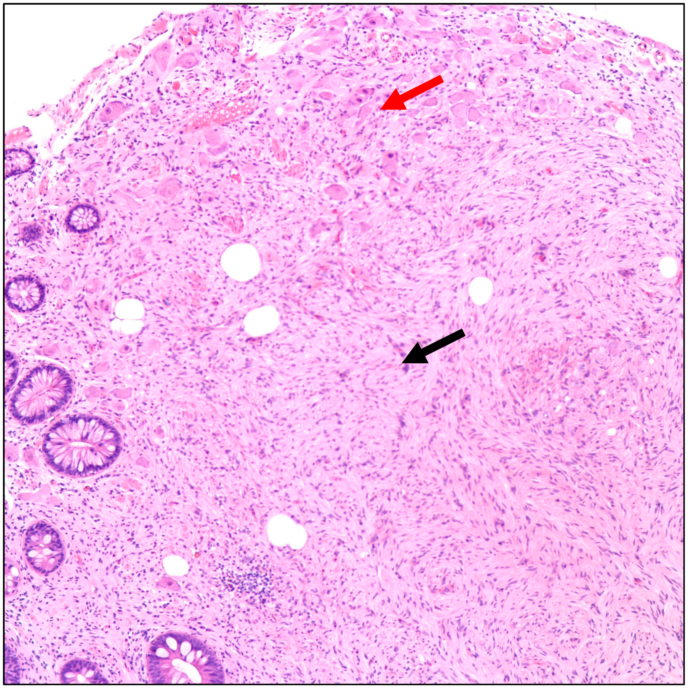
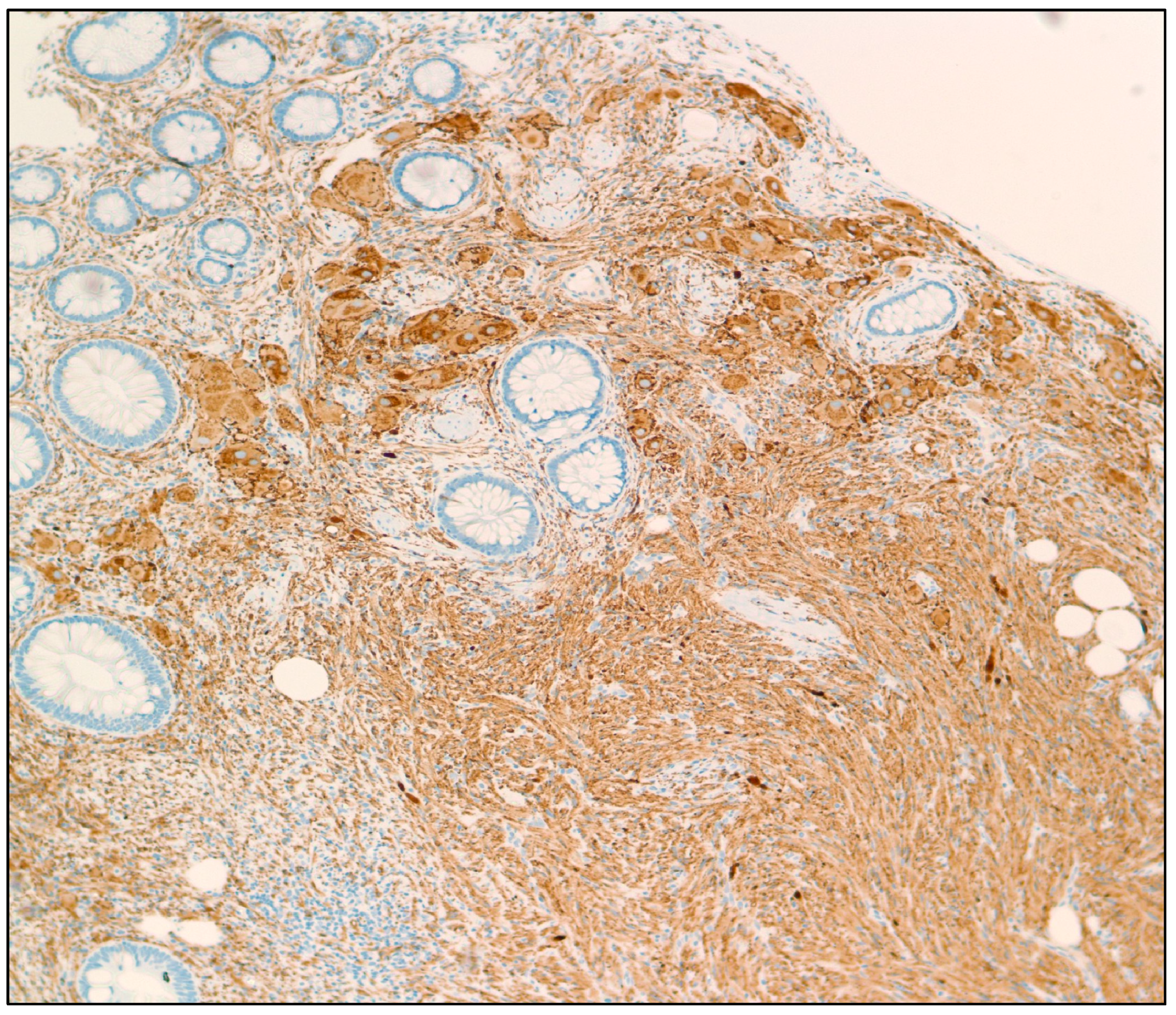
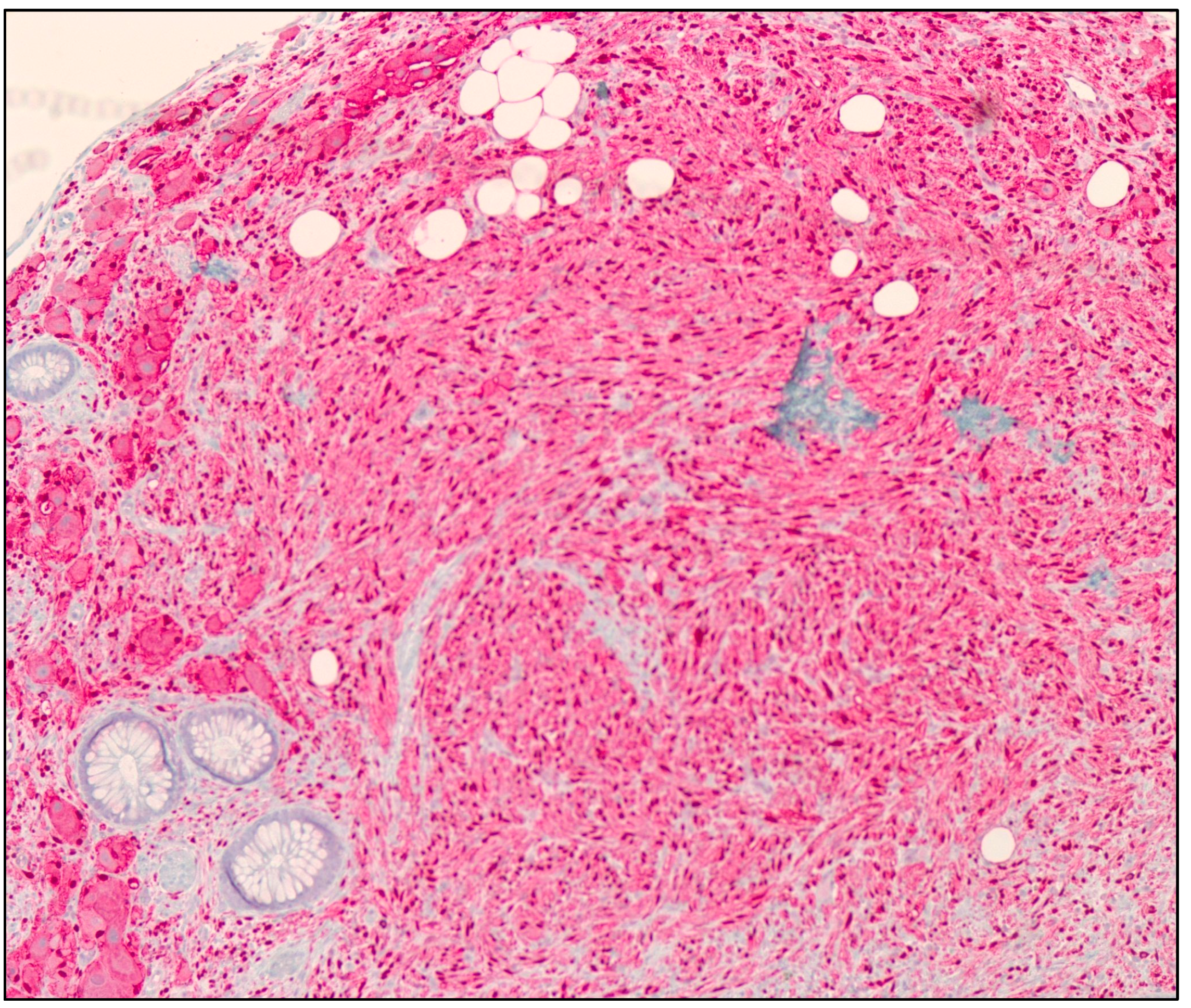
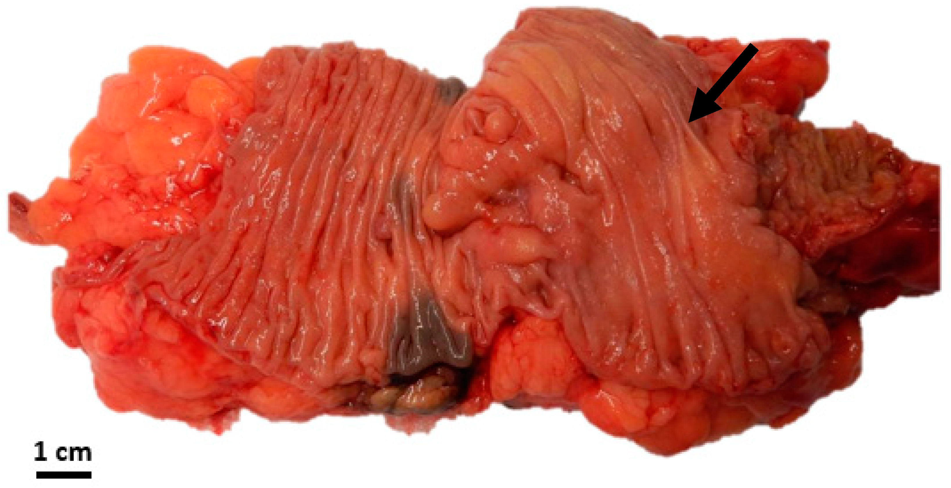
3.1. Mount Sinai Medical Center of Florida Experience
3.2. Comprehensive Review of Literature

| Ref. | Year | Age (Years) | Sex | Location in Colon | Category | Associated Symptoms | Associated Syndromes | Size | Associated Pathologies | IHC Stains | Management |
|---|---|---|---|---|---|---|---|---|---|---|---|
| [21] | 2022 | 38 | F | Sigmoid colon | DG | - | - | 30 mm | Adenocarcinoma | S100 protein (+) | Colectomy |
| [22] | 2021 | 73 | M | Splenic flexure | PG | - | - | 10 mm | - | - | Polypectomy |
| [16] | 2021 | 44 | M | Ascending colon and hepatic flexure | GP | Abdominal pain | - | - | Adenocarcinoma | Chromogranin, synaptophysin, CD56, and CK20 (+) | Colectomy |
| 43 | M | Ascending colon | PG | Abdominal pain | - | 8 mm | - | - | Polypectomy | ||
| [10] | 2021 | 9 | M | Cecum | DG | Abdominal pain | - | 5 cm | - | GFAP and S100 protein (+) | Polypectomy |
| [6] | 2020 | 84 | M | Ascending and descending colons | PG | Hematochezia | - | 3 to 4 mm range | - | - | Polypectomy |
| [23] | 2020 | 40 | M | Ascending colon | PG | Positive fecal occult blood | - | 15 mm | - | S100 protein and synaptophysin (+) | Polypectomy |
| [24] | 2020 | 68 | M | Sigmoid colon | PG | Positive fecal occult blood | - | 11 mm | Mucosal neurofibromas | S100 protein (+) | Colectomy |
| [25] | 2018 | 50 | M | Splenic flexure | PG | - | - | 7 mm | - | S100 protein and synaptophysin (+) | Polypectomy |
| [5] | 2017 | 65 | M | Ascending colon | PG | - | - | 6 mm | - | S100 protein (+) | Polypectomy |
| [17] | 2017 | 67 | M | Colon (diffuse) | DG | Abdominal pain | NF1 | - | - | S100 protein and synaptophysin (+) | Right hemicolectomy |
| [26] | 2017 | 43 | M | Colon (diffuse) | PG | Hematochezia | PHTS | 1 to 6 mm range | - | - | Polypectomy |
| [8] | 2016 | 51 | F | Descending colon | PG | - | - | 10 mm | - | S100 protein and NSE (+) | Polypectomy |
| [27] | 2016 | 60 | M | Colon (diffuse) | GP | - | Polyposis coli and Cowden | 0.3 to 2.3 cm range | - | S100 protein (+) | Total colectomy |
| [28] | 2016 | 51 | F | Colon (diffuse) | PG | - | - | 2 to 5 mm range | - | S100 protein and NSE (+) | Polypectomy |
| [15] | 2015 | 70 | M | Rectosigmoid colon | DG | Diarrhea | - | 8.5 cm | - | S100 protein and chromogranin (+) | Colectomy |
| 35 | M | Ascending colon | DG | - | - | 5.5 cm | - | S100 protein, chromogranin, and CD56 (+) | Right hemicolectomy | ||
| [29] | 2015 | 38 | M | Colon (diffuse) | DG | Diarrhea and weight loss | HPS | 3 to 5 mm range | - | S100 protein (+) | Polypectomy |
| [30] | 2015 | 43 | M | Cecum | PG | Hematochezia | - | 6 mm | - | S100 protein (+) | Polypectomy |
| [31] | 2015 | 71 | M | Colon (diffuse) | DG | Positive fecal occult blood | - | - | - | S100 protein, CD56, NSE, and Neurofilament protein (+) | Polypectomy |
| [32] | 2015 | 54 | M | Colon (diffuse) | DG | Hematochezia and abdominal pain | - | 0.1 to 8 cm range | - | S100 protein and Neurofilament protein (+) | Polypectomy |
| [33] | 2013 | 68 | F | Descending colon | PG | - | - | 16 cm | - | S100 protein and NSE (+) | Colectomy |
| [34] | 2012 | 57 | M | Colon (diffuse) | DG | Hematochezia | - | 7 mm | - | S100 protein, synaptophysin, and chromogranin (+) | Polypectomy |
| [35] | 2013 | 7 | M | Colon (diffuse) | DG | Abdominal pain | - | - | - | S100 protein (+) | Colectomy |
| [36] | 2012 | 61 | M | Descending colon | PG | - | - | 6 mm | - | S100 protein (+) | Polypectomy |
| [37] | 2012 | 42 | M | Colon (diffuse) | DG | Hematochezia | Cowden | - | Adenocarcinoma | - | Colectomy |
| [38] | 2009 | 38 | F | Transverse and sigmoid colon | DG | Abdominal pain | - | 6 to 7 cm range | Adenocarcinoma | S100 protein (+) | Colectomy |
| [39] | 2009 | 14 | F | Colon (diffuse) | DG | Diarrhea | WDHA | - | - | - | Colectomy |
| [40] | 2009 | 5 | F | Sigmoid colon | PG | Abdominal pain | - | - | - | - | Colectomy |
| [41] | 2008 | 48 | F | Sigmoid colon | PG | - | - | - | Diffuse melanosis coli | S100 protein and NSE (+) | - |
| [42] | 2006 | 41 | M | Colon (diffuse) | GP | Hematochezia | - | 1 to 2 mm range | - | S100 protein (+) | Polypectomy |
| [43] | 2005 | 29 | M | Colon (diffuse) | GP | Diarrhea | - | 5 to 15 mm range | - | S100 protein (+) | Polypectomy |
| [44] | 2006 | 42 | M | Cecum | DG | Constipation | - | 10 cm | - | S100 protein (+) | Ileocolectomy |
| [45] | 1999 | 77 | F | Cecum | GP | Abdominal pain | - | 3 to 4 cm range | - | S100 protein and NSE (+) | Polypectomy |
| [46] | 1998 | 54 | M | Hepatic flexure | PG | - | - | 5 mm | - | S100 protein (+) | - |
| Ref. | Year | Species | Age | Sex | Location in Colon | Category | Size | IHC Stains | Management |
|---|---|---|---|---|---|---|---|---|---|
| [20] | 1990 | Steer | 7 months old | - | Descending colon | DG | - | NSE and Neurofilament protein (+) | Colectomy |
| [19] | 2007 | Horse | 8 years old | - | Colon (40 cm from anus) | DG | 3 to 15 mm range | S100 protein and GFAP (+) | - |
| [18] | 2011 | Dog | 5 months old | F | Colon and rectum | DG | 2 to 8 mm range | - | Colectomy |
4. Discussion
5. Conclusions
Author Contributions
Funding
Institutional Review Board Statement
Informed Consent Statement
Data Availability Statement
Acknowledgments
Conflicts of Interest
References
- Luksch, R.; Castellani, M.R.; Collini, P.; De Bernardi, B.; Conte, M.; Gambini, C.; Gandola, L.; Garaventa, A.; Biasoni, D.; Podda, M.; et al. Neuroblastoma (Peripheral neuroblastic tumours). Crit. Rev. Oncol. Hematol. 2016, 107, 163–181. [Google Scholar] [CrossRef] [PubMed]
- Modha, A.; Paty, P.; Bilsky, M.H. Presacral ganglioneuromas. Report of five cases and review of the literature. J. Neurosurg. Spine 2005, 2, 366–371. [Google Scholar] [CrossRef] [PubMed]
- Kameneva, P.; Kastriti, M.E.; Adameyko, I. Neuronal lineages derived from the nerve-associated Schwann cell precursors. Cell. Mol. Life Sci. 2021, 78, 513–529. [Google Scholar] [CrossRef] [PubMed]
- Guan, J.; Hallberg, B.; Palmer, R.H. Chromosome Imbalances in Neuroblastoma–Recent Molecular Insight into Chromosome 1p-deletion, 2p-gain, and 11q-deletion Identifies New Friends and Foes for the Future. Cancers 2021, 13, 5897. [Google Scholar] [CrossRef] [PubMed]
- Ofori, E.; Ona, M.; Ramai, D.; Huang, T.; Xiao, P.; Reddy, M. Colonic Ganglioneuroma: A Rare Finding during Colorectal Cancer Screening. Case Rep. Gastroenterol. 2017, 11, 434–439. [Google Scholar] [CrossRef]
- Nehme, C.; Ghazaleh, S.; Patel, D.; Hasan, S.; Nawras, A. Incidental Ascending Colon Ganglioneuroma in the Setting of Hematochezia. Cureus 2020, 12, e9447. [Google Scholar] [CrossRef]
- Shekitka, K.M.; Sobin, L.H. Ganglioneuromas of the gastrointestinal tract. Relation to Von Recklinghausen disease and other multiple tumor syndromes. Am. J. Surg. Pathol. 1994, 18, 250–257. [Google Scholar] [CrossRef]
- Abdelfatah, M.; Sangah, G.; Harvin, G. What Do We Need to Know About Colonic Polypoid Ganglioneuroma? A Case Report and A Comprehensive Review. J. Gastrointest. Cancer 2018, 49, 327–332. [Google Scholar] [CrossRef]
- Skovronsky, D.M.; Oberholtzer, J.C. Pathologic classification of peripheral nerve tumors. Neurosurg. Clin. N. Am. 2004, 15, 157–166. [Google Scholar] [CrossRef]
- Mauro, A.; Zenzeri, L.; Esposito, F.; Gaglione, G.; Strisciuglio, C.; Pilozzi, E.; Corleto, V.D.; Ziparo, C.; Di Nardo, G. Isolated intestinal Ganglioneuromatosis: Case report and literature review. Ital. J. Pediatr. 2021, 47, 80. [Google Scholar] [CrossRef]
- Mulligan, L.M.; Ponder, B.A. Genetic basis of endocrine disease: Multiple endocrine neoplasia type 2. J. Clin. Endocrinol. Metab. 1995, 80, 1989–1995. [Google Scholar] [CrossRef] [PubMed]
- Carlson, K.M.; Bracamontes, J.; Jackson, C.E.; Clark, R.; Lacroix, A.; Wells, S.A., Jr.; Goodfellow, P.J. Parent-of-origin effects in multiple endocrine neoplasia type 2B. Am. J. Hum. Genet. 1994, 55, 1076–1082. [Google Scholar] [PubMed]
- Williams, A.J.; Doherty, E.S.; Hart, M.H.; Grider, D.J. Diffuse Gastric Ganglioneuromatosis: Novel Presentation of PTEN Hamartoma Syndrome-Case Report and Review of Gastric Ganglioneuromatous Proliferations and a Novel PTEN Gene Mutation. Case Rep. Med. 2018, 2018, 4319818. [Google Scholar] [CrossRef] [PubMed]
- Cimino, P.J.; Gutmann, D.H. Neurofibromatosis type 1. Handb. Clin. Neurol. 2018, 148, 799–811. [Google Scholar] [CrossRef]
- Kim, T.J.; Lim, H.; Kang, H.S.; Moon, S.H.; Kim, J.H.; Park, C.K.; Kwon, M.J.; Lee, B.H. Diffuse Ganglioneuromatosis of the Colon Presenting as a Large Subepithelial Tumor in Adults: Report of Two Cases. Korean J. Gastroenterol. 2015, 66, 111–115. [Google Scholar] [CrossRef]
- Baiomi, A.; Abbas, H.; Niazi, M.; Remotti, H.; Daniel, M.; Balar, B. Colonic Ganglioneuroma: A Rare Lesion with Extremely Different Presentations and Outcomes in Two Patients. Gastroenterol. Res. 2021, 14, 194–198. [Google Scholar] [CrossRef]
- Iwamuro, M.; Omote, R.; Tanaka, T.; Sunada, N.; Nada, T.; Kondo, Y.; Nose, S.; Kawaguchi, M.; Otsuka, F.; Okada, H. Diffuse Intestinal Ganglioneuromatosis Showing Multiple Large Bowel Ulcers in a Patient with Neurofibromatosis Type 1. Intern. Med. 2017, 56, 3287–3291. [Google Scholar] [CrossRef]
- Bemelmans, I.; Küry, S.; Albaric, O.; Hordeaux, J.; Bertrand, L.; Nguyen, F.; Abadie, J. Colorectal hamartomatous polyposis and ganglioneuromatosis in a dog. Vet. Pathol. 2011, 48, 1012–1015. [Google Scholar] [CrossRef]
- Porter, B.F.; Storts, R.W.; Payne, H.R.; Edwards, J.F. Colonic ganglioneuromatosis in a horse. Vet. Pathol. 2007, 44, 207–210. [Google Scholar] [CrossRef]
- Cole, D.E.; Migaki, G.; Leipold, H.W. Colonic ganglioneuromatosis in a steer. Vet. Pathol. 1990, 27, 461–462. [Google Scholar] [CrossRef]
- Nakamura, K.; Seishima, R.; Emoto, K.; Kawahara, T.; Matsui, S.; Shigeta, K.; Okabayashi, K.; Hosoe, N.; Kitagawa, Y. Development of Colon Adenocarcinoma on a Ganglioneuroma in a Patient with Nonhereditary Disease. Am. J. Gastroenterol. 2022, 117, 1028. [Google Scholar] [CrossRef] [PubMed]
- Agarwal, S.; Wang, Y.; Iyer, P.G.; Leggett, C.L. Incidental Colonic Ganglioneuroma on Surveillance Colonoscopy. ACG Case Rep. J. 2022, 9, e00727. [Google Scholar] [CrossRef] [PubMed]
- Arai, T.; Yamada, H.; Edagawa, T.; Yoshida, S.; Maekura, S.; Nakachi, K. Endoscopic Treatment of Solitary Colonic Ganglioneuroma. Case Rep. Gastroenterol. 2020, 14, 34–38. [Google Scholar] [CrossRef] [PubMed]
- Hashimoto, H.; Koda, H.; Horiuchi, H.; Takayama, M.; Toyoda, J.; Momiyama, M.; Harihara, Y.; Morikawa, T. Colonic Neurogenic Lesion: An Admixture of Mucosal Neurofibromatous Lesion and Submucosal Ganglioneuromatous Lesion with Transition. Int. J. Surg. Pathol. 2020, 28, 563–568. [Google Scholar] [CrossRef]
- Tayyem, O.; Bilal, M.; Reep, G. Every polyp is not an adenoma: Ganglioneuroma of the colon. Dig. Liver Dis. 2018, 50, 411. [Google Scholar] [CrossRef]
- Masood, U.; Pavelock, N.; Sharma, A.; Lebel, R.R.; Gupta, A.; Murthy, U. A novel PTEN mutation associated with colonic ganglioneuromatous polyps. QJM 2017, 110, 385–386. [Google Scholar] [CrossRef]
- Pistorius, S.; Klink, B.; Pablik, J.; Rump, A.; Aust, D.; Garzarolli, M.; Schröck, E.; Schackert, H.K. An unusual case of Cowden syndrome associated with ganglioneuromatous polyposis. Hered. Cancer Clin. Pract. 2016, 14, 11. [Google Scholar] [CrossRef]
- Kang, G.H.; Lee, B.S.; Kang, D.Y.; Choi, H. The polypoid ganglioneuroma associated with hyperplastic polyposis. Korean J. Intern. Med. 2016, 31, 788–790. [Google Scholar] [CrossRef]
- Ngeow, J.; Yu, W.; Yehia, L.; Niazi, F.; Chen, J.; Tang, X.; Heald, B.; Lei, J.; Romigh, T.; Tucker-Kellogg, L.; et al. Exome Sequencing Reveals Germline SMAD9 Mutation That Reduces Phosphatase and Tensin Homolog Expression and Is Associated with Hamartomatous Polyposis and Gastrointestinal Ganglioneuromas. Gastroenterology 2015, 149, 886–889.e5. [Google Scholar] [CrossRef]
- Abraham, G.; Prakash, S.R. Solitary Colonic Ganglioneuroma: A Rare Incidental Finding of Hematochezia. Case Rep. Gastrointest. Med. 2015, 2015, 794985. [Google Scholar] [CrossRef]
- Maruyama, H.; Torii, Y.; Enomoto, Y.; Nonomura, A.; Ohbayashi, C. A case of colonic solitary polypoid ganglioneuroma with a feature of inverted hyperplastic polyp. Pathol. Int. 2015, 65, 446–449. [Google Scholar] [CrossRef] [PubMed]
- Lu, C.; Qiu, Y.; Lu, X.; Li, G.; Bu, H. Synchronous diffuse ganglioneuromatosis and multiple schwannomas of the colon: A case report and literature review. Exp. Ther. Med. 2015, 9, 733–736. [Google Scholar] [CrossRef] [PubMed]
- Mateş, I.; Iosif, C.; Dinu, D.; Constantinoiu, S. Solitary ganglioneuromatosis of the descending colon, presenting as giant retroperitoneal tumour. Chirurgia 2013, 108, 584–588. [Google Scholar] [PubMed]
- Sayki Arslan, M.; Ekız, F.; Yilmaz, G.; Çoban, Ş.; Savaş, B.; Ensarı, A.; Örmecı, N. Ganglioneuromatous polyposis of the colon in a patient with multiple adenomatous polyps. Turk. J. Gastroenterol. 2012, 23, 780–783. [Google Scholar] [CrossRef]
- Rosenfeld, E.H.; Chumpitazi, B.P.; Castro, E.; Naik-Mathuria, B. Diffuse Intestinal Ganglioneuromatosis Causing Severe Intestinal Dysmotility in a Child with a PTEN Mutation. J. Pediatr. Gastroenterol. Nutr. 2019, 68, e35–e37. [Google Scholar] [CrossRef]
- Fiori, E.; Pozzessere, C.; Lamazza, A.; Leone, G.; Borrini, F.; Schillaci, A.; Mingazzini, P. Endoscopic treatment of ganglioneuroma of the colon associated with a lipoma: A case report. J. Med. Case Rep. 2012, 6, 304. [Google Scholar] [CrossRef]
- Trufant, J.W.; Greene, L.; Cook, D.L.; McKinnon, W.; Greenblatt, M.; Bosenberg, M.W. Colonic ganglioneuromatous polyposis and metastatic adenocarcinoma in the setting of Cowden syndrome: A case report and literature review. Hum. Pathol. 2012, 43, 601–604. [Google Scholar] [CrossRef]
- Qiao, S.; Iwashita, T.; Ichihara, M.; Murakumo, Y.; Yamaguchi, A.; Isogai, M.; Sakata, K.; Takahashi, M. Increased expression of glial cell line-derived neurotrophic factor and neurturin in a case of colon adenocarcinoma associated with diffuse ganglioneuromatosis. Clin. Neuropathol. 2009, 28, 105–112. [Google Scholar] [CrossRef]
- Moon, S.B.; Park, K.W.; Jung, S.E.; Lee, S.C. Vasoactive intestinal polypeptide-producing ganglioneuromatosis involving the entire colon and rectum. J. Pediatr. Surg. 2009, 44, e19–e21. [Google Scholar] [CrossRef]
- Soccorso, G.; Puls, F.; Richards, C.; Pringle, H.; Nour, S. A ganglioneuroma of the sigmoid colon presenting as leading point of intussusception in a child: A case report. J. Pediatr. Surg. 2009, 44, e17–e20. [Google Scholar] [CrossRef]
- Mendez, I.M.; Pereda, T.; Rodriguez, F.J.; Funez, R.; Sanchez, A. Solitary colonic polypoid ganglioneuroma. Diagn. Pathol. 2008, 3, 20. [Google Scholar] [CrossRef] [PubMed]
- Chan, O.T.; Haghighi, P. Hamartomatous polyps of the colon: Ganglioneuromatous, stromal, and lipomatous. Arch. Pathol. Lab. Med. 2006, 130, 1561–1566. [Google Scholar] [CrossRef] [PubMed]
- Rafiq, S.; Hameer, H.; Sitrin, M.D. Ganglioneuromatous polyposis associated with juvenile polyps and a tubular adenoma. Dig. Dis. Sci. 2005, 50, 506–508. [Google Scholar] [CrossRef]
- Tomita, H.; Miya, K.; Tanaka, H.; Shimokawa, K. Ganglioneuroma and adenocarcinoma associated with neurofibromatosis type 1 in the colorectal region. Int. J. Color. Dis. 2006, 21, 89–91. [Google Scholar] [CrossRef]
- Macenlle, R.; Fernández-Seara, J.; Pato, M.; Pereira, J.; Pascual, P.; Montero, M.; Miranda, C. Ganglioneuromatous polyposis of the colon associated with adenocarcinoma and primary hyperparathyroidism. Eur. J. Gastroenterol. Hepatol. 1999, 11, 447–450. [Google Scholar] [CrossRef] [PubMed]
- Srinivasan, R.; Mayle, J.E. Polypoid ganglioneuroma of colon. Dig. Dis. Sci. 1998, 43, 908–909. [Google Scholar] [CrossRef] [PubMed]
- Everhart, J.E.; Ruhl, C.E. Burden of digestive diseases in the United States part II: Lower gastrointestinal diseases. Gastroenterology 2009, 136, 741–754. [Google Scholar] [CrossRef]
- Sharara, A.I.; Ziade, N.; Shayto, R.H.; Rustom, L.B.O.; Chehab, H.; Rimmani, H.H.; Hanna, K.; Chalhoub, J.M.; Sarkis, F.S.; Rahal, M.A.; et al. The Natural History of Incidental Colonic Diverticulosis on Screening Colonoscopy. Can. J. Gastroenterol. Hepatol. 2018, 2018, 3690202. [Google Scholar] [CrossRef]
- Tao, T.; Shi, H.; Wang, M.; Perez-Atayde, A.R.; London, W.B.; Gutierrez, A.; Lemos, B.; Durbin, A.D.; Look, A.T. Ganglioneuromas are driven by activated AKT and can be therapeutically targeted with mTOR inhibitors. J. Exp. Med. 2020, 217, e20191871. [Google Scholar] [CrossRef]
- Lodish, M.B.; Stratakis, C.A. RET oncogene in MEN2, MEN2B, MTC and other forms of thyroid cancer. Expert Rev. Anticancer Ther. 2008, 8, 625–632. [Google Scholar] [CrossRef]
- Rudin, C.M.; Drilon, A.; Poirier, J.T. RET mutations in neuroendocrine tumors: Including small-cell lung cancer. J. Thorac. Oncol. 2014, 9, 1240–1242. [Google Scholar] [CrossRef] [PubMed]
- Bacci, C.; Sestini, R.; Ammannati, F.; Bianchini, E.; Palladino, T.; Carella, M.; Melchionda, S.; Zelante, L.; Papi, L. Multiple spinal ganglioneuromas in a patient harboring a pathogenic NF1 mutation. Clin. Genet. 2010, 77, 293–297. [Google Scholar] [CrossRef] [PubMed]
- Shimizu, T.; Hondo, N.; Miyagawa, Y.; Kitazawa, M.; Muranaka, F.; Tokumaru, S.; Nakamura, S.; Koyama, M.; Yamamoto, Y.; Ehara, T.; et al. A case of appendiceal ganglioneuroma in neurofibromatosis type 1. Surg. Case Rep. 2021, 7, 218. [Google Scholar] [CrossRef] [PubMed]
- Wilzén, A.; Krona, C.; Sveinbjörnsson, B.; Kristiansson, E.; Dalevi, D.; Øra, I.; De Preter, K.; Stallings, R.L.; Maris, J.; Versteeg, R.; et al. ERBB3 is a marker of a ganglioneuroblastoma/ganglioneuroma-like expression profile in neuroblastic tumours. Mol. Cancer 2013, 12, 70. [Google Scholar] [CrossRef]


| Clinicopathological Variables | Number of Patients |
|---|---|
| Mode of presentation | |
| Non-incidental | 0 |
| Incidental during routine screening colonoscopy | 8 |
| Sex | |
| Male | 5 |
| Female | 3 |
| Location | |
| Cecum | 1 |
| Ascending colon | 2 |
| Transverse colon | 2 |
| Descending colon | 1 |
| Sigmoid colon | 2 |
| Type | |
| PG | 8 |
| GP | 0 |
| DG | 0 |
| Associated diverticulosis | |
| No | 3 |
| Yes | 5 |
| Management | |
| Polypectomy | 7 |
| Colectomy | 1 |
| Clinicopathological Variables | Number of Patients (%) |
|---|---|
| Sex | |
| Male | 31 (72.1%) |
| Female | 12 (27.9%) |
| Location | |
| Ascending and descending colons | 1 (2.3%) |
| Ascending colon | 7 (16.3%) |
| Cecum | 5 (11.6%) |
| Hepatic flexure | 1 (2.3%) |
| Transverse colon | 2 (4.7%) |
| Splenic flexure | 2 (4.7%) |
| Descending colon | 4 (9.3%) |
| Sigmoid colon | 6 (14.0%) |
| Transverse and sigmoid colon | 1 (2.3%) |
| Rectosigmoid colon | 1 (2.3%) |
| Colon (diffuse) | 13 (30.2%) |
| Category | |
| PG | 24 (55.8%) |
| DG | 14 (32.6%) |
| GP | 5 (11.6%) |
| Associated pathologies | |
| None | 32 (74.5%) |
| Diverticulosis | 5 (11.6%) |
| Diffuse melanosis coli | 1 (2.3%) |
| Adenocarcinoma | 4 (9.3%) |
| Mucosal neurofibromas | 1 (2.3%) |
| Management | |
| Polypectomy | 28 (65.1%) |
| Colectomy | 15 (34.9%) |
Disclaimer/Publisher’s Note: The statements, opinions and data contained in all publications are solely those of the individual author(s) and contributor(s) and not of MDPI and/or the editor(s). MDPI and/or the editor(s) disclaim responsibility for any injury to people or property resulting from any ideas, methods, instructions or products referred to in the content. |
© 2023 by the authors. Licensee MDPI, Basel, Switzerland. This article is an open access article distributed under the terms and conditions of the Creative Commons Attribution (CC BY) license (https://creativecommons.org/licenses/by/4.0/).
Share and Cite
Bahmad, H.F.; Trinh, S.; Qian, L.; Terp, K.; Alloush, F.; Elajami, M.K.; Kilinc, E.; Poppiti, R. Colonic Ganglioneuroma: A Combined Single-Institution Experience and Review of the Literature of Forty-Three Patients. Diseases 2023, 11, 69. https://doi.org/10.3390/diseases11020069
Bahmad HF, Trinh S, Qian L, Terp K, Alloush F, Elajami MK, Kilinc E, Poppiti R. Colonic Ganglioneuroma: A Combined Single-Institution Experience and Review of the Literature of Forty-Three Patients. Diseases. 2023; 11(2):69. https://doi.org/10.3390/diseases11020069
Chicago/Turabian StyleBahmad, Hisham F., Sally Trinh, Linda Qian, Kristy Terp, Ferial Alloush, Mohamad K. Elajami, Ekim Kilinc, and Robert Poppiti. 2023. "Colonic Ganglioneuroma: A Combined Single-Institution Experience and Review of the Literature of Forty-Three Patients" Diseases 11, no. 2: 69. https://doi.org/10.3390/diseases11020069