Article

Recognition System of Human Fatigue State Based on Hip Gait Information in Gait Patterns

1
College of Telecommunications & Information Engineering, Nanjing University of Posts and Telecommunications, Nanjing 210003, China
2
Graduate College, Nanjing University of Posts and Telecommunications, Nanjing 210003, China
*
Author to whom correspondence should be addressed.
Electronics 2022, 11(21), 3514; https://doi.org/10.3390/electronics11213514
Submission received: 23 September 2022 / Revised: 23 October 2022 / Accepted: 27 October 2022 / Published: 28 October 2022
(This article belongs to the Section Artificial Intelligence Circuits and Systems (AICAS))

Abstract

:
Fatigue is a common phenomenon in sports and affects sports performance. The production of fatigue during running increases the risk of sports-related injury. People with high physical demands, such as construction workers, soldiers and athletes, are often in a state of muscle fatigue, which may have an adverse effect on health and safety. It is necessary to take effective preventive measures when muscle fatigue occurs. In this paper, a wearable system for monitoring hip dynamics during human walking is proposed, and a machine learning method is used to evaluate fatigue level. The fatigue level of each subject was determined by monitoring the percentage of maximum oxygen uptake. Different percentages of oxygen uptake correspond to different exercise levels. The hip joint angle sensor used herein can sense real-time changes in the angle of the human hip joint, and the data can be used to objectively evaluate the fatigue level of the human body to reduce the risk of running-related overuse injuries. This system can be applied to a human exoskeleton device without increasing the burden on the wearer.

1. Introduction

Most sports require lower extremity strength. Running is one of the most popular sports and a good way to exercise [1,2], but it is also associated with a high risk of injury [3,4]. The state of the lower extremities has a great influence on exercise performance, and gait changes during exercise are mainly caused by fatigue of the lower extremities [5,6]. Muscle fatigue accumulates as the amount of running increases, and the human body is unable to provide the power required for exercise, resulting in a reduction in the range of motion of the joints of the lower extremities. Increased fatigue may adversely affect health and safety [7]. Local fatigue of specific muscles affects the stability of gait. Balance and posture control are the basic mechanisms of exercise, and muscle fatigue can affect the stability and variability of muscles after exercise [8]. Increased fatigue causes changes in human gait [4,9,10]. These changes in gait can be observed, and they reflect the relationship between fatigue and gait. This study used running to simulate the fatigue level of people exercising and includes experiments regarding the prediction of fatigue status.
The methods currently used to predict human fatigue include wearable sensors, optical motion capture systems or force plate analyses. An optical motion capture device requires that a reflective marker be affixed to the body so that information such as the angle of motion of a subject’s joints can be captured by a camera. With the continuous development of machine learning, this method has been widely used. An optical motion capture system does not require a large number of wearable devices to predict fatigue, and it is relatively simple to use. The obvious shortcoming of this kind of method is that it requires a specific site and powerful computational hardware. Therefore, it is used more in a laboratory environment, and its practicability needs to be improved. Force plates monitor changes in fatigue by monitoring the ground reaction force of the human body during walking. Gerlach et al. studied how the ground reaction forces change as female runners exercise on a treadmill. Their study showed that fatigue caused by treadmills led to an average reduction in the peak impact and loading rate of all runners by 6% and 11%, respectively. Runners seem to adjust their running style when fatigued, resulting in changes in ground reaction force [11]. According to previous experiments, it has been shown that force plate analysis has better performance than motion capture analysis. The equipment is simple, and the amount of calculation is greatly reduced compared to an optical motion capture system. This method is also limited by location, and currently, experiments can only be carried out in the laboratory.
Wearable devices provide a noninvasive way to monitor the human body [12,13]; they are widely used in fatigue recognition and are not limited to specific application scenarios. Baghdadi et al. captured subtle changes in a subject’s gait by placing a single inertial measurement unit (IMU) on the right ankle. Participants underwent a three-hour carrying experiment, and the Borg rate of perceived exertion (RPE) score [14] was recorded every 10 min. The data were used to train a support vector machine (SVM), and the kernel function was optimized. Studies have shown that step length decreases with fatigue, and outputs from walking models of the leg in the sagittal plane also change with fatigue [15]. By using an SVM and a self-organizing map (SOM) to correctly classify gait models, Janssen et al. achieved 100% accuracy in people recognition, and fatigue recognition was 98.1% accurate [16]. Experiments showed the feasibility of SVMs to distinguish between different individuals and different levels of fatigue. Zhang et al. studied the performance of different SVM classifiers for predicting fatigue levels, and linear kernel and radial basis function (RBF) classifiers had the best performance [17].
Previous research on the recognition of human fatigue via gait mainly focused on position, posture, speed and other lower extremity data that was obtained by collection devices while subjects walked. No studies have directly collected hip angle changes during walking for fatigue state analysis. In this study, a novel and easy-processing recognition system of human fatigue state based on hip gait information in gait patterns is proposed. An angle sensor was placed on the hip joint to directly collect information on the angle of the hip joint, and the relationship between changes in hip joint angle and fatigue level was analyzed. Our system reduces dependence on the laboratory environment compared with an optical motion capture system and force plate analysis, and does not increase the burden on people who require wearable devices such as lower extremity exoskeleton devices. The main work of this study is shown in Figure 1. Angle sensors are small and easy to use; they can be used to directly measure changes in the angle of the human hip joint to monitor the fatigue state, though this has not yet been studied. In this study, an angle sensor was used to collect real-time changes in hip joint angle during human motion, the relationship between the hip joint angle sequence and the individual fatigue level was established by machine learning, and performance was evaluated. Ultimately, the system could accurately assess subject fatigue during running.

2. Materials and Methods

2.1. Three-Level Load Test

This study recruited 10 volunteers to participate in data collection. The volunteers were aged 20–25 years old. In many studies, volunteers were asked to run on a treadmill until they were fatigued. Fatigue is affected by a person’s subjective feelings, and there are problems with adopting the same set of fatigue inducement schemes for all subjects. The most widely used scheme in fatigue multiclassification research involves judging the level of fatigue the RPE [18,19,20,21]. The RPE includes both the psychological and physiological components of fatigue [22]. Another feasible scheme is to calculate an individual’s maximum exercise intensity during high-intensity exercise [16,17]. In the step-by-step load test, the subject’s exercise intensity is continuously increased until their limit is reached; then, the maximum exercise intensity is calculated. Taking into account the danger of this approach, our study adopted a suitable three-level load test.
The three-level load test requires the subject to run at three different speeds and slopes. The subject’s heart rate data is monitored for 30 s before the completion of the run, and the relationship between heart rate and exercise intensity is plotted. The relationship of exercise intensity, running speed and slope is as follows:
VO2 = 3.5 + 0.2 × v + 0.9 × v × A
where VO2 represents oxygen consumption per unit body weight, with units of ml/min/kg, v represents speed, with units of m/min, A represents the slope (altitude difference/distance), with units of %, and the exercise intensity is EC = VO2/3.5, with units of metabolic equivalents of task (METs).
The maximum resting heart rate of each subject is given by the formula HRmax = 208 − 0.7 × Age, and the maximum exercise intensity of the subject was obtained from the fitted curve. The corresponding speed, slope and sports grade in the three-level load test are shown in Table 1.

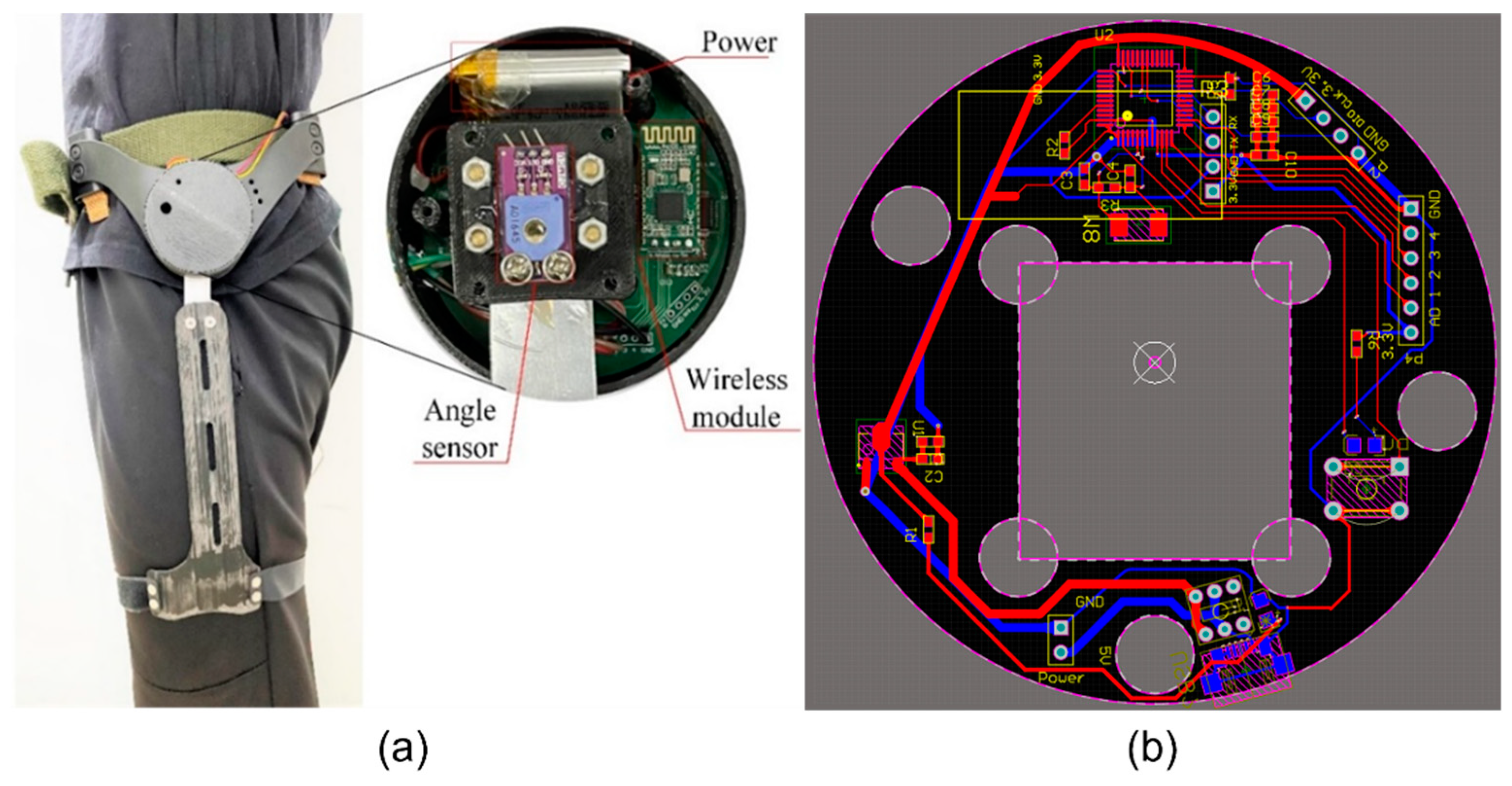
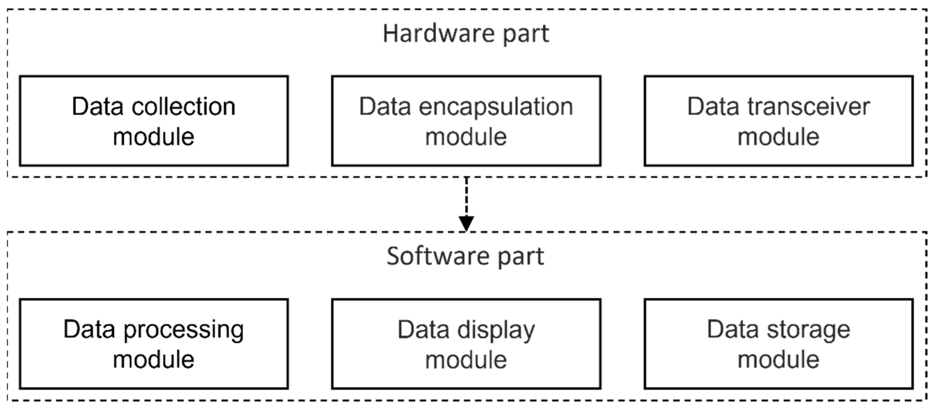
2.2. Gait Information Collection Device

The hip and knee joints of humans were used in this study to gather data on lower limb joint angles utilizing angle sensors. The data gathered by all four joints was transferred to the host computer via a module inside the left hip joint, where the control circuit of the collection system was housed. The left hip joint collection device is shown in Figure 2a. The device’s angle sensor features a pivot shaft attached to the thigh linkage to track the movement of the thigh. The hardware in this device is in the LQP48 package, which has excellent computational performance and low power consumption, and employs STM32F103C8T6 as the main control chip. To finish the data transfer from the lower computer to the upper computer, they were connected to a Bluetooth communication module via the serial port. The board was built with a 35 mm radius circular for simple installation in the housing, with the hollow part serving as the mounting place for the angle sensor (as illustrated in Figure 2b). An angle sensor with a measurement range of 0–333 degrees was chosen for the rotor of the wearable device to gather the angle data of the hip and knee joints in the range of 0–90 degrees rotation during the walking process. The device’s measured operating voltage, current, and total power consumption were 3.6 V, 6.7 mA, and 24.12 mW, respectively. A lithium battery with a 3.7 V and 500 mA standard served as the built-in power source. Figure 3a depicts the wearable collection device’s actual operation, showing how well it fits the human torso, thigh, and calf torso to enable accurate data collection from the angle sensor. The device’s bottom portion was fastened to the thigh with a bandage, while the upper portion was secured to the human torso using a belt. During the walking process, the lower arm movement was consistent with the thigh movement to achieve accurate measurement of hip joint angle information. The other component was the knee joint collection device, which measures the knee joint angle by having its top part fixed to the thigh and its lower part fixed to the calf. Figure 3b depicts the procedure for gathering information about the subject’s gait.

2.3. Experimental Program

Participants were told not to perform any strenuous exercise 48 h before the start of the experiment. All experiments were conducted between 11 am and 5 pm. The experimental process was carried out on a treadmill. Angle sensors were placed on both sides of the hip joint. The main body of the angle sensor was connected to the human body and remained relatively static, and the rotor of the angle sensor was kept in alignment with the thigh with a mechanical device. There was an angle sensor on each hip joint to ensure that information for both the left and right hip joints was collected. The device had a sampling frequency of 100 Hz to ensure the accuracy of the hip angle data. Figure 4 depicts the gait information collection system’s overall design. There are hardware and software components to the system. In the previous paragraph, the hardware component of the gait information collection system was thoroughly explained. This component is primarily in charge of analog-to-digital data conversion, data collection, data encapsulation into frames, and data sending and receiving. The gait information collection system’s software component was created using the Python programming language, and its features include data processing, data display, and data storage.

2.4. Fatigue Inducement

In this study, a four-stage experiment was designed to represent four fatigue levels. The four fatigue levels corresponded to 60, 70, 80 and 90% of the maximum exercise intensity, and there were five fatigue levels in total when including the resting state. The subjects were asked to wear a hip device and walk slowly on a treadmill for one minute while their gait information was recorded while they were in a non-fatigued state. In the four-stage experiment, each level of the experiment lasted 3 min. After the experiment, gait data were collected during slow walking for one minute. Five to ten minutes of rest between experimental levels was provided so that the subject did not feel tired, and heart rate had returned to resting levels.

2.5. Data Collection

The experimental data were transmitted to the computer through the built-in circuit of the hip joint device in real time, and the computer client displayed diagrams of the left and right hip joint angle changes. The collected gait information of the subjects contained high-frequency noise, which was filtered out with a 20 Hz Butterworth filter. The first and second derivatives of the angle signal are the angular velocity and angular acceleration data, respectively, of each signal (Figure 5). The experimental data were labeled with the corresponding fatigue level 0–4 and reported by exercise intensity level.

2.6. Data Segmentation

The most popular gait segmentation algorithm at present is based on the peak detection algorithm, which has high feasibility and is strongly dependent on the data. It can segment the gait cycle but cannot detect the type of human movement. In this study, the dynamic time warping (DTW) algorithm was used to divide the gait cycle not only to achieve higher accuracy of gait segmentation than that achieved with peak detection but also to identify the type of subject movement [23]. The DTW algorithm first constructs a sequence template of the process of motion, then the matching distance is calculated by matching the signal with the template and, finally, the local minimum value of the matching distance is selected as the starting point and end point for gait segmentation [24]. This study used a sequence from one heel strike to the next heel strike as a representative gait cycle (RGC). In the experiment, only the gait information for walking was collected, and there was no need to recognize gait type. The hip joint angles were segmented simultaneously using the signal of the right hip joint as a reference. After segmentation, the length of an RGC is different, which causes problems for subsequent machine learning. In this study, the data were normalized by length, and an RGC was represented by a 150-point sequence. Figure 6 shows the sequence of the hip joint angle over time, and the start of the RGC is defined as the time when the right heel touches the ground [25]. The angle represents the angle between the subject’s thigh and the back of the body.

2.7. Machine Learning Algorithm

There are two existing gait recognition methods. One is classical machine learning, such as logistic regression, SVM, decision tree, gradient boosting decision tree (GBDT), and random forest models, and the other is gait recognition based on a neural network model [26]. Logistic regression achieves the classification task for linear models and shows better performance for linearly separable models. SVMs are a kind of generalized linear classifier that classify data according to a supervised learning method, and the decision boundary is identified by finding the maximum distance hyperplane [27]. Compared with logistic regression, SVMs have greatly improved the classification performance of nonlinear models [28]. The random forest model involves a collection of decision trees. Compared with traditional machine learning algorithms, including SVMs and neural networks, this algorithm shows better results in regression analysis and classification applications of strain sensor data analysis. The random forest model is robust to outliers, nonlinear and unbalanced data and has low or medium bias. No neural network model was used in this study because deep learning usually requires a large amount of data to train an effective model.

2.8. Performance Assessment

In previous studies, the accuracy was used to assess the performance of the model. Gholami et al. [29] evaluated fatigue during running by using flexible variable materials, the performance of the model was compared using R-squared (R2) and root mean squared error (RMSE), and the hip sensor-only model had the highest overall accuracy. RMSE is necessary for the assessment of model performance. The distance between the predicted fatigue level and the actual fatigue level should be considered as one of the performance aspects of the model. The mean absolute error (MAE) is the mean value of the error between the predicted value and the true value of the discrete variable. The calculation formula is as follows:
M A E ( Y , Y ) = 1 n i = 0 n | Y i Y i |
where Y i represents the true fatigue level, Y i represents the predicted fatigue level, and n represents the total number of samples.
In the case of the same accuracy, the smaller the prediction deviation, the better the performance of the model. In this study, MAE and accuracy were used to assess the performance of the model. The distance between the predicted fatigue level and the real fatigue level affects the MAE. A better prediction model maintains a low MAE while ensuring accuracy.

3. Results and Discussion

The gait data were input into the machine learning algorithm, and a performance assessment of the model was conducted by using fivefold cross validation. The experiment compared the effects of different machine learning models on fatigue identification performance, and the performance of different kernels in the SVM model was assessed.

3.1. Analysis of the Gait Sequence Data

The sequence of hip angle, angular velocity and angular acceleration were used as input for the classification model, and the accuracy and average absolute errors of the model are shown in Table 2, where R, Rv, Ra, L, Lv, and La represent the right hip angle, angular velocity, and angular acceleration data and the left hip angle, angular velocity, and angular acceleration data, respectively. It can be seen from the results that logistic regression had low performance in predicting fatigue levels, indicating that the fatigue states caused by running are not distinguishable in a strictly linearly manner. In the random forest model, the accuracy of the left and right hip angles reached 87.23%, and the MAE was 0.21, which was close to that of the overall performance. The angular velocity and angular acceleration data represent data redundancy in the random forest model and do not contribute to the classification of fatigue level [30]. The performance of the GBDT achieved an accuracy of 79.50% and an MAE of 0.36. In this model, the addition of angular velocity brought about a performance decline, and the angle data accounted for most of the overall performance.
The influences of three different kernel schemes (i.e., linear, polynomial, and radial basis function) were investigated for SVM classification. The RBF kernel is a strong local kernel that can map a sample to a higher dimensional space, and had high performance in gait data classification, with an accuracy of 88.85% and an MAE of 0.17. The linear kernel was mainly applied to linearly separable data, so its performance was low, reaching 51.62% accuracy and an MAE of 0.79. The polynomial kernel also had a certain classification ability for nonlinear data, and its performance was slightly better than that of linear and sum functions, with 60.79% accuracy and an MAE of 0.60.
Performance assessment of the model showed that the angle information of the left and right hip joints had high performance in fatigue prediction. The use of acceleration and angular acceleration data did not significantly improve the performance of the model. The accuracy of the GBDT was improved by approximately 1%, and the accuracy of the SVM was improved by approximately 7%. The first and second derivatives of angle could not obtain better performance [31]. The MAE of each model was positively correlated with accuracy.
Figure 7 shows the distribution of predicted states from various models. Although the logistic regression model showed the worst performance assessment in predicting fatigue levels, it performed well in predicting fatigue level 0 and fatigue level 4. This model can be used for binary detection of fatigue and non-fatigue. Other models indicate that the number of error samples decreased as the distance between the actual state and the predicted state increased.

3.2. Statistical Analysis of the Gait Sequence Data

The above mainly introduced the performance of hip joint angle, angular velocity and angular acceleration information in various machine learning classification models. The SVM with an RBF kernel obtained the best performance, with an accuracy of 88.85% and an MAE of 0.17. The mean, maximum, minimum, peak-to-peak value, variance, kurtosis, and skewness of the hip angle, angular velocity, and angular acceleration data were examined. These statistical data were also used as model inputs, and their performance results are reported. Table 3 shows the performance of the statistical data in the different models.
According to Table 3, statistical data can also achieve high performance, and the logistic regression model still shows very poor performance. The performance of the random forest model was low when using the unilateral hip angle data, and the performance using the left and right hip angle data best reflected the overall performance. The statistical data of the gait sequence in the GBDT model were slightly better at predicting fatigue level than the gait sequence data. Random forest and GBDT models are more suitable for scenarios with small amounts of data and limited computational ability. The performance of the SVM using the statistical data of the gait sequence as the training input was low. SVMs require a large amount of data, and it is difficult to predict fatigue level using a small amount of data. The fatigue level predictions of various models are shown in the Figure 8.

4. Conclusions

In this study, a wearable hip recognition system was used to predict fatigue levels after running. An angle sensor was used to collect hip joint angle data for different fatigue levels. Performance assessment of various classification models in identifying fatigue states was also conducted. The experimental results show that although the fatigue state of the subject changed slightly during running, data preprocessing and machine learning algorithms enabled accurate fatigue state prediction. Fatigue caused by running could be reflected in the gait of the subject, and changes in gait could be effectively predicted by SVMs, which had the best performance. The performance of the SVM model shows that the accuracy of the prediction with data from one hip joint can be close to the accuracy achieved with all the data, and the first and second derivatives of the hip angle show the redundancy of the data. Using gait sequence data for model training is very computationally heavy. A few statistical variables on the gait sequence could still achieve performance similar to that of gait sequence data, which made fatigue prediction independent of computer hardware and has great significance for real-time prediction of fatigue level. In addition, a wearable device was used to predict the fatigue level of the lower extremity musculature during walking. An angle sensor was used to collect data on changes in the sagittal plane motion of the hip joint. The device can detect abnormal gait information during high-intensity work, especially in people performing lower extremity movements for long periods of time, to reduce the risk associated with fatigue. The wearable device provides a convenient fatigue-state monitoring solution for people who require lower extremity exoskeleton devices. The device is simple to install on a human exoskeleton device and offers a convenient way to collect data and less difficult way to assess fatigue level.

Author Contributions

Conceptualization, S.S. and Z.C.; methodology, S.S. and Z.C.; software, S.S. and H.L.; validation, S.S. and Z.C.; formal analysis, S.S. and C.D.; investigation, S.S. and Z.C.; resources, S.S. and Z.C.; data curation, S.S. and Y.L.; writing—original draft preparation, S.S. and Q.W.; writing—review and editing, S.S. and Z.C.; visualization, S.S.; supervision, Z.C.; project administration, S.S.; funding collection, Z.C. All authors have read and agreed to the published version of the manuscript.

Funding

This work was financially supported by National Natural Science Foundation of China (Grant No.61372044).

Data Availability Statement

All data included in this study are available upon request by contact with the corresponding author.

Conflicts of Interest

The authors declare no conflict of interest.

References

  1. Pedisic, Z.; Shrestha, N.; Kovalchik, S.; Stamatakis, E.; Liangruenrom, N.; Grgic, J.; Titze, S.; Biddle, S.J.; Bauman, A.E.; Oja, P. Is running associated with a lower risk of all-cause, cardiovascular and cancer mortality, and is the more the better? A systematic review and meta-analysis. Br. J. Sports Med. 2019, 54, 898–905. [Google Scholar] [CrossRef] [PubMed]
  2. Oja, P.; Titze, S.; Kokko, S.; Kujala, U.M.; Heinonen, A.; Kelly, P.; Koski, P.; Foster, C. Health benefits of different sport disciplines for adults: Systematic review of observational and intervention studies with meta-analysis. Br. J. Sport. Med. 2015, 49, 434–440. [Google Scholar] [CrossRef] [PubMed] [Green Version]
  3. Van Gent, R.N.; Siem, D.; van Middelkoop, M.; Van Os, A.G.; Bierma-Zeinstra, S.M.A.; Koes, B.W. Incidence and determinants of lower extremity running injuries in long distance runners: A systematic review. Br. J. Sport. Med. 2007, 41, 469–480. [Google Scholar] [CrossRef] [PubMed] [Green Version]
  4. Derrick, T.R.; Dereu, D.; McLean, S.P. Impacts and kinematic adjustments during an exhaustive run. Med. Sci. Sports Exerc. 2002, 34, 998–1002. [Google Scholar] [CrossRef]
  5. Mizrahi, J.; Verbitsky, O.; Isakov, E.; Daily, D. Effect of fatigue on leg kinematics and impact acceleration in long distance running. Hum. Mov. Sci. 2000, 19, 139–151. [Google Scholar] [CrossRef]
  6. Clansey, A.C.; Hanlon, M.; Wallace, E.S.; Lake, M.J. Effects of Fatigue on Running Mechanics Associated with Tibial Stress Fracture Risk. Med. Sci. Sports Exerc. 2012, 44, 1917–1923. [Google Scholar] [CrossRef]
  7. Winter, S.; Gordon, S.; Watt, K. Effects of fatigue on kinematics and kinetics during overground running: A systematic review. J. Sports Med. Phys. Fit. 2017, 57, 887–899. [Google Scholar] [CrossRef]
  8. Donelan, J.M.; Naing, V.; Li, Q. Biomechanical energy harvesting. In Proceedings of the 2009 IEEE Radio and Wireless Symposium, San Diego, CA, USA, 18–22 January 2009; IEEE: Piscataway, NJ, USA, 2009. [Google Scholar]
  9. Szeidl, C.A. Consumption Commitments and Risk Preferences. Q. J. Econ. 2007, 122, 831–877. [Google Scholar]
  10. Koblbauer, I.F.; van Schooten, K.S.; Verhagen, E.A.; van Dieen, J.H. Kinematic changes during running-induced fatigue and relations with core endurance in novice runners. J. Sci. Med. Sport 2014, 17, 419–424. [Google Scholar] [CrossRef]
  11. Gerlach, K.E.; White, S.C.; Burton, H.W.; Dorn, J.M.; Leddy, J.J.; Horvath, P.J. Kinetic changes with fatigue and relationship to injury in female runners. Med. Sci. Sports Exerc. 2005, 37, 657–663. [Google Scholar] [CrossRef]
  12. Hussain, S.; Xie, S.Q.; Jamwal, P.K. Robust nonlinear control of an intrinsically compliant robotic gait training orthosis. IEEE Trans. Syst. Man Cybern. Syst. 2013, 43, 655–665. [Google Scholar] [CrossRef]
  13. Hussain, S.; Xie, S.Q.; Jamwal, P.K. Adaptive impedance control of a robotic orthosis for gait rehabilitation. IEEE Trans. Cybern. 2013, 43, 1025–1034. [Google Scholar] [CrossRef] [PubMed]
  14. Borg, G. Perceived exertion as an indicator of somatic stress. Scand. J. Rehabil. Med. 1970, 2, 92–98. [Google Scholar] [PubMed]
  15. Baghdadi, A.; Megahed, F.M.; Esfahani, E.T.; Cavuoto, L.A. A machine learning approach to detect changes in gait parameters following a fatiguing occupational task. Ergonomics 2018, 61, 1116–1129. [Google Scholar] [CrossRef]
  16. Janssen, D.; Schollhorn, W.I.; Newell, K.M.; Jager, J.M.; Rost, F.; Vehof, K. Diagnosing fatigue in gait patterns by support vector machines and self-organizing maps. Hum. Mov. Sci. 2011, 30, 966–975. [Google Scholar] [CrossRef]
  17. Zhang, J.; Lockhart, T.E.; Soangra, R. Classifying Lower Extremity Muscle Fatigue During Walking Using Machine Learning and Inertial Sensors. Ann. Biomed. Eng. 2014, 42, 600–612. [Google Scholar] [CrossRef] [Green Version]
  18. Hajifar, S.; Sun, H.Y.; Megahed, F.M.; Jones-Farmer, L.A.; Rashedi, E.; Cavuoto, L.A. A forecasting framework for predicting perceived fatigue: Using time series methods to forecast ratings of perceived exertion with features from wearable sensors. Appl. Ergon. 2021, 90, 103262. [Google Scholar] [CrossRef]
  19. Noehren, B.; Sanchez, Z.; Cunningham, T.; McKeon, P.O. The effect of pain on hip and knee kinematics during running in females with chronic patellofemoral pain. Gait Posture 2012, 36, 596–599. [Google Scholar] [CrossRef]
  20. Bazett-Jones, D.M.; Cobb, S.C.; Huddleston, W.E.; O’Connor, K.M.; Armstrong, B.S.R.; Earl-Boehm, J.E. Effect of Patellofemoral Pain on Strength and Mechanics after an Exhaustive Run. Med. Sci. Sports Exerc. 2013, 45, 1331–1339. [Google Scholar] [CrossRef]
  21. Dierks, T.A.; Manal, K.T.; Hamill, J.; Davis, I. Lower Extremity Kinematics in Runners with Patellofemoral Pain during a Prolonged Run. Med. Sci. Sports Exerc. 2011, 43, 693–700. [Google Scholar] [CrossRef]
  22. Impellizzeri, F.M.; Marcora, S.M.; Coutts, A.J. Internal and External Training Load: 15 Years On. Int. J. Sports Physiol. Perform. 2019, 14, 270–273. [Google Scholar] [CrossRef] [PubMed]
  23. Barth, J.; Oberndorfer, C.; Pasluosta, C.; Schulein, S.; Gassner, H.; Reinfelder, S.; Kugler, P.; Schuldhaus, D.; Winkler, J.; Klucken, J.; et al. Stride Segmentation during Free Walk Movements Using Multi-Dimensional Subsequence Dynamic Time Warping on Inertial Sensor Data. Sensors 2015, 15, 6419–6440. [Google Scholar] [CrossRef] [PubMed] [Green Version]
  24. Ghassemi, N.H.; Hannink, J.; Martindale, C.F.; Gassner, H.; Muller, M.; Klucken, J.; Eskofier, B.M. Segmentation of Gait Sequences in Sensor-Based Movement Analysis: A Comparison of Methods in Parkinson’s Disease. Sensors 2018, 18, 145. [Google Scholar] [CrossRef] [Green Version]
  25. Barton, J.G.; Lees, A. An application of neural networks for distinguishing gait patterns on the basis of hip-knee joint angle diagrams. Gait Posture 1997, 5, 28–33. [Google Scholar] [CrossRef]
  26. Wang, X.H.; Yan, W.Q. Non-local gait feature extraction and human identification. Multimedia Tools Appl. 2021, 80, 6065–6078. [Google Scholar] [CrossRef]
  27. Figueiredo, J.; Santos, C.P.; Moreno, J.C. Automatic recognition of gait patterns in human motor disorders using machine learning: A review. Med. Eng. Phys. 2018, 53, 1–12. [Google Scholar] [CrossRef]
  28. Lau, H.Y.; Tong, K.Y.; Zhu, H.L. Support vector machine for classification of walking conditions of persons after stroke with dropped foot. Hum. Mov. Sci. 2009, 28, 504–514. [Google Scholar] [CrossRef]
  29. Gholami, M.; Napier, C.; Patino, A.G.; Cuthbert, T.J.; Menon, C. Fatigue Monitoring in Running Using Flexible Textile Wearable Sensors. Sensors 2020, 20, 5573. [Google Scholar] [CrossRef]
  30. Slijepcevic, D.; Zeppelzauer, M.; Schwab, C.; Raberger, A.M.; Breiteneder, C.; Horsak, B. Input representations and classification strategies for automated human gait analysis. Gait Posture 2020, 76, 198–203. [Google Scholar] [CrossRef]
  31. Burdack, J.; Horst, F.; Giesselbach, S.; Hassan, I.; Daffner, S.; Schollhorn, W.I. Systematic Comparison of the Influence of Different Data Preprocessing Methods on the Performance of Gait Classifications Using Machine Learning. Front. Bioeng. Biotechnol. 2020, 8, 260. [Google Scholar] [CrossRef]
Figure 1. System flowchart of hip gait data activity analysis. The wearable device collects hip angle data. The data are preprocessed and used for model learning. A performance assessment of various classification models in identifying fatigue states is given.
Electronics 11 03514 g001
Figure 2. (a) Wearable hip gait collection device. (b) Circuit design of the collection system.
Electronics 11 03514 g002
Figure 3. (a) The overall wearing effect of the collection device. (b) Subject’s gait information collection process.
Electronics 11 03514 g003
Figure 4. Collection system design.
Electronics 11 03514 g004
Figure 5. Hip gait data and filtered data.
Electronics 11 03514 g005
Figure 6. Gait data segmentation base on DTW.
Electronics 11 03514 g006
Figure 7. (a) Logistic regression prediction results. (b) Random forest prediction results. (c) GBDT prediction results. (d) SVM RBF prediction results. (e) SVM linear prediction results. (f) SVM poly prediction results.
Electronics 11 03514 g007
Figure 8. (a) Logistic regression prediction results. (b) Random forest prediction results. (c) GBDT prediction results. (d) SVM RBF prediction results. (e) SVM linear prediction results. (f) SVM poly prediction results.
Electronics 11 03514 g008
Table 1. Correspondence between exercise intensity and speed gradient in the three-level load experiment.
First LevelSecond LevelThird Level
ManWomanManWomanManWoman
EC (METs)5476108
V (m/min) +A (%)4.9 + 44.64 + 26.3 + 05.5 + 58.3 + 87.0 + 1
Table 2. Performance of hip joint angle, angular velocity and angular acceleration sequences in different models.
RLR LR RvR Rv L LsR Rv Ra L Lv La
Logistic regressionAccuracy (%)25.5431.8334.7125.5425.5438.31
MAE1.601.291.241.601.601.11
Random forestAccuracy (%)80.0477.5287.2380.0480.0487.95
MAE0.330.380.210.330.330.19
GBDTAccuracy (%)69.9666.9178.4269.9669.9679.50
MAE (%)0.510.570.370.510.510.36
SVM RBFAccuracy (%)81.1281.1281.6581.6581.2988.85
MAE0.350.350.340.340.330.17
SVM linearAccuracy (%)37.2342.6349.1039.3951.6251.62
MAE1.161.010.841.100.780.79
SVM polyAccuracy (%)39.7540.2960.4341.9161.3360.97
MAE1.111.020.631.040.610.60
Table 3. Performance of the statistical data for hip joint angle, angular velocity and angular acceleration in different models.
RLR LR RvR Rv L LsR Rv Ra L Lv La
Logistic regressionAccuracy (%)21.2223.0231.8324.1030.4028.60
MAE1.551.231.261.451.271.35
Random forestAccuracy (%)69.7869.9687.5974.4689.0387.41
MAE0.530.530.220.410.160.18
GBDTAccuracy (%)61.8766.1978.6068.7183.0982.01
MAE (%)0.670.620.350.500.250.28
SVM RBFAccuracy (%)28.4228.7837.5931.4737.7739.03
MAE1.291.351.211.251.191.21
SVM linearAccuracy (%)19.2425.9029.1430.2226.0822.66
MAE1.431.321.171.161.631.56
SVM polyAccuracy (%)22.1222.6633.6325.0035.9737.59
MAE1.561.291.281.511.171.17
Publisher’s Note: MDPI stays neutral with regard to jurisdictional claims in published maps and institutional affiliations.

