1. Introduction
Knee osteoarthritis (OA) is a common chronic degenerative disease [
1] and is a leading cause of global disability [
2,
3]. It is estimated that, by 2030, the prevalence of symptomatic knee OA will reach 30%, attributed to increasing life expectancy and the rising number of persons with obesity [
4]. Knee OA is typically a progressive disease of the whole synovial joint characterized by subchondral bone proliferation, bone misalignment, and synovitis [
5,
6]. As a consequence of these changes, joint pain and functional disability may ensue [
7].
While patients and health providers desire effective knee OA therapy, few effective long-term remedies have been identified. Treatments of knee OA commonly rely on traditional non-pharmacologic management (e.g., weight loss, exercise), which is advocated by most therapeutic guidelines, to relieve pain, improve function, prevent deformities, and slow the progression of the disease [
8]. Unfortunately, results are inconsistent across patient populations with these treatment options, and there are not set devices facilitating these treatments. Some other knee OA therapy methods currently being studied include acupuncture [
9,
10], transcutaneous electrical nerve stimulation (TENS) [
11,
12,
13], and low-level laser therapy (LLLT) [
14,
15,
16,
17,
18].
Phototherapy has been applied clinically in the treatment of soft tissue injuries and accelerate inflammation healing for more than 40 years [
19,
20]. Light-emitting diodes (LEDs) are light sources based on the semiconductor, and effects of different wavelengths of LEDs have showed efficacy in a variety of clinical conditions. Phototherapy using red LED arrays has also been reported to have anti-inflammatory properties, by influencing the release of cytokines from various kinds of cells. At the cellular level, the mechanism of phototherapy has been ascribed to the activation of mitochondrial respiratory chain components, resulting in initiation of a signaling cascade that promotes cellular proliferation and cytoprotecton [
21].
Researchers have proposed many phototherapy devices for knee OA in recent years. Gomes et al. presented a phototherapy with a nine-diode cluster device: one 905-nm super-pulsed diode laser, four 875-nm LED, and four 640-nm LED [
22]. Yasushi et al. used a therapeutic device for the treatment of knee OA, which had high-intensity red LEDs. The LEDs were mounted to the underside of the custom-designed brace, the brace was fitted on the knee joint, and then wrapped with bandaging [
23]. However, it is not portable due to its large size, resulting in poor efficiency in the clinic.
In this paper, we present an innovative device designed to help individuals with knee OA. Unlike many of the existing knee OA therapy devices, this device is wearable, flexible, and does not require physical activity. Patients can be treated standing or lying down as the device is attached to the knee, but does not generate any motion. SolidWorks, an outstanding 3D design tool, is utilized to build the 3D model of the phototherapy device. The red LED with emission wavelength of 630 nm is chosen as a light source, and the structure and parameters of the LED array are optimized by simulating optical analysis with TracePro. The hardware control system is constructed by an STM32 microcontroller. This article presents a constant current drive circuit based on an MOS tube, and provides a circuit design and system implementation.
2. Materials and Methods
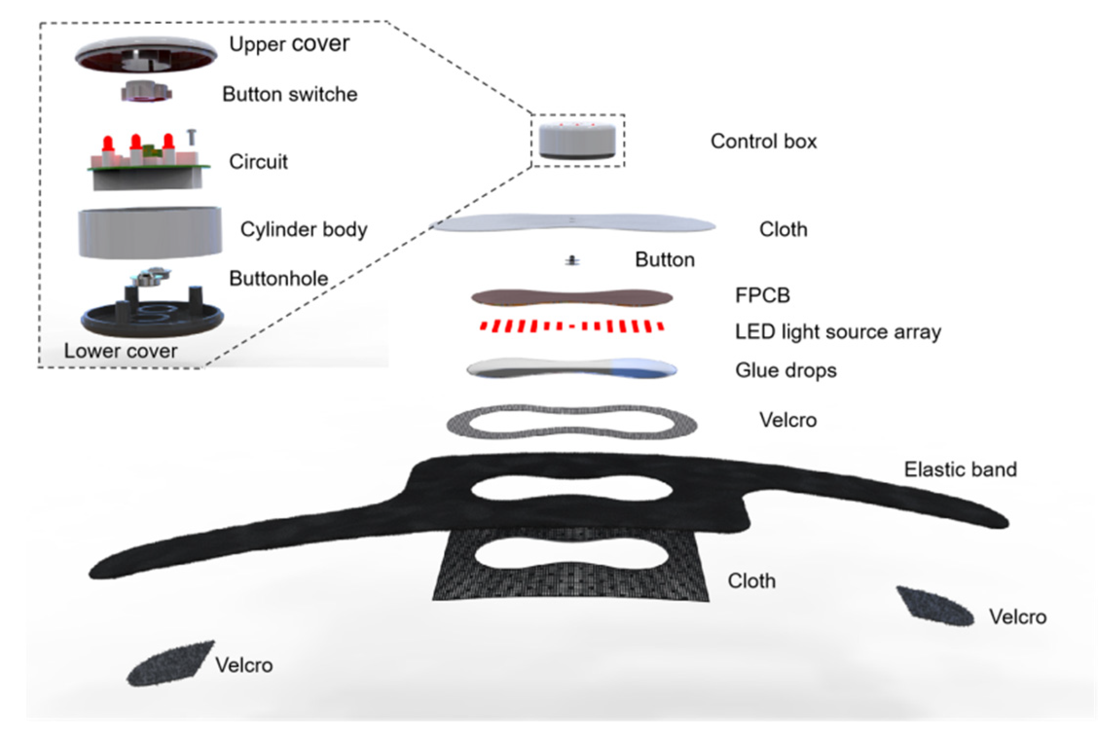
The phototherapy device is a set of device designs, electronic circuit designs, and system layouts for control box and phototherapy adhesive patch that incorporate arrays of centimeter-scale LEDs in soft sheets of flexible circuit board that laminate directly onto the skin in a simple, non-invasive manner.
Figure 1 demonstrates the 3D model of the phototherapy device for knee OA. The mechanical structure contains a control box and a phototherapy adhesive patch. The shell of the control box was created with 3D-printed molds using PVC materials. The overall structure is similar to a cylinder with a diameter of 50 mm and a height of 25 mm. It has the advantages of simplicity and practicality.
The control box comprises an upper cover, a cylinder body, and a lower cover. The upper cover was designed with two button switches and three LED indicators. The two button switches used mechanical elastic button switches, respectively, to achieve power switch and irradiance adjustment. The three LED indicators were associated with three brightness levels with irradiance; the greater the irradiance, the more LED indicators that light up. The cylinder body was equipped with a Type-C data interface, which was used to charge the built-in battery. In order to provide the necessary control systems and power supplies for the phototherapy adhesive patch, the lower cover was designed with two buttonhole, which could be connected with the buttons on the phototherapy adhesive patch.
The phototherapy adhesive patch included two buttons and a LED flexible printed circuit board. Through the connection between the button of the phototherapy adhesive patch and the buttonhole of the lower cover of the control box, the two modules were integrated to realize the function of phototherapy. The LED flexible printed circuit board (FPCB) is a highly reliable and flexible printed circuit made from a flexible insulating substrate, such as polyimide or polyester film. Thanks to the characteristics of light weight, thin thickness, and good bending performance, it could be closed to the curved surfaces of the knee skin.
2.1. The LED Array
Based on the analysis and summary of the pathogenesis and treatment principle of knee OA, the red LED with emission wavelength of 630 nm and rated power of 0.2 W was chosen as light source.
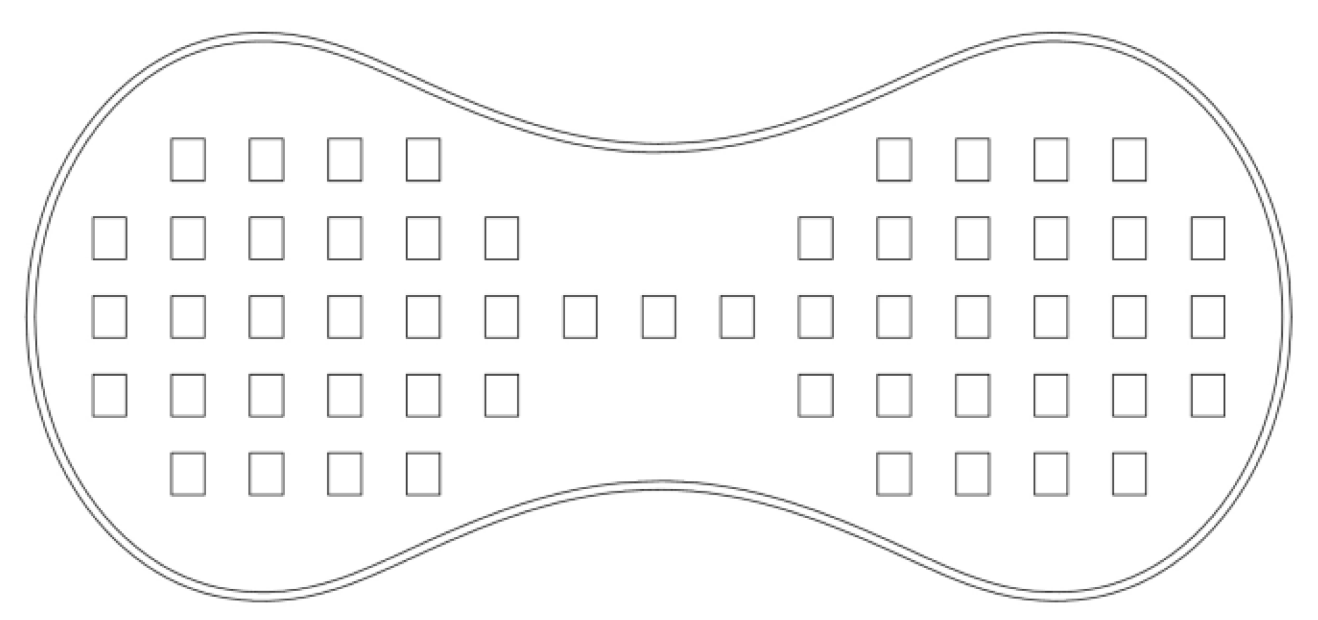
Figure 2 shows the LED array, consisting of 55 LEDs which were driven by a constant current drive module. The output power could be adjusted continuously through the pulse width modulation (PWM) signal, which were applied to dimming strategies using their feature of the dimming behavior is more linear. The PWM dimming strategy utilized a pulsed current to carry out the control. While the pulsed value of the current was a constant value, the average value of the current, and hence the average value of the luminous flux, depended on the duty ratio of the pulsed current [
24].
In order to improve the irradiance and uniformity on target plane. On the one hand, the LED array was constructed by the way of red LED interval arrangement, which could not only make up for the lack of irradiance through the superposition of illumination, but also effectively expand the irradiance area [
25,
26]. On the other hand, as the knee is a free-form curved surface, it was easy to laminate onto the knee skin by FPCB compared to traditional printed circuit board (PCB). Moreover, secondary optics based on freeform-surface lens would result in a poor user experience. In this paper, the integrated glue dropping process was adopted to cover the surface of the FPCB with glue drops evenly, which could improve the uniformity of irradiance and have the effect of waterproof [
27].
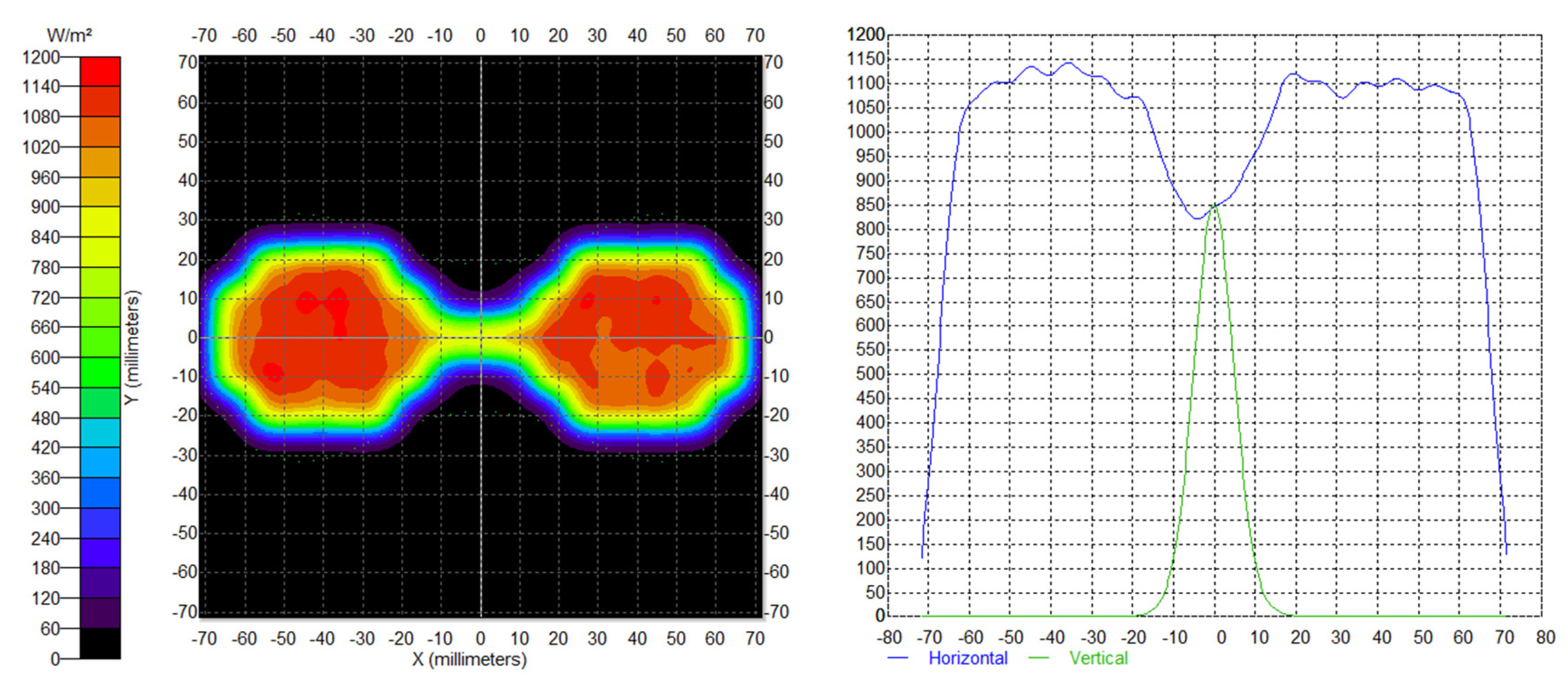
The optical model of the LED array was established by TracePro software, and the ray-tracing simulations of LED array was performed. The irradiation distance was set to 3 mm.
Figure 3 shows the simulating results. The maximum irradiance of the target plane was 1160.3 W/m
2, Average irradiance was 703.66 W/m
2. The uniform irradiance of the target plane reached 60.64%.
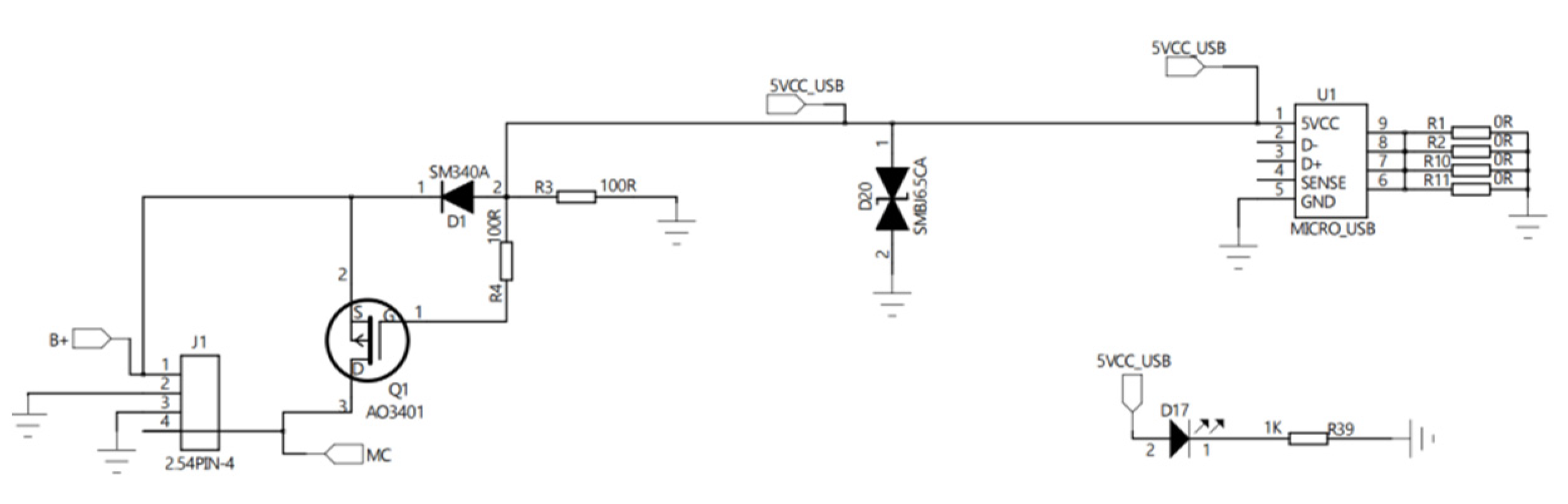
2.2. The Constant Current Drive Module
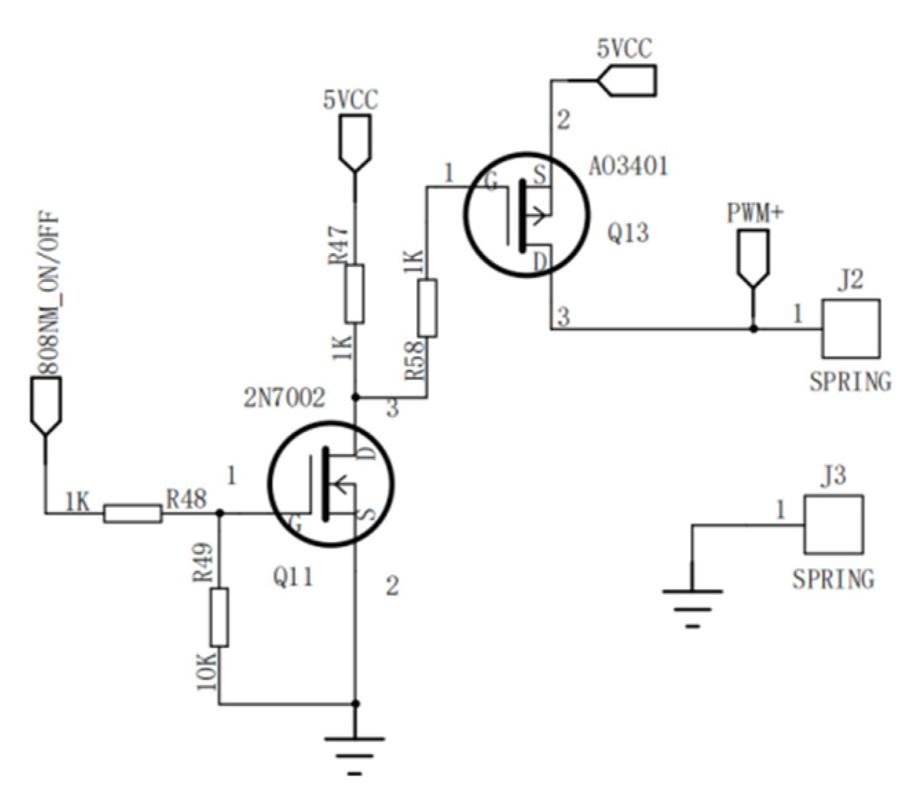
The constant current drive module used two MOS tubes to drive the LED array. The circuit outputted high and low levels through the STM32 microcontroller to control the time of MOS tube opening and closing, and then outputted an PWM signal to drive the LED array. When 80NM_ON/OFF outputted a high level, Q11 was ON, Q13 was ON, and the output was high. When 80NM_ON/OFF outputted a low level, Q11 was OFF, Q13 was OFF, and the output was low-level. Thus, the PWM signal was generated to control the LED array.
Figure 4 is a schematic diagram of the constant current drive module.
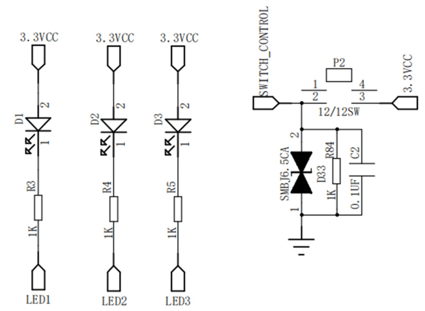
2.3. The Button Control Module
The button control module included two button switches and three LED indicators. The two button switches with mechanical elastic button switches, respectively, were used to achieve power switch and irradiance adjustment. The mechanical elastic button switch was equivalent to send a single pulse signal to the controller, which closed when pressed and automatically disconnected when released. The three LED indicators associated three brightness levels with irradiance. Different irradiance could be changed by adjusting different duty ratio.
Figure 5 is a schematic diagram of the button control module.
2.4. The Power Supply Module
In order to improve the portability and durability of the device, it was powered by a 3.7-V rechargeable lithium battery, which was charged by USB port. A SMBJ6.5CA diode was used as charging protection. When the 5-V power supply was connected, the D7 diode was switched on, the 5 V was connected to the positive electrode of the battery, and finally the battery began to charge.
Figure 6 is a schematic diagram of the power supply module.
3. Testing and Results
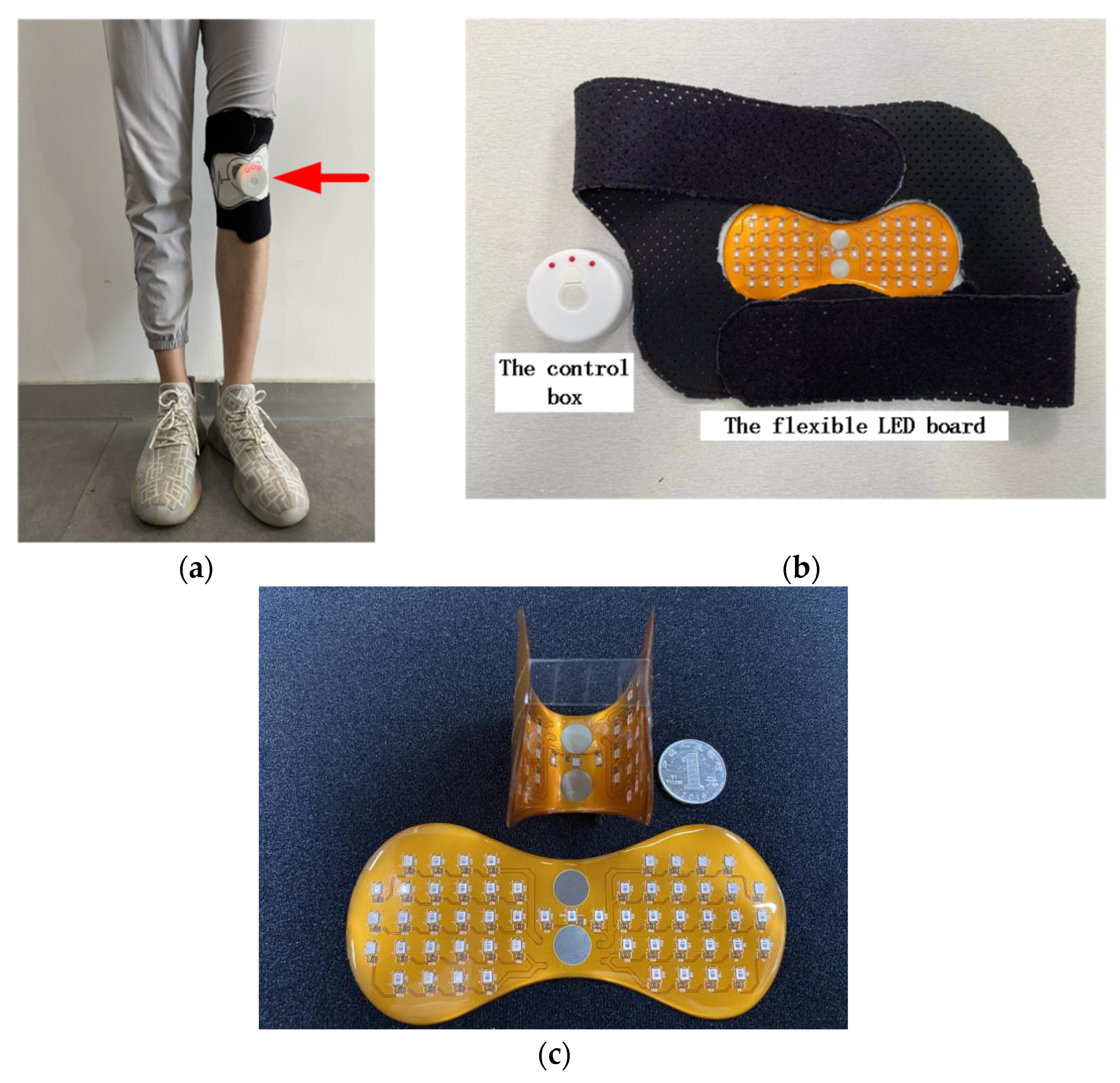
Figure 7b shows the wearable flexible phototherapy device for knee OA, incorporating a flexible LED board and a control box, the weight of which was only 101.8 g. The elastic band was worn on the patient’s knee. Because FPCB has the advantages of light weight, thin thickness, and good bending performance, the flexible LED board had excellent flexibility and stretchability. The bending angle of the flexible LED board could adjust at will between 0–90°, as shown in
Figure 7c.
Because the phototherapy adhesive patch was applied to the patient skin surface, the distance between the probe of spectral radiometer and the LED was small when measuring the irradiance. To ensure measurement accuracy, we selected appropriate measuring points which were vertical below the LED, and the measured irradiance was 13 mW/cm2.
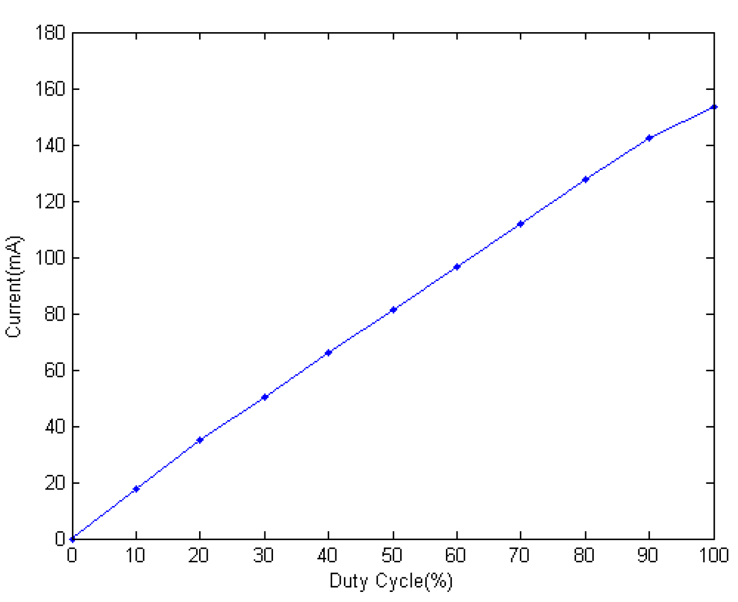
When setting a shift of 10% duty cycle over a total of 10 shifts, a multimeter was used to test and record the output current of the phototherapy adhesive patch.
Figure 8 demonstrates the photoelectric characteristics of the constant current drive module.
The irradiance had a linear relationship with the PWM duty cycle. When the PWM duty cycle was 0, the light irradiance was the minimum, and when the PWM duty cycle was 100%, the light irradiance was the maximum. The linear adjustment of the irradiance was realized, and the adjustment range was 0–13 mW/cm2, which met the requirements of design parameter. When the duty cycle increased, the output current also increased, and the maximum current reached 153.6 mA.
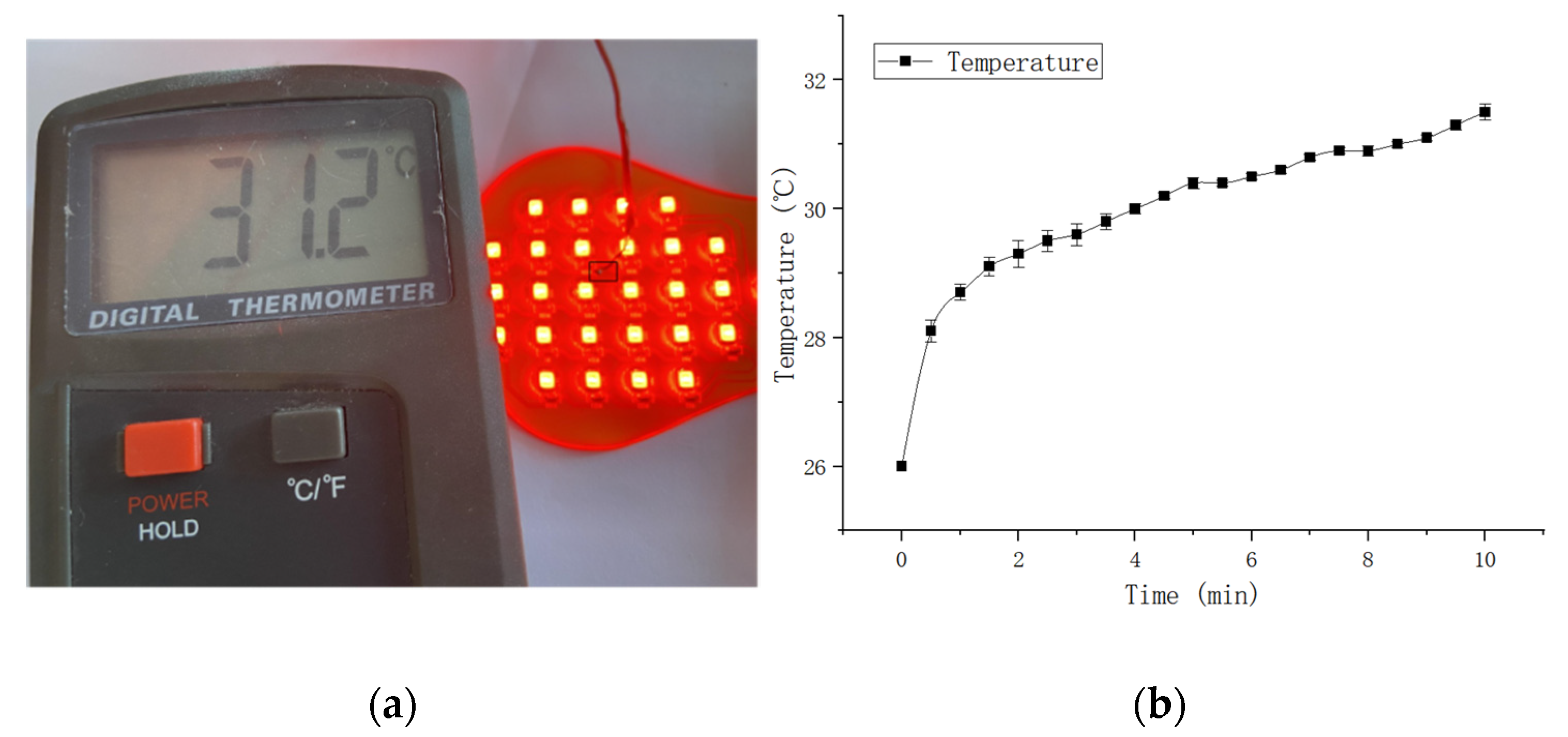
When the device is working, the current generates heat in the LED. According to the national standard GB/T 18153-2000, the main structure of the molded material and contact with the human body for about 10 min of the surface temperature of the equipment should be less than 48 °C. To verify the safety on the human body, the temperature of the device was tested. The device working at maximum irradiance is shown in
Figure 9. When the ambient temperature was about 26 °C, the maximum temperature of the surface of the light source was 31.2 °C during the system operation at the state of maximum light irradiance for 10 min, which is in the safe range.
Compared with other currently available devices, the weight of the device is light and the irradiance could be adjusted linearly. The flexible LED board has good elasticity, as it is made from FPCB which is more pliable than PCB, thus it can be worn on the patient’s knee. To verify the device’s effectiveness of phototherapy on the human body, we will focus on the research in our next-step work clinically.
4. Discussion
In summary, this work presents a wearable flexible phototherapy device worn on the knee for osteoarthritis. In order to improve the irradiance and uniformity, the LED array was constructed by the way of red LED interval arrangement, and the integrated glue dropping process was adopted to cover the surface of the FPCB with glue drops evenly. The design parameters of the LED array was optimized by simulating optical analysis with TracePro. This article presents a constant current drive circuit based on an MOS tube. The linear adjustment of the irradiance was realized, the adjustment range was 0–13 mW/cm2, and the maximum current reached 153.6 mA. The maximum temperature of the surface of the light source was 31.2 °C during the system operation at the state of maximum light irradiance for 10 min, which was in the safe range. As such, the phototherapy device in this paper has broad application prospects in knee OA therapy.
Author Contributions
Investigation, K.L., H.C., Y.W., M.W. and J.T.; methodology, K.L., H.C., Y.W., M.W. and J.T.; software, K.L.; supervision, H.C., Y.W. and J.T.; writing—original draft, K.L. All authors have read and agreed to the published version of the manuscript.
Funding
This research was funded by Science and Technology Service Network Initiative (No. KFJ-STS-QYZD-2021-02-004); the National Key Research and Development Program of China (No. 2016YFC0105604).
Informed Consent Statement
Informed consent was obtained from all subjects involved in the study.
Data Availability Statement
The data presented in this study are available on request from the corresponding author.
Conflicts of Interest
The authors declare no conflict of interest.
References
- Kudo, M.; Watanabe, K.; Otsubo, H.; Kamiya, T.; Kaneko, F.; Katayose, M.; Yamashita, T. Analysis of effectiveness of therapeutic exercise for knee osteoarthritis and possible factors affecting outcome. J. Orthop. Sci. 2013, 18, 932–939. [Google Scholar] [CrossRef] [PubMed]
- Vos, T.; Flaxman, A.D.; Naghavi, M.; Lozano, R.; Michaud, C.; Ezzati, M.; Shibuya, K.; Salomon, J.A.; Abdalla, S.; Aboyans, V.; et al. Years lived with disability (YLDs) for 1160 sequelae of 289 diseases and injuries 1990–2010: A systematic analysis for the Global Burden of Disease Study 2010. Lancet 2012, 380, 2163–2196. [Google Scholar] [CrossRef]
- Murray, C.J.; Vos, T.; Lozano, R.; Naghavi, M.; Flaxman, A.D.; Michaud, C.; Ezzati, M.; Shibuya, K.; Salomon, J.A.; Abdalla, S.; et al. Disability-adjusted life years (DALYs) for 291 diseases and injuries in 21 regions, 1990–2010: A systematic analysis for the Global Burden of Disease Study 2010. Lancet 2012, 380, 2197–2223. [Google Scholar] [CrossRef]
- Croft, P. The epidemiology of osteoarthritis: Manchester and beyond. Rheumatology 2005, 44 (Suppl. 4), iv27–iv32. [Google Scholar] [CrossRef] [PubMed] [Green Version]
- Goldring, S.R. Alterations in periarticular bone and cross talk between subchondral bone and articular cartilage in osteoarthritis. Ther. Adv. Musculoskelet. Dis. 2012, 4, 249–258. [Google Scholar] [CrossRef] [PubMed] [Green Version]
- Burgos-Vargas, R.; Cardiel, M.H.; Loyola-Sánchez, A.; De Abreu, M.M.; Pons-Estel, B.A.; Rossignol, M.; Avouac, B.; Ferraz, M.B.; Halhol, H. Characterization of knee osteoarthritis in latin America. A comparative analysis of clinical and health care utilization in Argentina, Brazil, and Mexico. Reumatol. Clin. 2014, 10, 152–159. [Google Scholar] [CrossRef]
- Hunter, D.J. Osteoarthritis. Best Pract. Res. Clin. Rheumatol. 2011, 25, 801–814. [Google Scholar] [CrossRef]
- Huleatt, J.B.; Campbell, K.J.; La Prade, R.F. Nonoperative treatment approach to knee osteoarthritis in the master athlete. Sports Health 2014, 6, 56–63. [Google Scholar] [CrossRef] [Green Version]
- Kwon, Y.D.; Pittler, M.H.; Ernst, E. Acupuncture for peripheral joint osteoarthritis: A systematic review and meta-analysis. Rheumatology 2006, 45, 1331–1337. [Google Scholar] [CrossRef] [Green Version]
- Larmer, P.J.; Reay, N.D.; Aubert, E.R.; Kersten, P. Systematic review of guidelines for the physical management of osteoarthritis. Arch. Phys. Med. Rehabil. 2014, 95, 375–389. [Google Scholar] [CrossRef]
- Hulme, J.M.; Welch, V.; de Bie, R.; Judd, M.; Tugwell, P. Electromagnetic fields for the treatment of osteoarthritis. Cochrane Database Syst. Rev. 2002, 1, CD003523. [Google Scholar]
- Osiri, M.; Welch, V.; Brosseau, L.; Shea, B.; McGowan, J.L.; Tugwell, P.; Wells, G.A. Transcutaneous electrical nerve stimulation for knee osteoarthritis. Cochrane Database Syst. Rev. 2000, 4, CD002823. [Google Scholar]
- Marks, R.; Ungar, M.; Ghasemmi, M. Electrical muscle stimulation for osteoarthritis of the knee: Biological basis and systematic review. J. Physiother. 2000, 28, 6–20. [Google Scholar]
- Clijsen, R.; Brunner, A.; Barbero, M.; Clarys, P.; Taeymans, J. Effects of low-level laser therapy on pain in patients with musculoskeletal disorders: A systematic review and meta-analysis. Eur. J. Phys. Rehabil. Med. 2017, 53, 603–610. [Google Scholar] [CrossRef]
- Ucurum, S.G.; Kayali, Y. The relationship between pain, muscle strength and lower extremity function in patients with knee osteoarthritis. J. Basic Clin. Health Sci. 2020, 4, 72–76. [Google Scholar]
- Alayat, M.S.M.; Aly, T.H.A.; Elsayed, A.E.M.; Fadil, A.S.M. Efficacy of pulsed Nd: YAG laser in the treatment of patients with knee osteoarthritis: A randomized controlled trial. Lasers Med. Sci. 2017, 32, 503–511. [Google Scholar] [CrossRef]
- Rayegani, S.M.; Raeissadat, S.A.; Heidari, S.; MoradiJoo, M. Safety and effectiveness of low-level laser therapy in patients with knee osteoarthritis: A systematic review and meta-analysis. J. Lasers Med. Sci. 2017, 8, S12–S19. [Google Scholar] [CrossRef] [PubMed] [Green Version]
- Stausholm, M.B.; Naterstad, I.F.; Couppé, C.; Fersum, K.V.; Leal-Junior, E.C.P.; Lopes-Martins, R.Á.B.; Joensen, J. Effectiveness of Low-Level Laser Therapy Associated with Strength Training in Knee Osteoarthritis: Protocol for a Randomized Placebo-Controlled Trial. Methods Protoc. 2021, 4, 19. [Google Scholar] [CrossRef] [PubMed]
- Karu, T. Primary and secondary mechanisms of action of visible to near-IR radiation on cells. J. Photochem. Photobiol. B Biol. 1999, 49, 1–17. [Google Scholar] [CrossRef]
- Karu, T. ChMB: Low Power Laser Therapy. In Biomedical Photonics Handbook; CRC Press: Boca Raton, FL, USA, 2003; Volume 48, pp. 1–25. [Google Scholar]
- Yeager, R.L.; Franzosa, J.A.; Millsap, D.S.; Angell-Yeager, J.L.; Heise, S.S.; Wakhungu, P.; Lim, J.; Whelan, H.T.; Eells, J.T.; Henshel, D.S. Effects of 670-nm phototherapy on development. Photomed. Laser Surg. 2005, 23, 268–272. [Google Scholar] [CrossRef] [PubMed] [Green Version]
- de Paula Gomes, C.A.; Leal-Junior, E.C.; Dibai-Filho, A.V.; de Oliveira, A.R.; Bley, A.S.; Biasotto-Gonzalez, D.A.; de Tarso Camillo de Carvalho, P. Incorporation of photobiomodulation therapy into a therapeutic exercise program for knee osteoarthritis: A placebo-controlled, randomized, clinical trial. Lasers Surg. Med. 2018, 50, 819–828. [Google Scholar] [CrossRef] [PubMed]
- Oshima, Y.; Coutts, R.D.; Badlani, N.M.; Healey, R.M.; Kubo, T.; Amiel, D. Effect of light-emitting diode (LED) therapy on the development of osteoarthritis (OA) in a rabbit model. Biomed. Pharmacother. 2011, 65, 224–229. [Google Scholar] [CrossRef] [PubMed]
- Garcia, J.; Da Lla-Costa, M.A.; Cardesin, J.; Alonso, J.M.; Rico-Secades, M. Dimming of High-Brightness LEDs by Means of Luminous Flux Thermal Estimation. IEEE Trans. Power Electron. 2009, 24, 1107–1114. [Google Scholar] [CrossRef]
- Gan, Y.; Jia, C.Y. Design of LED uniform lighting source based on tracepro. Sci. Technol. Eng. 2020, 20, 12808–12813. [Google Scholar]
- Bai, J.; Li, X.; Hu, L.; Wei, Y.; Gao, T.; Xu, X.; Sun, X. Research on illumination uniformity in edible mushrooms incubator with genetic algorithm-ScienceDirect. Optik 2021, 239, 166862. [Google Scholar] [CrossRef]
- Cheng, T.K.; Chen, C.C.; Chang, C.P. White Light Emitting Diodes Package Containing Plural Blue Light-Emitting Diodes. U.S. Patent 9,224,718, 29 October 2015. [Google Scholar]
| Publisher’s Note: MDPI stays neutral with regard to jurisdictional claims in published maps and institutional affiliations. |
© 2021 by the authors. Licensee MDPI, Basel, Switzerland. This article is an open access article distributed under the terms and conditions of the Creative Commons Attribution (CC BY) license (https://creativecommons.org/licenses/by/4.0/).


