Article

Genetic Variants in Protein Tyrosine Phosphatase Non-Receptor Type 23 Are Responsible for Mesiodens Formation

1
Center of Excellence in Medical Genetics Research, Faculty of Dentistry, Chiang Mai University, Chiang Mai 50200, Thailand
2
Division of Pediatric Dentistry, Department of Orthodontics and Pediatric Dentistry, Faculty of Dentistry, Chiang Mai University, Chiang Mai 50200, Thailand
3
Dental Department, Wiang Kaen Hospital, Wiang Kaen, Chiang Rai 57310, Thailand
4
Department of Anatomy and Cell Biology and the Craniofacial Anomalies Research Center, The University of Iowa, Iowa City, IA 52242, USA
5
Centre for Craniofacial and Regenerative Biology, King’s College London, Floor 27 Guy’ Hospital, London Bridge, London SE1 9RT, UK
6
National Biobank of Thailand, National Science and Technology Development Agency, Thailand Science Park, Pathum Thani 12120, Thailand
7
Department of Developmental Biology, Harvard School of Dental Medicine, Harvard University, Boston, MA 02115, USA
8
Computational Bioscience Research Center, Biological and Environmental Science and Engineering, King Abdullah University of Science and Technology, Thuwal 23955-6900, Saudi Arabia
9
Center for Structural Biology, National Institute of Health and Medical Research, National Centre for Scientific Research, University of Montpellier, 34090 Montpellier, France
10
Iowa Institute of Oral Health Research, University of Iowa, Iowa City, IA 52242, USA
*
Author to whom correspondence should be addressed.
Biology 2023, 12(3), 393; https://doi.org/10.3390/biology12030393
Submission received: 30 January 2023 / Revised: 24 February 2023 / Accepted: 28 February 2023 / Published: 1 March 2023
(This article belongs to the Special Issue Molecular Mechanisms behind the Wnt Signalling Pathways)

Abstract

:

Simple Summary

A mesiodens is an extra tooth located in the midline of the upper jaw. To investigate the genetic cause of mesiodens, clinical and radiographic examination were performed on 23 family members of a two-generation Hmong family. Whole exome or Sanger sequencing were performed in 22 family members. We found an extremely rare mutation (c.1807G>A;p.Glu603Lys) in the PTPN23 gene in seven family members who had mesiodens. However, six family members who did not have mesiodens also carried the mutation. The mode of inheritance appears to be autosomal dominance with incomplete penetrance (53.84%). The finding of a PTPN23 mutation as a cause of mesiodens phenotype is supported by the findings of two additional rare PTPN23 mutations in two unrelated Thai patients with mesiodens. PTPN23 is a regulator of endosomal trafficking functioning to move activated membrane receptors, such as EGFR, from the endosomal sorting complex for multivesicular body biogenesis, lysosomal degradation and subsequent downregulation of receptor signaling. Our immunohistochemical study and RNAscope on developing mouse embryos showed broad expression of PTPN23 in oral tissues, while immunofluorescence showed that EGFR was specifically concentrated in the midline epithelium. Our study showed for the first time that genetic variants in PTPN23 caused reduced phosphatase activity, disrupted midline signaling, and subsequent mesiodens formation.

Abstract

A mesiodens is a supernumerary tooth located in the midline of the premaxilla. To investigate the genetic cause of mesiodens, clinical and radiographic examination were performed on 23 family members of a two-generation Hmong family. Whole exome sequencing (WES) or Sanger sequencing were performed in 22 family members and two unrelated Thai patients with mesiodens. WES in the Hmong family revealed a missense mutation (c.1807G>A;p.Glu603Lys) in PTPN23 in seven affected members and six unaffected members. The mode of inheritance was autosomal dominance with incomplete penetrance (53.84%). Two additional mutations in PTPN23, c.2248C>G;p.Pro750Ala and c.3298C>T;p.Arg1100Cys were identified in two unrelated patients with mesiodens. PTPN23 is a regulator of endosomal trafficking functioning to move activated membrane receptors, such as EGFR, from the endosomal sorting complex towards the ESCRT-III complex for multivesicular body biogenesis, lysosomal degradation, and subsequent downregulation of receptor signaling. Immunohistochemical study and RNAscope on developing mouse embryos showed broad expression of PTPN23 in oral tissues, while immunofluorescence showed that EGFR was specifically concentrated in the midline epithelium. Importantly, PTPN23 mutant protein was shown to have reduced phosphatase activity. In conclusion, mesiodens were associated with genetic variants in PTPN23, suggesting that mesiodens may form due to defects in endosomal trafficking, leading to disrupted midline signaling.

Graphical Abstract

1. Introduction

A mesiodens, a term first used by Bolk in 1917, is a supernumerary tooth located in the midline of the premaxilla region [1]. The prevalence of mesiodens ranges from 0.1 to 5%, depending on the population studied [2,3,4,5,6,7,8,9]. Both the highest and lowest prevalences were reported in the Turkish population, with 5.04% [8] and 0.1% [9], respectively (Table 1). Males are affected approximately twice as often as females [3,5,6,7,8,10,11,12], especially in Japan (2.8:1) [13] and Switzerland (2.6:1) [14].
Most mesiodens are found as single supernumerary teeth with a vertical orientation; however, mesiodens may appear as double or multiple (mesiodentes) in different orientations (Table 2) [3,5,6,7,8,9,11,12,13,15]. Interestingly, more than 53% and up to 78.8% of mesiodens do not erupt into the oral cavity; therefore, radiographic examination is required in order to make a diagnosis and identify the orientation of mesiodens for treatment planning [2,3,4,5,6,7,8,9,10,13,14]. Mesiodens are classified according to their morphology: conical, tuberculate, and supplemental type [16]. Mesiodens do not exfoliate and are, therefore, considered permanent teeth.
Several hypotheses have been proposed as to the etiology of mesiodens, including atavism, dichotomy, and hyperactivity of the dental lamina, with genetic factors believed to be important [17,18]. The occurrence of mesiodens in siblings [11,19,20,21,22,23,24], in family members of more than one generation [24,25,26,27], and in monozygotic twins support the role of genetic factors in the pathogenesis of mesiodens. Previous studies in monozygotic twins found five pairs of concordant, four pairs of mirror imaged discordant, and eleven pairs of discordant mesiodens [28,29,30,31,32,33,34,35,36,37,38,39,40]. Mesiodens are observed to skip a generation in affected families, favoring an autosomal dominant mode of inheritance with incomplete penetrance (Table 3) [27].
Notably, mesiodens have been reported in a number of patients with Nance–Horan syndrome [41,42,43,44] and holoprosencephaly [45]. Recently genetic variants in the WNT/β-catenin signaling pathway, including LRP4, LRP5, LRP6, WLS, and DKK1, have been demonstrated to be implicated in mesiodens phenotype [46,47,48,49,50]. The objective of our study was to investigate the genetic etiology of mesiodens in a two-generation Hmong family.
We performed whole exome sequencing (WES) in a large Hmong family affected with mesiodens and two additional unrelated Thai patients. Three rare variants in protein tyrosine phosphatase non-receptor type 23 (PTPN23; MIM 606584), a regulator of endosomal trafficking, were identified as causes of the mesiodens phenotype.

2. Materials and Methods

Ethical approval: This study was approved by the Human Experimentation Committee of the Faculty of Dentistry, Chiang Mai University (no. 71/2020) and was performed in accordance with the ethical standards of the Declaration of Helsinki. Written informed consent was obtained from all participants or from a legal guardian of children younger than 18 years old.

2.1. Clinical Examination, Sample Collection, and DNA Extraction

Clinical and radiographic examinations were performed on 23 family members of a Hmong family and two unrelated Thai patients with mesiodens.
Depending on the availability, either saliva or blood was used as a source of genomic DNA. Saliva was collected according to the Oragene DNA OG-575 kit (DNA Genotek Incorporated, Ottawa, ON, Canada). Genomic DNA was extracted and purified following the prepIT L2P reagent protocol (DNA Genotek Incorporated, Ottawa, ON, Canada). When blood was used, collection of 4 mL of blood in EDTA tubes was performed for some family members. The genomic DNA from whole blood was extracted according to the protocol of the QuickGene DNA whole blood kit (Kurabo industries Limited, Osaka, Japan).
The DNA samples were tested for protein contamination and concentration. Each DNA sample should contain double-strand DNA over 1 µg in quantity and 50 ng/µL in concentration. The DNA samples were then sent for WES (Macrogen Incorporated, Seoul, Republic of Korea).

2.2. Whole Exome Sequencing and Sanger Direct Sequencing

For 17 participants of the Hmong family (I-1, I-5, I-7, I-8, I-9, I-10, I-11, II-2, II-6, II-8, II-9, II-10, II-11, II-12, II-13, II-14, and II-15), the genomic DNA samples were subjected to exome capture using the SureSelect V6+UTR-post kit (Agilent Technologies, Santa Clara, CA, USA). The captured DNA underwent high throughput sequencing using the Illumina HiSeq platform (Illumina, San Diego, CA, USA). All prepared flow cells were run using paired-end 150-basepair reads. Reads were aligned to the human reference sequences (GRCh37) in the Burrows–Wheeler Alignment (BWA) tool version 0.7.17 to generated BAM files [51]. Using Genome Analysis Toolkit (GATK) version 3.8.1 software (Broad Institute, Cambridge, MA, USA) [52], both single nucleotide variants (SNVs) and small insertions/deletions (Indels) were identified in individual genome variant call format (gVCF) files. Then, the genotyped data of all samples were called by GenotypeGVCFs, and high-quality variants were obtained after a variant quality score recalibration (VQSR) step following the GATK best practice. All variants were assigned their pathogenic effect scores using the variant effect predictor (VEP) tool version 99 [53], as well as cross-checking against the database for nonsynonymous SNPs’ functional predictions (dbNSFP), version 3.5a [54]. General variant filtering was completed by (1) excluding the intron, intergenic and non-coding regions, (2) excluding synonymous variants, and (3) excluding common variants with allele frequencies of 1000 Genome project (http://www.1000genomes.org/ accessed on 8 January 2022), gnomAD exome and gnomAD genome (https://gnomad.broadinstitute.org/ accessed on 8 January 2022) greater than 0.01, 0.05, and 0.05, respectively.
Using combined VCFs, a combination file from 17 Hmong individual files with a list of all genotypes in separate columns was created. According to the pedigree, we hypothesized that the affected are inherited in an autosomal dominant mode, with some being non-penetrant; thereby, we specified filtering to identify candidate variants by conditioning seven affected members (I-5, II-8, II-9, II-11, II-12, II-14, and II-15) as heterozygous, eight unaffected members (I-1, I-8, I-9, I-11, II-2, II-6, II-10, and II-13) as wild-type or heterozygous (because not having mesiodens does not mean not having the genotype) and two unaffected and unrelated members (I-7 and I-10) as wild-type. After we identified a candidate gene, Sanger direct sequencing was performed on all DNA samples from the family using forward primer sequences: AGA CCC CAT TGG GAG ACT CG, and reverse primer sequences: AGG ACT GGG CAC TGA CTT TT). Sequencher 4.8 Sequence analysis software (Genecodes, Ann Arbor, Michigan, United States of America) was used to analyze variant presence.
Amino acid sequences of the protein in various vertebrate species were aligned with PLALINE multiple sequence alignment software (https://www.ibi.vu.nl/programs/pralinewww/ accessed on 8 January 2022) to calculate conservation consistency. MutationTaster (http://www.mutationtaster.org/ accessed on 8 January 2022), PolyPhen-2 (http://genetics.bwh.harvard.edu/pph2/ accessed on 8 January 2022), and sorting intolerant from tolerant (SIFT) software (http://provean.jcvi.org/index.php/ accessed on 8 January 2022) were used to predict the functional effects of the mutation.

2.3. Gene and Protein Expression

Expression of PTPN23 was analyzed by RNAscope multiplex fluorescence assay (Advance cell diagnostics, ACD, a BioTechne brand) following the manufacturer’s instructions. RNAscope was used for PTPN23 as it provides a very accurate and quantitative assessment of gene expression levels. PTPN23 targets the availability of EGFR protein. Therefore, EGFR protein expression was analyzed using immunofluorescence to focus on the spatial distribution of this receptor. Sections were dewaxed 3 × 10 min in xylene and rehydrated in ethanol series (100%, 90%, 70%, 50%, and 30%) for a period of 2 min for each step. Antigen retrieval was performed in citric acid 0.01 M at 90 °C for 30 min or Tris EDTA at 90 °C for 30 min. Blocking was achieved in PBS with 0.025% Tween20, 1% BSA, and 10% serum. Primary antibody (anti-pEGFR) was applied with an overnight incubation at 4 °C. Secondary Goat anti-Rabbit biotin was used 1/800 (Dako, E0432) followed by Streptavidin–HRP (Abcam, ab64269). The color reaction was performed in TSA buffer (100 mM Borate buffer with 0.0003% hydrogen peroxidase) with Opal-570 1/300 (Akoya Bioscience, OP001003) for 10 min. Slides were mounted with fluoroshield with DAPI as a nuclei stain.

2.4. Computational Structural Analysis of Mutants

Sequence and sequence-related information were retrieved from the Uniprot database [55]. For structural analysis, crystallographic models for the Bro1 (PDB accession 5crv) and CC domain (PDB accession 5l mL) and the AlphaFold [56] theoretical model were used. Models were manually inspected, and mutations were evaluated using the Pymol program (http://pymol.org accessed on 8 January 2022). Linear sequence motifs were identified using the Eukaryotic Linear Motif (ELM) database [57].

2.5. PTPN23 Mutant Protein Stability

To evaluate the stability of PTPN23 in cells, we purchased a PTPN23 clone from Genescript in the plasmid pCDNA3.1(+). The c.1807 G>A; p.Glu603Lys; rs141113890 mutation was inserted by overlapping PCR and verified by sequence analysis. WT and Mutant PTPN23 were transfected into dental and oral cell lines (HEK293, MDPC, and LS-8 cells) using Polyethylenimine (PEI). After transfection of HEK293 cells, lysates were made in RIPA buffer plus protease inhibitors and run on 8% SDS Page gels. Primary antibodies toward PTPN23 (LS Bio, Inc., Seattle, WA, USA) and GAPDH (Santa Cruz Biotechnology, Dallas, TX, USA) were used for the detection of proteins. Identical lysates were run on different gels due to differences in acrylamide concentrations used to detect the proteins.

2.6. PTPN23 Mutant Phosphatase Activity

Phosphatase activity was measured using the EnzCheck Phosphatase assay kit (Molecular Probes) in samples without transfection, and those transfected with control DNA, WT PTPN23 and Mutant PTPN23. Lysates were made in Reporter lysis buffer (Promega) and incubated with substrate DiFMUP and measured using an excitation of ~360 nm and an emission detection of 460 nm. Potato Acid Phosphatase, provided by the manufacturer, was used as a control. Each bar is an n = 3 on independent transfections, and each control was an independent dilution of the Potato Acid Phosphatase, n = 3. Results are expressed as a fold increase over untransfected, where values are set to 1.

3. Results

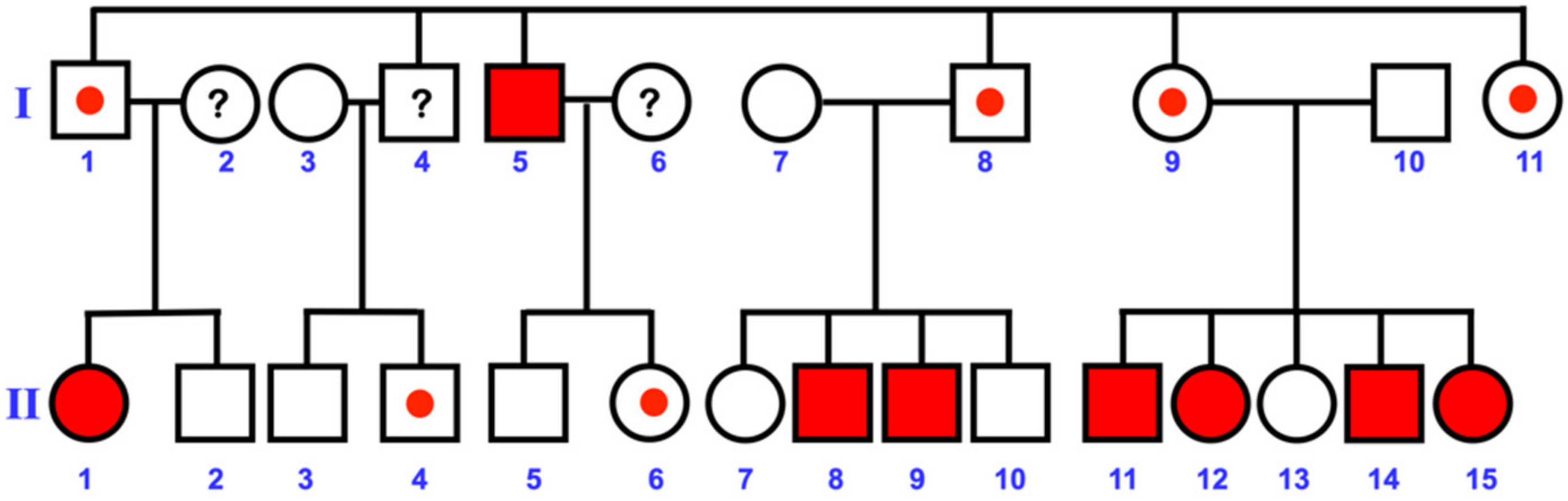
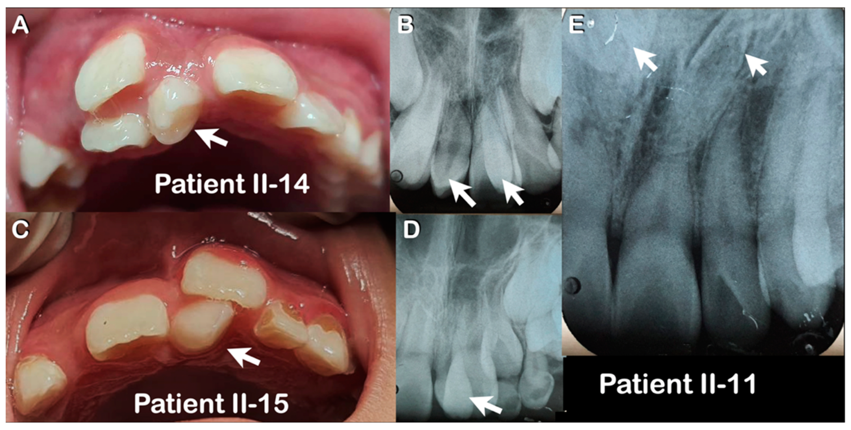
3.1. Varied Mesiodens Phenotypes within the Hmong Family (Family 1)

A two-generation large Hmong family living in the Wiang Kaen city of Chiang Rai province in Thailand with an age range from 7–62 years participated in the study. This family consisted of 26 members, of which eight were affected by mesiodens and 18 were unaffected (Figure 1). However, four members (I-2, I-4, I-6, and II-1) were not available for genetic study. Clinical and radiographic examinations were performed on all participants (Table 4). Of the eight members with mesiodens, six had erupted midline teeth (75%), while two remained unerupted (25%). Six (75%) had single mesiodens and two (25%) had double mesiodentes (Figure 2A,B). Two (25%) family members had inverted mesiodens (Figure 2E). This highlights the phenotypic variability in mesiodens formation even within the same family.

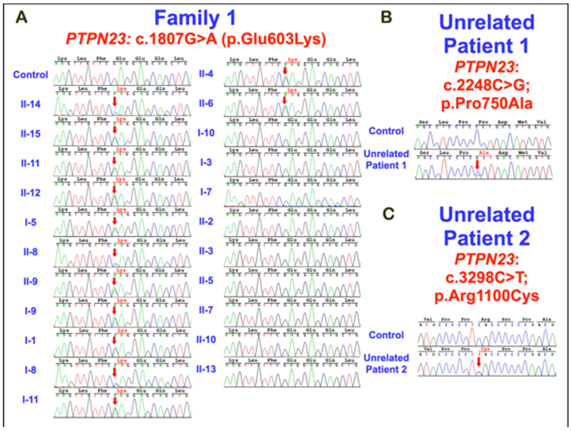
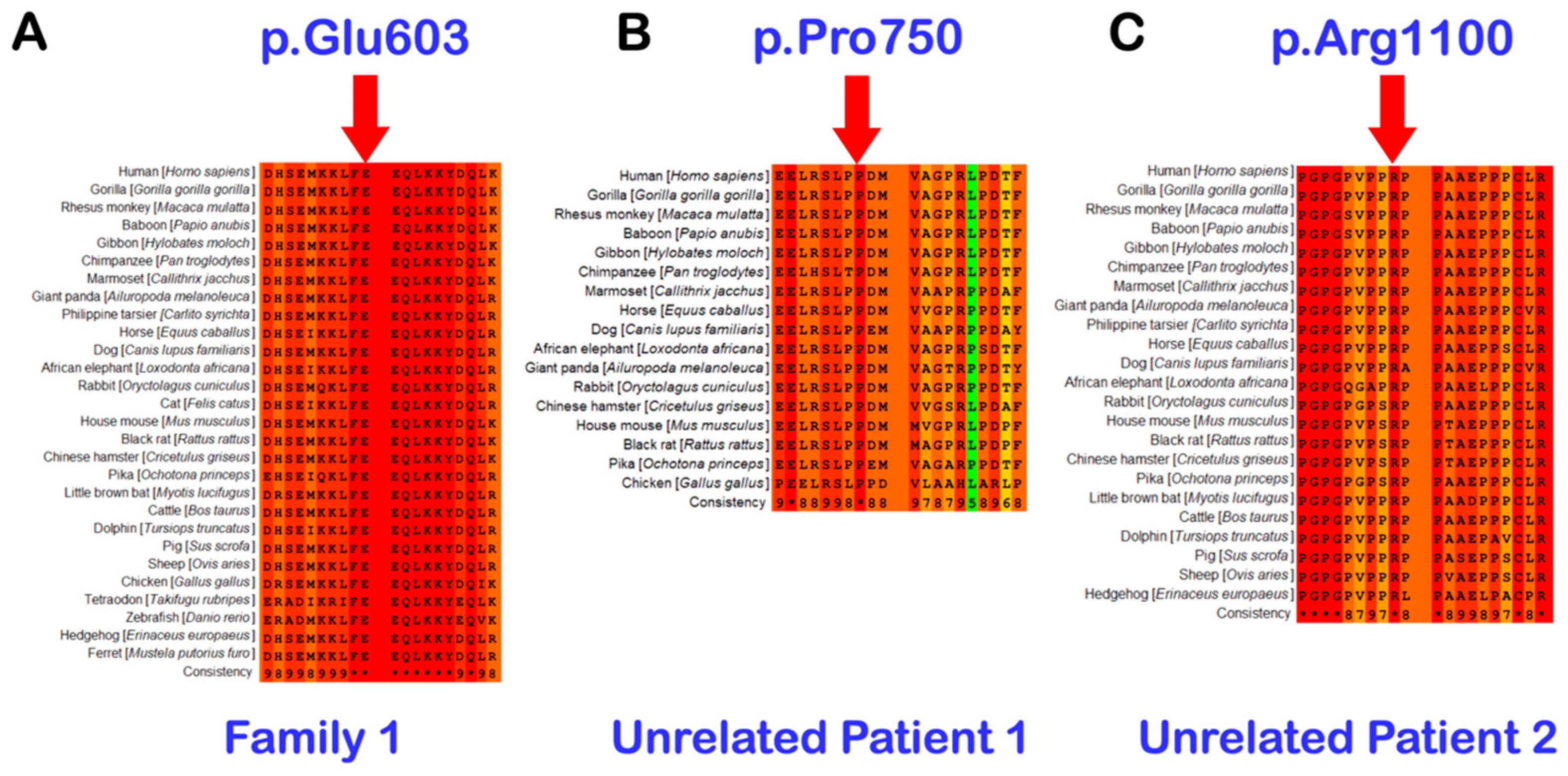
3.2. Whole Exome Sequencing, Sanger Direct Sequencing, and Bioinformatic Analysis

Whole exome and Sanger direct sequencing revealed a missense variant in the PTPN23 gene (chr3:g.47450916G>A; NM_015466.4:c.1807G>A; NP_056281.1:p.Glu603Lys; rs141113890) in all seven affected individuals analyzed (I-5, II-8, II-9, II-11, II-12, II-14, and II-15) and additionally in six unaffected relatives (I-1, I-8, I-9, I-11, II-4, and II-6). Six unaffected family members (II-2, II-3, II-5, II-7, II-10, and II-13) and three unaffected unrelated members (I-3, I-7, and I-10) did not carry the variant (Figure 1 and Figure 3, Table 4).
PTPN23 c.1807G>A (p.Glu603Lys) is most likely the pathogenic variant in this family because it was the only variant that all eight affected family members had in common. Moreover, this variant is extremely rare in the general population. According to gnomAD, the variant is not seen in over 30,608 alleles in the South Asian, Finnish, Jewish, Latin American, and African American populations. It is seen in five of 18,372 alleles in the East Asian population (allele frequency = 0.00027) and four of 113,444 alleles in the European population (allele frequency = 0.00003526). In the general population, it is seen in only 10 of 250,702 alleles, with an allele frequency of 0.00003989 and without any homozygous variation. The mutation is predicted as disease-causing with a probability of 0.99973 (MutationTaster), possibly damaging with a score of 0.488 (PolyPhen-2) and tolerated with a score of 0.516 (SIFT). The combined annotation-dependent depletion (CADD) score, a tool for scoring the deleteriousness of single nucleotide variants, of the p.Glu603Lys variant is 22.5, suggesting that this variant is predicted to be among the 1.0% most deleterious possible substitutions in the human genome (https://genome.ucsc.edu accessed on 8 January 2022). The multiple alignments of PTPN23 amino acid presented that the amino acid Glu603 is highly conserved in many vertebrate species (Figure 4).

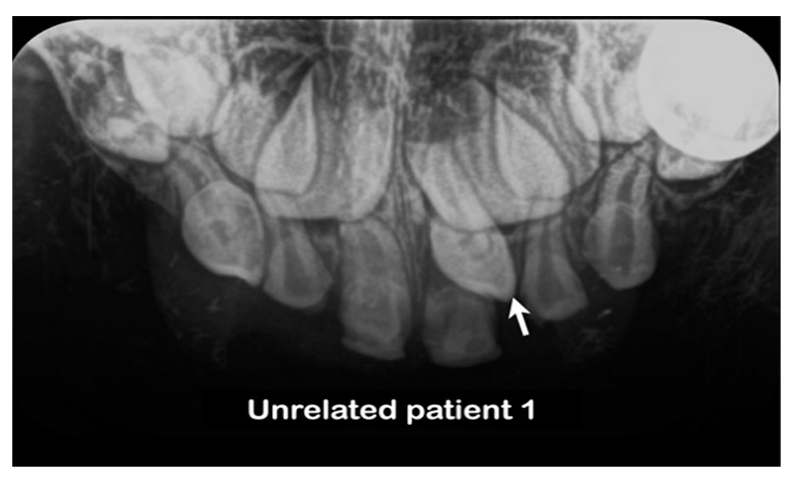
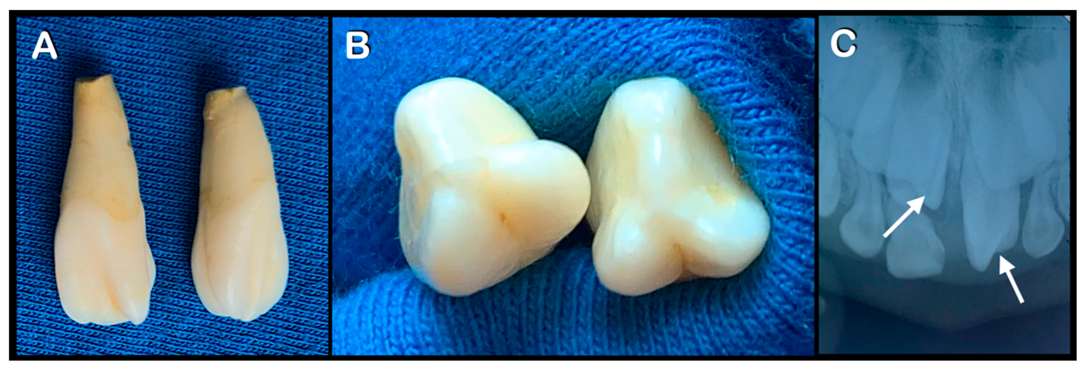
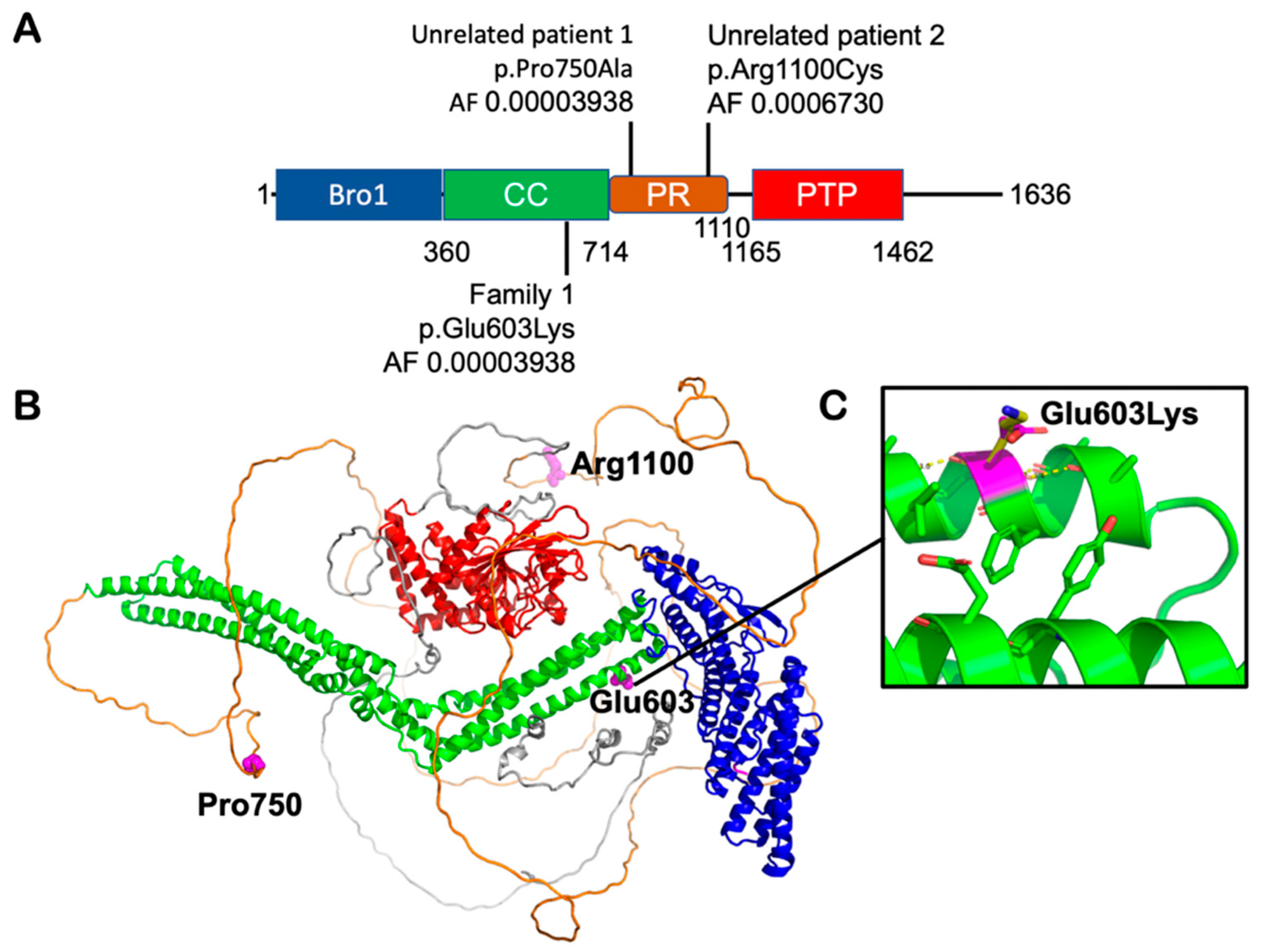
3.3. Unrelated Mesiodens Patients Identified with Rare PTPN23 Variants

Having identified the c.1807G>A; p.Glu603Lys variant in PTPN23 was the likely pathogenic variant in family 1 by WES, we cross-checked our in-house exome bank of 720 people affected with various disorders in order to identify patients in our cohort who had rare PTPN23 variants. We identified two rare variants in unrelated patients, both of whom displayed mesiodens. The chr3: g.47451536C>G; NM_015466.4: c.2248C>G; NP_056281.1: p.Pro750Ala (rs199549354) variant in the PTPN23 gene was identified in an unrelated Thai patient 1 with mesiodens (Figure 3 and Figure 5, Table 4). According to gnomAD, the allele frequency of this variant is 0.001436 with 43 alternative alleles in a total of 29,938 alleles and two homozygotes in South Asians, allele frequency = 0.000 with none of the alternative alleles in a total of 19,702 alleles in East Asians and a total allele frequency = 0.0001639 with 45 alternative alleles in 274,536 alleles from global populations. This amino acid is conserved in various vertebrate species (Figure 4). The mutation is predicted as disease-causing with a probability of 0.99989 (MutationTaster), possibly damaging with a score of 0.888 (PolyPhen-2) and damaging with a score of 0.016 (SIFT).
The chr3: g.47452586C>T; NM_015466.4: c.3298C>T; NP_056281.1: p.Arg1100Cys (rs201946361) variant was identified in the second unrelated Thai patient 2 with double mesiodentes (Figure 3 and Figure 6, Table 4). According to gnomAD, the allele frequency of this variant in the general population is 0.0006730, with 169 alternative alleles in a total of 251,122 alleles and no homozygotes in the East Asian population. In the South Asian population, six alleles were found in a total of 27,774 alleles with an allele frequency of 0.0002160. The mutation is predicted as a polymorphism with a probability of 0.99999 (MutationTaster), possibly damaging with a score of 0.946 (PolyPhen-2) and damaging with a score of 0.003 (SIFT). The CADD scores of p.Pro750Ala and p.Arg1100Cys variants are 20.8 and 20.3, respectively, suggesting that these variants are predicted to be among the 1.0% most deleterious possible substitutions in the human genome (https://genome.ucsc.edu accessed on 8 January 2022).

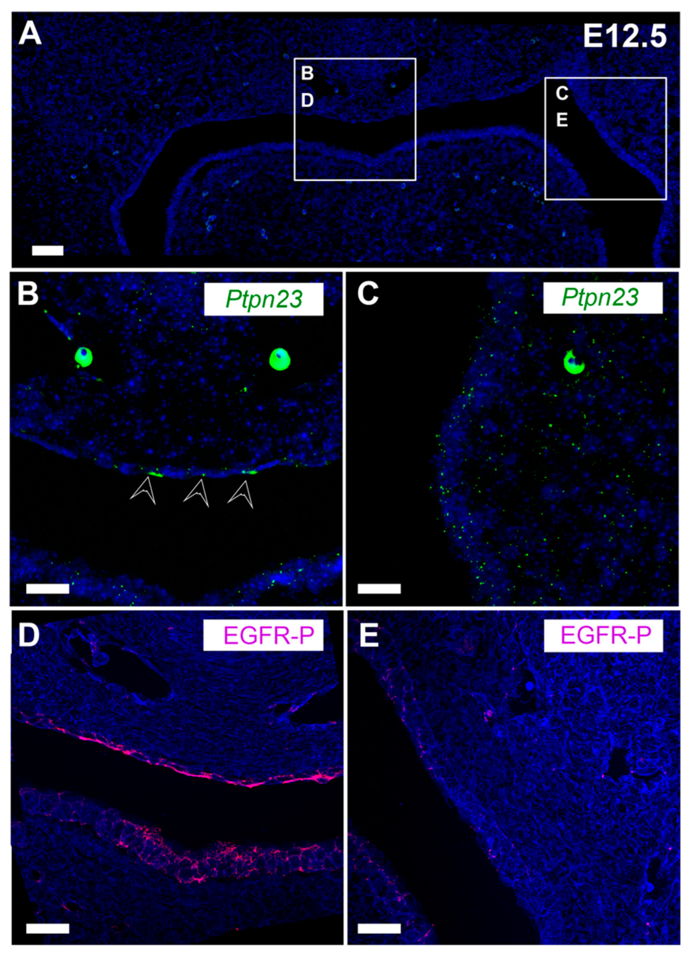
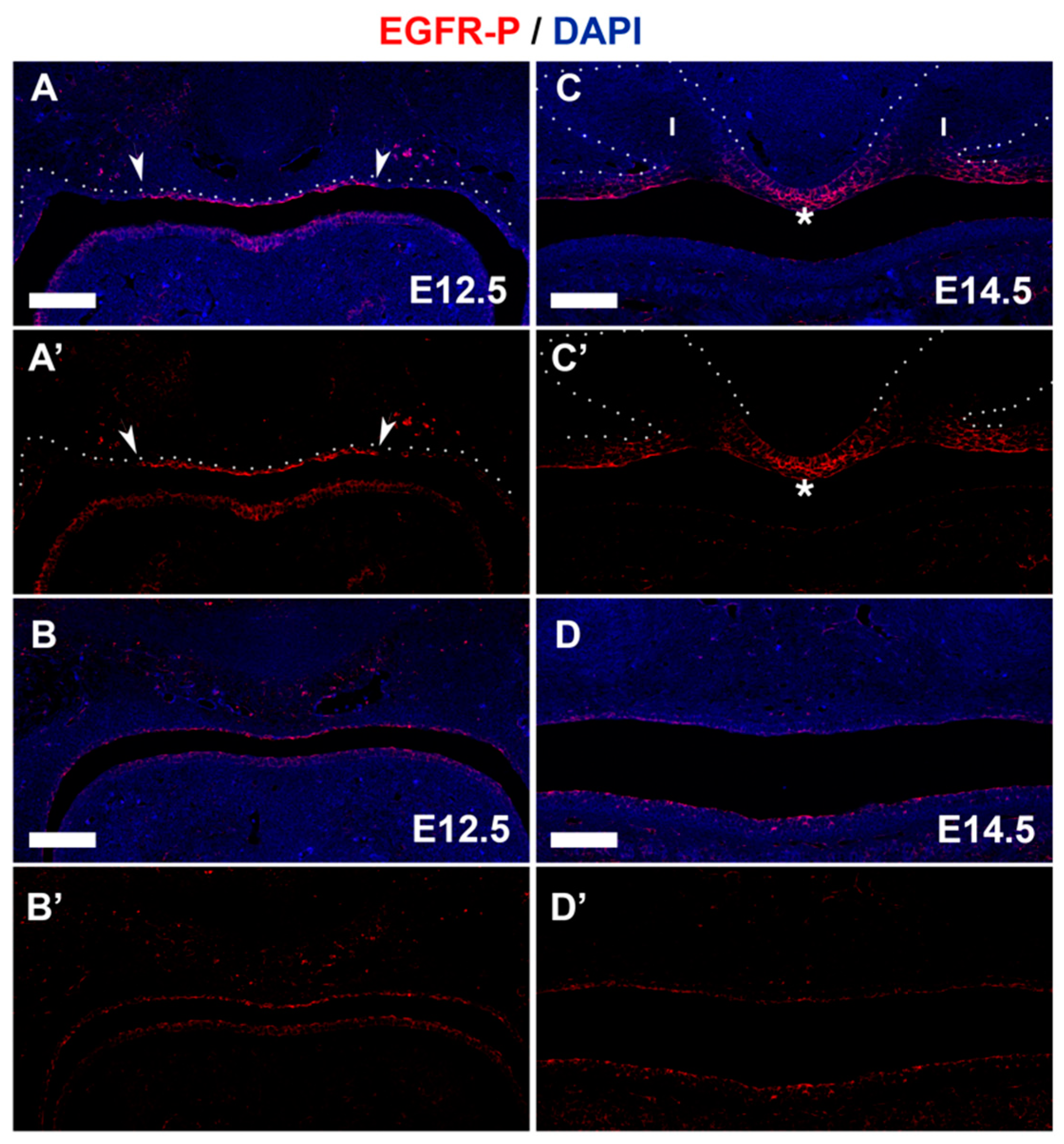
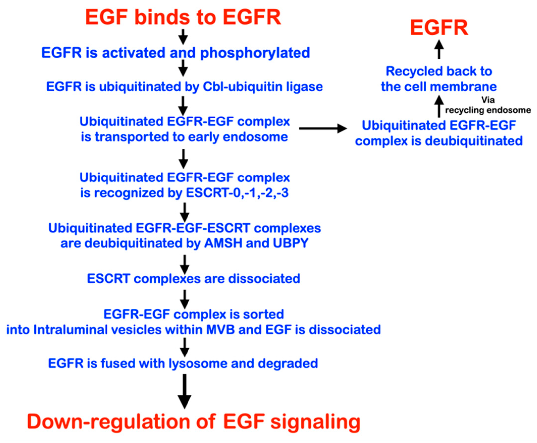
3.4. PTPN23 and Its Relationship to EGFR during Early Murine Tooth Development

To understand how changes in PTPN23 function might impact tooth development, we analyzed its expression during tooth development in the mouse using RNAscope. Previously a LacZ reporter has been used to analyze the expression of PTPN23 in the developing mouse embryo, with robust expression in the brain, vertebrae, and submandibular salivary glands [58]. At E12.5, PTPN23 was observed in the midline oral epithelium and higher levels, more laterally in both the epithelium and mesenchyme (Figure 7A,C, green dots). PTPN23 is a known regulator of endosomal trafficking and functions to deactivate membrane receptors, such as EGFR [59], with mutations in PTPN23 in patients potentially impacting EGFR signaling due to the availability of the receptor. The expression of EGFR protein was, therefore, followed in the mouse during the early stages of tooth development by immunofluorescence using serial sections to those used for PTPN23. Interestingly, EGFR was expressed at high levels in the midline epithelium at E12.5 (Figure 7D and Figure 8A,A’), with a reduced expression more laterally and further back in the mouth (Figure 7E and Figure 8B,B’). At E14.5, when the incisor tooth germs are at the cap stage, EGFR retained its high expression in the midline between the incisors, with a lack of expression in the main body of the incisors (Figure 8C,C’) and further back in the oral cavity (Figure 8D,D’). EGFR was, therefore, expressed in the midline, where mesiodens teeth originate.

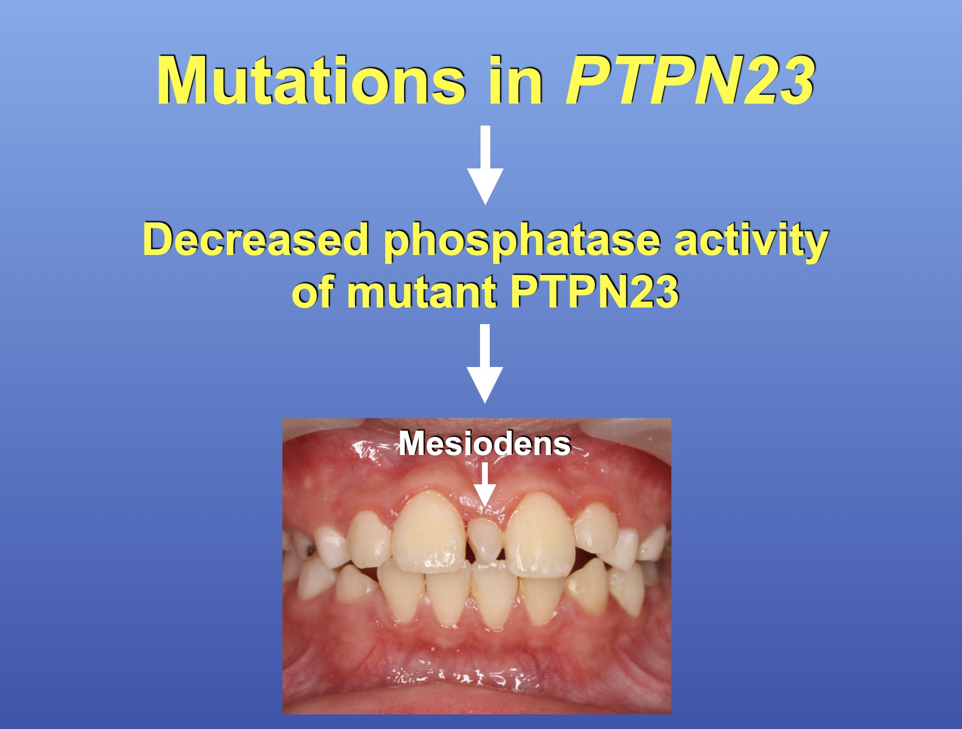
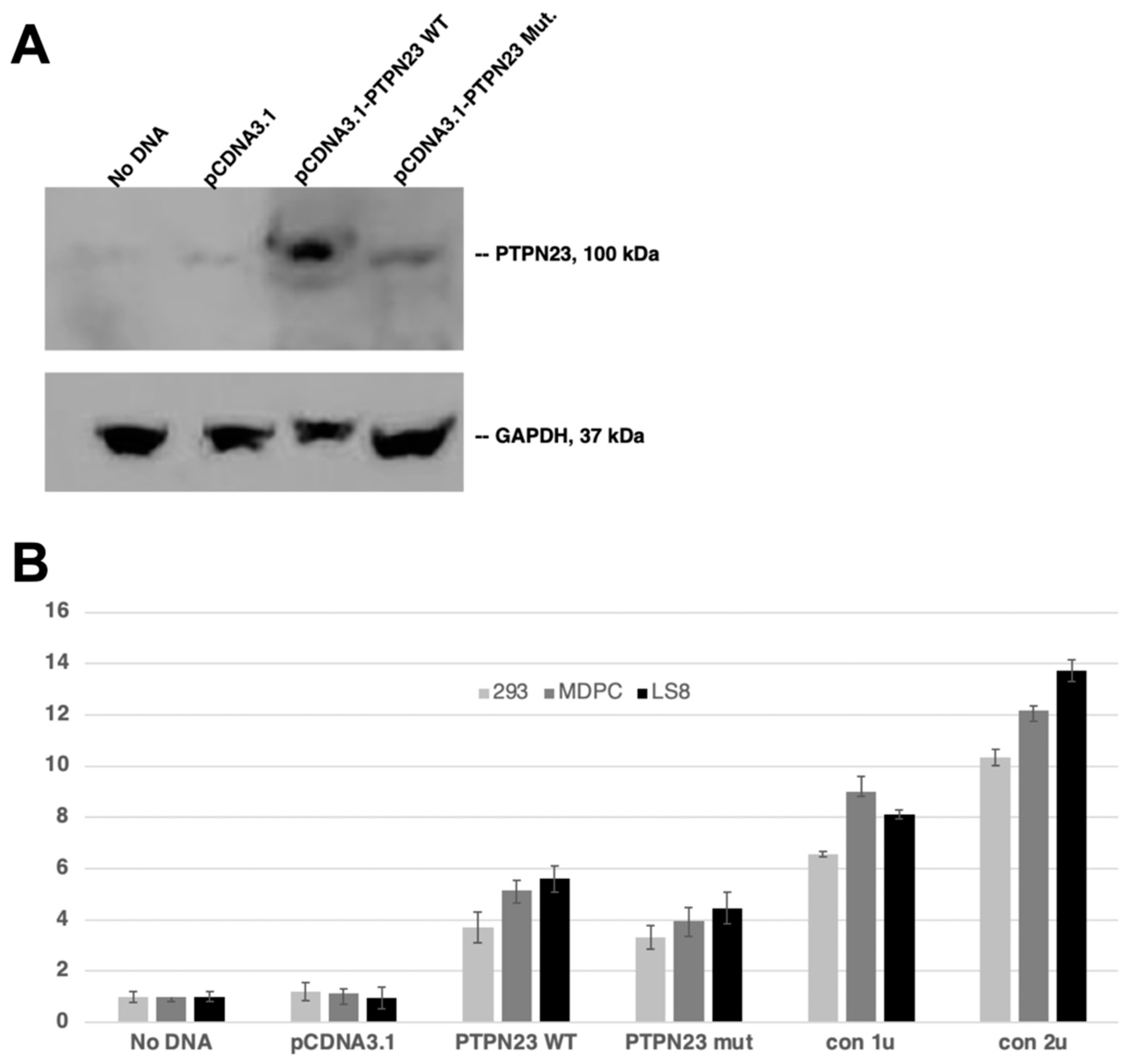
3.5. Decreased Phosphatase Activity of Mutant PTPN23
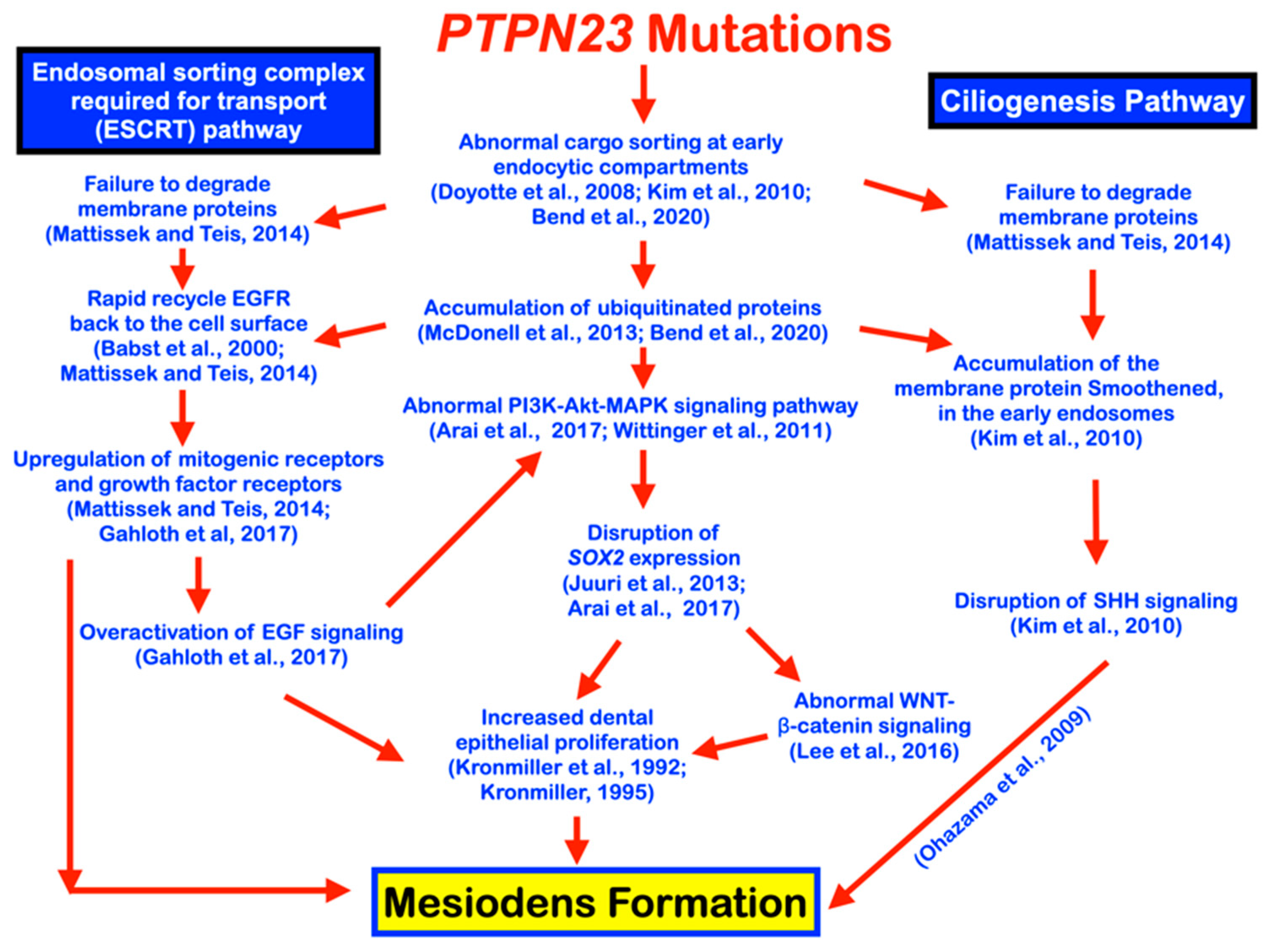
To understand if the mutation in PTPN23 affected protein stability, equal amounts of transfected lysates were analyzed for protein expression levels. Interestingly, untransfected and empty vector-transfected cells showed little PTPN23 endogenous protein expression (Figure 9A). HEK293 cell lysates transfected with wild type (WT) and mutant PTPN23 showed an increase in PTPN23 protein expression compared to controls, with the c.1807 G>A; p.Glu603Lys; rs141113890 mutation potentially influencing protein stability (Figure 9A). To determine if the mutation affected phosphatase activity, we analyzed both WT and mutant proteins in three different cell lines (HEK293, MDPC dental pulp cells, and LS-8 oral epithelial cells). As expected, the no transfection and empty vector transfection controls showed limited phosphatase activity, while transfected WT PTPN23 led to an approximately five- to six-fold increase in phosphatase activity (Figure 9B). Importantly, mutant PTPN23 phosphatase activity was decreased compared to WT (Figure 9B). However, both the WT and mutant PTPN23 proteins demonstrated less activity than 1 unit of Potato Acid Phosphatase, used as a positive control (Figure 9B).

4. Discussion

A mesiodens is a supernumerary tooth located in the midline of the premaxilla. It has been reported to be associated with Nance–Horan syndrome [41,42,43,44] and holoprosencephaly [45]. Nance–Horan syndrome (NHS; MIM 302350) is a rare X-linked developmental disorder caused by mutations in NHS. Clinical findings of the patients include congenital cataracts, intellectual disability, autism, and dental anomalies, including screwdriver blade-shaped incisors, supernumerary teeth, and mesiodens [41,42,43,44,60].
A number of research groups have tried to find the gene responsible for the development of isolated mesiodens. Unfortunately, this had not been successful, likely because most cases of mesiodens are sporadic. Even though familial cases have been reported, the number of those affected was not enough to locate the gene [61]. One of the major obstacles in trying to find the gene responsible for mesiodens is its well-known non-penetrance of inheritance (approximately 50% penetrance). This means that if you see a child with mesiodens, other family members may carry the same mutation even though they do not have mesiodens. In addition, a large number of cases (53.8-78.8%) of mesiodentes do not erupt into the oral cavities (Table 2) [3,5,6,7,8,9]. Without radiographic examination, those with unerupted mesiodens might have mistakenly been considered unaffected. These problems have made gene hunting for mesiodens phenotypes complicated and unsuccessful. However, recently genetic variants in LRP4, LRP5, LRP6, WLS, and DKK1 have been reported to be implicated in mesiodens with or without oral exostoses, including torus palatinus, torus mandibularis, and buccal exosteses [46,47,48,49,50]. We were very fortunate to meet a large Hmong family living in Chiang Rai, a province at the border of Thailand and Myanmar. This family, comprised of 26 members, had eight affected and 18 unaffected individuals. Mesiodens were found to be more common in males than females, with a ratio of 1.7:1. The morphology of mesiodens of the patients was not classified because some of them were already extracted, and some were unerupted. To our best knowledge, this is the largest family affected by mesiodens that has been reported in the literature.
Whole exome and Sanger direct sequencing identified the heterozygous missense c.1807G>A; p.Glu603Lys, c.2248C>G; p.Pro750Ala, and c.3298C>T; p.Arg1100Cys variants in the PTPN23 gene as the molecular etiologies of the mesiodens phenotypes in family 1, unrelated patient 1, and unrelated patient 2, respectively. For family 1, the mode of inheritance was autosomal dominance with incomplete penetrance. Many affected were the children of non-penetrant parents. Of the 13 family members analyzed who carried the mutation, only seven were found to have mesiodens; therefore, the penetrance was 53.84%. Despite the family having the same variant, the mesiodens phenotype (erupted/unerupted, inverted/normal, single/double) in those affected was highly variable, highlighting that other factors must impact these differences. We were not able to report the morphology of the mesiodentes found in the Hmong family because most of their mesiodentes were extracted, and some of them were unerupted. It was, therefore, impossible to know the exact morphology without using three-dimension computed tomography.
PTPN23 is located in chromosome 3p21.3 and is composed of 25 exons. It encodes a 1636 amino acid non-receptor protein tyrosine phosphatase type 23 (PTPN23) or His-domain protein tyrosine phosphatase (HDPTP) [62,63]. Bi-allelic mutations in PTPN23 in humans are associated with autosomal recessive neurodevelopmental disorder and structural brain anomalies with or without seizures and spasticity (NEDBASS; MIM 618890) [62] and autosomal recessive microcephalic complex hereditary spastic paraplegia [64]. In keeping with this, PTPN23 is expressed in the cerebral cortex, thalamus, and hypothalamus of adult mice and in the embryonic nervous system [59,65]. Unfortunately, mesiodens or other dental anomalies were not mentioned in these studies, and unerupted mesiodens could have been missed [62,64]. Our patients with mono-allelic PTPN23 mutations were healthy with no neuromuscular disorders. Evidently, the results of our study and the previous reports of patients with neuromuscular disorders [62,64] suggest that the phenotypes of patients with PTPN23 mutations depend on the context of the mutations and/or other factors, including modifying genes.
PTPN23 is a catalytically inactive phosphatase that binds to tyrosine-phosphorylated proteins in order to prevent them from dephosphorylation [66,67]. Homozygous PTPN23 knockout mouse embryos display an accumulation of ubiquitinated proteins in endosomes, disruption of the MVB biogenesis, smaller body size, significant malformations, and subsequent embryonic lethality at E8.5 [58,59,62,68]. The mouse embryos, therefore, die prior to any signs of tooth development. The function of PTPN23 is restricted to the early endosome at the initiation of the ESCRT–MVB pathway, where it binds to ESCRT-0 to downregulate ubiquitinated cargoes and promotes forward movement of receptors from the early endosome towards the lysosomes, thereby leading to downregulation of the signal [68,69,70] (Figure 10). PTPN23 is crucial for releasing EGFR from ESCRT-0 and allowing it to engage ESCRT-III [71]. Aberrant interaction between PTPN23 and ESCRT-0 is expected to result in enhanced recycling of endocytosed EGFR [72,73,74] and subsequent overactivation of EGFR and MAPK signaling [73,75]. Depletion of PTPN23 has been shown to cause the accumulation of ubiquitin–protein conjugates in tubulo-vesicular endosomal compartments and reduction of EGFR sorting to endosomal lumen [59,64,71] (Figure 10 and Figure 11). The decision for EGFR to be recycled to the cell membrane and retain its function or sorted to the endosomes for subsequent lysosomal degradation depends on the strength of the signals [74,76] (Figure 10).
PTPN23 comprises several functional domains, including the Bro1 domain, the coiled-coil (CC) domain, a long proline-rich region (PR), and the inactive protein tyrosine phosphatase (PTP) domain [62,77] (Figure 12). The p.Glu603Lys mutation, which was found in family 1, is located in the CC domain (Figure 12). Glu603 is solvent exposed and its substitution to a lysine is not perturbing the structure
However, the substitution of a negatively charged glutamic acid with a positively charged lysine would affect the electrodynamics of this region. Given that the CC is a scaffolding domain, such a change in surface charge may influence its intra- or intermolecular interactions. The p.Glu603Lys mutation may affect protein stability and function [62,77]. Alternatively, non-structural effects (linked to splicing or translation, for example) are possible. The p.Pro750Ala and p.Arg1100Cys mutations, identified in unrelated patients 1 and 2, respectively, are located in the unstructured and flexible PR (Figure 12). Even though these variants will not impact the structural integrity or stability of the protein, they may affect post-translational modifications (PTMs) or ligand binding sites. To date, no PTMs have been reported in the vicinity of these three mutations, but PR motifs are known to bind to SH3 or WW domains. The PTPN23 CC has indeed been shown to bind to the EGFR adaptor protein Grb2 [78]. Therefore, collectively, these mutations are predicted to disrupt EGFR signaling by hampering protein–protein interactions.
We have provided evidence that the c.1807 G>A; p.Glu603Lys; rs141113890 PTPN23 mutation associated with family 1 may affect protein stability. According to a Western blot, transfected mutant PTPN23 cell lysates had less protein expression compared to WT transfected cell lysates (Figure 9A). In addition, a lower level of PTPN23 phosphatase activity was shown in all mutant transfected cell lines compared to WT transfected cell lines (Figure 9B). The results suggest that either less expression and/or decreased activity of mutant PTPN23 protein activity could be causative of the mesiodens in the patients.
PTPN23 mutations in our patients are predicted to result in the accumulation of ubiquitin–protein conjugates in vesiculotubular endosomal compartments and reduction of EGFR sorting to the endosomal lumen, decreased degradation of EGFR and subsequent overactivation of EGFR signaling (Figure 11) [59,78]. Egf is expressed in developing jaws immediately before the formation of the dental lamina [79,80]. In culture, the addition of Egf has been demonstrated to result in both inhibitions of normal tooth formation and induction of ectopic supernumerary teeth in the diastema [79,80]. Here, we show that while PTPN23 has a relatively broad expression domain in the murine oral cavity, its target EGFR is expressed at high levels in the forming midline. Given the restricted expression of EGFR, and the ability of ectopic EGF signaling to generate ectopic teeth, this pathway is a likely target for mesiodens formation (Figure 7 and Figure 8).
Notably, patients with bi-allelic variants in NHS and PTPN23 share phenotypes, including developmental brain disorders, intellectual disability, cataracts, and autism [62,81]. The presence of mesiodens in patients with PTPN23 variants and patients with NHS-associated Nance–Horan syndrome [41,42,43,44] raises a question if there is an association between the functions of NHS and PTPN23. The role of NHS is to maintain the integrity of the actin at the cell membrane, which is important for cell shape, migration, and intercellular junction [82,83,84]. Actin plays an important role in producing force to form an endosome [85]. As previously mentioned, PTPN23 binds with ESCRT-0 to encourage the forward movement of ubiquitinated EGFR endosomes toward the lysosomes, leading to the downregulation of the signal [68,69]. Therefore, mesiodens formation in patients with NHS or PTPN23 mutations might relate to the disruptive endocytosis process.
It is hypothesized that disruption of EGFR signaling, as a result of PTPN23 mutations and subsequent decreased mutant PTPN23 phosphatase activity, would lead to disruption in the expression of the transcription factor SOX2 [86,87] via the PI3K–Akt signaling pathway [86,88] (Figure 11). The finding of mutations in the phosphatase gene (PTPRH) resulting in aberrant EGFR activity supports our hypothesis [89]. The PI3K–Akt signaling pathway is likely to be involved in mesiodens pathogenesis because Sox2-positive odontogenic epithelial stem cells have been demonstrated to contribute to supernumerary tooth formation [87,90] and mutations in SOX2 have been reported to be associated with syndromic supernumerary teeth in SOX2 anophthalmia syndrome [91,92]. Sox2 is crucial for the initiation of tooth formation and regulates the progenitor state of dental epithelial cells [87,93,94,95,96,97]. The SOX2 lineage has been shown to give rise to successional teeth [87]. Sox2 disrupts Wnt signaling by binding to β-catenin, a central regulator of the Wnt signaling pathway [94]. The Wnt–β-catenin signaling pathway is known to be crucial for tooth development and overactivation of Wnt/β-catenin signaling results in supernumerary tooth formation or odontoma [96] (Figure 11). The association of genetic variants in Wnt/β-catenin signaling pathway and mesiodens formation [46,47,48] suggests the mutations in PTPN23 in our patients were upstream of the Wnt/β-catenin signaling pathway in the pathogenetic process.
Additionally, abnormal BMP signaling may be involved in mesiodens patients with PTPN23 mutations because mutations in PTPN23 may lead to disruptive ESCRT recruitment, MVB sorting, degradation of BMP receptors, hyperactivation of BMP signaling, overactivation of WNT signaling, and mesiodens formation [98].
Recycling endosomes are a dynamic vesiculotubular compartment exporting endocytosed membrane proteins and lipids to the cell surface via vesicular intermediates [99,100]. The endocytic recycling pathway has also been associated with ciliogenesis. PTPN23 has a specific role in ciliary vesicle targeting. Silencing of PTPN23 has been shown to significantly reduce the number of ciliated cells [99]. Knockdown PTPN23 has been demonstrated to result in the accumulation of the transmembrane protein Smoothened in early endosomes [99]. It is hypothesized that mutations in PTPN23 would result in abnormal endocytic trafficking and accumulation of Smoothened in the early endosomes, leading to a disruption of SHH signaling [101], which could contribute to mesiodens formation (Figure 11). Disruption to a number of signaling pathways driven by PTPN23 mutations could, therefore, account for the formation of mesiodens.

Limitations of the Study

Mesiodens are frequently unerupted, and therefore, their incidence can be hidden. In our study, it was not possible to use cone beam computed tomography (CBCT) on each patient due to the additional exposure to radiation, but this would have provided more information on the morphology of the mesiodens observed. We used mouse embryos for gene and protein expression, presuming conservation of the pathway in mammals, but human embryonic and fetal tissue would have beneficial. Finally, it was not possible to test the impact of the loss of function of PTPN23 in the mouse as the null mice are early lethals; therefore, conditional models would need to be generated.

5. Conclusions

In conclusion, PTPN23 is a regulator of endosomal trafficking, and its function is to move activated membrane receptors forward from ESCRT-0 towards ESCRT-III [59], thereby regulating the activity of a number of signaling pathways. We show that mutations in PTPN23 are associated with the formation of midline upper supernumerary teeth, known as mesiodens. We hypothesize that these mutations disrupt the accumulation of activated EGFR and other signaling pathways in early endosomes, leading to abnormal signaling at the early stages of tooth development and subsequent supernumerary tooth formation.

Author Contributions

Conceptualization, P.A., R.C., S.T., B.O., S.T.A., C.N., B.A.A., A.S.T. and P.K.; methodology, software, validation, P.A., R.C., S.E., J.M.F., W.I., S.T., B.O., S.T.A., C.N., B.A.A., A.S.T. and P.K.; formal analysis, investigation, data curation, P.A., R.C., S.E., J.M.F., W.I., S.T., B.O., S.T.A., C.N., B.A.A., A.S.T. and P.K.; writing—original draft preparation, P.A., R.C., S.E., J.M.F., W.I., S.T., B.O., S.T.A., C.N., B.A.A., A.S.T. and P.K.; writing—review and editing, P.A., R.C., S.E., J.M.F., W.I., S.T., B.O., S.T.A., C.N., B.A.A., A.S.T. and P.K.; visualization, supervision, resources, project administration, P.K. All authors have read and agreed to the published version of the manuscript.

Funding

This work was supported by the Genomics Thailand Research Grant of Health Systems Research Institute (Grant no. 64-123), the King Abdullah University of Science and Technology (KAUST) through the baseline fund and the Award No. FCC/1/1976-25 and REI/1/4446-01 from the Office of Sponsored Research (OSR). The funders had no role in study design, data collection and analysis, the decision to publish, or the preparation of the manuscript.

Institutional Review Board Statement

This study was approved by the Human Experimentation Committee of the Faculty of Dentistry, Chiang Mai University (no. 71/2020) and was performed in accordance with the ethical standards of the Declaration of Helsinki. Written informed consent was obtained from all participants or from a legal guardian of children younger than 18 years old.

Informed Consent Statement

Written informed consent has been obtained from the patients or their parents to publish this paper.

Data Availability Statement

Not applicable.

Acknowledgments

We thank our patients and their families for their kind cooperation and for allowing us to use their medical and dental information for the benefit of other patients.

Conflicts of Interest

The authors declare no conflict of interest.

References

  1. Bolk, L. Die Uberzahligen oberen incisivi des Menschen. Dtsch. Mscf. 2hk 1917, 35, 185. [Google Scholar]
  2. Thilander, B.; Myrberg, N. The prevalence of malocclusion in Swedish schoolchildren. Eur. J. Oral Sci. 1973, 81, 12–21. [Google Scholar] [CrossRef]
  3. Gündüz, K.; Çelenk, P.; Zengin, Z.; Sümer, P. Mesiodens: A radiographic study in children. J. Oral Sci. 2008, 50, 287–291. [Google Scholar] [CrossRef]
  4. Kositbowornchai, S.; Keinprasit, C.; Poomat, N. Prevalence and distribution of dental anomalies in pretreatment orthodontic Thai patients. Khon Kaen Dent. J. 2010, 13, 92–100. [Google Scholar]
  5. Mukhopadhyay, S. Mesiodens: A clinical and radiographic study in children. J. Indian Soc. Pedod. Prev. Dent. 2011, 29, 34–38. [Google Scholar] [CrossRef]
  6. Kazanci, F.; Celikoglu, M.; Miloglu, O.; Yildirim, H.; Ceylan, I. The frequency and characteristics of mesiodens in a Turkish patient population. Eur. J. Dent. 2011, 5, 361–365. [Google Scholar] [CrossRef]
  7. Lara, T.S.; Lancia, M.; Silva Filho, O.G.d.; Garib, D.G.; Ozawa, T.O. Prevalence of mesiodens in orthodontic patients with deciduous and mixed dentition and its association with other dental anomalies. Dent. Press J. Orthod. 2013, 18, 93–99. [Google Scholar] [CrossRef] [PubMed]
  8. Goksel, S.; Agirgol, E.; Karabas, H.C.; Ozcan, I. Evaluation of Prevalence and Positions of Mesiodens Using Cone-Beam Computed Tomography. J. Oral Maxillofac. Res. 2018, 9, e1. [Google Scholar] [CrossRef] [PubMed]
  9. Aren, G.; Erdem, A.P.; Onur, Ö.D.; Ak, G. The prevelance of mesiodens in a group of non-syndromic Turkish children: A radiographic study. Eur. Oral Res. 2018, 52, 162–166. [Google Scholar] [CrossRef] [PubMed]
  10. Ramesh, K.; Venkataraghavan, K.; Kunjappan, S.; Ramesh, M. Mesiodens: A clinical and radiographic study of 82 teeth in 55 children below 14 years. J. Pharm. Bioallied Sci. 2013, 5, S60. [Google Scholar] [CrossRef] [PubMed]
  11. Almeida, J.D.; Guimarães Cabral, L.A.; Martins Gomes, A.P.; Moraes, E. Supernumerary mesiodentes with familial character: A clinical report. Quintessence Int. 1995, 26, 343–345. [Google Scholar] [PubMed]
  12. Sykaras, S.N. Mesiodens in primary and permanent dentitions: Report of a case. Oral Surg. Oral Med. Oral Pathol. 1975, 39, 870–874. [Google Scholar] [CrossRef] [PubMed]
  13. Asaumi, J.-I.; Shibata, Y.; Yanagi, Y.; Hisatomi, M.; Matsuzaki, H.; Konouchi, H.; Kishi, K. Radiographic examination of mesiodens and their associated complications. Dentomaxillofac. Radiol. 2004, 33, 125–127. [Google Scholar] [CrossRef] [PubMed]
  14. von Arx, T. Anterior maxillary supernumerary teeth: A clinical and radiographic study. Aust. Dent. J. 1992, 37, 189–195. [Google Scholar] [CrossRef]
  15. Khandelwal, V.; Nayak, A.; Naveen, R.; Ninawe, N.; Nayak, P.; Prasad, S.S. Prevalence of mesiodens among six-to seventeen-year-old school going children of Indore. J. Indian Soc. Pedod. Prev. Dent. 2011, 29, 288–293. [Google Scholar] [CrossRef]
  16. Mitchell, L. Supernumerary teeth. Dent. Update 1989, 16, 65–66, 68–69. [Google Scholar]
  17. Anthonappa, R.; King, N.; Rabie, A. Aetiology of supernumerary teeth: A literature review. Eur. Arch. Paediatr. Dent. 2013, 14, 279–288. [Google Scholar] [CrossRef]
  18. Peterkova, R.; Hovorakova, M.; Peterka, M.; Lesot, H. Three-dimensional analysis of the early development of the dentition. Aust. Dent. J. 2014, 59, 55–80. [Google Scholar] [CrossRef]
  19. Sedano, H.O.; Gorlin, R.J. Familial occurrence of mesiodens. Oral Surg. Oral Med. Oral Pathol. 1969, 27, 360–361. [Google Scholar] [CrossRef]
  20. Cadenat, H.; Combelles, R.; Fabert, G.; Clouet, M. Mesiodens and heredity. Rev. Stomatol. Chir. Maxillofac. 1977, 78, 341–346. [Google Scholar]
  21. Desai, R.S.; Shah, N.P. Multiple supernumerary teeth in two brothers: A case report. J. Oral Pathol. Med. 1998, 27, 411–413. [Google Scholar] [CrossRef] [PubMed]
  22. Marya, C.M.; Kumar, B. Familial occurrence of mesiodentes with unusual findings. Quintessence Int. 1998, 29, 49–51. [Google Scholar] [PubMed]
  23. Gallas, M.M.; Garcia, A. Retention of permanent incisors by mesiodens: A family affair. Br. Dent. J. 2000, 188, 63–64. [Google Scholar] [CrossRef] [PubMed]
  24. Takahashi, M.; Hosomichi, K.; Yamaguchi, T.; Yano, K.; Funatsu, T.; Adel, M.; Haga, S.; Maki, K.; Tajima, A. Whole-exome sequencing analysis of supernumerary teeth occurrence in Japanese individuals. Hum. Genome Var. 2017, 4, hgv201646. [Google Scholar] [CrossRef] [PubMed]
  25. Mason, C.; Rule, D. Midline supernumeraries: A family affair. Dent. Update 1995, 22, 34–35. [Google Scholar] [PubMed]
  26. Sharma, A. Familial occurence of mesiodens--a case report. J. Indian Soc. Pedod. Prev. Dent. 2003, 21, 84–85. [Google Scholar] [PubMed]
  27. Severin, E.; Purcărea, R.; Albu, C.; Albu, D.F.; Ionescu, E.; Stanciu, D. Familial mesiodens. Rom. J. Stomatol. 2009, 55, 293–296. [Google Scholar]
  28. Hunstadbråten, K. Anomalies in twins. Quintessenz 1965, 16, 71–72. [Google Scholar]
  29. Schön, F. Supernumerary incisors in uniovular twins, and their treatment by means of electrosurgery. Quintessence Int. Dent. Dig. 1974, 5, 13–18. [Google Scholar]
  30. Bucci, E.; Martina, R. True hyperdontia in monochorial twins. Clinical case. Arch. Stomatol. 1975, 16, 305–313. [Google Scholar]
  31. Carton, A.; Rees, R. Mirror image dental anomalies in identical twins. Br. Dent. J. 1987, 162, 193–194. [Google Scholar] [CrossRef]
  32. Choi, W.; Chang, R.; Chuang, S. Bilateral mesiodentes of identical twins--a case report. Zhonghua Ya Yi Xue Hui Za Zhi 1990, 9, 116–121. [Google Scholar] [PubMed]
  33. Beere, D.; Hargreaves, J.; Sperber, G.; Cleaton-Jones, P. Mirror image supplemental primary incisor teeth in twins: Case report and review. Pediatr. Dent. 1990, 12, 390–392. [Google Scholar] [PubMed]
  34. Seddon, R.P.; Johnstone, S.C.; Smith, P.B. Mesiodentes in twins: A case report and a review of the literature. Int. J. Paediatr. Dent. 1997, 7, 177–184. [Google Scholar] [CrossRef] [PubMed]
  35. Brand, A.; Akhavan, M.; Tong, H.; Kooka, Y.; Zernik, J.H. Orthodontic, genetic, and periodontal considerations in the treatment of impacted maxillary central incisors: A study of twins. Am. J. Orthod. Dentofac. Orthop. 2000, 117, 68–74. [Google Scholar] [CrossRef]
  36. Townsend, G.; Richards, L.; Hughes, T.; Pinkerton, S.; Schwerdt, W. Epigenetic influences may explain dental differences in monozygotic twin pairs. Aust. Dent. J. 2005, 50, 95–100. [Google Scholar] [CrossRef]
  37. Sharma, A. A rare case of concomitant hypo-hyperdontia in identical twins. J. Indian Soc. Pedod. Prev. Dent. 2008, 26, S79–S81. [Google Scholar]
  38. Babacan, H.; Öztürk, F.; Polat, H.B. Identical unerupted maxillary incisors in monozygotic twins. Am. J. Orthod. Dentofac. Orthop. 2010, 138, 498–509. [Google Scholar] [CrossRef]
  39. Gurgel, C.V.; Cota, A.L.S.A.L.S.; Kobayashi, T.Y.; Silva, S.M.B.; Machado, M.A.A.M.; Rios, D.; Garib, D.G.; Oliveira, T.M. Bilateral mesiodens in monozygotic twins: 3D diagnostic and management. Case Rep. Dent. 2013, 2013, 193614. [Google Scholar] [CrossRef]
  40. Reddy, G.S.M.; Mahajan, B.; Desai, R.S. Mesiodens In Twins: A Case Report. Indian J. Dent. Sci. 2013, 5, 088–089. [Google Scholar]
  41. Nance, W.; Warburg, M.; Bixler, D.; Helveston, E. Congenital X-linked cataract, dental anomalies and brachymetacarpalia. Birth Defects Orig. Artic. Ser. 1974, 10, 285–291. [Google Scholar]
  42. Bixler, D.; Higgins, M.; Hartsfield, J., Jr. The Nance-Horan syndrome: A rare X-linked ocular-dental trait with expression in heterozygous females. Clin. Genet. 1984, 26, 30–35. [Google Scholar] [CrossRef] [PubMed]
  43. Walpole, I.R.; Hockey, A.; Nicoll, A. The Nance-Horan syndrome. J. Med. Genet. 1990, 27, 632. [Google Scholar] [CrossRef] [PubMed]
  44. Gjørup, H.; Haubek, D.; Jacobsen, P.; Ostergaard, J.R. Nance–Horan syndrome—The oral perspective on a rare disease. Am. J. Med. Genet. A 2017, 173, 88–98. [Google Scholar] [CrossRef]
  45. Chan, M.Y.; Shifteh, K.; Shanske, A.L. Mesiodens, a new microform of holoprosencephaly? Am. J. Med. Genet. A 2009, 149, 268–271. [Google Scholar] [CrossRef]
  46. Kantaputra, P.; Jatooratthawichot, P.; Chintakanon, K.; Intachai, W.; Pradermdutsadeeporn, P.; Adisornkanj, P.; Tongsima, S.; Ngamphiw, C.; Olsen, B.; Tucker, A.S. Mutations in LRP6 highlight the role of WNT signaling in oral exostoses and dental anomalies. Arch. Oral Biol. 2022, 142, 105514. [Google Scholar] [CrossRef] [PubMed]
  47. Kantaputra, P.N.; Guven, Y.; Tripuwabhrut, K.; Adisornkanj, P.; Hatsadaloi, A.; Kaewgahya, M.; Olsen, B.; Ngamphiw, C.; Jatooratthawichot, P.; Tongsima, S. Mutations in LRP5 and BMP4 are associated with mesiodens, tooth agenesis, root malformation, and oral exostoses. Clin. Genet. 2022, 102, 333–338. [Google Scholar] [CrossRef]
  48. Kantaputra, P.N.; Tripuwabhrut, K.; Jatooratthawichot, P.; Adisornkanj, P.; Hatsadaloi, A.; Nop Porntrakoolsaree, N.; Kaewgaya, M.; Olsen, B.; Tongsima, S.; Ngamphiw, C.; et al. Genetic variants in the WLS are associated with dental anomalies, torus palatinus, and torus mandibularis. Eur. J. Orthod. 2022. Epub ahead of printing. [Google Scholar] [CrossRef]
  49. Kantaputra, P.; Jatooratthawichot, P.; Kottege, N.; Anthonappa, R.P.; Kaewgahya, M.; Tongsima, S.; Ngamphiw, C.; Cairns, J.R.K.; Predes, D.; He, X. DKK1 is a strong candidate for mesiodens and taurodontism. Clin. Genet. 2023. Epub ahead of printing. [Google Scholar] [CrossRef]
  50. Kantaputra, P.; Jatooratthawichot, P.; Adisornkanj, P.; Kitsadayurach, P.; Kaewgahya, M.; Olsen, B.; Ohazama, A.; Ngamphiw, C.; Tongsima, S.; Cox, T.C.; et al. Rare variants in LRP4 are associated with mesiodens, root maldevelopment, and oral exostoses in humans. Biology 2023, 12, 220. [Google Scholar] [CrossRef]
  51. Li, H.; Durbin, R. Fast and accurate short read alignment with Burrows–Wheeler transform. Bioinformatics 2009, 25, 1754–1760. [Google Scholar] [CrossRef]
  52. McKenna, A.; Hanna, M.; Banks, E.; Sivachenko, A.; Cibulskis, K.; Kernytsky, A.; Garimella, K.; Altshuler, D.; Gabriel, S.; Daly, M. The Genome Analysis Toolkit: A MapReduce framework for analyzing next-generation DNA sequencing data. Genome Res. 2010, 20, 1297–1303. [Google Scholar] [CrossRef] [PubMed]
  53. McLaren, W.; Gil, L.; Hunt, S.E.; Riat, H.S.; Ritchie, G.R.; Thormann, A.; Flicek, P.; Cunningham, F. The ensembl variant effect predictor. Genome Biol. 2016, 17, 122. [Google Scholar] [CrossRef] [PubMed]
  54. Liu, X.; Wu, C.; Li, C.; Boerwinkle, E. dbNSFP v3. 0: A one-stop database of functional predictions and annotations for human nonsynonymous and splice-site SNVs. Hum. Mutat. 2016, 37, 235–241. [Google Scholar] [CrossRef]
  55. Consortium, T.U. UniProt: The universal protein knowledgebase in 2021. Nucleic Acids Res. 2020, 49, D480–D489. [Google Scholar] [CrossRef] [PubMed]
  56. Jumper, J.; Evans, R.; Pritzel, A.; Green, T.; Figurnov, M.; Ronneberger, O.; Tunyasuvunakool, K.; Bates, R.; Žídek, A.; Potapenko, A.; et al. Highly accurate protein structure prediction with AlphaFold. Nature 2021, 596, 583–589. [Google Scholar] [CrossRef] [PubMed]
  57. Kumar, M.; Gouw, M.; Michael, S.; Sámano-Sánchez, H.; Pancsa, R.; Glavina, J.; Diakogianni, A.; Valverde, J.A.; Bukirova, D.; Čalyševa, J.; et al. ELM—The eukaryotic linear motif resource in 2020. Nucleic Acids Res. 2019, 48, D296–D306. [Google Scholar] [CrossRef]
  58. Gingras, M.C.; Kharitidi, D.; Chénard, V.; Uetani, N.; Bouchard, M.; Tremblay, M.L.; Pause, A. Expression analysis and essential role of the putative tyrosine phosphatase His-domain-containing protein tyrosine phosphatase (HD-PTP). Int. J. Dev. Biol. 2009, 53, 1069–1074. [Google Scholar] [CrossRef]
  59. Doyotte, A.; Mironov, A.; McKenzie, E.; Woodman, P. The Bro1-related protein HD-PTP/PTPN23 is required for endosomal cargo sorting and multivesicular body morphogenesis. Proc. Natl. Acad. Sci. USA 2008, 105, 6308–6313. [Google Scholar] [CrossRef]
  60. Burdon, K.P.; McKay, J.D.; Sale, M.M.; Russell-Eggitt, I.M.; Mackey, D.A.; Wirth, M.G.; Elder, J.E.; Nicoll, A.; Clarke, M.P.; FitzGerald, L.M.; et al. Mutations in a novel gene, NHS, cause the pleiotropic effects of Nance-Horan syndrome, including severe congenital cataract, dental anomalies, and mental retardation. Am. J. Hum. Genet. 2003, 73, 1120–1130. [Google Scholar] [CrossRef]
  61. Kim, Y.Y.; Hwang, J.; Kim, H.-S.; Kwon, H.J.; Kim, S.; Lee, J.H.; Lee, J.H. Genetic alterations in mesiodens as revealed by targeted NGS and gene co-occurrence network analysis. Oral Dis. 2017, 23, 966–972. [Google Scholar] [CrossRef]
  62. Bend, R.; Cohen, L.; Carter, M.T.; Lyons, M.J.; Niyazov, D.; Mikati, M.A.; Rojas, S.K.; Person, R.E.; Si, Y.; Wentzensen, I.M. Phenotype and mutation expansion of the PTPN23 associated disorder characterized by neurodevelopmental delay and structural brain abnormalities. Eur. J. Hum. Genet. 2020, 28, 76–87. [Google Scholar] [CrossRef] [PubMed]
  63. Toyooka, S.-I.; Ouchida, M.; Jitsumori, Y.; Tsukuda, K.; Sakai, A.; Nakamura, A.; Shimizu, N.; Shimizu, K. HD-PTP: A novel protein tyrosine phosphatase gene on human chromosome 3p21.3. Biochem. Biophys. Res. Commun. 2000, 278, 671–678. [Google Scholar] [CrossRef] [PubMed]
  64. Khalaf-Nazzal, R.; Fasham, J.; Ubeyratna, N.; Evans, D.J.; Leslie, J.S.; Warner, T.T.; Al-Hijawi, F.; Alshaer, S.; Baker, W.; Turnpenny, P.D. Final Exon Frameshift Biallelic PTPN23 Variants Are Associated with Microcephalic Complex Hereditary Spastic Paraplegia. Brain Sci. 2021, 11, 614. [Google Scholar] [CrossRef] [PubMed]
  65. Husedzinovic, A.; Neumann, B.; Reymann, J.; Draeger-Meurer, S.; Chari, A.; Erfle, H.; Fischer, U.; Gruss, O.J. The catalytically inactive tyrosine phosphatase HD-PTP/PTPN23 is a novel regulator of SMN complex localization. Mol. Biol. Cell 2015, 26, 161–171. [Google Scholar] [CrossRef] [PubMed]
  66. Barr, A.J.; Ugochukwu, E.; Lee, W.H.; King, O.N.; Filippakopoulos, P.; Alfano, I.; Savitsky, P.; Burgess-Brown, N.A.; Müller, S.; Knapp, S. Large-scale structural analysis of the classical human protein tyrosine phosphatome. Cell 2009, 136, 352–363. [Google Scholar] [CrossRef]
  67. Gingras, M.C.; Zhang, Y.L.; Kharitidi, D.; Barr, A.J.; Knapp, S.; Tremblay, M.L.; Pause, A. HD-PTP is a catalytically inactive tyrosine phosphatase due to a conserved divergence in its phosphatase domain. PLoS ONE 2009, 4, e5105. [Google Scholar] [CrossRef]
  68. Tabernero, L.; Woodman, P. Dissecting the role of His domain protein tyrosine phosphatase/PTPN23 and ESCRTs in sorting activated epidermal growth factor receptor to the multivesicular body. Biochem. Soc. Trans. 2018, 46, 1037–1046. [Google Scholar] [CrossRef]
  69. Gahloth, D.; Levy, C.; Heaven, G.; Stefani, F.; Wunderley, L.; Mould, P.; Cliff, M.J.; Bella, J.; Fielding, A.J.; Woodman, P. Structural basis for selective interaction between the ESCRT regulator HD-PTP and UBAP1. Structure 2016, 24, 2115–2126. [Google Scholar] [CrossRef]
  70. McDonell, L.M.; Mirzaa, G.M.; Alcantara, D.; Schwartzentruber, J.; Carter, M.T.; Lee, L.J.; Clericuzio, C.L.; Graham, J.M.; Morris-Rosendahl, D.J.; Polster, T. Mutations in STAMBP, encoding a deubiquitinating enzyme, cause microcephaly–capillary malformation syndrome. Nat. Genet. 2013, 45, 556–562. [Google Scholar] [CrossRef]
  71. Ali, N.; Zhang, L.; Taylor, S.; Mironov, A.; Urbé, S.; Woodman, P. Recruitment of UBPY and ESCRT exchange drive HD-PTP-dependent sorting of EGFR to the MVB. Curr. Biol. 2013, 23, 453–461. [Google Scholar] [CrossRef] [PubMed]
  72. Babst, M.; Odorizzi, G.; Estepa, E.J.; Emr, S.D. Mammalian tumor susceptibility gene 101 (TSG101) and the yeast homologue, Vps23p, both function in late endosomal trafficking. Traffic 2000, 1, 248–258. [Google Scholar] [CrossRef] [PubMed]
  73. Mattissek, C.; Teis, D. The role of the endosomal sorting complexes required for transport (ESCRT) in tumorigenesis. Mol. Membr. Biol. 2014, 31, 111–119. [Google Scholar] [CrossRef] [PubMed]
  74. Tanaka, N.; Kyuuma, M.; Sugamura, K. Endosomal sorting complex required for transport proteins in cancer pathogenesis, vesicular transport, and non-endosomal functions. Cancer Sci. 2008, 99, 1293–1303. [Google Scholar] [CrossRef] [PubMed]
  75. Raiborg, C.; Bache, K.G.; Gillooly, D.J.; Madshus, I.H.; Stang, E.; Stenmark, H. Hrs sorts ubiquitinated proteins into clathrin-coated microdomains of early endosomes. Nat. Cell Biol. 2002, 4, 394–398. [Google Scholar] [CrossRef]
  76. Sigismund, S.; Woelk, T.; Puri, C.; Maspero, E.; Tacchetti, C.; Transidico, P.; Di Fiore, P.P.; Polo, S. Clathrin-independent endocytosis of ubiquitinated cargos. Proc. Natl. Acad. Sci. USA 2005, 102, 2760–2765. [Google Scholar] [CrossRef] [PubMed]
  77. Gahloth, D.; Heaven, G.; Jowitt, T.A.; Mould, A.P.; Bella, J.; Baldock, C.; Woodman, P.; Tabernero, L. The open architecture of HD-PTP phosphatase provides new insights into the mechanism of regulation of ESCRT function. Sci. Rep. 2017, 7, 9151. [Google Scholar] [CrossRef]
  78. Tanase, C.-A. Histidine domain-protein tyrosine phosphatase interacts with Grb2 and GrpL. PLoS ONE 2010, 5, e14339. [Google Scholar] [CrossRef]
  79. Kronmiller, J.E. Spatial distribution of epidermal growth-factor transcripts and effects of exogenous epidermal growth factor on the pattern of the mouse dental lamina. Arch. Oral Biol. 1995, 40, 137–143. [Google Scholar] [CrossRef]
  80. Kronmiller, J.E.; Upholt, W.B.; Kollar, E.J. Alteration of murine odontogenic patterning and prolongation of expression of epidermal growth factor mRNA by retinol in vitro. Arch. Oral Biol. 1992, 37, 129–138. [Google Scholar] [CrossRef]
  81. Seow, W.K.; Brown, J.; Romaniuk, K. The Nance-Horan syndrome of dental anomalies, congenital cataracts, microphthalmia, and anteverted pinna: Case report. Pediatr. Dent. 1985, 7, 307–311. [Google Scholar] [PubMed]
  82. Brooks, S.P.; Coccia, M.; Tang, H.R.; Kanuga, N.; Machesky, L.M.; Bailly, M.; Cheetham, M.E.; Hardcastle, A.J. The Nance–Horan syndrome protein encodes a functional WAVE homology domain (WHD) and is important for co-ordinating actin remodelling and maintaining cell morphology. Hum. Mol. Genet. 2010, 19, 2421–2432. [Google Scholar] [CrossRef] [PubMed]
  83. Sharma, S.; Ang, S.L.; Shaw, M.; Mackey, D.A.; Gécz, J.; McAvoy, J.W.; Craig, J.E. Nance–Horan syndrome protein, NHS, associates with epithelial cell junctions. Hum. Mol. Genet. 2006, 15, 1972–1983. [Google Scholar] [CrossRef] [PubMed]
  84. Sharma, S.; Burdon, K.P.; Dave, A.; Jamieson, R.V.; Yaron, Y.; Billson, F.; Van Maldergem, L.; Lorenz, B.; Gécz, J.; Craig, J.E. Novel causative mutations in patients with Nance-Horan syndrome and altered localization of the mutant NHS-A protein isoform. Mol. Vis. 2008, 14, 1856. [Google Scholar]
  85. Akamatsu, M.; Vasan, R.; Serwas, D.; Ferrin, M.A.; Rangamani, P.; Drubin, D.G. Principles of self-organization and load adaptation by the actin cytoskeleton during clathrin-mediated endocytosis. Elife 2020, 9, e49840. [Google Scholar] [CrossRef]
  86. Arai, C.; Yoshizaki, K.; Miyazaki, K.; Saito, K.; Yamada, A.; Han, X.; Funada, K.; Fukumoto, E.; Haruyama, N.; Iwamoto, T. Nephronectin plays critical roles in Sox2 expression and proliferation in dental epithelial stem cells via EGF-like repeat domains. Sci. Rep. 2017, 7, 45181. [Google Scholar] [CrossRef]
  87. Juuri, E.; Jussila, M.; Seidel, K.; Holmes, S.; Wu, P.; Richman, J.; Heikinheimo, K.; Chuong, C.-M.; Arnold, K.; Hochedlinger, K. Sox2 marks epithelial competence to generate teeth in mammals and reptiles. Development 2013, 140, 1424–1432. [Google Scholar] [CrossRef]
  88. Wittinger, M.; Vanhara, P.; El-Gazzar, A.; Savarese-Brenner, B.; Pils, D.; Anees, M.; Grunt, T.W.; Sibilia, M.; Holcmann, M.; Horvat, R. hVps37A Status affects prognosis and cetuximab sensitivity in ovarian cancer. Clin. Cancer Res. 2011, 17, 7816–7827. [Google Scholar] [CrossRef]
  89. Swiatnicki, M.R.; Rennhack, J.P.; Ortiz, M.M.; Hollern, D.P.; Perry, A.V.; Kubiak, R.; Riveria Riveria, S.M.; O’Reilly, S.; Andrechek, E.R. Elevated phosphorylation of EGFR in NSCLC due to mutations in PTPRH. PLoS Genet. 2022, 18, e1010362. [Google Scholar] [CrossRef]
  90. Saito, K.; Takahashi, K.; Huang, B.; Asahara, M.; Kiso, H.; Togo, Y.; Tsukamoto, H.; Mishima, S.; Nagata, M.; Iida, M.; et al. Loss of Stemness, EMT, and Supernumerary Tooth Formation in Cebpb−/−Runx2+/− Murine Incisors. Sci. Rep. 2018, 8, 5169. [Google Scholar] [CrossRef]
  91. Chacon-Camacho, O.F.; Fuerte-Flores, B.I.; Ricardez-Marcial, E.F.; Zenteno, J.C. SOX2 anophthalmia syndrome and dental anomalies. Am. J. Med. Genet. A 2015, 167, 2830–2833. [Google Scholar] [CrossRef] [PubMed]
  92. Numakura, C.; Kitanaka, S.; Kato, M.; Ishikawa, S.; Hamamoto, Y.; Katsushima, Y.; Kimura, T.; Hayasaka, K. Supernumerary impacted teeth in a patient with SOX2 anophthalmia syndrome. Am. J. Med. Genet. A 2010, 152, 2355–2359. [Google Scholar] [CrossRef] [PubMed]
  93. Kim, E.J.; Jung, S.Y.; Wu, Z.; Zhang, S.; Jung, H.S. Sox2 maintains epithelial cell proliferation in the successional dental lamina. Cell Prolif. 2020, 53, e12729. [Google Scholar] [CrossRef] [PubMed]
  94. Lee, M.-J.; Kim, E.-J.; Otsu, K.; Harada, H.; Jung, H.-S. Sox2 contributes to tooth development via Wnt signaling. Cell Tissue Res. 2016, 365, 77–84. [Google Scholar] [CrossRef] [PubMed]
  95. Martin, K.J.; Rasch, L.J.; Cooper, R.L.; Metscher, B.D.; Johanson, Z.; Fraser, G.J. Sox2+ progenitors in sharks link taste development with the evolution of regenerative teeth from denticles. Proc. Natl. Acad. Sci. USA 2016, 113, 14769–14774. [Google Scholar] [CrossRef] [PubMed]
  96. Xavier, G.M.; Patist, A.L.; Healy, C.; Pagrut, A.; Carreno, G.; Sharpe, P.T.; Martinez-Barbera, J.P.; Thavaraj, S.; Cobourne, M.T.; Andoniadou, C.L. Activated WNT signaling in postnatal SOX2-positive dental stem cells can drive odontoma formation. Sci. Rep. 2015, 5, 14479. [Google Scholar] [CrossRef] [PubMed]
  97. Zhang, L.; Yuan, G.; Liu, H.; Lin, H.; Wan, C.; Chen, Z. Expression pattern of Sox2 during mouse tooth development. Gene Expr. Patterns 2012, 12, 273–281. [Google Scholar] [CrossRef]
  98. Gahloth, D.; Levy, C.; Walker, L.; Wunderley, L.; Mould, A.P.; Taylor, S.; Woodman, P.; Tabernero, L. Structural basis for specific interaction of TGFβ signaling regulators SARA/Endofin with HD-PTP. Structure 2017, 25, 1011–1024.e1014. [Google Scholar] [CrossRef]
  99. Kim, J.; Lee, J.E.; Heynen-Genel, S.; Suyama, E.; Ono, K.; Lee, K.; Ideker, T.; Aza-Blanc, P.; Gleeson, J.G. Functional genomic screen for modulators of ciliogenesis and cilium length. Nature 2010, 464, 1048–1051. [Google Scholar] [CrossRef]
  100. Maxfield, F.R.; McGraw, T.E. Endocytic recycling. Nat. Rev. Mol. Cell Biol. 2004, 5, 121–132. [Google Scholar] [CrossRef]
  101. Ohazama, A.; Haycraft, C.J.; Seppala, M.; Blackburn, J.; Ghafoor, S.; Cobourne, M.; Martinelli, D.C.; Fan, C.-M.; Peterkova, R.; Lesot, H. Primary cilia regulate Shh activity in the control of molar tooth number. Development 2009, 136, 897–903. [Google Scholar] [CrossRef] [PubMed]
Figure 1. Pedigree of the two-generation Hmong family (Family 1). This family consists of 26 members, of which eight are affected with mesiodens (block red) and 18 are unaffected (white). Red dots represent non-penetrant individuals who have the variant p.Glu603Lys but not mesiodens. A number of the affected are children of the non-penetrant. Family members I-2, I-4, I-6, and II-1 were not available for genetic study. The phenotypes of the parents of I-1, I-4, I-5, I-8, I-9, and I-11 are unknown.
Biology 12 00393 g001
Figure 2. Clinical and radiographic pictures of mesiodens (arrows) in (A) Patient II-14. (B) Periapical radiograph in (A) shows double mesiodentes (arrows). (C) Patient II-15. (D) Periapical radiograph in (C) shows mesiodens. (E) Periapical radiograph of patient II-11. Note double inverted mesiodentes (arrows).
Biology 12 00393 g002
Figure 3. Chromatograms of the heterozygous missense (A) c.1807G>A; p.Glu603Lys, (B) c.2248C>G; p.Pro750Ala, and (C) c.3298C>T; p.Arg1100Cys mutations in PTPN23 gene in family 1, unrelated patient 1 and unrelated patient 2, respectively.
Biology 12 00393 g003
Figure 4. Conservation of amino acid residues (A) p.Glu603, (B) p.Pro750, and (C) p.Arg1100 across vertebrate species. All amino acids are highly conserved.
Biology 12 00393 g004
Figure 5. Periapical radiograph of mesiodens of the unrelated patient 1 (arrow).
Biology 12 00393 g005
Figure 6. Unrelated patient 2. Extracted double mesiodentes in (A) Frontal view. (B) Top view. (C) Periapical radiograph prior to extraction (arrows). Note screwdriver-shaped crown morphology of both mesiodentes.
Biology 12 00393 g006
Figure 7. Expression of PTPN23 and EGFR in the oral cavity. Murine E12.5 frontal sections at the midline at the upper incisors. Serial sections used for PTPN23 and EGFR. (A) DAPI (blue) stained section highlighting regions shown in (BE). (B,C) PTPN23 RNAscope. Signal shows up as green dots. Large circular patches are autofluorescence from blood cells. (D,E) Immunofluorescence EGFR protein in pink. (B) PTPN23 is expressed in the midline epithelium (arrows) and (C) robustly in the more lateral epithelium and mesenchyme. (D) EGFR is expressed in the midline in the forming incisor region, similar to PTPN23 expression. (E) EGFR is weakly expressed more laterally. Scale bar in (A): 100 μm. Scale bar in (BE): 50 μm.
Biology 12 00393 g007
Figure 8. EGFR is expressed at high levels in the forming midline. Murine frontal sections. (AB’) E12.5. (CD’) E14.5. (AD) Immunofluorescence EGFR (pink/red) plus DAPI (blue). (A’,B’,C’,D’) EGFR only. (A,A’,C,C’) Anterior region. (B,B’,D,D’) More posterior region. Arrows in (A,A’) highlight border of expression of EGFR in the midline at E12.5. Asterix in (C,C’) highlights midline between cap stage incisors (outlined with white dots). I = incisor. Scale bar in (A,C): 100 μm (same scale in all images).
Biology 12 00393 g008
Figure 9. PTPN23 mutant protein has reduced phosphatase activity. (A) Cell lysates from transfected and untransfected cells were probed for PTPN23 expression. Both untransfected and control transfected cells showed low levels of endogenous PTPN23 protein expression. WT and mutant PTPN23 transfected lysates showed increased expression (n = 3). (B) Phosphatase activity was measured after no transfection and transfection of control DNA, WT PTPN23, and mutant PTPN23 (Glu603Lys). Three different cell lines were used to measure phosphatase activity: HEK293, MDPC (dental pulp cells), and LS-8 (oral epithelial cells). WT PTPN23 has low phosphatase activity in the three cell lines, compared to Potato Acid Phosphatase controls with either 1 unit (u) or 2 units. However, mutant PTPN23 has decreased phosphatase activity compared to WT PTPN23 (n = 3).
Biology 12 00393 g009
Figure 10. EGFR–Multivesicular Biogenesis–Lysosomal Degradation pathway.
Biology 12 00393 g010
Figure 11. Proposed pathogenetic pathways as a result of PTPN23 mutations that lead to mesiodens formation. Refs: [59,62,70,72,73,77,78,79,80,81,82,83,84,85].
Biology 12 00393 g011
Figure 12. Organization of PTPN23 domains and location of the variants. (A) Residue numbers indicate domain boundaries, and arrows show locations of the mutations found in our patients. CC: coiled-coil domain; PR: proline-rich region; PTP inactive phosphatase. (B) Theoretical Alpha fold model of the PTPN23 protein structure. Colors as in (A). The mutated residues are highlighted in magenta as sphere models. (C) Close-up view of the Glu603 wild-type residues (magenta stick model) and its lysine substitution (yellow).
Biology 12 00393 g012
Table 1. The prevalence of mesiodens in different populations.
AuthorsEthnicNumber of PatientsPrevalence (%)Radiographic ExaminationMale:Female
Thilander and Myrberg, 1973 [2]Swedish63980.9yesNot mentioned
von Arx, 1992 [14]SwissNot mentionedNot mentionedyes2.6:1
Asaumi et al., 2004 [13]JapaneseNot mentionedNot mentionedyes2.8:1
Gunduz et al., 2008 [3]Turkish230000.3yes2.1:1
Kositbowornchai et al., 2010 [4]Thai5701.05yesNot mentioned
Mukhopadhyay, 2011 [5]Indian79320.8yes1.78:1
Kazanci et al., 2011 [6]Turkish33510.3yes1.5:1
Lara et al., 2013 [7]Brazilian19951.5yes1.5:1
Ramesh et al., 2013 [10]IndianNot mentionedNot mentionedyes2.05:1
Goksel et al., 2018 [8]Turkish19025.04yes1.9:1
Aren et al., 2018 [9]Turkish581420.1yesNot mentioned
Table 2. Characteristics of mesiodens.
AuthorsN
(Patients)
N (Teeth)Number%Unerupted %Orientation%
Asaumi et al., 2004 [13]200256Single73Not mentionedVertical27
Double26Inverted67
>31Transverse6
Gunduz et al., 2008 [3]6985Single76.878.8Vertical55.2
Double23.1Inverted37.6
Transverse7
Mukhopadhyay, 2011 [5]6478Single78.153.8Vertical62.8
Double21.9Inverted30.8
Transverse6.4
Kazanci et al., 2011 [6]1012Single8066.7Vertical58.3
Double20Inverted33.3
>30Transverse8.4
Lara et al., 2013 [7]3036Single8075Vertical75
Double20Inverted13.9
>30Transverse11.1
Goksel et al., 2018 [8]101130Single76.2378.46Vertical63.1
Double18.81Inverted20
>34.95Transverse16.9
Aren et al., 2018 [9] 5983Single6168.7Vertical77.1
Double37.3Inverted22.9
>31Transverse0
Table 3. Published reports of familial occurrence of mesiodens.
Types of
Familial Occurrences
ReferencesDescriptions
SiblingsSedano and Gorlin, 1969 [19]2 siblings affected (normal parents)
Cadenat et al., 1977 [20]Discordant dizygotic twin (parents’ phenotype: N/A)
Almeida et al., 1995 [11]3 siblings affected (parents’ phenotype: N/A)
Desai and Shah, 1998 [21]2 siblings affected (parents’ phenotype: N/A)
Marya and Kumar, 1998 [22]2 siblings affected (parents’ phenotype: N/A)
Gallas and Garcia, 2000 [23] 2 siblings affected (parents’ phenotype: N/A)
Takahashi et al., 2017 [24]2 siblings affected (parents’ phenotype: N/A)
Monozygotic twinsHunstadbråten, 1965 [28]Mirror imaged discordant
Schön, 1974 [29]Concordant
Bucci and Martina, 1975 [30]Discordant
Carton and Rees, 1987 [31]Mirror imaged discordant
Choi et al., 1990 [32]Near concordant
Beere et al.,1990 [33]Mirror imaged discordant
Seddon et al., 1997 [34]Near concordant
Brand et al., 2000 [35]Mirror imaged discordant
Townsend et al., 2005 [36]8 pairs of discordant
Sharma, 2008 [37]Discordant
Babacan et al., 2010 [38]Concordant
Gurgel et al., 2013 [39]Concordant
Reddy et al., 2013 [40]Discordant
More than one generationMason and Rule, 1995 [25]Father, mother and 2 sons
Sharma, 2003 [26] Father and daughter
Severin et al., 2009 [27]Family 1: Mother, 2 daughters, uncle, and cousin
Family 2: Father, daughter, and son
Family 1: Mother and son
Takahashi et al., 2017 [24]Family 2: Father and son
Family 3: Mother and 3 sons
Skip generationSeverin et al., 2009 [27]Grandmother and grandson
Table 4. Clinical and molecular findings of patients and their family members (NM_015466.4; NP_056281.1).
FamiliesPatientsEthnicGenderPhenotypesOrientationEruptionPTPN23 Mutations
Family
1
II-14HmongMaleDouble mesiodentesNormalEruptedc.1807G>A;
p.Glu603Lys; rs141113890
II-15HmongFemaleSingle mesiodensNormalErupted
II-11HmongMaleDouble mesiodentesInvertedUnerupted
II-12HmongFemaleSingle mesiodensInvertedUnerupted
I-5HmongMaleSingle mesiodensNormalErupted
II-8HmongMaleSingle mesiodensNormalErupted
II-9HmongMaleSingle mesiodensNormalErupted
II-1HmongFemaleSingle mesiodensNormalEruptedNot available
I-9HmongFemaleUnaffected--c.1807G>A;
p.Glu603Lys; rs141113890
I-1HmongMaleUnaffected--
I-8HmongMaleUnaffected--
I-11HmongFemaleUnaffected--
II-4HmongMaleUnaffected--
II-6HmongFemaleUnaffected--
I-10HmongMaleUnaffected--No mutation
I-3HmongFemaleUnaffected--
I-7HmongFemaleUnaffected--
II-2HmongMaleUnaffected--
II-3HmongMaleUnaffected--
II-5HmongMaleUnaffected--
II-7HmongFemaleUnaffected--
II-10HmongMaleUnaffected--
II-13HmongFemaleUnaffected--
Male: Female
with mesiodens
= 1.67:1 (5:3)
Single: Double
mesiodens cases
= 6:2 (3:1)
Inverted
mesiodens cases
= 25% (2/8)
Unerupted mesiodens cases
= 25% (2/8)
Penetrance
= 53.84% (7/13)
Family
2
Unrelated patient 1ThaiFemaleSingle mesiodensNormalEruptedc.2248C>G
p.Pro750Ala
rs199549354
Family
3
Unrelated patient 2ThaiMaleDouble mesiodentesNormalEruptedc.3298C>T
p.Arg1100Cys
rs201946361
Disclaimer/Publisher’s Note: The statements, opinions and data contained in all publications are solely those of the individual author(s) and contributor(s) and not of MDPI and/or the editor(s). MDPI and/or the editor(s) disclaim responsibility for any injury to people or property resulting from any ideas, methods, instructions or products referred to in the content.

