Article

Role of Alternative Elicitor Transporters in the Onset of Plant Host Colonization by Streptomyces scabiei 87-22

1
Department of Biology, California State University, Bakersfield, CA 93311-1022, USA
2
InBioS-Center for Protein Engineering, University of Liège, Institut de Chimie, B6a, B-4000 Liège, Belgium
3
Department of Plant Pathology, University of Florida, Gainesville, FL 32611-0180, USA
*
Authors to whom correspondence should be addressed.
Biology 2023, 12(2), 234; https://doi.org/10.3390/biology12020234
Submission received: 31 December 2022 / Revised: 26 January 2023 / Accepted: 30 January 2023 / Published: 1 February 2023

Abstract

:

Simple Summary

The bacterium Streptomyces scabiei is the main causative agent of common scab disease on economically important root and tuber crops. Our work investigated if and how this pathogen uses multiple sugar transport systems to sense the presence of a living plant host through perception and import of cello-oligosaccharides, the elicitors that trigger the onset of the pathogenic lifestyle of S. scabiei.

Abstract

Plant colonization by Streptomyces scabiei, the main cause of common scab disease on root and tuber crops, is triggered by cello-oligosaccharides, cellotriose being the most efficient elicitor. The import of cello-oligosaccharides via the ATP-binding cassette (ABC) transporter CebEFG-MsiK induces the production of thaxtomin phytotoxins, the central virulence determinants of this species, as well as many other metabolites that compose the ‘virulome’ of S. scabiei. Homology searches revealed paralogues of the CebEFG proteins, encoded by the cebEFG2 cluster, while another ABC-type transporter, PitEFG, is encoded on the pathogenicity island (PAI). We investigated the gene expression of these candidate alternative elicitor importers in S. scabiei 87-22 upon cello-oligosaccharide supply by transcriptomic analysis, which revealed that cebEFG2 expression is highly activated by both cellobiose and cellotriose, while pitEFG expression was barely induced. Accordingly, deletion of pitE had no impact on virulence and thaxtomin production under the conditions tested, while the deletion of cebEFG2 reduced virulence and thaxtomin production, though not as strong as the mutants of the main cello-oligosaccharide transporter cebEFG1. Our results thus suggest that both ceb clusters participate, at different levels, in importing the virulence elicitors, while PitEFG plays no role in this process under the conditions tested. Interestingly, under more complex culture conditions, the addition of cellobiose restored thaxtomin production when both ceb clusters were disabled, suggesting the existence of an additional mechanism that is involved in sensing or importing the elicitor of the onset of the pathogenic lifestyle of S. scabiei.

1. Introduction

In order to optimally benefit from opportunities that will enhance their competition, survival, and spread, soil borne phytopathogens need to be able to respond adequately to their environment. Hereby, it is crucial that phytopathogens can distinguish common soil elements from signals indicating the presence of a plant host for the onset of their virulence mechanisms. Plant cell wall components such as cellulose, xylan, and their respective oligosaccharide degradation products are common triggers for saprophytic and plant pathogenic microorganisms [1]. Pathogenicity by the plant pathogenic streptomycetes Streptomyces scabiei, S. acidiscabies, and S. turgidiscabies, the main causative agents of common scab disease on various economically important root and tuber crops [2], is also triggered by plant cell wall components [3], more specifically, cellobiose and cellotriose are known to induce the production of their main virulence factor, thaxtomin A [3,4]. This phytotoxin targets the cellulose synthase enzyme that is active in expanding and dividing plant cell tissue, leading to stunted plant growth, cell hypertrophy, and tissue necrosis [5,6]. Apart from thaxtomin phytotoxins, cellobiose and particularly cellotriose, also induce the production of many other specialized metabolites, such as concanamycin phytotoxins, siderophores to acquire iron for housekeeping functions, ectoine, possibly for protection against osmotic shock once inside the host, and bottromycin and concanamycin antimicrobials, most likely to prevent other microorganisms from colonizing the same niche [7].
Considering that cellulose is the most abundant polysaccharide on earth, this adaptation, however, comes with the difficulty of distinguishing between cello-oligosaccharides originating from the saprophytic degradation of dead plant material and cello-oligosaccharides indicating the presence of a nearby living host. We hypothesized previously that the ability of S. scabiei to discriminate between dead and living plants to initiate its main virulence strategy would in part reside in the fact that this strain has disabled its cellulolytic system and therefore cannot generate cellobiose, which is by far the major byproduct released by cellulases on dead plant cell walls [8]. On the other hand, cellotriose (and not cellobiose) has been shown to emanate from living plants upon the action of thaxtomins [4]. Cellotriose, which is also the best elicitor of the virulome [7], would therefore signal the presence of a living host to colonize while cellobiose can only be generated by the soil microbial community [8].
The main ATP-binding cassette (ABC) transporter responsible for actively importing cellobiose and cellotriose has been identified [9]. It consists of CebE as a cello-oligosaccharide-binding protein, CebF and CebG as membrane proteins that form the permease, and MsiK, the ATPase that provides the energy for this active transport. The imported sugars are then hydrolyzed by the beta-glucosidase BglC to feed the glycolysis with glucose [10]. Importantly, protein–ligand interaction studies revealed that the CebE protein of S. scabiei possesses KD values in the nanomolar range towards cellotriose and cellobiose, whereas the CebE protein of the highly cellulolytic strain S. reticuli displays KD values in the micromolar range [9]. The higher affinity of CebE of S. scabiei to cello-oligosaccharides and especially to cellotriose could be a key adaptation for this bacterium to perceive this molecule as a signal instead of a nutrient and trigger appropriate responses such as thaxtomin production. Cellobiose and cellotriose induce the production of thaxtomin, not only through interaction with the pathway-specific activator of the thaxtomin biosynthetic cluster, TxtR [11], but also mainly by relieving inhibition of txtR transcription by the cellulose utilization regulator CebR [12]. Indeed, plant pathogenic streptomycetes seem to have evolved to recruit a regulator associated with primary metabolism in nonpathogenic Streptomyces species as the major gatekeeper of the production of their most important virulence factor. Interestingly, except for thaxtomins, none of the biosynthetic gene clusters involved in the production of the specialized metabolites induced by the import of cello-oligosaccharides is under direct control of CebR, suggesting the existence of a yet unknown mechanism for switching on the other components of the virulome [7].
In an earlier study, we observed the unexpected survival of the deletion mutant of bglC on minimal medium supplied with cellobiose as a unique carbon source, revealing that S. scabiei 87-22 possesses at least one alternative beta-glucosidase (BcpE1) that resides in a second cebR-cebEFG cluster [13]. In addition, genes encoding an ABC-type sugar transport system are also found in the pathogenicity island (PAI) of thaxtomin-producing pathogenic Streptomyces species [9,14]. In this study, we evaluated the potential role of these two candidate alternative cello-oligosaccharide importers in the onset of the pathogenic lifestyle of the common scab causing model species S. scabiei 87-22.

2. Materials and Methods

2.1. Bacterial Strains, Media, and Culture Conditions

All strains and plasmids used in this study are described in Table A1. Escherichia coli strains were cultured in Luria–Bertani (LB) medium at 37 °C. Streptomyces strains were routinely grown at 28 °C in Tryptic Soy Broth (TSB) or on International Streptomyces Project medium 4 (ISP-4, BD Biosciences, Franklin Lakes, NJ, USA). When required, the medium was supplemented with the antibiotics apramycin (100 µg/mL), kanamycin (50 µg/mL), chloramphenicol (25 µg/mL), thiostrepton (25 µg/mL), spectinomycin (50 µg/mL), streptomycin (100 µg/mL), and/or nalidixic acid (50 µg/mL).
Mycelial suspensions for the different assays were prepared by growing the strains as precultures in 5 mL TSB for 2 days at 28 °C while shaking at 220 rpm. Pellets were harvested and washed twice with sterile distilled water and resuspended in sterile distilled water to reach an optical density of 1.0 at 600 nm (OD600).

2.2. In Silico Analysis

A search for the orthologues of SCAB57751 (CebE1) [9], SCAB2421 (CebE2), and SCAB77271 (PitE) in various pathogenic Streptomyces species was performed via the online BLASTP tool [15]. Retrieved amino acid sequences were aligned and phylogenetic trees were constructed through the ‘Advanced’ workflow of NGPhylogeny.fr (https://ngphylogeny.fr/workflows/advanced/fastme-oneclick? accessed on 23 January 2023) with all parameters and methods set by default [16] with the addition of the option ‘Bootstrap branch supports’. Computational prediction of the binding sites of CebR2 was performed using the PREDetector software available at http://predetector.hedera22.com/ [17], with a training set of sequences obtained via the AURTHO approach [18]. The in silico analyzed strains and genome accession numbers are listed in Table A2.

2.3. Transcriptomics

S. scabiei 87-22 cultivation, RNA preparation, and RNA-seq mapping and analyses were performed as previously described [7]. RNAseq data were publicly deposited, and our experimental and analytical pipelines are described in the GEO database repository (accession number: GSE181490). Values (Log2 fold-change) express the transcriptional response after 1 h and 2 h post addition of cellobiose or cellotriose. Genes of the thaxtomin biosynthetic cluster were used as positive controls, whereas genes of housekeeping function and of other sugar transporters were used as negative controls.

2.4. Construction of Deletion Mutants

Deletion mutants in S. scabiei 87-22 were created using the REDIRECT© PCR targeting methodology [19] that replaced the gene(s) of interest by an antibiotic resistance deletion cassette, as described previously [12]. Double mutants were generated using deletion cassettes conferring resistance to different antibiotics. The primers used to generate the deletion cassettes and verification of the created gene deletions are listed in Table A3. The protocol for genomic DNA extraction is detailed in Appendix B.

2.5. Analysis of Thaxtomin Production

For visual comparison of growth phenotypes and quantification of thaxtomin production, small agar plates (35 mm diameter, 5 mL) with either TDM (thaxtomin defined medium) [4] supplemented with 1% cellobiose or OBA (oat bran agar) [4], with or without 1% cellobiose, were inoculated with 40 µL of a mycelial suspension at OD600 1.0 (mycelia of 48 h TSB cultures, washed twice in distilled water) of the S. scabiei 87-22 wild-type and the deletion mutants and incubated for at 28 °C. Plates were evaluated and thaxtomin was extracted from the agar plates at 3 and 7 days post inoculation (dpi). Thaxtomin extraction and HPLC analysis of the samples were performed as described previously [9]. The assays were performed at least two times with two technical repeats of each strain and with at least two isolates of each mutant.

2.6. Virulence Assays

The virulence phenotype of the mutant isolates was evaluated through in vitro radish seedling assays as described previously [9]. The assays were performed three times with at least two isolates of each mutant.

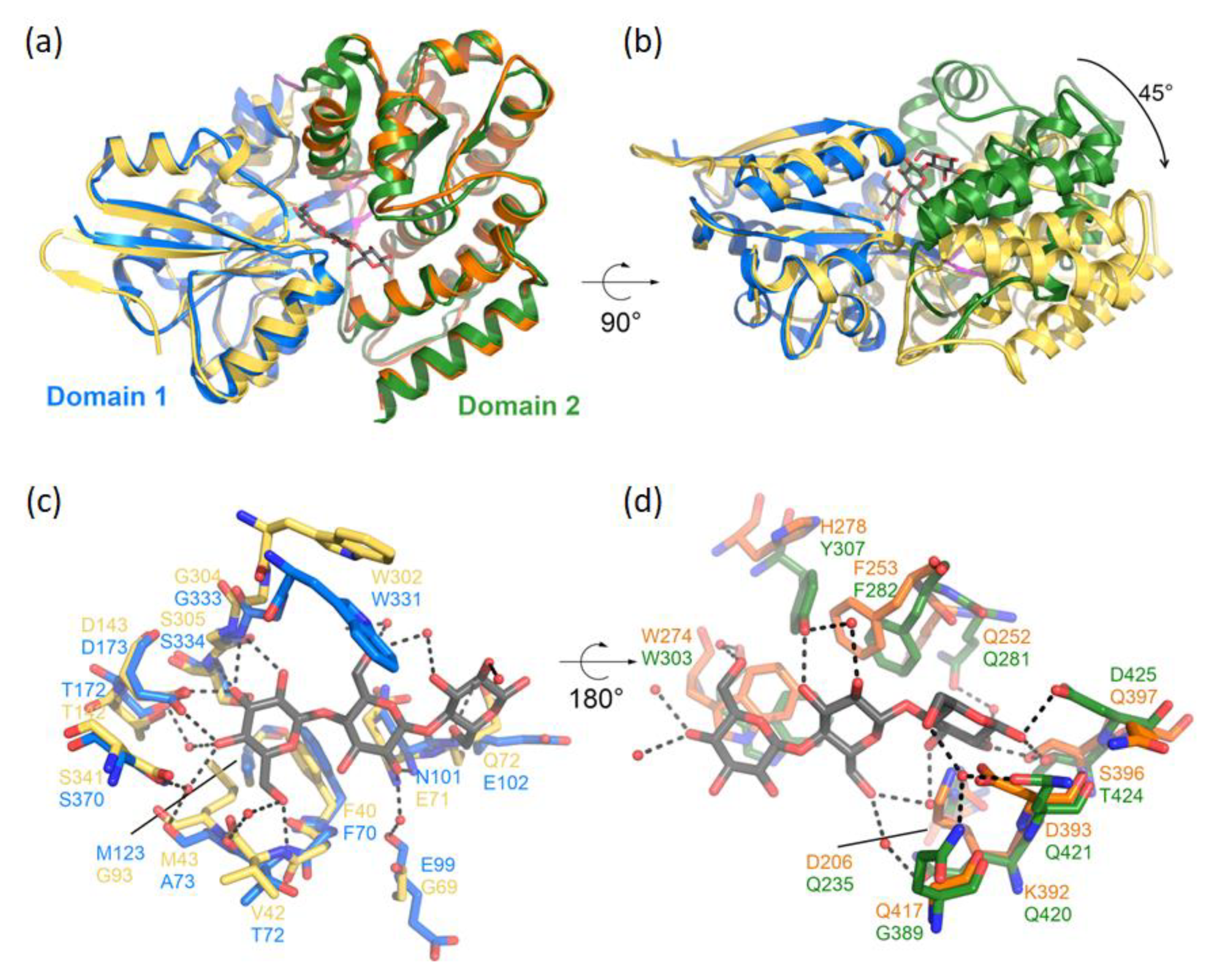
2.7. Modeling of CebE2

The 3D model of CebE2 was generated using AlphaFold v2.3.0 [20], as implemented in the AlphaFold Colab notebook (https://colab.research.google.com/github/deepmind/alphafold/blob/main/notebooks/AlphaFold.ipynb#scrollTo=pc5-mbsX9PZC accessed on 30 December 2022).

3. Results

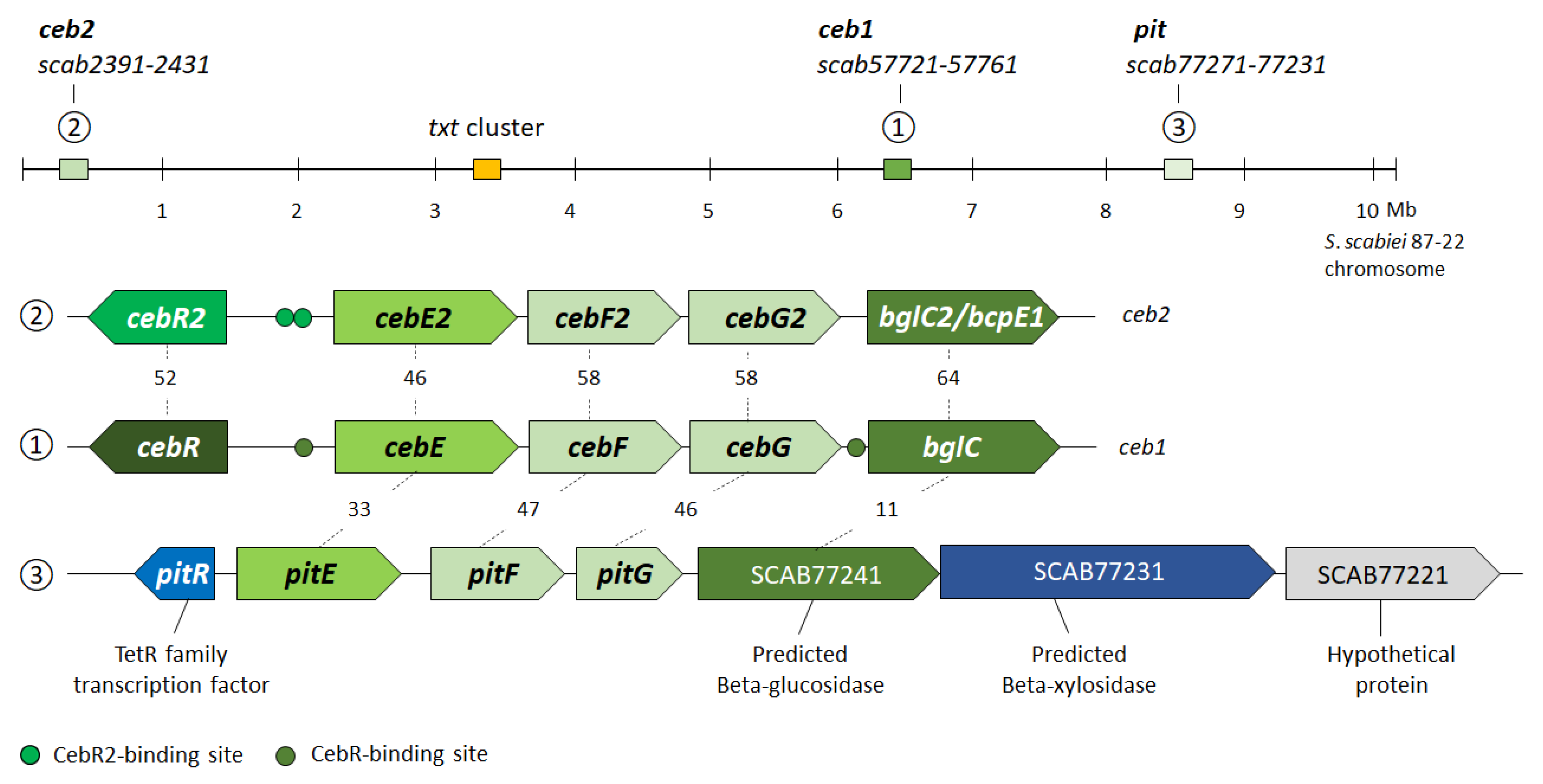
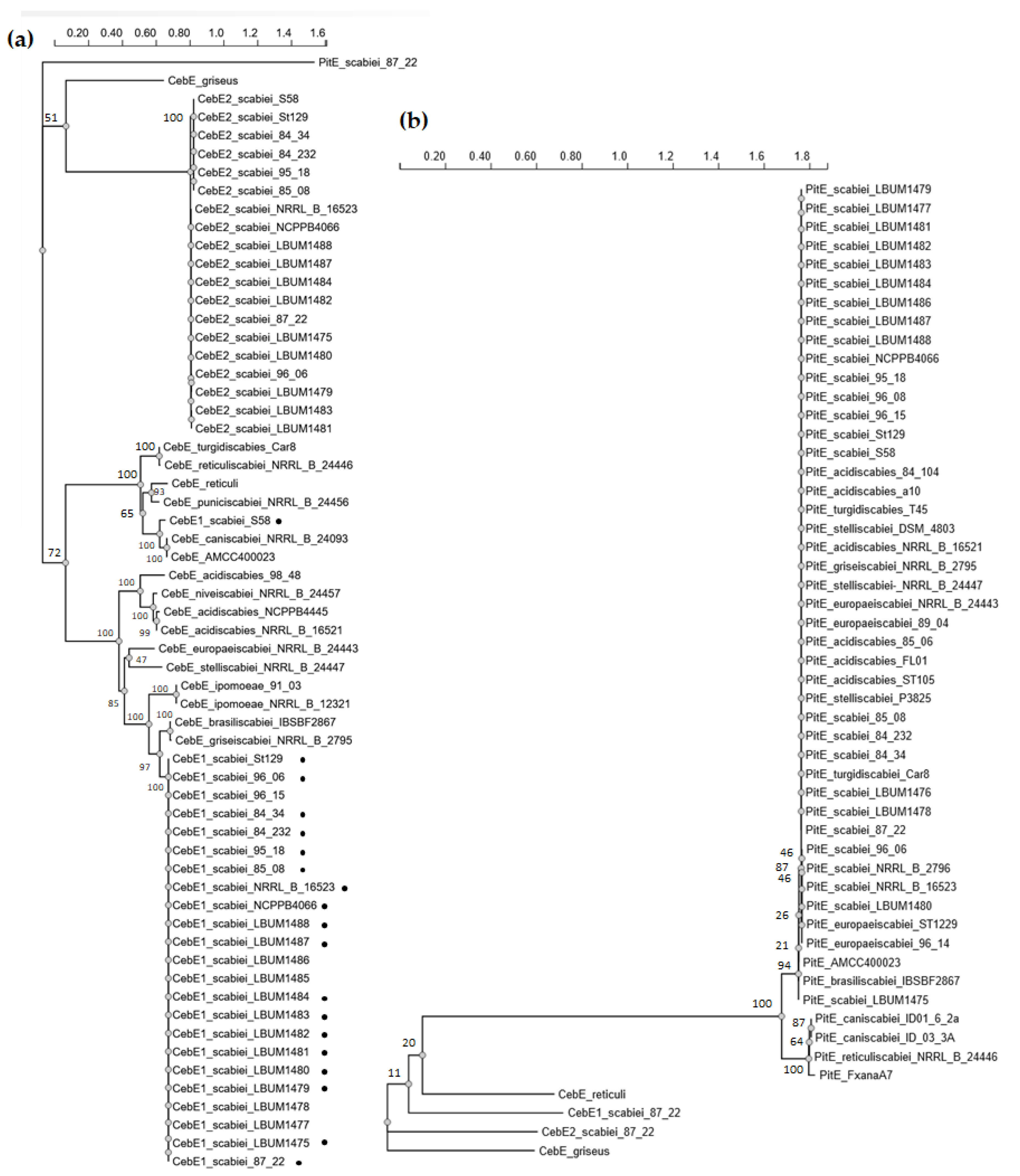
3.1. In Silico Analysis of CebE Homologs of Streptomyces scabiei

In our initial search for the cellobiose/cellotriose transporter of S. scabiei based on sequence homology with the proteins of the known cellobiose ABC-type transporter of S. reticuli (CebEreti) [21] and of S. griseus (CebEgris) [22], three gene clusters were identified, which is in accordance with what was mentioned in previous reports [9,11,14]. The cluster with the highest homology to both CebEreti and CebEgris (scab57761-57721, Figure 1) was previously demonstrated to code for the high-affinity cellobiose/cellotriose-specific CebEFG-MsiK transporter of S. scabiei [9]. This gene cluster also contains the gene for the transcriptional repressor CebR that controls both cello-oligosaccharide utilization and thaxtomin production [12], and the gene encoding the beta-glucosidase BglC for cello-oligosaccharide catabolism [10]. This first cebR-cebEFG-bglC gene locus is highly conserved among pathogenic streptomycetes, including the species S. acidiscabies, S. ipomoeae, S. stelliscabiei, S. griseiscabiei, S. brasiliscabiei, S. europaeiscabiei, and S. niveiscabiei (Figure 2a). The CebEs of pathogenic strains belonging to the species S. caniscabiei, S. reticuliscabiei, S. turgidiscabies, and S. puniscabiei are phylogenetically related and grouped with the CebEreti (Figure 2a). It has to be noted that none of the isolated pathogenic species possess a CebE protein that is orthologous to CebEgris (Figure 2a).
The second gene cluster that was identified contains the paralogues (from now on called cebR2, cebE2, cebF2, cebG2, and bglC2/bcpE1) of the first ceb gene locus with identical synteny (scab2391-2431, Figure 1). The putative role of this second ceb cluster in cello-oligosaccharide transport and utilization is supported by the recent biochemical characterization of the gene encoding the beta-glucosidase BcpE1 (BlgC2), which displayed similar kinetic properties for cellobiose hydrolysis and that was proven to compensate for the genetic loss of bglC of the first ceb cluster [13]. The search for cis-acting elements in the upstream region of scab2421 (cebE2) identified two sequences (TGGGAACGTgCCCA and TGGGcACGTTCCCA) that displayed only one mismatch (at symmetrical positions 9 and 6, respectively) with the 14 nt-long TGGGAACGTTCCCA palindromic consensus sequence deduced for CebR2 proteins via the AURTHO approach [18]. This high similarity between CebR (TGGGACGCGTCCCA) and CebR2 cis-acting elements further supports the hypothesis that these ceb clusters have originated from a common ancestor. Surprisingly, the 19 pathogenic strains that possess this putative second cello-oligosaccharide transporter all belong to the species S. scabiei (Figure 2a, Table A2).
Finally, a third sugar ABC-type transporter with partial homology, scab77271-77231, is located within the pathogenicity island (PAI) and has been identified among all the thaxtomin A-producing pathogenic Streptomyces, except for strains belonging to S. ipomoeae (Figure 2b, Table A2). From now on, genes of this third transporter will be called pitE, pitF, and pitG, with pit standing for pathogenicity island transporter. Importantly, neither CebR- nor CebR2-binding sites were identified upstream of pitE (scab77271), suggesting that the expression of the pit cluster responds to other environmental elicitors.

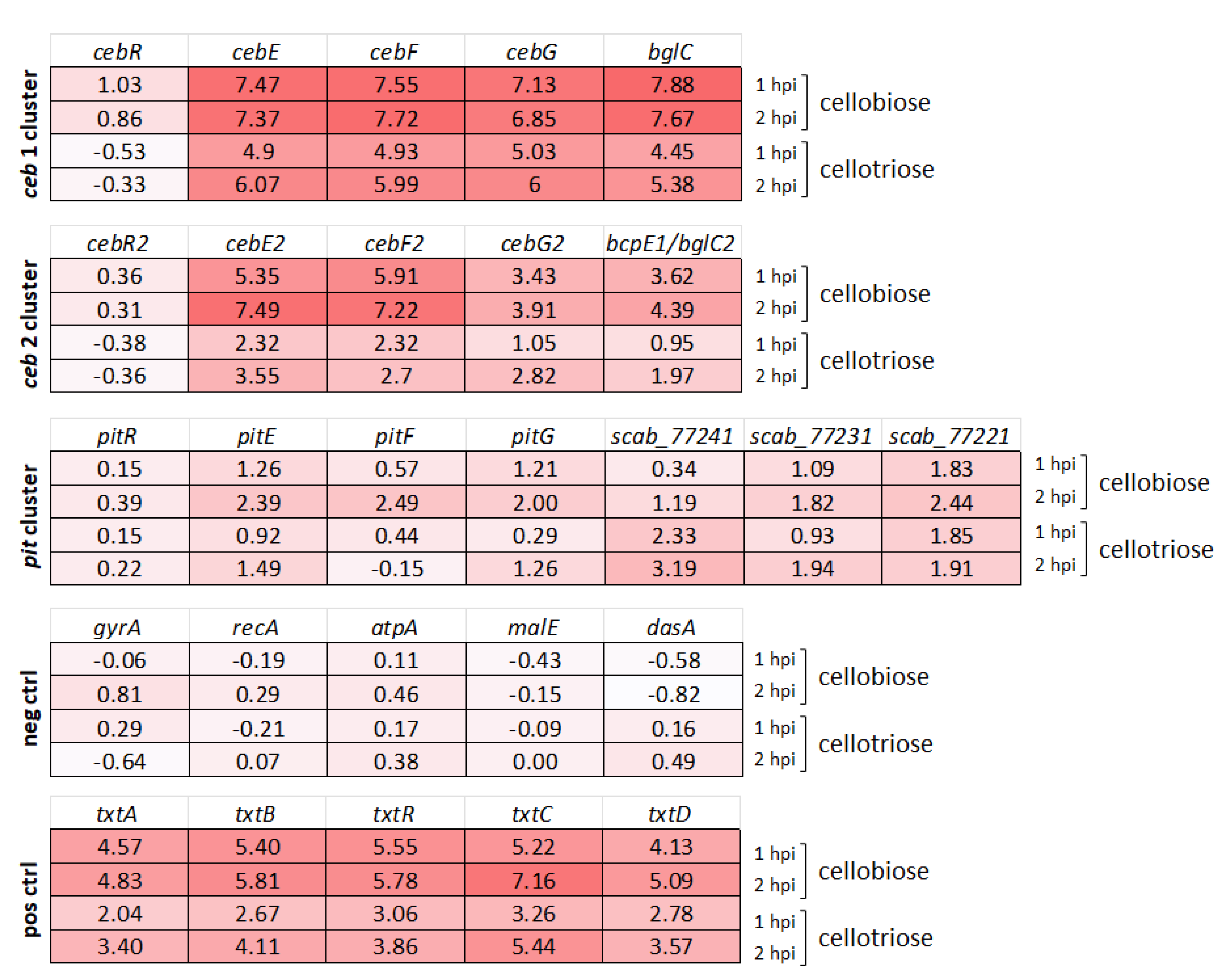
3.2. Transcriptional Response of Alternative Cello-Oligosaccharide Transporters to Cellobiose and Cellotriose

To evaluate if these two alternative transporters are potentially also involved in the import of cello-oligosaccharide elicitors by S. scabiei 87-22, we assessed their expression response post addition of cellobiose and cellotriose. RNA samples were collected 1 and 2 h post-addition of each elicitor [7]. As shown in Figure 3, the expression of the genes of the second ceb operon (cebE2, cebF2, cebG2, and bcpE1/bglC2) was drastically induced by cellobiose, with an average Log2-fold change (LFC) of 4.6 and 5.75 at 1 and 2 h post-addition of cellobiose, respectively. These values are in the same order of magnitude, although somewhat lower, as observed for the first ceb operon with an average LFC of 7.5 and 7.4. The genes of the pitEFG transcription unit showed an average LFC of 1.0 and 2.3 at 1 and 2 h post-addition of cellobiose, respectively. Although these fold changes suggest a significant response compared to the negative control genes (1.0 LFC as threshold fixed for a significant response), they are much lower than what is observed for the other two transporters, and for the genes of the thaxtomin biosynthetic gene cluster (positive controls) (Figure 3). Regarding the effect of cellotriose, gene expression of cebE2, cebF2, and cebG2 displayed an average LFC of 1.7 and 2.8 at 1 and 2 h post-addition of cellotriose, respectively. These values are weaker than those observed for the first transporter, with an average LFC of 4.8 and 5.9, but still representing a highly significant upregulation. The expression of the three transporter genes of the pit operon did not show a significant response with an average LFC of 0.55 and 0.87 at 1 and 2 h post-addition of cellotriose, respectively. Interestingly, the gene encoding the predicted beta-glucosidase (scab_77241) of the pit cluster showed a stronger expression response to cellotriose compared to the rest of the predicted transcription unit, with an LCF of 3.19 at 2 h post-addition of the elicitor.
Overall, our transcriptomic study revealed that the genes encoding a putative alternative transporter of cello-oligosaccharides (cebEFG2) are significantly transcriptionally activated by the presence of cellobiose and cellotriose, whereas the expression response of the genes of the transporter located on the pathogenicity island is much weaker, barely significant. These transcriptomic data, post-elicitor induction, are in line with earlier qRT-PCR results performed on RNA samples collected after 24, 48, and 72 h of growth of S. scabiei EF-35 in minimal medium supplemented with cellobiose (0.5%) [14].

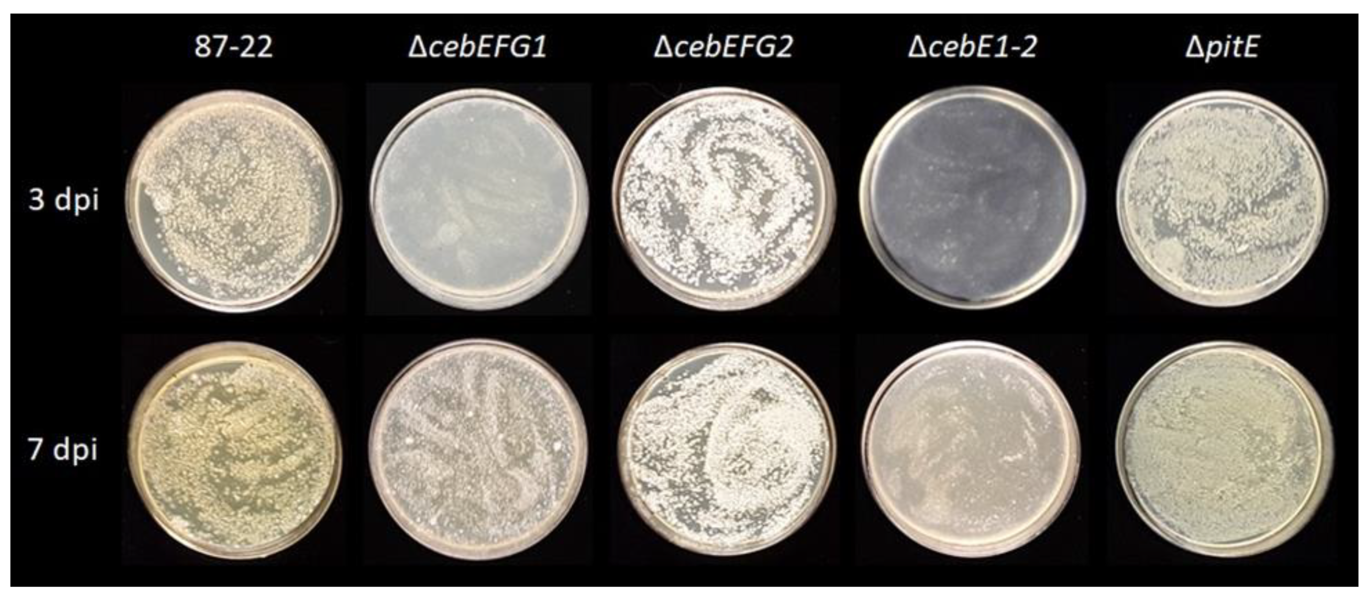
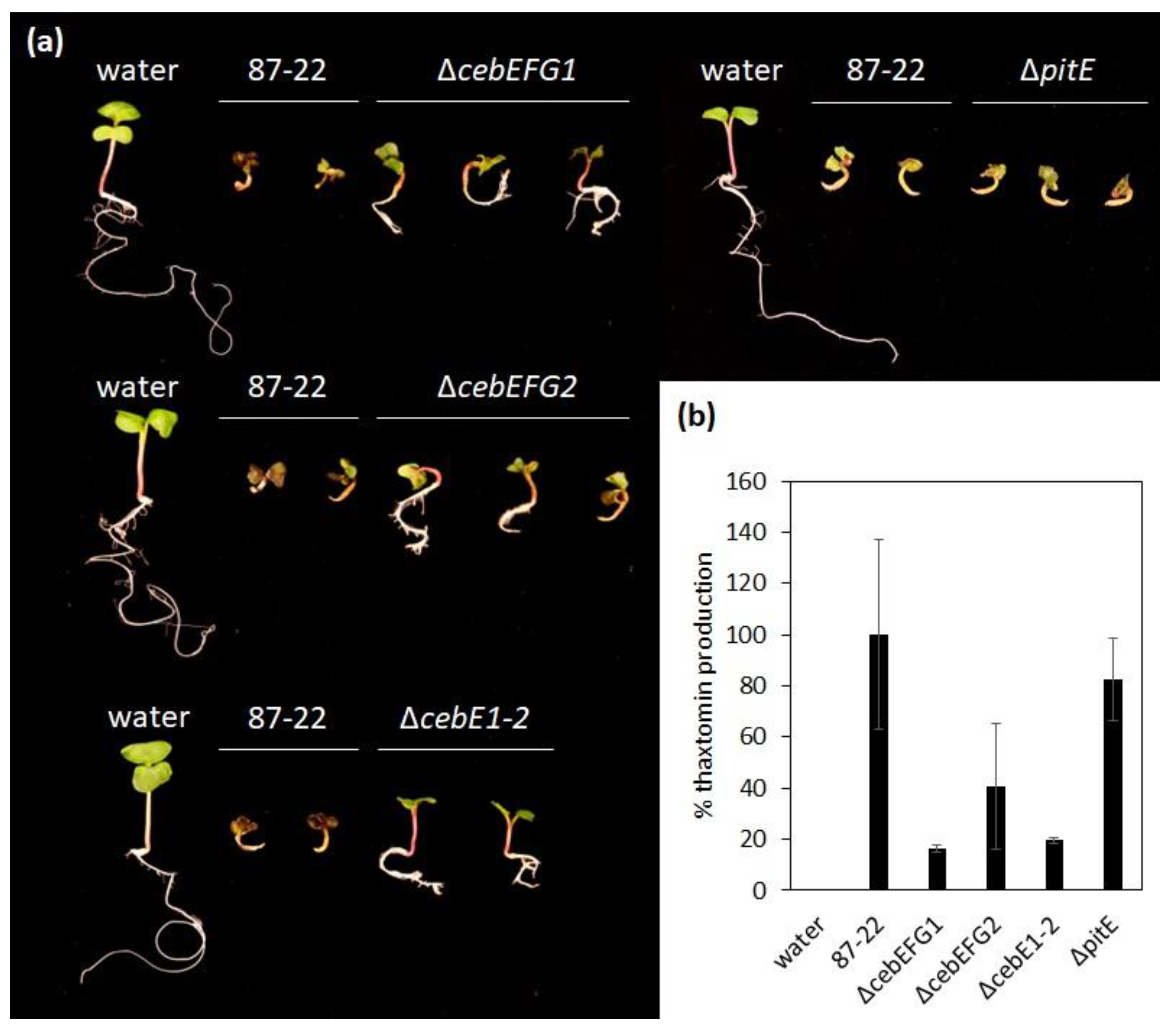
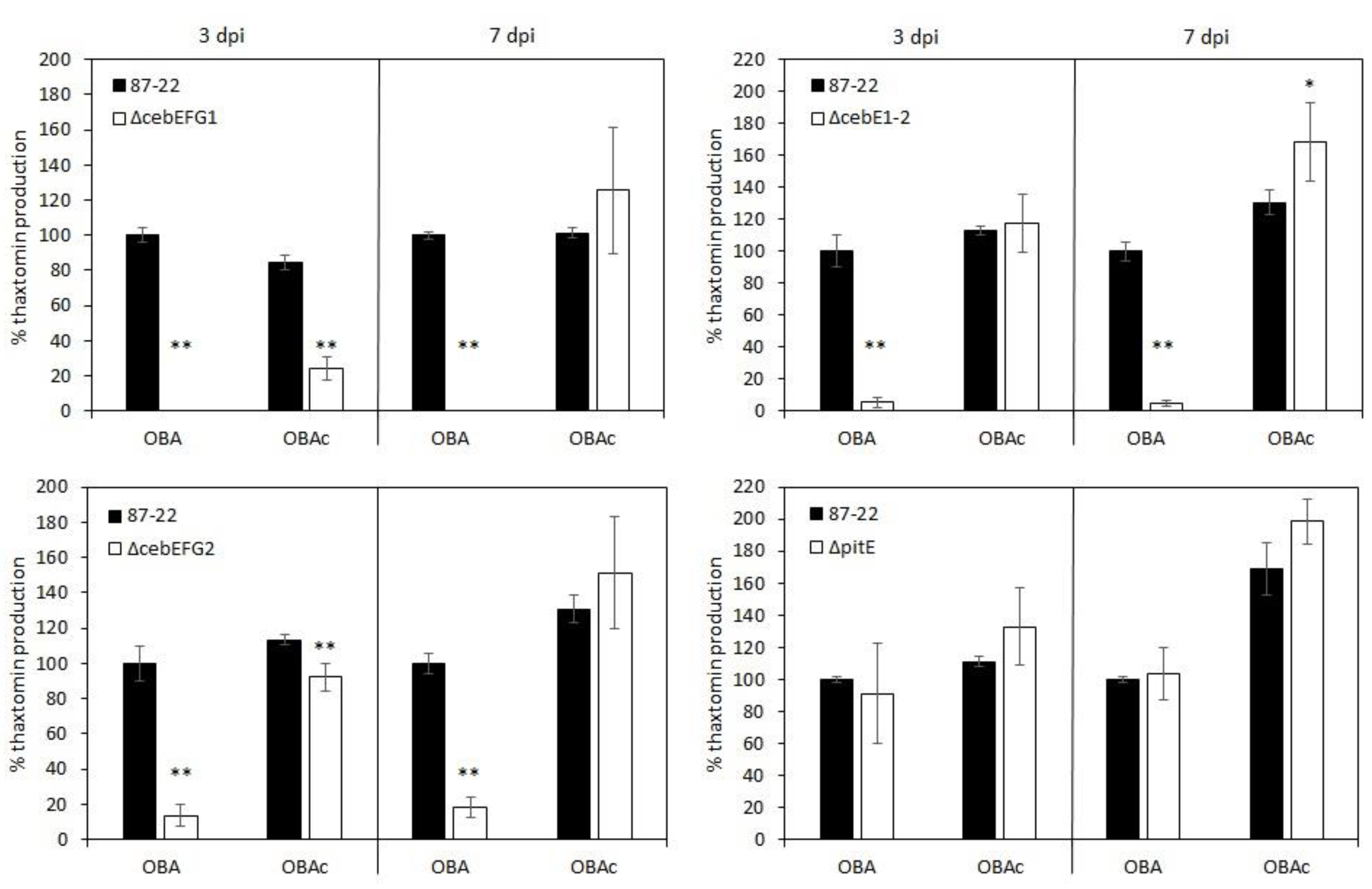
3.3. Role of the CebEFG2 and PitEFG Transporters in Virulence Elicitor Utilization by Streptomyces scabiei
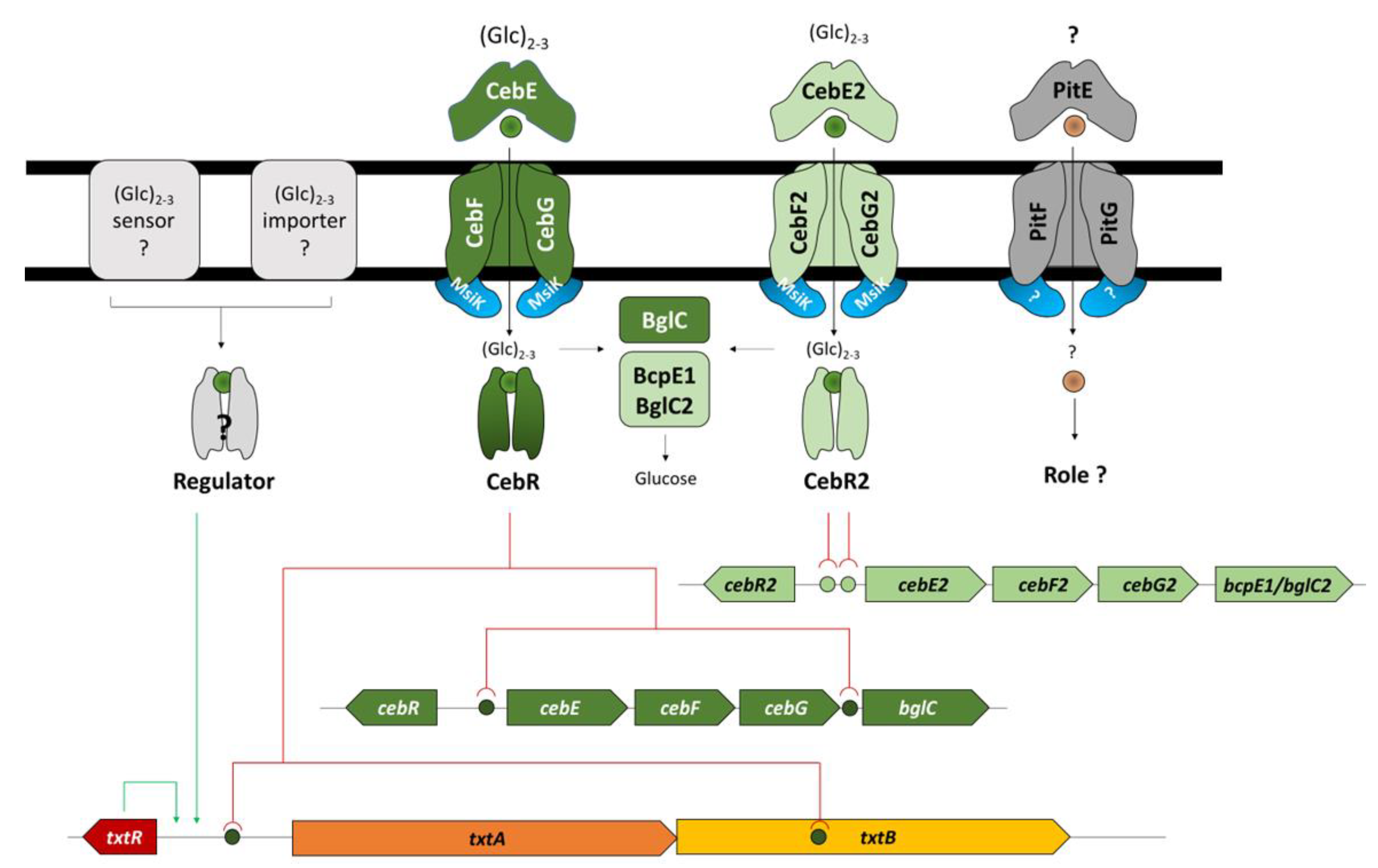
Specific deletion mutants in the genes encoding either the solute-binding proteins and/or all three proteins of the transporter were created to evaluate the role of the homologous transporters for growth on cellobiose and in the S. scabiei pathology. Figure 4 shows the ability of the various ceb and pit gene cluster deletion mutants to grow on minimal medium supplemented with cellobiose as the unique carbon source (TDMc). The differences compared to the S. scabiei wild-type strain (87-22) are optimally observed at three days post-inoculation, where the cebEFG1 deletion mutant (ΔcebEFG1) showed a drastic growth delay, which is partially made up at seven days post-inoculation (Figure 4). The restored growth of mutant ΔcebEFG1 at seven days post-inoculation is most likely due to the strong activation of the second ceb cluster by cello-oligosaccharides (Figure 3), and which also codes for a beta-glucosidase BcpE1 (BglC2) that possesses hydrolytic properties towards cellobiose similar to the BglC connected to CebEFG1 [13]. Instead, deletion of either the transporter encoding genes of the second ceb cluster (ΔcebEFG2) or the sugar-binding protein of the pathogenicity island transporter (ΔpitE) did not significantly affect growth on TDMc. The double mutant for both cebE genes (ΔcebE1-2) was even more impaired in growth on TDMc, further confirming that both transporters, CebEFG1 and CebEFG2, participate in concert in cellobiose uptake.
Upon inoculation of radish seedlings, S. scabiei typically causes strong root and shoot stunting, swelling of the hypocotyl, and necrosis of the root tip (Figure 5a). Disabling of either one or both ceb clusters resulted in less virulent strains. Seedlings inoculated with the mutants were able to partially develop compared to the wild type 87-22 (Figure 5a). This attenuated virulence phenotype is most likely due to reduced thaxtomin production, measured when the toxin was extracted from the agar–water medium the plants were grown in (Figure 5b). Despite the conservation and localization of the pitEFG transporter genes within the PAI of the pathogenic streptomycetes, the loss of its solute-binding protein did not seem to affect the virulence capacity under the conditions tested (Figure 5).
As displayed in Figure 6, reduced toxin production was also measured when growing the ceb mutants on OBA, a plant-based medium known to induce thaxtomin production [4]. Thaxtomin production levels were, however, restored when cellobiose was added to the medium (OBAc), even for the mutant strains where both cellobiose transporters were disabled (Figure 6). These results confirm the observations for the reduced virulence capacity of the mutants when inoculated onto radish seedlings and that under more complex conditions, as there would be in plant–microbe interactions, the loss of the CebEFG2 transporter cannot be entirely compensated for by the CebEFG1 transporter.

4. Discussion

In this work, we showed that S. scabiei, the most widespread and oldest potato scab-causing pathogen [23], harbors paralogues to its main cello-oligosaccharide transporter that was previously identified (CebEFG1) [9]. Expression of cebEFG2, encoding the transporter with the highest homology to CebEFG1, responds to the same main cello-oligosaccharide virulence elicitors, cellobiose and cellotriose. This is in line with qRT-PCR results from earlier work on S. scabiei EF-35 [14] and indicates that this transporter is indeed an alternative to CebEFG1 and therefore could serve as a safeguard for the loss of the main transporter. However, when grown on the complex plant-based medium OBA or during plant–microbe interaction, CebEFG2 seems to have a significant role.
Considering that cellulose is the most abundant polysaccharide on earth, the uptake and consumption of cello-oligosaccharides was anticipated to be a key feature for soil-dwelling streptomycetes. Hence, it is surprising that multiple gene loci exist that are dedicated to this specific function with at least four different ceb gene clusters, namely the main cebR-cebEFG-bglC gene clusters of S. scabiei [9], S. reticuli [21,24], and S. griseus [22], and the paralogous cebR2-cebEFG2-bcpE1/bglC2 gene cluster found in most S. scabiei species. These three main types of ceb clusters are not orthologous as they differ from one another by (i) their low level of homology (the percentages of amino acid identity between CebE proteins are below 50%, much lower than the values usually found for orthologous genes/proteins), (ii) their gene organization (cebR is the last gene of the transcriptional unit in S. griseus, while it is divergently transcribed in S. scabiei and S. reticuli), and (iii) their affinity for binding cellobiose and cellotriose (at the nano- and micromolar levels for CebEscab and CebEreti, respectively) [9,21]. Importantly, the molecular mechanism governing the expression control of these genes has been conserved as the transcriptional CebR repressors of S. scabiei, S. reticuli, and S. griseus all recognize the same cis-acting element with the palindromic TGGGACGCGTCCCA as consensus sequence [12,22,24], which allows them to also control the transcription of the cellulase encoding genes [25].
The second and alternative cello-oligosaccharide gene cluster (cebEFG2) seems to only be present in the S. scabiei group of pathogenic Streptomyces species. Even the phylogenetically closest pathogenic strains to the S. scabiei group, such as S. ipomoeae, S. brasiliscabiei, and S. griseoscabiei, do not possess this second cello-oligosaccharide transporter (CebEFG2scab). This CebEFG2scab transporter is not exclusive to pathogenic S. scabiei species as it is also found in diverse non-pathogenic Streptomyces species, including S. coelicolor [26] and S. lividans [27] as the best-studied species, amongst many others according to BlastP results (not shown). The question remains whether or not other pathogenic streptomycetes that have a CebEscab (species S. acidiscabies, S. ipomoeae, S. europaeiscabiei, S. niviscabiei, S. stelliscabiei, S. brasiliscabiei, and S. griseiscabiei) or a CebEreti (S. turgidiscabies, S. caniscabiei, S. reticuliscabiei, and S. puniscabiei) background potentially possess an additional and yet unidentified ceb cluster for cello-oligosaccharide import and/or to safeguard the possible loss of the main ceb cluster. Another intriguing observation is that the PAI that contains the genes essential for plant host colonization has never been transferred and integrated into the chromosome of Streptomyces species that possess a CebEgris background. Why this has never occurred remains unexplained. Maybe these species lack other genetic features that are indirectly essential for plant host colonization, which would minimize the selection pressure to keep the PAI if integrated in these species, unless scab-causing Streptomyces species containing CebEgris have not been isolated yet.
The reason why CebEFG2 is less efficient than the main CebEFG1 transporter at both using cello-oligosaccharides as a nutrient source or at importing the virulence elicitors remains to be studied at the molecular level. We recently obtained the crystallographic structure of CebE1scab in complex with cellotriose and are currently investigating the amino acids involved in sugar recognition (publication in preparation). Based on the crystallographic structure of CebE1scab, we generated the 3D model of CebE2 using AlphaFold v2.3.0 [20], as implemented in the AlphaFold Colab notebook. The CebE2 model is characterized by an overall good quality with an average pLDDT value of 95.9 for the folded region (aa 23-425). This model corresponds to an open conformation of the protein. However, when superimposed separately, each domain is similar to the structure of CebE1 in complex with cellotriose, with rms deviation of 1.01 Å over 135 Cα and 0.93 Å over 190 Cα for Domain 1 and 2, respectively (Figure 7a). The opening of the ligand binding pocket corresponds to an approximate 45° rotation of Domain 2 compared to Domain 1 (Figure 7b). According to the structural model of CebE2 (Figure 7), Domain 1 interacts with the ligand through seven hydrogen bonds (H-bonds) that are conserved between CebE1 and CebE2 (sidechain of D143 and W302; mainchain of V42, E71, Q72, and G304), and 3 hydrophobic interactions, of which one is conserved (F40) and two are equivalent (N101 sidechain in CebE1 vs. Q71 in CebE2; the A73-M123 pair in CebE1 replaced by the M43-G93 pair in CebE2) (Figure 7c). In Domain 2 (Figure 7d), two major hydrophobic interactions are conserved in between CebE1 and CebE2 (F253 and W274) as well as two H-bonds involving the T424 side chain in CebE1 (S396 in CebE2). For the H-bonds involving the sidechains of Y307 and D425, the model does not establish whether H278 and Q397 can be substituent. The interaction between CebE1 and cellotriose is also mediated by a network of eight water molecules, of which four are fully conserved and the four are only partially conserved. Overall, because of the large number of interactions conserved, CebE2 is clearly compatible with the binding of cellotriose and cellobiose, such as CebE1. It is, however, difficult to extrapolate a difference in binding affinities between CebE1 and CebE2 for these ligands, as well as different import efficiencies of the entire machinery, and therefore how they individually contribute to informing S. scabiei of the presence of the inducer of its pathogenic lifestyle.
Another major difference between the ceb1 and ceb2 clusters of S. scabiei is the sequence of the consensus cis-acting elements recognized by CebR1 (TGGGACGCGTCCCA) and CebR2 (TGGGAACGTTCCCA), respectively. Although these two CebR-binding sites only differ in two symmetrical positions of their 14-nt inverted repeat consensus sequence, these nucleotide replacements may result in different binding affinities, which would ultimately lead to a different transcriptional response in terms of concentration of the allosteric effector required to dissociate the protein-DNA complex. This small modification also suggests that only CebR1 directly controls the thaxtomin biosynthetic gene cluster and cellulase encoding genes as the computational prediction of the CebR2 regulon seems to be strictly limited to the intergenic region between cebR2 and cebEFG2-bcpE1/bglC2.
Perhaps the most surprising result of our study is the conserved ability of cellobiose to induce thaxtomin production in mutants where both cello-oligosaccharide transporters were disabled (strain ΔcebE1-2) when they are inoculated on OBAc (Figure 6, top right panel). This was unexpected as it therefore strongly suggests the existence of another mechanism to either import or sense cello-oligosaccharides in S. scabiei 87-22. We previously hypothesized the existence of another regulator than CebR able to sense and respond to cellobiose and cellotriose as, except for the thaxtomin, all other metabolites that are a part of the virulome of S. scabiei 87-22 have their expression induced by both elicitors despite the absence of CebR cis-acting elements in their biosynthetic gene clusters [7]. The identity of this additional transcriptional regulator of cello-oligosaccharide utilization is unknown and could be part of the numerous regulatory protein encoding genes that display a strong response to both cellobiose and cellotriose, as observed in our transcriptomic analysis [7].
Finally, we demonstrated here that genes coding for the PitEFG ABC-transporter that are located on the PAI would play no role in the onset of pathogenicity of S. scabiei 87-22 (Figure 5). This result raises the question why these sugar-transporter genes are systematically maintained on the PAI if they do not play a role in plant colonization. This is maybe due to the experimental set up of plant bioassays where the controlled conditions in which the radish seedlings grow do not generate the panel of carbohydrates that common scab causing Streptomyces species encounter in the rhizosphere. Once the substrate of PitE is identified, it will be easier to deduce or propose a role for this transporter in plant colonization.

5. Conclusions

In this work, we refined the proposed signaling pathway from cello-oligosaccharide transport to the production of thaxtomin and the onset of the pathogenic lifestyle of S. scabiei 87-22 (Figure 8). In addition to the main CebEFG-MsiK transporter, cello-oligosaccharides, can be imported by the alternative CebEFG2 that also recruits MsiK as ATPase as deletion of msiK fully abolished growth on TDMc [9]. The imported sugars are both hydrolyzed by beta-glucosidase BglC and BcpE1/BglC2 to feed the glycolysis with glucose and serve as allosteric effectors to inhibit the transcriptional repression of txtR by CebR, the pathway-specific activator of the thaxtomin biosynthetic genes. As cellobiose is still able to induce thaxtomin in OBAc when both ceb clusters are disabled (Figure 6, top right panel), we suggest the existence of an additional importer or sensor of cello-oligosaccharides that would directly or indirectly (via a yet unknown regulator) induce thaxtomin production. The role and the substrate specificity of the pathogenicity island-associated transporter PitEFG is still unknown.

Author Contributions

Conceptualization, I.M.F. and S.R.; methodology, I.M.F. and S.R.; software, S.R.M.; validation, I.M.F. and S.R.; formal analysis, I.M.F., B.D., F.K. and S.R.; investigation, I.M.F., D.B., B.D., S.G., J.J.C.S., R.V., N.S., S.R.M., F.K. and S.R.; resources, I.M.F., R.L. and S.R.; data curation, F.K. and S.R.; writing—original draft preparation, I.M.F. and S.R.; writing—review and editing, I.M.F. and S.R.; visualization, I.M.F., F.K. and S.R.; supervision, I.M.F. and S.R.; project administration, I.M.F. and S.R.; funding acquisition, I.M.F. and S.R. All authors have read and agreed to the published version of the manuscript.

Funding

This research was funded by the CSUB Research Council of the University (RCU), CSUB Faculty Teaching and Learning Center (FTLC), and the Louis Stokes Alliance for Minority Participation (LSAMP-NSF). The work of S.R. and B.D was supported by an Aspirant grants from the FNRS (grant 1.A618.18) and a FRIA grant from the FNRS for S.R. and N.S. (FRIA 1.E.116.21). Transcriptomic analysis was supported by a ‘Crédit De Recherche FNRS grant’ (grant R.FNRS.5240 J.0158.21-CDR).

Institutional Review Board Statement

Not applicable.

Informed Consent Statement

Not applicable.

Data Availability Statement

The data presented in this study are openly available. RNAseq data were publicly deposited, and our experimental and analytical pipelines are described in the GEO database repository (accession number: GSE181490). The CebE structure we used for modelization can be found at https://www.wwpdb.org/pdb?id=pdb_00008bfy accessed on 30 December 2022.

Acknowledgments

S.R. and F.K. are Fonds de la Recherche Scientifique (FRS-FNRS) senior and research associates, respectively.

Conflicts of Interest

The authors declare no conflict of interest.

Appendix A

Table A1. Bacterial strains, plasmids, and cosmids used in this study.
Description Source or Reference
Plasmids/cosmids
pIJ790 λ Red plasmid (tS, CmlR)[19]
pUZ8002 Supplies transfer functions for mobilization of oriT-containing vectors from E. coli to Streptomyces (KanR)[19]
pIJ773 Template for REDIRECT© PCR targeting system, contains the [aac(3)IV + oriT] disruption cassette (AmpR, ApraR)[19]
pIJ778 Template for REDIRECT© PCR targeting system, contains the [aadA + oriT] disruption cassette (AmpR, StrepR, SpecR)[19]
SuperCos1 Cosmid cloning vector (AmpR, KanR)Stratagene
Cosmid 833 SuperCos1 derivative containing the S. scabiei 87-22 cellobiose/cellotriose-specific ABC-transporter locus (ceb1) (KanR, AmpR)[12]
Cosmid 2852 SuperCos1 derivative containing the S. scabiei 87-22 ceb2 ABC-transporter locus (KanR, AmpR)This study
Cosmid 1998 SuperCos1 derivative containing the S. scabiei 87-22 pit ABC-transporter locus (KanR, AmpR)This study
E. coli strains
DH5α General cloning hostGibco-BRL
BW25113 Host for Redirect PCR targeting system[19]
ET12567 dam, dcm, hsdS; non-methylating host for transfer of DNA into Streptomyces spp. (CmlR, TetR)[28]
Streptomyces strains
87-22 S. scabiei wild type strain[29]
cebEFG187-22 derivative with a deletion of the region from scab57751 (cebE1) through scab57731 (cebG1) (ApraR)This study
cebEFG287-22 derivative with a deletion of the region from scab2421 (cebE2) through scab2401 (cebG2) (ApraR)This study
pitE87-22 derivative with a deletion of the scab77271 (pitE) gene (ApraR)This study
† CmlR, chloramphenicol resistance; KanR, kanamycin resistance; AmpR, ampicillin resistance; ApraR, apramycin resistance; StrepR, streptomycin resistance; SpecR, spectinomycin resistance; tS, temperature sensitive.
Table A2. Genomes and protein accession numbers of strains investigated in this study.
StreptomycesABC-Type Sugar-Binding Proteins
SpeciesStrainCebE1CebE2PitEGenBank AssemblyRef.
scabiei87-22WP_013003368.1WP_037723613.1WP_231885169.1GCA_000091305.1[30,31]
scabieiLBUM 1475WP_013003368.1WP_037723613.1WP_237311104.1GCA_017933255.1[32]
scabieiLBUM 1477WP_013003368.1NFWP_006378806.1GCA_017969045.1[32]
scabieiLBUM 1478WP_013003368.1NFMBP5904699.1GCA_017969065.1[32]
scabieiLBUM 1479WP_013003368.1WP_060905683.1WP_006378806.1GCA_017969075.1[32]
scabieiLBUM 1480WP_013003368.1WP_037723613.1WP_231885169.1GCA_017933235.1[32]
scabieiLBUM 1481WP_013003368.1WP_210550546.1WP_006378806.1GCA_017969025.1[32]
scabieiLBUM 1482WP_013003368.1WP_037723613.1WP_231885169.1GCA_017933115.1[32]
scabieiLBUM 1483WP_013003368.1WP_210550546.1WP_006378806.1GCA_017969005.1[32]
scabieiLBUM 1484WP_013003368.1WP_037723613.1WP_231885169.1GCA_017968985.1[32]
scabieiLBUM 1485WP_013003368.1NFNFGCA_017968925.1[32]
scabieiLBUM 1486WP_013003368.1NFWP_006378806.1GCA_017968965.1[32]
scabieiLBUM 1487WP_013003368.1WP_037723613.1WP_006378806.1GCA_017968905.1[32]
scabieiLBUM 1488WP_013003368.1WP_037723613.1WP_006378806.1GCA_017968935.1[32]
scabieiNCPPB 4066WP_013003368.1WP_037723613.1WP_006378806.1GCA_000738715.1[33]
scabieiNRRL B-16523WP_013003368.1WP_037723613.1WP_231885169.1GCA_001005405.1[34]
scabieiS58GAQ65654.1WP_059084709.1WP_006378806.1GCF_001485125.1[35]
scabieiSt129WP_013003368.1WP_037723613.1WP_006378806.1GCA_003584815.1[36]
scabiei84-34WP_013003368.1WP_037723613.1WP_231885169.1GCA_001550245.1[33]
scabiei84-232WP_013003368.1WP_037723613.1WP_231885169.1GCA_001572115.1[33]
scabiei85-08WP_013003368.1WP_037723613.1WP_231885169.1GCA_001550215.1[33]
scabiei95-18WP_013003368.1WP_037723613.1WP_006378806.1GCA_001550225.1[33]
scabiei96-06WP_013003368.1WP_060905683.1WP_231885169.1GCA_001579685.1[33]
scabiei96-08NFWP_060905683.1WP_006378806.1GCA_001550235.1[33]
scabiei96-15WP_013003368.1NFWP_006378806.1GCA_001550295.1[33]
europaeiscabieiNCPPB 4086WP_037703405.1-NFGCF_000738695.1[33]
brasiliscabieiIBSBF2867WP_216591689.1-WP_210534218.1GCF_018927715.1[37]
griseiscabieiNRRL B-2795MBZ3900963.1-WP_006378806.1GCA_020010925.1[38]
ipomoeaeNRRL-B-12321WP_141569662.1-NFGCF_006547165.1
ipomoeae78-51WP_009295889.1-NFGCF_006547175.1[39]
ipomoeae88-35WP_009295889.1-NFGCF_006547185.1[39]
ipomoeae91-03WP_009295889.1-NFGCF_000317595.1[40]
europaeiscabieiNRRL B-24443WP_046704818.1-WP_006378806.1GCF_000988945.1[41]
europaeiscabiei89-04WP_046704818.1-WP_006378806.1GCF_001550325.1[33]
europaeiscabieiSt1229WP_046704818.1-WP_231885169.1GCF_003584825.1
europaeiscabiei96-14WP_060891518.1-WP_231885169.1GCF_001550375.1[36]
stelliscabieiDSM 41803WP_046918411.1-WP_006378806.1GCF_014873495.1
stelliscabieiP3825WP_046918411.1-WP_231885169.1GCF_001189035.1
stelliscabieiNRRL B-24447WP_046918411.1-WP_006378806.1GCF_001008135.1[42]
stelliscabiei1222.2WP_046918411.1-NFGCF_900215595.1
acidiscabiesNRRL B-16521WP_029183343.1-WP_006378806.1GCA_020010905.1[38]
acidiscabies84-104WP_029183343.1-WP_006378806.1GCA_000242715.2[43]
acidiscabies85-06WP_029183343.1-WP_006378806.1GCA_001941285.1[44]
acidiscabies98-49WP_029183343.1-NFGCA_001941265.1[44]
acidiscabiesa2WP_029183343.1-NFGCA_001949645.1
acidiscabiesa10WP_029183343.1-WP_006378806.1GCA_001485105.1[35]
acidiscabiesFL01WP_029183343.1-WP_006378806.1GCA_001941325.1[44]
acidiscabiesSt105WP_029183343.1-WP_006378806.1
GCA_003584805.1
acidiscabiesLBUM 1476WP_029183343.1-WP_006378806.1GCA_017969125.1
acidiscabiesNCPPB 4445WP_050369574-NFGCA_001189015.1
acidiscabies98-48WP_075734941.1-NFGCA_001941275.1[44]
niveiscabieiNRRL B-24457WP_055721858.1-NFGCF_001419795.1
puniciscabieiNRRL B-24456WP_055706423.1-NFGCA_001419685.1[45]
puniciscabieiDSM 41929WP_055706423.1-NFGCA_006715785.1[45]
caniscabiei96-12WP_060884585.1-NFGCA_001550315.1[33]
caniscabieiNRRL B-24093WP_060884585.1-NFGCA_002155765.1[34]
caniscabieiND05-01CWP_193381044.1-NFGCA_014930395.1
caniscabieiND05-13AWP_193381044.1-NFGCA_014930415.1
caniscabieiND05-3BWP_193381044.1-NFGCA_014930355.1
caniscabieiNE06-02DWP_193381044.1-NFGCA_014930365.1[46]
caniscabieiNE06-02FWP_193381044.1-NFGCA_014930405.1
caniscabieiID-03-3AWP_086802801.1-WP_086802416.1GCA_014852565.1
caniscabieiNRRL B-2801WP_086802801.1-WP_237277203.1GCA_002155725.1
caniscabieiID01-6.2aWP_086802801.1-WP_086802416.1GCA_014852655.1
caniscabieiID01-12cWP_086802801.1-NFGCA_014852675.1
spAMCC400023WP_045557721.1-WP_237311104.1GCA_021665875.1
spCF124WP_045557721.1-NFGCA_900114955.1
spFxanaA7WP_045557721.1-WP_234442873.1GCA_000958545.1
turgidiscabiesT45WP_059073075.1-WP_006378806.1GCA_001485145.1[35]
reticuliscabieiNRRL B-24446WP_059073075.1-WP_245879130.1GCF_002154675.1[47]
turgidiscabiesCar8ELP70267.1-WP_006378806.1GCA_000331005.1[48]
reticuli-CAB46342.1--GCA_001511815.1-
griseusNBRC 13350WP_012379731.1-- [49]
NF, Not found (either ORF not present or not found due to incomplete genome sequence); -, not present.
Table A3. Primers used in this study.
PrimerSequence (5′→3′) *Use
sc052cggggggtcggcacgtcagggcatcaggaggacgcaatgattccggggatccgtcgaccRedirect deletion cassette for scab57751-scab57731 (cebEFG1)
imf271ccccggtgagaagacgtacggcggcgaaccggggcctcatgtaggctggagctgcttcRedirect deletion cassette for scab57751-scab57731 (cebEFG1)
sc056cttacacatcccgcacgttcccVerification of ∆scab57751-scab57731 (∆cebEFG1)
imf272gcgtgtgactgaaggtgtccVerification of ∆scab57751-scab57731 (∆cebEFG1)
sc050tccatgggcacgttcggttggtcgcacagaaaggcgatgattccggggatccgtcgaccRedirect deletion cassette for scab2421-scab2401 (cebEFG2)
imf296ggcggggctcggctgtgcgggggcggtgggggcgggtcatgtaggctggagctgcttcRedirect deletion cassette for scab2421-scab2401 (cebEFG2)
sc058cgttcggttggtcgcacagaaaggVerification of ∆scab2421-scab2401 (∆cebEFG2)
imf297acgggctggatgttcttctcVerification of ∆scab2421-scab2401 (∆cebEFG2)
mjk1gcaacatcgcggcacaccgcctggaaataaggagtacagatgattccggggatccgtcgaccRedirect deletion cassette for scab77271 (pitE)
mjk2cgtccgcggagggcgcccccgagcgcgcggacgaaccgatcatgtaggctggagctgcttcRedirect deletion cassette for scab77271 (pitE)
mjk3gccatgccttcaacatacagVerification of ∆scab77271 (∆pitE)
mjk4gctggtcgaggataaaggtgVerification of ∆scab77271 (∆pitE)
* Non-homologous extensions are underlined, while engineered restriction sites are indicated in bold.

Appendix B

Genomic DNA Extraction

Mycelia of 48–72 h cultures were collected, washed twice with distilled water, and frozen at −20 °C for 60 min. Pellets were resuspended in 500 µL TE25S buffer (25 mM Tris-HCl pH 8, 25 mM EDTA, 0.3 M sucrose) and treated with 20 µL of 100 mg/mL lysozyme for 1h at 37 °C while mixing gently. To each sample, 5 µL Proteinase K (20 mg/mL) and 30 µL of 10% SDS were added and mixed by inversion. The tubes were incubated at 55 °C while occasionally mixing gently. After 1h, 100 µL of 5M NaCl was added and mixed thoroughly, followed by the addition of 65 µL of CTAB/NaCl (preheat 0.7M NaCl to 55 °C and slowly add CTAB to reach a 10% end concentration). The samples were incubated at 55 °C for 10 min and allowed to cool to 37 °C before cooling on ice for 10 min. Subsequently, 500 µL chloroform:isoamylalcohol (24:1) was added, mixed thoroughly, and the tubes were centrifuged at >10,000× g for 10 min at 4 °C. The upper phase was transferred to a clean microcentrifuge tube and cooled on ice for at least 10 min. The genomic DNA was precipitated upon the addition of one volume of cold isopropanol, inversion of the tubes to mix, and incubation on ice for at least 10 min or overnight at −20 °C. The DNA was pelleted by centrifugation at >10,000× g for 10 min at 4 °C. Pellets were washed with 70% ethanol and air-dried before resuspension in 100 µL of clean DI water. Samples were stored at −20 °C until use.

References

  1. Wilson, D.B. Microbial diversity of cellulose hydrolysis. Curr. Opin. Microbiol. 2011, 14, 259–263. [Google Scholar] [CrossRef]
  2. Loria, R.; Kers, J.; Joshi, M. Evolution of plant pathogenicity in Streptomyces. Annu. Rev. Phytopathol. 2006, 44, 469–487. [Google Scholar] [CrossRef] [PubMed]
  3. Wach, J.M.; Krasnoff, S.B.; Loria, R.; Gibson, D.M. Effect of carbohydrates on the production of thaxtomin A by Streptomyces scabies. Arch. Microbiol. 2007, 18, 81–88. [Google Scholar] [CrossRef] [PubMed]
  4. Johnson, E.G.; Joshi, M.V.; Gibson, D.M.; Loria, R. Cello-oligosaccharides released from host plants induce pathogenicity in scab-causing Streptomyces species. Physiol. Mol. Plant Pathol. 2007, 71, 18–25. [Google Scholar] [CrossRef]
  5. Bischoff, V.; Cookson, S.J.; Wu, S.; Scheible, W.R. Thaxtomin A affects CESA-complex density, expression of cell wall genes, cell wall composition, and causes ectopic lignification in Arabidopsis thaliana seedlings. J. Exp. Bot. 2009, 60, 955–965. [Google Scholar] [CrossRef] [PubMed]
  6. Scheible, W.R.; Fry, B.; Kochevenko, A.; Schindelasch, D.; Zimmerli, L.; Somerville, S.; Loria, R.; Somerville, C.R. An Arabidopsis mutant resistant to thaxtomin A, a cellulose synthesis inhibitor from Streptomyces species. Plant Cell 2003, 15, 1781–1794. [Google Scholar] [CrossRef]
  7. Deflandre, B.; Stulanovic, N.; Planckaert, S.; Anderssen, S.; Bonometti, B.; Krim, L.; Coppieters, W.; Devreese, B.; Rigali, S. The virulome of Streptomyces scabiei in response to cello-olligasaccharides. Microb. Genom. 2022, 8, 000760. [Google Scholar] [CrossRef]
  8. Jourdan, S.; Francis, I.M.; Deflandre, B.; Loria, R.; Rigali, S. Tracking the subtle mutations driving host sensing by the plant pathogen Streptomyces scabies. mSphere 2017, 2, 27144. [Google Scholar] [CrossRef]
  9. Jourdan, S.; Francis, I.M.; Kim, M.J.; Salazar, J.J.C.; Planckaert, S.; Frère, J.M.; Matagne, A.; Kerff, F.; Devreese, B.; Loria, R.; et al. The CebE/MsiK transporter is a doorway to the cello-oligosaccharide-mediated induction of Streptomyces scabies pathogenicity. Sci. Rep. 2016, 2, e00367-16. [Google Scholar] [CrossRef]
  10. Jourdan, S.; Francis, I.M.; Deflandre, B.; Tenconi, E.; Riley, J.; Planckaert, S.; Tocquin, P.; Martinet, L.; Devreese, B.; Loria, R.; et al. Contribution of the β-glucosidase BglC to the onset of the pathogenic lifestyle of Streptomyces scabies. Mol. Plant Pathol. 2018, 19, 1480–1490. [Google Scholar] [CrossRef] [Green Version]
  11. Joshi, M.V.; Bignell, D.R.D.; Johnson, E.G.; Sparks, J.P.; Gibson, D.M.; Loria, R. The AraC/XylS regulator TxtR modulates thaxtomin biosynthesis and virulence in Streptomyces scabies. Mol. Microbiol. 2007, 66, 633–642. [Google Scholar] [CrossRef]
  12. Francis, I.M.; Jourdan, S.; Fanara, S.; Loria, R.; Rigali, S. The cellobiose sensor CebR is the gatekeeper of Streptomyces scabies pathogenicity. mBio 2015, 6, e02018. [Google Scholar] [CrossRef]
  13. Deflandre, B.; Thiébaut, N.; Planckaert, S.; Jourdan, S.; Anderssen, S.; Hanikenne, M.; Devreese, B.; Francis, I.; Rigali, S. Deletion of bglC triggers a genetic compensation response by awakening the expression of alternative beta-glucosidase. BBA Gene Regul. Mech. 2020, 1863, 194615. [Google Scholar] [CrossRef] [PubMed]
  14. Lerat, S.; Simao-Beaunoir, A.M.; Wu, R.; Beaudoin, N.; Beaulieu, C. Involvement of the plant polymer suberin and the disaccharide cellobiose in triggering thaxtomin A biosynthesis, a phytotoxin produced by the pathogenic agent Streptomyces scabies. Phytopathology 2010, 100, 91–96. [Google Scholar] [CrossRef]
  15. Altschul, S.F.; Gish, W.; Miller, W.; Myers, E.W.; Lipman, D.J. Basic local alignment search tool. J. Mol. Biol. 1990, 215, 403–410. [Google Scholar] [CrossRef]
  16. Lemoine, F.; Correia, D.; Lefort, V.; Doppelt-Azeroual, O.; Mareuil, F.; Cohen-Boulakia, S.; Gascuel, O. NGPhylogeny.fr: New generation phylogenetic services for non-specialists. Nucleic Acids Res. 2019, 47, W260–W265. [Google Scholar] [CrossRef] [PubMed]
  17. Hiard, S.; Marée, R.; Colson, S.; Hoskisson, P.; Titgemeyer, F.; van Wezel, G.V.; Joris, B.; Wehenkel, L.; Rigali, S. PREDetector: A new tool to identify regulatory elements in bacterial genomes. Biochem. Biophys. Res. Commun. 2007, 357, 861–864. [Google Scholar] [CrossRef] [PubMed]
  18. Anderssen, S.; Naômé, A.; Jadot, C.; Brans, A.; Tocquin, P.; Rigali, S. AURTHO: Autoregulation of transcription factors as facilitator of cis-acting element discovery. BBA Gene Regul. Mech. 2022, 1865, 194847. [Google Scholar] [CrossRef] [PubMed]
  19. Gust, B.; Challis, G.L.; Fowler, K.; Kieser, T.; Chater, K.F. PCR-targeted Streptomyces gene replacement identifies a protein domain needed for biosynthesis of the sesquiterpene soil odor geosmin. Proc. Natl. Acad. Sci. USA 2003, 100, 1541–1546. [Google Scholar] [CrossRef]
  20. Jumper, J.; Evans, R.; Pritzel, A.; Green, T.; Figurnov, M.; Ronneberger, O.; Tunyasuvunakool, K.; Bates, R.; Žídek, A.; Potapenko, A.; et al. Highly accurate protein structure prediction with AlphaFold. Nature 2021, 596, 583–589. [Google Scholar] [CrossRef]
  21. Schlösser, A.; Jantos, J.; Hackmann, K.; Schrempf, H. Characterization of the binding protein-dependent cellobiose and cellotriose transport system of the cellulose degrader Streptomyces reticuli. Appl. Environ. Microbiol. 1999, 65, 2636–2643. [Google Scholar] [CrossRef] [PubMed]
  22. Marushima, K.; Ohnishi, Y.; Horinouchi, S. CebR as a master regulator for cellulose/cellooligosaccharide catabolism affects morphological development in Streptomyces griseus. J. Bacteriol. 2009, 191, 5930. [Google Scholar] [CrossRef] [PubMed]
  23. Bukhalid, R.A.; Takeuchi, T.; Labeda, D.; Loria, R. Horizontal transfer of the plant virulence gene, nec1, and flanking sequences among genetically distinct Streptomyces strains in the Diastatochromogenes cluster. Appl. Environ. Microbiol. 2002, 68, 738–744. [Google Scholar] [CrossRef] [PubMed]
  24. Schlösser, A.; Aldekamp, T.; Schrempf, H. Binding characteristics of CebR, the regulator of the ceb operon required for cellobiose/cellotriose uptake in Streptomyces reticuli. FEMS Microbiol. Lett. 2000, 190, 127–132. [Google Scholar] [CrossRef]
  25. Book, A.J.; Lewin, G.R.; McDonald, B.R.; Takasuka, T.E.; Doering, D.T.; Adams, A.S.; Blodgett, J.A.; Clardy, J.; Raffa, K.F.; Fox, B.G.; et al. Cellulolytic Streptomyces strains associated with herbivorous insects share a phylogenetically linked capacity to degrade lignocellulose. Appl. Environ. Microbiol. 2014, 80, 4692–4701. [Google Scholar] [CrossRef]
  26. Bertram, R.; Schlicht, M.; Mahr, K.; Nothaft, H.; Saier, M.H.; Titgemeyer, F. In silico and transcriptional analysis of carbohydrate uptake systems of Streptomyces coelicolor A3(2). J. Bacteriol. 2004, 186, 1362–1373. [Google Scholar] [CrossRef]
  27. Hurtubise, Y.; Shareck, F.; Kluepfel, D.; Morosoli, R. A cellulase/xylanase-negative mutant of Streptomyces lividans 1326 defective in cellobiose and xylobiose uptake is mutated in a gene encoding a protein homologous to ATP-binding proteins. Mol. Microbiol. 1995, 17, 367–377. [Google Scholar] [CrossRef]
  28. MacNeil, D.J.; Gewain, K.M.; Ruby, C.L.; Dezeny, G.; Gibbons, P.H.; MacNeil, T. Analysis of Streptomyces avermitilis genes required for avermectin biosynthesis utilizing a novel integrative vector. Gene 1992, 111, 61–68. [Google Scholar] [CrossRef]
  29. Loria, R.; Bukhalid, R.A.; Creath, R.A.; Leiner, R.H.; Olivier, M.; Steffens, J.C. Differential production of thaxtomins by pathogenic Streptomyces species in vitro. Phytopathology 1995, 85, 537–541. [Google Scholar] [CrossRef]
  30. Bignell, D.R.D.; Huguet-Tapia, J.C.; Joshi, M.V.; Pettis, G.S.; Loria, R. What does it take to be a plant pathogen: Genomic insights from Streptomyces species. Antonie Van Leeuwenhoek 2010, 98, 179–194. [Google Scholar] [CrossRef]
  31. Bignell, D.R.D.; Seipke, R.F.; Huguet-Tapia, J.C.; Chambers, A.H.; Parry, R.J.; Loria, R. Streptomyces scabies 87-22 contains a coronafacic acid-like biosynthetic cluster that contributes to plant-microbe interactions. Mol. Plant Microbe Interact. 2010, 23, 161–175. [Google Scholar] [CrossRef] [PubMed]
  32. Hudec, C.; Biessy, A.; Novinscak, A.; St-Onge, R.J.; Lamarre, S.; Blom, J.; Fillion, M. Comparative genomics of potato common scab-causing Streptomyces spp. displaying varying virulence. Front. Microbiol. 2021, 12, 716522. [Google Scholar] [CrossRef] [PubMed]
  33. Zhang, Y.; Bignell, D.R.D.; Zuo, R.; Fan, Q.; Huguet-Tapia, J.C.; Ding, Y.; Loria, R. Promiscuous pathogenicity islands and phylogeny of pathogenic Streptomyces spp. Mol. Plant Microb. Interact. 2016, 29, 640–650. [Google Scholar] [CrossRef]
  34. Labeda, D.P.; Goodfellow, M.; Brown, R.; Ward, A.C.; Lanoot, B.; Vanncanneyt, M.; Swings, J.; Kim, S.B.; Liu, Z.; Chun, J.; et al. Phylogenetic study of the species within the family Streptomycetaceae. Antonie Van Leeuwenhoek 2012, 101, 73–104. [Google Scholar] [CrossRef]
  35. Tomihama, T.; Nishi, Y.; Sakai, M.; Ikenaga, M.; Okubo, T.; Ikeda, S. Draft genome sequences of Streptomyces scabiei S58, Streptomyces turgidiscabies T45, and Streptomyces acidiscabies a10, the pathogens of potato common scab, isolated in Japan. Genome Announc. 2016, 4, e00062-16. [Google Scholar] [CrossRef] [PubMed]
  36. Croce, V.; López-Radcenco, A.; Lapaz, M.I.; Pianzzola, M.J.; Moyna, G.; Siri, M.I. An integrative approach for the characterization of plant-pathogenic Streptomyces spp. strains based on metabolomic, bioactivity, and phylogenetic analysis. Front. Microbiol. 2021, 12, 643792. [Google Scholar] [CrossRef] [PubMed]
  37. Corrêa, D.B.A.; do Amaral, D.T.; da Silva, M.J.; Destéfano, S.A.L. Sterptomyces brasiliscabiei, a new species causing potato scab in south Brazil. Antonie Van Leeuwenhoek 2021, 114, 913–931. [Google Scholar] [CrossRef]
  38. USDA Agricultural Research Service. Whole-Genome Sequencing NRRL B-16521 and NRRL B-2795. National Center for Biotechnology Information. 2022. Available online: https://data.nal.usda.gov/dataset/whole-genome-sequencing-nrrl-b-16521-and-nrrl-b-2795 (accessed on 30 December 2022).
  39. Clark, C.; Chen, C.; Ward-Rainey, N.; Pettis, G.S. Diversity within Streptomyces ipomoeae based on inhibitory interactions, rep-PCR, and plasmid profiles. Phytopathology 1998, 88, 1179–1186. [Google Scholar] [CrossRef]
  40. Huguet-Tapia, J.C.; Lefébure, T.; Badger, J.H.; Guan, D.; Pettis, G.S.; Stanhope, M.J.; Loria, R. Genome content and phylogenomics reveal both ancestral and lateral evolutionary pathways in plant pathogenic Streptomyces species. Appl. Environ. Microbiol. 2016, 82, 2146–2155. [Google Scholar] [CrossRef]
  41. USDA/ARS/NCAUR. Streptomyces Europaeiscabiei Strain: NRRL B-24443 Genome Sequencing and Assembly. National Center for Biotechnology Information. 2022. Available online: https://data.nal.usda.gov/dataset/streptomyces-europaeiscabiei-strainnrrl-b-24443-genome-sequencing-and-assembly (accessed on 30 December 2022).
  42. USDA/ARS/NCAUR. Streptomyces Stelliscabiei Strain: NRRL B-24447 Genome Sequencing and Assembly. National Center for Biotechnology Information. 2022. Available online: https://data.nal.usda.gov/dataset/streptomyces-stelliscabiei-strainnrrl-b-24447-genome-sequencing-and-assembly (accessed on 30 December 2022).
  43. Huguet-Tapia, J.C.; Loria, R. Draft genome sequence of Streptomyces acidiscabies 84-104, an emergent plant pathogen. J. Bacteriol. 2012, 194, 1847. [Google Scholar] [CrossRef] [Green Version]
  44. Zhang, Y.; Loria, R. Emergence of novel pathogenic Streptomyces by site-specific accretion and cis-mobilization of pathogenicity islands. Mol. Plant Microbe Interact. 2017, 30, 72–82. [Google Scholar] [CrossRef] [PubMed]
  45. Park, D.H.; Kim, J.S.; Kwon, S.W.; Wilson, C.; Yu, Y.M.; Hur, J.H.; Lim, C.K. Streptomyces luridiscabiei sp. nov. Streptomyces puniciscabiei sp. nov. and Streptomyces niveiscabiei sp. nov. which cause potato common scab disease in Korea. Int. J. Syst. Evol. Microbiol. 2003, 53, 2049–2054. [Google Scholar] [CrossRef] [PubMed]
  46. Nguyen, H.; Weisberg, A.J.; Chang, J.H.; Clarke, C.R. Streptomyces caniscabiei sp. nov., which causes potato common scab and is distributed across the world. Int. J. Syst. Evol. Microbiol. 2022, 72, 005225. [Google Scholar] [CrossRef] [PubMed]
  47. Bouchek-Mechiche, K.; Gardan, L.; Normand, P.; Jouan, B. DNA relatedness among strains of Streptomyces pathogenic to potato in France: Description of three new species, S. europaeiscabiei sp. nov. and S. stelliscabiei sp. nov. associated with common scab, and S. reticuliscabiei sp. nov. associated with netted scab. Int. J. Syst. Evol. Microbiol. 2000, 50, 91–99. [Google Scholar] [CrossRef]
  48. Huguet-Tapia, J.C.; Badger, J.H.; Loria, R.; Pettis, G.S. Streptomyces turgidiscabies Car8 contains a modular pathogenicity island that shares virulence genes with other actinobacterial plant pathogens. Plasmid 2011, 65, 118–124. [Google Scholar] [CrossRef] [PubMed]
  49. Ohnishi, Y.; Ishikawa, J.; Hara, H.; Suzuki, H.; Ikenoya, M.; Ikeda, H.; Yamashita, A.; Hattori, M.; Horinouchi, S. Genome sequence of the streptomycin-producing microorganism Streptomyces griseus IFO 13350. J. Bacteriol. 2008, 190, 4050–4060. [Google Scholar] [CrossRef] [Green Version]
Figure 1. Chromosome position, gene organization, and identity levels of the three sugar ABC-type transporters presumed to interact with cello-oligosaccharide elicitors in S. scabiei 87-22. Numbers between ORFs are the percentage of amino acid identity between the corresponding homologous proteins.
Biology 12 00234 g001
Figure 2. Phylogenetic analysis of CebE and PitE homologues. (a) Phylogram with CebE1 and CebE2 homologues. Black dots indicate the strains that possess both CebE1 and CebE2 proteins. Except for S. scabiei strains, only one representative of proteins with identical accession numbers was used to generate the tree; (b) Phylogram of PitE homologues. Bootstrap values of main nodes are indicated. The scale bar indicates the number of amino acid sequence substitutions per site. All accession numbers of proteins used to generate the phylograms are listed in Table A2 in Appendix A.
Biology 12 00234 g002
Figure 3. Transcriptional response of known and putative elicitor transporter clusters upon cellobiose and cellotriose supply. Values in the heatmap are the mean of Log2 fold-change. The expression changes of housekeeping genes gyrA, recA, atpA, and of bxlE and dasA encoding the xylobiose and N-N’-diacetylchitobiose ABC-type sugar-binding proteins, respectively, were used as negative controls (for which the expression is known to be independent of either cellobiose or cellotriose). The expression changes of five genes of the thaxtomin biosynthetic gene cluster (txt) were used as positive controls. abbreviations: neg ctrl, negative controls; pos ctrl, positive controls; hpi, hour(s) post induction by either cellobiose or cellotriose.
Biology 12 00234 g003
Figure 4. Growth of wild type (87-22) and transporter deletion mutant strains on minimal medium supplemented with cellobiose as the sole carbon source at 3 and 7 dpi. Note that the growth delay was only observed when genes of the ceb1 cluster are deleted (mutants ΔcebEFG1 and ΔcebE1-2).
Biology 12 00234 g004
Figure 5. (a) Radish seedling assays showing the reduced virulence of the transporter deletion mutant strains compared to the wild type (87-22) at 7 dpi; (b) Thaxtomin levels as extracted from the radish seedling assay at 7 dpi and analyzed by HPLC. Thaxtomin levels produced by the wild type were set as 100%.
Biology 12 00234 g005
Figure 6. Thaxtomin production levels of the mutant strains compared to the wild type (87-22) on a plant-based medium without (OBA) and with (OBAc) the addition of cellobiose. Thaxtomin was extracted from agar plates at 3 and 7 dpi and analyzed by HPLC. Thaxtomin levels produced by the wild type were set as 100%. For the mutant strains, the means (± standard deviation) of at least three independently generated mutant isolates plated in duplicate are shown. Mutants that produced statistically different thaxtomin levels compared to the wild type grown under the same conditions are indicated by * if p < 0.05 and ** if p < 0.025.
Biology 12 00234 g006
Figure 7. 3D model of CebE2. (a) Cartoon representing the CebE1 (Domain 1 in blue and Domain 2 in green) structure in complex with cellotriose (grey sticks) with the two domains of CebE2 superimposed separately (Domain 1 in yellow and Domain 2 in orange). (b) CebE1 (same coloring scheme as in (a)) and CebE2 (yellow) with their Domain 1 superimposed, highlighting the 45° rotation observed between Domain 2 of both proteins. (c) Interactions (black dashed lines) between cellotriose (grey sticks) and Domain 1 of CebE1 (blue sticks), including through water molecules (red spheres). Equivalent residues in CebE2 are displayed as yellow sticks. (d) Same as (c) for Domain 2 with CebE1 in green and CebE2 in orange. The figure was prepared using PyMOL (The PyMOL Molecular Graphics System v2.5.2 Enhanced for Mac OS X, Schrödinger, LLC).
Biology 12 00234 g007
Figure 8. Model of cellobiose and cellotriose transport and thaxtomin production for the onset of pathogenicity in S. scabiei 87-22. Red lines, transcription repression; green lines, transcription activation.
Biology 12 00234 g008
Disclaimer/Publisher’s Note: The statements, opinions and data contained in all publications are solely those of the individual author(s) and contributor(s) and not of MDPI and/or the editor(s). MDPI and/or the editor(s) disclaim responsibility for any injury to people or property resulting from any ideas, methods, instructions or products referred to in the content.

