Article

A Novel Method to Screen Strong Constitutive Promoters in Escherichia coli and Serratia marcescens for Industrial Applications

1
Key Laboratory of Industrial Biotechnology of the Ministry of Education, Laboratory of Applied Microorganisms and Metabolic Engineering, School of Biotechnology, Jiangnan University, Wuxi 214122, China
2
School of Food Science and Technology, Jiangnan University, Wuxi 214122, China
*
Author to whom correspondence should be addressed.
Biology 2023, 12(1), 71; https://doi.org/10.3390/biology12010071
Submission received: 16 November 2022 / Revised: 18 December 2022 / Accepted: 28 December 2022 / Published: 30 December 2022
(This article belongs to the Section Microbiology)

Abstract

:

Simple Summary

With the advancement of synthetic biology and metabolic engineering, regulatory elements applied for the accurate expression of target genes have become more important. Among them, due to their important role in regulating gene expression at the transcription level, a number of homologous or heterologous promoters have been used to improve the yield of target metabolites in different microorganisms. However, the method to isolate strong constitutive promoters in different microorganisms is still limited. Our work describes a novel approach to identify strong constitutive promoters in Escherichia coli and Serratia marcescens. The identified promoters were further used for fine-tuning gene expression and reprogramming the metabolic flux of L-valine and prodigiosin in E. coli and S. marcescens, respectively, and finally, the higher-level L-valine synthesis strain and prodigiosin production strain were isolated. The method shown in our study can also be a useful strategy to identify strong constitutive promoters in other bacteria and isolate other effective genetic regulatory elements, such as ribosome binding sites, terminators, and N-terminal coding sequences (NCS), for tuning gene expression in different microorganisms.

Abstract

Promoters serve as the switch of gene transcription, playing an important role in regulating gene expression and metabolites production. However, the approach to screening strong constitutive promoters in microorganisms is still limited. In this study, a novel method was designed to identify strong constitutive promoters in E. coli and S. marcescens based on random genomic interruption and fluorescence-activated cell sorting (FACS) technology. First, genomes of E. coli, Bacillus subtilis, and Corynebacterium glutamicum were randomly interrupted and inserted into the upstream of reporter gene gfp to construct three promoter libraries, and a potential strong constitutive promoter (PBS) suitable for E. coli was screened via FACS technology. Second, the core promoter sequence (PBS76) of the screened promoter was identified by sequence truncation. Third, a promoter library of PBS76 was constructed by installing degenerate bases via chemical synthesis for further improving its strength, and the intensity of the produced promoter PBS76-100 was 59.56 times higher than that of the promoter PBBa_J23118. Subsequently, promoters PBBa_J23118, PBS76, PBS76-50, PBS76-75, PBS76-85, and PBS76-100 with different strengths were applied to enhance the metabolic flux of L-valine synthesis, and the L-valine yield was significantly improved. Finally, a strong constitutive promoter suitable for S. marcescens was screened by a similar method and applied to enhance prodigiosin production by 34.81%. Taken together, the construction of a promoter library based on random genomic interruption was effective to screen the strong constitutive promoters for fine-tuning gene expression and reprogramming metabolic flux in various microorganisms.

1. Introduction

Microorganisms have been widely used to synthesize numerous chemicals and materials that have previously been produced from fossil resources, especially in the fermentative production of amino acids, antibiofiguretics, enzymes, and biofuels [1,2,3,4]. To achieve product accumulation at the desired levels, efficient genetic tools for controlling the key gene expression of related biosynthetic pathways are indispensable [3,5,6]. Hence, with the demand for the precise quantification of target gene expression in the dynamic and fine-tuned regulation of metabolic flux in synthetic and systems biotechnology, gene expression tools and systems have been rapidly established and developed for mediating gene expression and balancing metabolic flux in various organisms [7,8]. Genetic regulatory elements are of great importance for metabolic engineering and synthetic biological applications, allowing the precise regulation of gene expression at the desired levels [9,10]. At present, genetic regulatory elements for mediating gene expression mainly rely on the transcriptional, post-transcriptional, translational, and protein degradation levels, and include promoters, ribosome binding sites, terminators, small regulatory RNAs, and so on [11,12,13,14,15]. Among them, promoter engineering has emerged as a powerful tool to strongly control gene expression at the level of transcription and has a significant role in tuning the expression of downstream genes; thus, it is widely employed for improving the yield of target metabolites in different microorganisms [16,17].
Generally, transcriptional regulation of key genes is a direct and resultful approach to balance metabolic flux and optimize gene expression [18,19]. To accurately regulate the metabolic flux of the products, it is essential to construct an ingenious method to generate a promoter library covering a wide range of promoters of various intensities, from which promoters of appropriate strength can be screened. In recent years, promoter libraries have been created by promoter modification, omics screening, de novo design, and so on [20,21]. Moreover, constitutive promoters have commonly acted as the regulatory element to realize the regulation of a metabolic network in multiple organisms, such as E. coli, C. glutamicum, B. subtilis, Streptomyces, and some non-model strains [22,23,24,25]. In Halomonas bluephagenesis, the promoter (Pporin) of the strongest-expressed protein porin was used to construct a library with a wide range of intensities. The promoter library was constructed via saturation mutagenesis in the core region of the promoter, covering the relative transcriptional strengths from 40 to 14,000, and could work in both E. coli and H. bluephagenesis for the expression of heterologous genes [26]. Due to the deficiency of available promoters in Burkholderiales, an approach was carried out to screen strong constitutive promoters based on transcriptome sequencing. Thirty-seven promoters identified from the omics data were then cloned and characterized with a firefly luciferase reporter and applied to drive the 56 kb epothilone BGC and 23 kb rhizomide BGC for the efficient production of epothilone and rhizomide [27]. In Bacillus licheniformis DW2, a gradient strength promoter library was obtained by coupling the bacitracin synthetase gene cluster promoter PbacA with various 5′-UTRs. The screened promoters with strong, middle, and weakest intensities were then successfully used in protein expression and metabolic pathway optimization [28]. On the basis of the endogenous tandem promoter Pldh, a promoter library was developed to fine-tune the expression of the gene pyc to enhance malic acid biosynthesis in Bacillus coagulans [29]. Moreover, a flow cytometry-based quantitative method was constructed for gene expression, and 200 native or synthetic promoters were identified in a high-throughput platform in Streptomyces [17]. In addition, to realize the dynamic regulation of gene expression, exogenous inducers are required to control the timing of gene expression, but the inducers are usually expensive and unstable, making theme unsuitable for industrial application. Alternatively, self-regulated promoters are workable to dynamically modulate gene expression without the addition of any triggers, and have thus been widely used in balancing cell growth and product synthesis [30]. Above all, although promoters suitable for target gene expression could be successfully screened from the designed promoter libraries, the methods to screen strong constitutive promoters in different microorganisms are still limited, and most were obtained by random mutations on the reported promoters or by identification from the omics data [31,32,33,34,35,36]. Hence, more effort should be made to design a novel method to identify strong constitutive promoters in different bacteria.
In this study, an approach based on random genomic interruption and FACS technology was designed to isolate strong constitutive promoters in E. coli and S. marcescens. Green fluorescent protein (GFP) was used as a reporter gene, and FACS technology was applied to sort high-intensity promoters. After identification, characterization, and modification of the strong constitutive promoters screened from these libraries, the final regulatory elements were applied to enhance the metabolic flux in the biosynthesis of L-valine and prodigiosin to confirm the strength of the identified promoters. The main workflow involved: (1) Randomly interrupting the genomes of E. coli, B. subtilis, and C. glutamicum to construct three different promoter libraries, respectively; (2) Characterizing of the identified strong constitutive promoter; (3) Constructing a gradient strength promoter library based on the modification of promoter PBS76; (4) Applying the regulatory element to enhance the expression of ilvCDE genes for L-valine overproduction in E. coli W3110; (5) Screening for strong constitutive promoters applicable for S. marcescens and utilizing the promoter to optimize the metabolic flux of prodigiosin synthesis in S. marcescens JNB5-1. Therefore, the method shown in our study can also be a useful strategy to identify strong constitutive promoters in other bacteria.

2. Materials and Methods

2.1. Strains, Plasmids, and Cultivation

Strains and plasmids used in this study are listed in Table S1. E. coli JM109 was used as the host for vector construction. E. coli MG1655, B. subtilis 168, and C. glutamicum ATCC13032 were employed as the targets for random genomic interruption manipulation. S. marcescens JNB5-1 was used to screen strong constitutive promoters for application in prodigiosin synthesis. Among them, E. coli and B. subtilis were grown on lysogeny broth (LB) medium (yeast extract 5 g/L, tryptone 10 g/L, NaCl 10 g/L) at 37 °C for 8–12 h. C. glutamicum was cultured in brain heart infusion (BHI) medium at 30 °C for 16–24 h. S. marcescens was cultivated overnight in LB medium at 30 °C. When necessary, 100 µg/mL ampicillin was added into the medium for selections.

2.2. Design and Construction of Random Library to Isolate Strong Constitutive Promoters Based on Random Genomic Interruption

To construct random libraries to isolate strong constitutive promoters in E. coli and S. marcescens, genome interruption was carried out. First, genomes of E. coli MG1655, B. subtilis 168, C. glutamicum ATCC13032, and S. marcescens JNB5-1 were extracted using TIANamp Bacteria DNA Kit. Here, two approaches (ultrasound and enzyme digestion) were performed to randomly interrupt genomes. In this study, genomes of E. coli MG1655 and S. marcescens JNB5-1 were interrupted by ultrasonic. For genome fragmentation using an ultrasonic crusher, the output power was set at 20%, the ultrasonic time was 1 s, and the ultrasonic interval was set at 3 s. Ultrasound was performed a total of 30 times to break the genomes into random fragments ranging from 100 to 250 bp. Then, Klenow Fragment (Takara, Beijing, China) was used to repair and smooth the 5′ protruding end of the double-stranded DNA. Specifically, a 20 µL reaction system is described below: Template DNA 25 ng, 10 × Klenow Fragment buffer 2.5 µL, dNTP 2.5 µL, Klenow Fragment 1 µL, and ddH2O to 20 µL. After reaction at 37 °C for 3 h, the solution was then incubated at 65 °C for 5 min. Finally, the treated mixed fragment was inserted into the upstream of the report gene gfp, and then cloned into the vector pUC19 and pUCP18, to form the plasmid libraries pUC19-PEC-gfp and pUCP18-PSM-gfp, respectively.
Genomes of B. subtilis 168 and C. glutamicum ATCC13032 were interrupted by enzyme digestion. Mbo I (Takara, Beijing, China) was used to achieve the interruption of B. subtilis 168 and C. glutamicum ATCC13032 genomes. The restriction site identified by Mbo I was GATC, and linear DNA fragments of different sizes would be obtained in the places with this restriction site in the genome. In the 20 μL reaction solution, the amount of Mbo I added was 1 μL, the amount of 10× K buffer added was 2 μL, DNA fragments added were less than 1 μg, and the rest was supplemented with ddH2O. Then, the reaction solution was incubated at 37 °C for 20 min. The linearized fragments obtained by enzyme digestion need dephosphorylation before being cloned into the vector. Thus, alkaline phosphatase (Takara, Beijing, China) was used to remove the phosphate group at the 5′ end of DNA fragments. DNA fragments (1–10 pmol), 5 μL of alkaline phosphatase, and 10× SAP buffer were added to the reaction system, which was made up to 50 μL with sterile water. First, the solution was reacted at 37 °C for 15–30 min, and then it was incubated at 65 °C for 15 min to inactivate it. At this point, 2.5 μL of 3 M NaCl and precooled absolute ethanol were added, and the solution was placed at −20 °C for 1 h. The solution was centrifuged and washed by 200 μL of precooled 70% ethanol, and finally, it was dried and dissolved with TE buffer. Similarly, random DNA fragments of B. subtilis 168 and C. glutamicum ATCC13032 genomes were inserted into the plasmid pUC19-gfp to obtained plasmids libraries pUC19-PBS-gfp and pUC19-PCG-gfp, respectively. Then, plasmids pUC19-PEC-gfp, pUC19-PBS-gfp, and pUC19-PCG-gfp were transformed into E. coli JM109 to isolate strong constitutive promoters in E. coli, as the expression of fluorescent GFP is driven by the random genomic regions. pUCP18-PSM-gfp was transformed into S. marcescens JNB5-1 to isolate strong constitutive promoters in S. marcescens. The transformants incubated on the LB plates were collected and washed with PBS buffer three times, and then the OD600 was diluted to about 0.3, followed by FACS sorting. The recombinant strains E. coli and S. marcescens with higher fluorescence were coated on the plates after FACS sorting for overnight culture. All the colonies grown on the plates were further inoculated into 96-deep-well plates and cultured at 37 °C. GFP fluorescence and optical density (OD600) were detected using the microplate reader after 10 h of incubation.

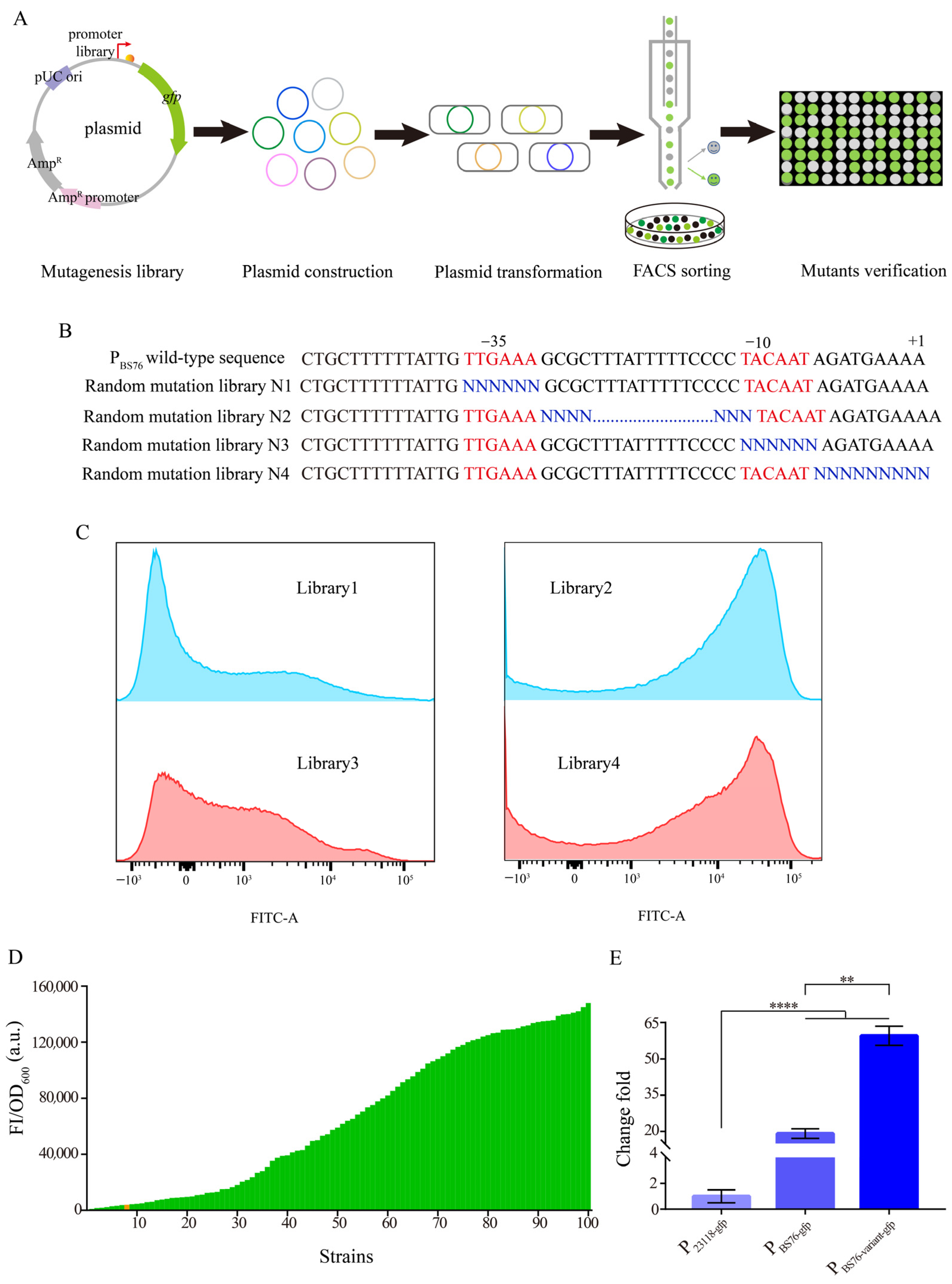
2.3. Construction of the Promoter PBS76 Library

The truncated promoter PBS76 was applied to construct four random mutation libraries. Thus, PBS76 was ligated with the reporter gene gfp and cloned into vector pUC19 to construct the basic plasmid for subsequent library construction. Four mutation libraries were constructed using the basic plasmid pUC19-PBS76-gfp as a template through PCR with primers containing degenerate bases. The PCR ligation product was then transformed into the competent cells of E. coli JM109. All the colonies on the LB plates were collected and subjected to plasmid extraction, and then the plasmid libraries were electroporated into E. coli MG1655. The strains incubated on the plates were collected and washed with PBS buffer three times, and then the OD600 was diluted to about 0.3, followed by FACS sorting. Colonies with higher fluorescence intensity than that of the control were selected and spread on the LB plates containing ampicillin (100 µg/mL). The recombinant E. coli strains containing the fluorescence plasmids grown on the LB agar plates were collected and cultured in 96-deep-well plates at 37 °C for the second-round screening with enhanced GFP fluorescence. After overnight incubation, GFP fluorescence and optical density (OD600) were measured using the microplate reader.

2.4. Construction of Recombinant Strains

The vector used for promoter screening and validation was pUC19, and the reporter gene gfp was used to characterize the promoter strength. The construction procedure of gfp expression plasmid pUC19-PBBa_23118-gfp served as an example. In brief, promoter PBBa_23118 and gene gfp were amplified by corresponding primers (Table S2) and fused by overlap extension PCR. Then, the fused fragment was inserted into the linearized vector pUC19 using ClonExpress One Step Cloning Kit (Vazyme Biotech Co., Nanjing, China), resulting in plasmid pUC19-PBBa_23118-gfp. The recombinant plasmid was then transformed into E. coli JM109 and selected on an LB agar plate containing ampicillin (100 µg/mL). Similarly, the other GFP expression vectors under the control of different promoters were constructed using this approach. As for the gene expression plasmids constructed based on expression vectors pTrc99a and pUCP18 for the overexpression of genes ilvCDE and pigFN, respectively, the genes ilvCDE and pigFN were amplified and purified, and then the purified PCR products were ligated with the linearized plasmids pTrc99a and pUCP18 through Gibson assembly (ClonExpress® II One Step Cloning Kit, Vazyme Bio Inc., Nanjing, China). The recombinant plasmids were transformed into strains E. coli W3110 and S. marcescens JNB5-1 by electroporation, and selected on LB agar plate containing ampicillin (100 µg/mL).

2.5. Fluorescence Assays

The recombinant E. coli strains that contained the fluorescence plasmids or the plasmid libraries were inoculated in LB medium and cultured at 37 °C, 200 rpm for 10 h in the plate shaker. The cultures were diluted with 0.1 M phosphate-buffered saline (PBS, pH 7.4) to guarantee an OD600 value of about 0.5 before fluorescence detection. Then, 200 µL of each culture was transferred into the 96-deep-well black plates, and the whole plate was measured using the microplate reader (Cytation 3, BioTek Instruments, Inc., Winooski, VT, USA) under an excitation wavelength of 490 nm and an emission wavelength of 530 nm at 25 °C. Fluorescence intensity was determined by dividing the fluorescence by OD600. The background fluorescence value of strains without fluorescence expression (FPbg) and the background OD600 of the medium (ODbg) were corrected, and the following Equation (1) was used to calculated the relative fluorescence intensities.
( FP OD ) = ( FP FP bg OD OD bg )

2.6. Cultivation in Shake Flasks

The engineered strains modified for L-valine production were first activated on LB agar plates, then inoculated into 30 mL seed medium in the 500 mL shake flask and cultivated for 10 h at 37 °C and 230 rpm. The seed medium contained 20 g/L glucose, 10 g/L yeast extract, 5 g/L peptone, 1.2 g/L KH2PO4, 1.2 g/L MgSO4·7H2O, 10 mg/L FeSO4·7H2O, 10 mg/L MnSO4·H2O, 1.3 mg/L VB1, and 0.3 mg/L VH. Then, 5 mL seed culture was transferred into 25 mL fermentation medium in a 500 mL shake flask and cultivated for 24 h at 37 °C and 230 rpm. The fermentation medium contained 20 g/L glucose, 2 g/L yeast extract, 4 g/L peptone, 1 g/L NaCl, 2 g/L KH2PO4, 0.7 g/L MgSO4·7H2O, 0.1 g/L FeSO4·7H2O, 0.1 g/L MnSO4·H2O, 0.8 mg/L VB1, and 0.2 mg/L VH. During the fermentation process, pH was maintained at 7.0 by adding NH4OH with phenol red as an indicator and adding 60% glucose solution when the glucose concentration was low.
For prodigiosin production, the engineered strains were grown overnight at 30 °C and 200 rpm in a rotary shaker, then transferred into the 250 mL shake flask containing 30 mL fermentation medium for 72 h cultivation at 30 °C. The fermentation medium contained 20 g/L sucrose, 15 g/L beef extract, 10 g/L CaCl2, 7.5 g/L L-proline, 0.2 g/L MgSO4·7H2O, and 6 mg/L FeSO4·7H2O.

2.7. Analytical Methods

Cell growth was measured by the UV spectrophotometer at OD600. The L-valine production was determined by HPLC using acetonitrile/water (10⁚90 v/v) as the mobile phase. The flow rate was 1 mL/min, and detection wavelength was set as 278 nm. Glucose concentration was detected by using a glucose biosensor (SBA-40C, Shandong, China). Prodigiosin was measured as previously reported with minor modification [37]. The fermentation medium was dissolved in acidic ethanol (pH 3.0) and placed for 8 h until fully dissolved. Then, the samples were centrifuged at 10,000 rpm for 10 min. Finally, the supernatant was taken for determination at the absorbance of A535. Fluorescence characterization with cytometry was evaluated with a BD FACS AriaII cell sorter (BD FACSDiscover™ S8, Becton, Dickinson and Company, Franklin Lakes, NJ, USA). The collected cells were diluted into cold PBS before detection, loaded, and run at the rate of 0.5 µL/s. A total of 10,000 samples were captured for subsequent sorting.
All the experiments were carried out in triplicate, and all the data are expressed as the mean ± standard deviation. One-way ANOVA was used for comparing statistical difference between the groups of experimental data.

3. Results

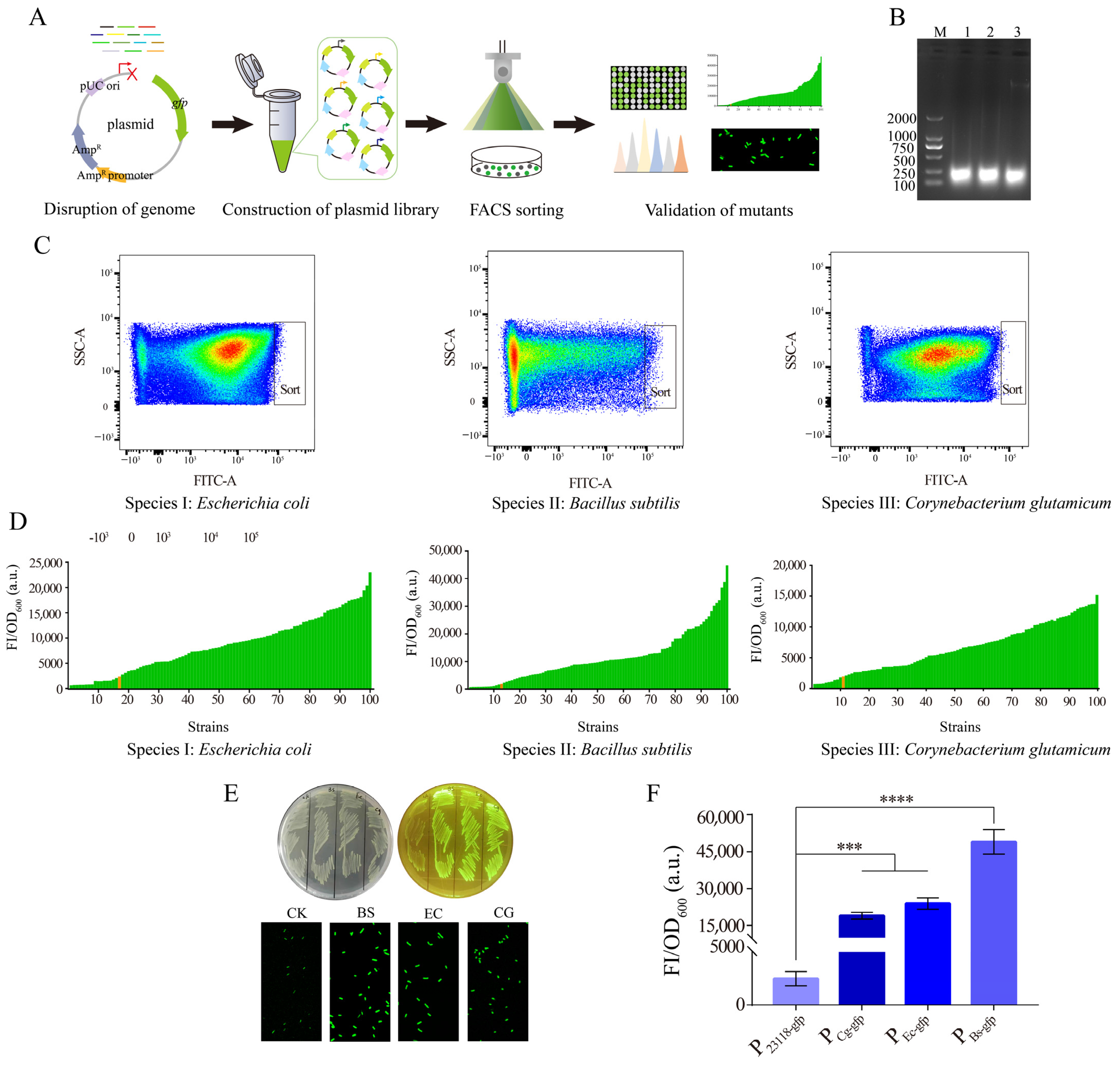
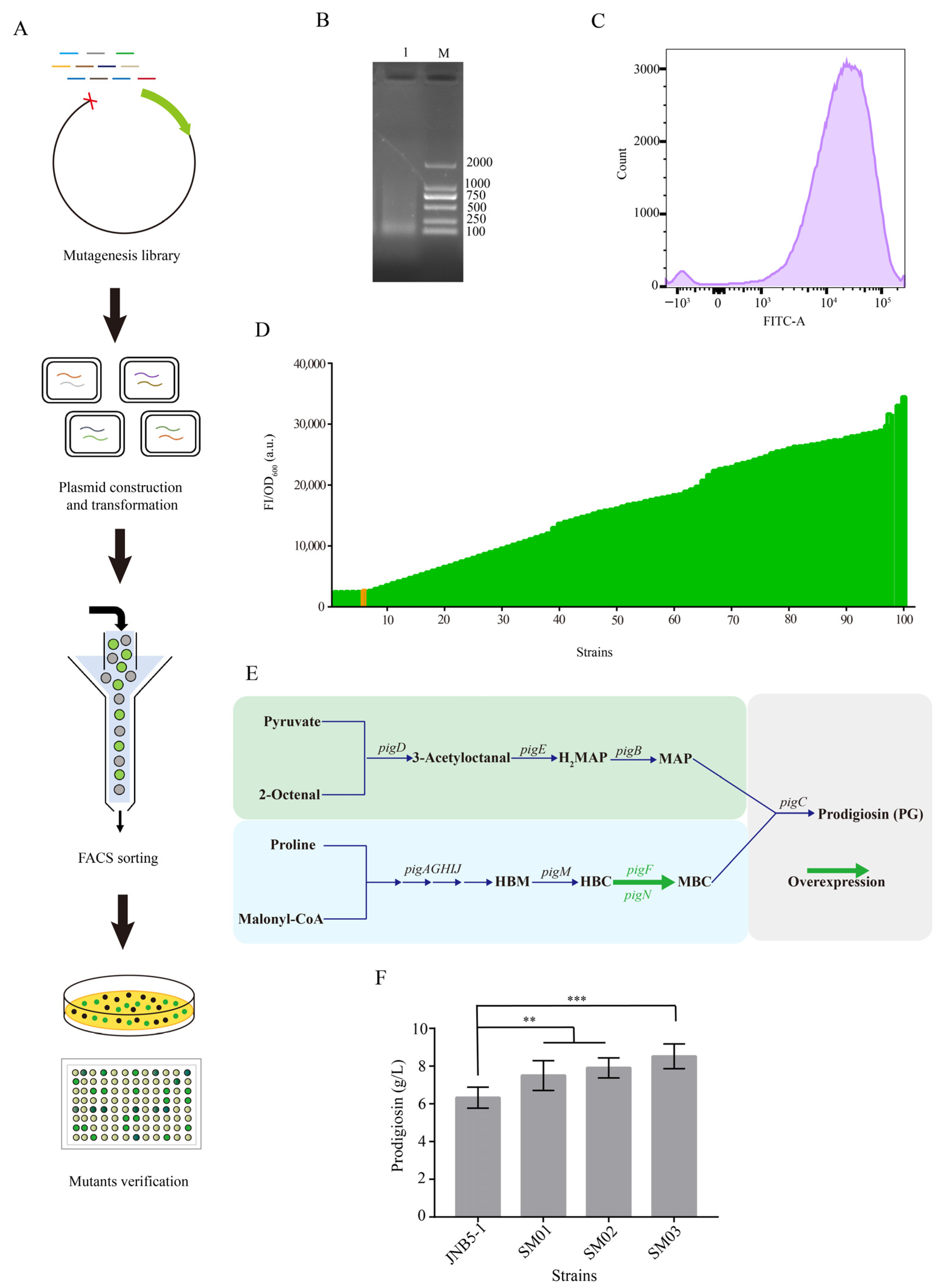
3.1. Screening of Potential Strong Constitutive Promoters Based on a Novel Approach of Random Genomic Interruption and FACS Technology

In microorganisms, protein expression level is affected by many factors, such as transcription initiation, transcription termination, translation regulation, and RNA stability, among which transcription initiation plays an essential role. Promoters, as the switch to control transcription initiation, play a key role in regulating the output level of genes. Therefore, promoters are often modified to achieve the appropriate expression levels of the target gene. Currently, screening promoters of appropriate strength by constructing various promoter libraries is the most common approach. However, the methods for isolating strong constitutive promoters in microorganisms are still limited, and are mainly based on promoter modification and omics screening. In this study, a novel approach to screen strong constitutive promoters based on random genomic disruption and FACS technology was designed (Figure 1A). Genomes of E. coli MG1655, B. subtilis 168, and C. glutamicum ATCC13032 were extracted, randomly interrupted, and inserted into the upstream of the reporter gene gfp for further construction of promoter libraries. Specifically, genomes of B. subtilis 168 and C. glutamicum ATCC13032 were first digested using the enzyme Mob I, and the reaction solution was reacted at 37 °C for 20 min. As shown in Figure 1B, the broken genomic fragments were mainly concentrated around 100–250 bp, shown in the lane 2 and 3. The genome of E. coli MG1655 was fragmented by ultrasound, and the power was set to 20%. After 30 s of fragmentation, the band presented in lane 1 was obtained. The resulting fragments from the three sources were inserted into vector pUC19, upstream of the reporter gene gfp, respectively. At this point, as the expression of fluorescent GFP was driven by the random genomic regions, three random promoter libraries were generated (Library1-EC, Library2-BS, Library3-CG). Then, all transformants grown on the LB plates were collected and subjected to FACS sorting. E. coli/pUC19-PBBa_J23118-gfp was used as the control strain to screen mutants with significantly higher fluorescence intensity. In Library1-EC, the proportion of strains with significantly higher fluorescence value accounted for 0.88% of the total library, while in Library2-BS and Library3-CG, the proportion of strains with significantly higher fluorescence value accounted for 1.52% and 0.1% of the total library, respectively (Figure 1C). The sorted strains were then coated on the LB plates and inoculated in 96-deep-well plates for rescreening. Obviously, the fluorescence values of about 90% of the mutants were higher than that of the control strain. Moreover, in each library, there was a mutant with significantly higher fluorescence intensity than that of the other mutants (Figure 1D). As shown in Figure 1E, on the LB plates, the color of the three mutants with the stronger fluorescence intensities was significantly greener than that of the control. Fluorescence microscopy also showed that the fluorescence intensity of the mutant cells was significantly higher than that of control strain. Finally, the fluorescence values of the three mutants that contained the gfp gene under the control of promoters from different sources were compared. The results showed that the fluorescence intensity of the mutant containing the gfp gene under the control of the promoter from B. subtilis was significantly higher than that of the other two mutants, and about 19.7 times higher than that of the control strain (Figure 1F). Thus, the screened random genomic region that contained sequences of the strong constitutive promoter from B. subtilis 168 was selected for further study. In conclusion, the construction of the promoter library based on random genomic interruption and FACS technology could be effectively applied in the screening of strong constitutive promoters in E. coli.

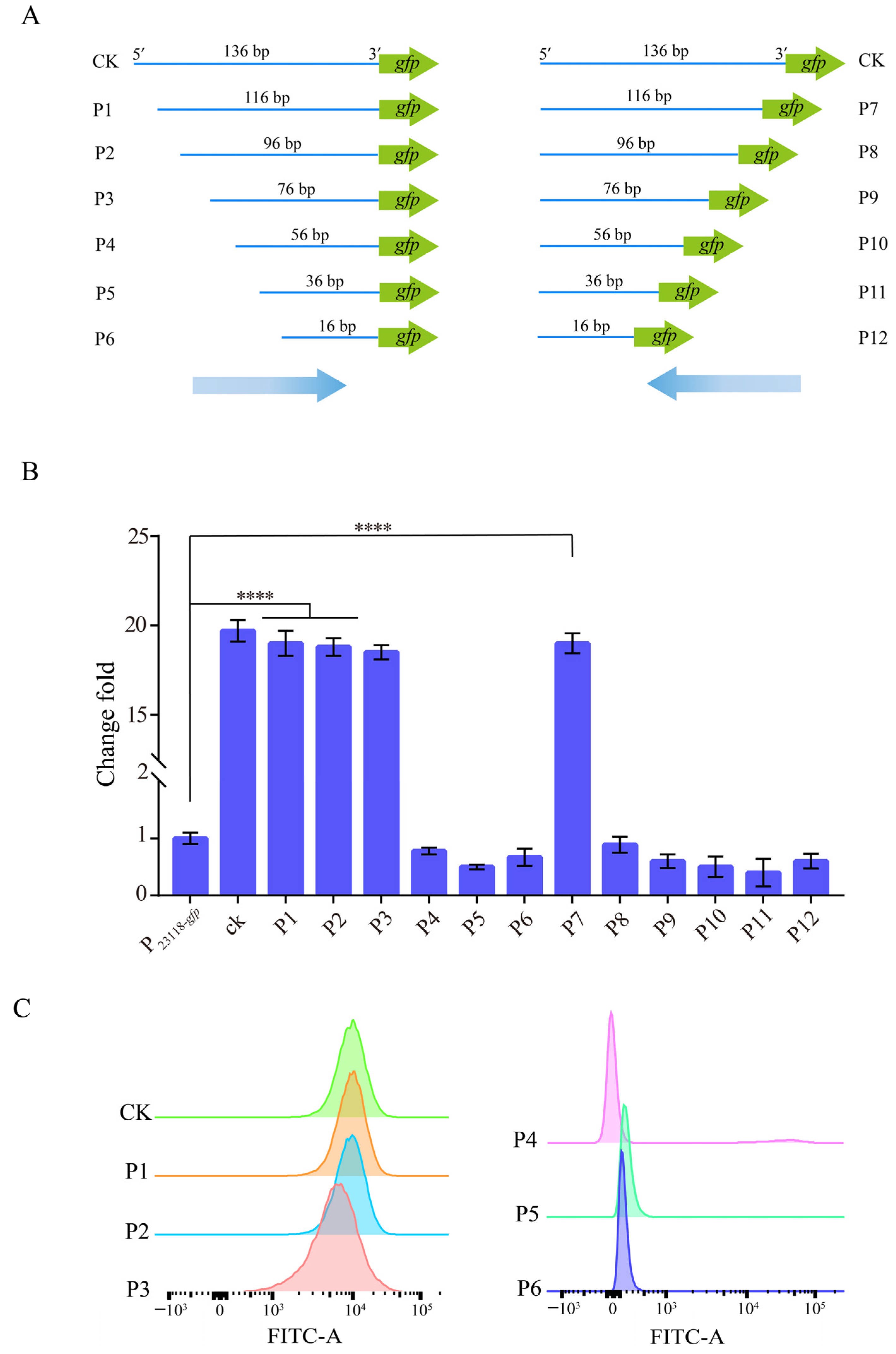
3.2. Characterization of the Identified Strong Constitutive Promoter from B. subtilis 168

In order to further analyze and identify the random sequence obtained from B. subtilis 168, the plasmid of the mutant was sequenced. The Results show that the random sequence upstream of the reporter gene gfp was the 136 bp sequence upstream of the transcription factor AcoR, which regulates acetoin synthesis in B. subtilis. Subsequently, the 136 bp sequence was shortened from both ends to identify the sequences of the promoter region of the acoR gene. As shown in Figure 2A, the sequence was truncated from the 5′ end to the 3′ end, with each truncation being 20 bp, until the sequence was truncated to 16 bp, and a total of six truncated DNA fragments (P1–P6) were obtained. Similarly, the sequence was truncated from the 3′ end to the 5′ end by 20 bp each time, and six other DNA fragments (P7–P12) were also obtained. The truncated DNA fragments were ligated before the reporter gene gfp by PCR amplification, cloned into the vector pUC19, and then transformed into E. coli JM109 for verification. Data showed that as the sequence was truncated from 5′ end to 3′ end to 116 bp, 96 bp, and 76 bp, the fluorescence intensity was basically consistent with that of the original DNA fragment. However, when the sequence truncated to 56 bp, 36 bp, and 16 bp, the expression level of GFP decreased significantly. In addition, when the sequence was truncated from 3′ end to 5′ end, only DNA fragment P7 had the same fluorescence intensity as that of the original DNA fragment. After that, the other truncated sequences showed a significantly lower fluorescence value than that of the original sequence. (Figure 2B). Subsequently, the fluorescence intensity of DNA fragments P1–P6 was further analyzed by FACS analysis to verify the above experimental results. The fluorescence intensity of P1, P2, and P3 was consistent with that of the original DNA fragment, while the fluorescence value of P4, P5, and P6 was significantly weaker (Figure 2C). These results indicated that the sequences located between positions −82 and −7 relative to the TIS of the acoR gene contained the identified strong constitutive promoter (here we named PBS76). These data are consistent with the result of N O Ali et al. [38]. The identified PBS76 was used as the subsequent research object. Above all, sequence truncation is an effective way to identify the promoter regions of the screened sequence.

3.3. Construction of a Gradient Promoter Library via Modifying Promoter PBS76

To achieve high yields of target metabolites, appropriate promoters should be screened and used for fine-tuning gene expression and reprogramming the metabolic flux of target products. Theoretically, the strength of the promoter is determined by adjacent sequences from the −35 region to the transcription start site. Hence, to better regulate the expression of key enzymes and enhance the metabolic flux of target product synthesis, the screened strong constitutive promoter PBS76 was further modified by the construction of promoter libraries. The workflow of the construction and screening of the promoter random mutagenesis library is shown in Figure 3A. As shown in Figure 3B, four promoter PBS76 mutation libraries were designed and constructed with sequences of −35 region (Library 1), spacer sequences between −35 and −10 regions (Library 2), sequences of −10 region (Library 3), and sequences between −10 region and transcription start site (Library 4) modified by the introduction of random base “N”, respectively. Firstly, random base “N” was introduced into the four regions of PBS76 promoter in plasmid pUC19-PBS76-gfp by inverse PCR, and the resulting plasmids containing mutation libraries were transferred into E. coli JM109, respectively. Then, FACS sorting technology was applied to screen the mutant strains with a higher fluorescence intensity than that of the control. The results showed that the portion with a higher fluorescence intensity in Library 1, Library 2, Library 3, and Library 4 accounted for 0.9%, 1.6%, 1.1% and 2.1% of the total library, respectively. As shown in Figure 3C, the fluorescence intensity of Library 1 and Library 3 mainly fluctuated in the range of 0–104 (a.u.), and the activity of most promoters was concentrated between 0 and 103 (a.u.). The proportion of mutant strains with a high fluorescence value in Library 1 and Library 3 was relatively small. The fluorescence range of mutants in Library 2 and Library 4 was mainly in the range of 103–105 (a.u.), which was higher than that in Library 1 and Library 3. Thus, mutants with a significantly higher fluorescence intensity than that of the control could be easier to screen in Library 2 and Library 4. The mutants with a higher fluorescence intensity sorted from Library 2 and Library 4 were coated on LB plates for overnight cultivation. The grown colonies were randomly selected and inoculated in 96-deep-well plates, and fluorescence intensity was measured after 8 h of incubation. The results indicated that a mutant (PBS76-variant) with a significantly higher fluorescence intensity than that of others was screened using promoter PBS76 as the control (Figure 3D). The intensity of PBS76-variant was 59.56 times higher than that of promoter PBBa_J23118 (Figure 3E). In total, four promoter libraries were constructed to obtain a series of promoters with a gradient of intensity, among which the strongest one was 59.56 times higher than that of promoter PBBa_J23118, and 3.03 times higher than that of promoter PBS76. In the following experiments, to further confirm the strength of the promoters identified in our study, six promoters, PBBa_J23118, PBS76, PBS76-50, PBS76-75, PBS76-85, and PBS76-100, with different strengths were selected for the regulation of expression levels of key genes and metabolic flux in L-valine synthesis.

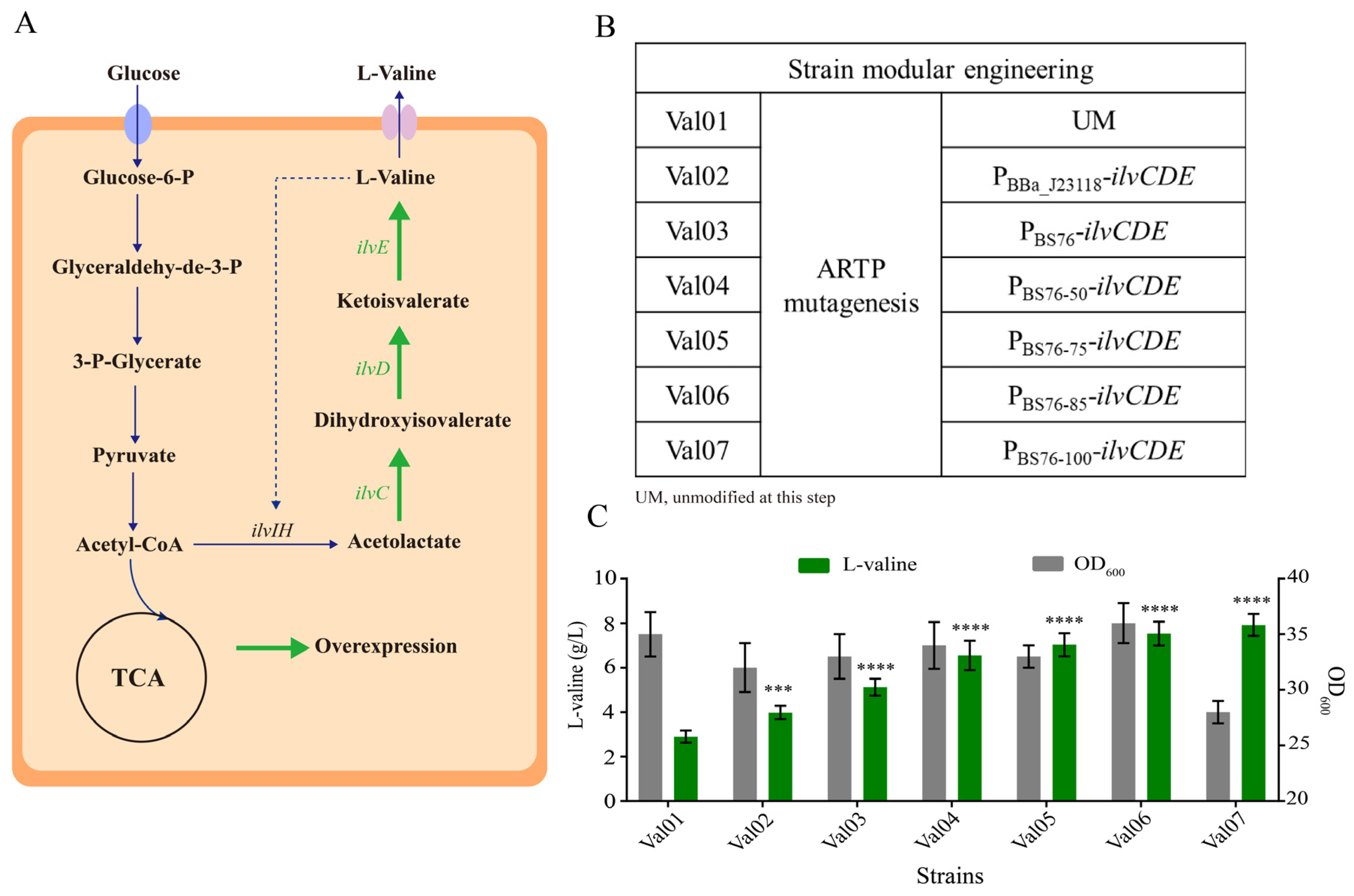
3.4. Application of the Identified Regulatory Elements for L-Valine Overproduction in E. coli W3110

To obtain the higher-level titer of target products, proper metabolic engineering should be carried out for the target metabolite. Here, to further validate the promoter activities identified in our study, L-valine, a branched-chain amino acid, which is widely used in nutrient supplements and the pharmaceutical industry, served as the target metabolite [39,40]. Based on the synthetic pathway of L-valine, three key enzymes (acetohydroxy acid isomeroreductase, encoding by ilvC; branched-chain amino acid aminotransferase, encoding by ilvE; dihydroxy acid dehydratase, encoding by ilvD) involved in L-valine biosynthesis were subjected to manipulate for better production of L-valine (Figure 4A). A L-valine-producing strain (Val01) obtained by ARTP mutagenesis of E. coli W3110 was used as the starting strain [41]. The L-valine production of Val01 could reach 2.90 g/L after 24 h fermentation in shake flask. Promoters PBBa_J23118, PBS76, PBS76-50, PBS76-75, PBS76-85, and PBS76-100 with different strength were applied to enhance the metabolic flux of L-valine synthesis. Specifically, the key ilvCDE genes were cloned into the vector pTrc99a under the control of promoters PBBa_J23118, PBS76, PBS76-50, PBS76-75, PBS76-85, and PBS76-100, respectively, and then transformed into E. coli JM109. The transformants were selected and verified by colony PCR and sequencing. The successful transformants were transformed into strain Val01, resulting in recombinant strains Val02, Val03, Val04, Val05, Val06, and Val07 (Figure 4B). Subsequently, the recombinant strains were subjected to shake flask fermentation for 24 h to compare their ability to produce L-valine. Based on our results, the cell biomass of strains Val02, Val03, Val04, Val05, and Val06 was not different from that of strain Val01 after 24 h shake-flask fermentation, while the OD600 of strain Val07 only reached 28. Obviously, the intensity of promoters was positively correlated with the L-valine yield produced by these seven strains. Among them, the L-valine yield produced by Val07 reached 7.92 g/L, while amounts of 3.98, 5.12, 6.55, 7.03, and 7.53 g/L L-valine were attained by strains Val02, Val03, Val04, Val05, and Val06, respectively (Figure 4C). Strain Val07 owned the highest L-valine yield, 173.1% higher than that of strain Val01. The strength of promoter PBS76-100 was too strong, which caused the disorder of intracellular metabolism and the burden on cell growth, so the cell growth was severely inhibited. However, due to the enhanced metabolic flux of L-valine synthesis driven by promoter PBS76-100, there was an improvement in L-valine yield. Taken together, these results further confirmed that promoters PBS76, PBS76-50, PBS76-75, PBS76-85, and PBS76-100 are strong promoters compared to promoter PBBa_J23118 and could be used to improve the production of target products.

3.5. Screening of Strong Constitutive Promoter Applicable for S. marcescens by Random Genomic Disruption and FACS Technology

Prodigiosin is a red pigment with great economic value and of widely promising application due to its immunosuppressive and anticancer activities [37]. S. marcescens is the main producing strain for prodigiosin synthesis. Research of the model organism for prodigiosin synthesis has mainly focused on transcriptional regulation, the function of a two-component system and a quorum sensing system [42,43,44]. However, due to the unclear genetic background and the lack of gene-editing approaches of S. marcescens, the improvement in prodigiosin production has been severely limited. Moreover, the shortage of an effective synthetic biology toolbox to precisely regulate gene expression has impeded its development seriously. Here, to further confirm the general applicability of the method conducted in our study to screen strong constitutive promoters in different bacteria, a gradient strength promoter library was constructed via random genomic disruption for screening strong constitutive promoters in S. marcescens as mentioned above (Figure 5A). First, the genome of S. marcescens JNB5-1 was extracted and interrupted by ultrasonic. The output power of the ultrasonic crusher was set at 20%, the ultrasonic time was 1 s, and the ultrasonic interval set at 3 s. A total of 30 repeats of ultrasound were achieved to break the genome into random fragments ranging from 100 to 300 bp (Figure 5B). The 5′ end of the fragments was repaired and smoothed by the Klenow Fragment. Then, the random fragments were inserted into the upstream of the reporter gene gfp and cloned into the vector pUCP18, resulting in the plasmid library pUCP18-PSM-gfp. The constructed plasmid was then transformed into S. marcescens JNB5-1, and the transformants grown on the LB plates were collected for further FACS sorting. S. marcescens JNB5-1 containing plasmid pUCP18-Ppig-gfp was used as the control, which was used to express the gene gfp under the control of the native promoter Ppig of prodigiosin synthesis gene cluster. Mutants with a higher GFP fluorescence than that of the control were collected for further analysis. The results showed that the fluorescence intensity varied over a 1000-fold range from 102 to 105 (a.u.), and the activities of most promoters were mainly concentrated between 103 and 104 (a.u.) (Figure 5C). Moreover, the sorted strains were then coated on LB plates, and the colonies were inoculated into 96-deep-well plates for the second round of screening. Among them, the GFP expression intensity of 95% mutants was higher than that of the control (Figure 5D). The fluorescence intensity of one of the mutants containing the gfp gene under the control of the promoter PSM (the promoter of the operon rpsQ-rpsJ) was significantly higher than that of the others. Therefore, the random sequence of this mutant was selected for further research.
Nowadays, prodigiosin attracts much attention due to its multiple biological activities. In S. marcescens, the synthesis of prodigiosin is achieved by the pig gene cluster. 2-methyl-3-n-amyl-pyrrole (MAP) and 4-methoxy-2,2′-bipyrrole-5-carbaldehyde (MBC) are two major precursors and are condensed into prodigiosin via the PEP-utilizing enzyme. Importantly, O-methyl transferase (encoded by pigF) and oxidoreductase (encoded by pigN) involved in catalyzing 4-hydroxy-2,2′-bipyrrole-5-carbaldehyde (HBC) to the formation of 4-methoxy-2,2′-bipyrrole-5-carbaldehyde (MBC) have been confirmed to play the most significant role in prodigiosin synthesis among the pig gene cluster in our previous study (Figure 5E). In addition, substituting the promoter of a gene cluster with strong constitutive promoters has been proven as a feasible strategy for metabolite production. Here, to study the application of the strong constitutive promoter PSM screened from S. marcescens in the optimization of the prodigiosin metabolic pathway, the gene expression of pigFN was regulated by replacing promoters with those of different strengths. The native promoter Ppig of the pig gene cluster, an endogenous promoter PRplJ from S. marcescens found using RNA-Seq [37], and the novel constitutive promoter PSM that was screened based on random genomic interruption were selected to mediate the expression of pigFN. Thus, the expression vectors of pigFN genes mediated by promoters Ppig, PRplJ, and PSM were then transferred into S. marcescens JNB5-1, resulting in strains SM01, SM02, and SM03. Then, these strains were cultivated in fermentation medium for 72 h. As shown in Figure 5F, after 72 h of fermentation, the prodigiosin production of the wild-type JNB5-1 was 6.32 g/L. The yield of prodigiosin in strains SM01 and SM02 reached 7.50 g/L and 7.91 g/L, respectively. The maximum prodigiosin titer of strain SM03 reached 8.52 g/L, which was 34.81% higher than that produced by the wild-type strain. Meanwhile, the activity of promoter PSM was stronger than that of promoter PRplJ in the S. marcescens expression system. Altogether, these results suggested that the method based on random genomic disruption and FACS technology could also be a valuable approach to screen strong constitutive promoters in S. marcescens.

4. Discussion

The transcription of DNA to RNA is the first step in protein expression, and promoter, RNA polymerase, and sigma factor are typically involved in this process. Among them, promoters directly affect the transcription rate of protein, and hence they are common targets used for engineering the biosynthesis of natural and non-natural products in model or non-model strains [45]. To improve L-proline production in C. glutamicum, tailored promoter libraries were used to fine-tuned the expression levels of the target gdh, pyc, and proB genes, and finally, L-proline production was significantly increased [46]. To balance the metabolic flux of the naringenin biosynthesis pathway in E. coli, constitutive promoters with a gradient of intensity were randomly selected to control the expression levels of the pathway genes of naringenin. After screening more than 1200 candidates via the ultraviolet spectrophotometry–fluorescence spectrophotometry high-throughput method, the metabolic flux of the naringenin synthetic pathway was appropriately balanced, and finally, the naringenin was significantly improved [47]. Song et al. screened promoters from B. subtilis in various conditions, and a strong promoter, PtrnQ, in comparison to P43, was isolated and used to elevate the final production of both cytoplasmic BgaB and secreted protein α-amylase [36]. Promoter engineering has been also used to improve the production of 3-aminopropionic acid [48] and myo-inositol in E. coli [49], pullulanase [31], methyl parathion hydrolase, and chlorothalonil hydrolytic dehalogenase in B. subtilis [50], L-lysine in C. glutamicum [51], medium-chain-length polyhydroxyalkanoates in Pseudomonas mendocina [32], co-enzyme Q10 in Rhodobacter sphaeroides [33], polycyclic tetramate macrolactam in Streptomyces albus [34], co-enzyme A in Corynebacterium ammoniagenes [35], H2 in Thermococcus onnurineus [52], and so on. However, although promoters suitable for target gene expression and products production could be successfully screened from random mutation on the reported promoters or identified from the omics data, the current methods to screen strong constitutive promoters in different microorganisms are limited. In this study, a novel approach based on random genomic interruption was designed for the construction of promoter libraries, and strong constitutive promoters suitable for E. coli and S. marcescens were obtained by using FACS technology to sort high-intensity promoter sequences (Figure 1 and Figure 5). Further, to confirm the strength of the identified promoters, the promoters obtained in our study were applied to enhance the expression of ilvCDE genes in E. coli W3110 to enhance L-valine production, and to increase expression of the pigFN genes in S. marcescens JNB5-1 to improve prodigiosin synthesis. As shown in Figure 4 and Figure 5, the L-valine production in strain Val07 and prodigiosin synthesis in strain SM03 were significantly increased compared to E. coli Val01 and S. marcescens JNB5-1 (Figure 4 and Figure 5). As far as we know, our study is the first to identify strong constitutive promoters through the method of random genomic interruption and FACS technology.
L-valine, an essential amino acid, is one of the three branched-chain amino acids (BCAAs), and studies on L-valine have shown that it is widely used in many industrial fields, such as pharmaceuticals, cosmetics, food, and feed [53]. Moreover, with the growing world market for L-valine, the microbial cell factory for the efficient synthesis of L-valine has become of increasing interest. C. glutamicum is the most commonly used industrial microorganism for producing L-valine. Due to the advantages of clear genetic background and easy genetic manipulation, E. coli has become another important host for the synthesis of L-valine in recent years [54,55]. However, possibly due to the more complex regulatory mechanism for L-valine biosynthesis in E. coli, there are fewer reports of L-valine-producing E. coli strains than there are of L-valine-producing C. glutamicum strains. Park et al. developed an engineered L-valine production strain through systematic metabolic engineering of E. coli W, and finally, a high-level production of L-valine (60.7 g/L) with a yield of 0.22 g/g glucose was obtained [54]. Considering that cofactor balance is another important factor required for L-valine biosynthesis, Savrasova et al. constructed an E. coli MG1655-based L-valine-producing strain by replacing the native NADPH-dependent aminotransferase with a heterologous NADH-dependent leucine dehydrogenase [56]. By systematic metabolic engineering, Hao et al. constructed a chromosomally engineered L-valine-producing strain, and the final strain could produce 84 g/L L-valine with a yield and productivity of 0.41 g/g glucose and 2.33 g/L/h, respectively, in a 5 L bioreactor through a two-stage fed-batch fermentation [57]. In our previous study, we constructed a high-level L-valine production E. coli strain (92 g/L) using multimodular engineering [41]. In this study, to further confirm the strength of the promoters identified in our study, promoters PBBa_J23118, PBS76 (identified by the method based on random genomic interruption and FACS technology), and four promoters, PBS76-50 (weakest), PBS76-75 (middle), PBS76-85 (strong), and PBS76-100 (strongest), from our isolated PBS76 promoter library were applied to increase the expression levels of ilvCDE genes, and strains Val02, Val03, Val04, Val05, Val06, and Val07 were obtained, respectively. The results of shake flask fermentation showed that the L-valine production in strains Val02, Val03, Val04, Val05, Val06, and Val07 was significantly enhanced compared to the original strain Val01 (Figure 4). These results further support that the method based on random genomic interruption and FACS technology is a very effective method to screen strong constitutive promoters in different bacteria.
Prodigiosin (PG), a red linear tripyrrole pigment, is the most prominent member of the prodiginine family and is produced by S. marcescens, Serratia rubidaea, Streptomyces coelicolor, Streptomyces griseoviridis, Serratia nematodiphila, and so on [58]. Among them, S. marcescens is the most widely studied prodigiosin-producing strain. Studies on prodigiosin have shown that it has important antimicrobial, anticancer, and immunosuppressive properties, and hence prodigiosin has received widespread attention in the last few decades [59]. Due to being economical and more environmentally friendly, the biotechnological production of prodigiosin by S. marcescens has recently attracted a great deal of interest. However, the high-efficiency production of prodigiosin by S. marcescens for commercial purposes is still a challenge. The prodigiosin biosynthesis pathway in S. marcescens is encoded by the pigABCDEFGHIJKLMN genes, a total of 14 genes, which are transcribed as a polycistronic mRNA from a promoter upstream of the pigA gene. Among them, the genes pigB, pigD, and pigE are involved in the biosynthesis of 2-methyl-3-n-amyl-pyrrole (MAP), while the genes pigA, pigF, pigG, pigH, pigI, pigJ, pigK, pigL, pigM, and pigN use L-proline as the substrate to synthesize 4-methoxy-2,2′-bipyrrole-5-carbaldehyde (MBC). Finally, the terminal condensing enzyme PigC encoding by the pigC gene condenses MAP and MBC to prodigiosin [58]. In our previous study, via transcriptomics and proteomics, we identified that the genes pigN and pigF probably play the most important role in prodigiosin biosynthesis in the pig gene cluster [60]. In addition, through the introduction of a polynucleotide fragment into the pigN 3′ untranslated region and disulfide bonds into the O-methyl transferase (PigF), the prodigiosin production in strain S. marcescens JNB5-1 was significantly enhanced [61]. In this study, to verify the universality of the method of screening strong constitutive promoters based on the random genomic interruption and FACS technology, a strong constitutive promoter PSM was firstly isolated based on the approach shown in our study. Then, to improve the production of prodigiosin in strain JNB5-1, the genes pigN and pigF were overexpressed under the control of the promoters Ppig, PRplJ, and PSM, and strains SM01, SM02, and SM03 were obtained, respectively. The results showed that the prodigiosin titer of these strain was positively correlated with the strength of the promoters. Among them, the prodigiosin titer of strain SM03 increased to 8.52 g/L, which was 1.35-times greater than that of the original strain JNB5-1 (6.32 g/L; Figure 5). This result further suggests that random genomic interruption and FACS technology is an effective method to identify strong constitutive promoter in the host that we are interested in.

5. Conclusions

This work describes a novel method to identify strong constitutive promoters in E. coli and S. marcescens based on random genomic interruption and FACS technology, and the identified promoters were further used in fine-tuning gene expression and reprogramming metabolic flux for higher-level production of L-valine and prodigiosin in E. coli and S. marcescens, respectively. The method shown in our study can also be a useful strategy for isolating other effective genetic regulatory elements, such as ribosome binding sites, terminators, and N-terminal coding sequences (NCS), in different microorganisms.

Supplementary Materials

The following supporting information can be downloaded at: https://www.mdpi.com/article/10.3390/biology12010071/s1, Table S1: Strains and plasmids used in this study; Table S2: Primers used in this study.

Author Contributions

Conceptualization, X.P. and Z.R.; methodology, X.P., M.T., J.Y. and Y.H.; software, X.Z. and T.Y.; validation, X.P. and Z.R.; formal analysis, X.P. and M.T.; investigation, X.P., M.T. and J.Y.; resources, X.P.; data curation, Z.R.; writing—original draft preparation, X.P. and M.T.; writing—review and editing, X.P. and Z.R.; visualization, Z.R.; supervision, Z.R.; project administration, X.P., T.Y. and Z.R.; funding acquisition, X.P., T.Y. and Z.R. All authors have read and agreed to the published version of the manuscript.

Funding

This work was supported by the National Key Research and Development Program of China (2021YFC2100900), the National Natural Science Foundation of China (32100055 and 31870066), the Natural Science Foundation of Jiangsu Province (BK20210464, BK20221537 and BK20221080), the project funded by China Postdoctoral Science Foundation (2021M691280), the Jiangsu Planned Projects for Postdoctoral Research Funds (2021K296B), and the Fundamental Research Funds for the Central Universities (JUSRP12119).

Institutional Review Board Statement

Not applicable.

Informed Consent Statement

Not applicable.

Data Availability Statement

The data that support the findings of this study are available from the corresponding author upon reasonable request.

Conflicts of Interest

The authors declare no conflict of interest.

References

  1. Ko, Y.S.; Kim, J.W.; Lee, J.A.; Han, T.; Kim, G.B.; Park, J.E.; Lee, S.Y. Tools and strategies of systems metabolic engineering for the development of microbial cell factories for chemical production. Chem. Soc. Rev. 2020, 49, 4615–4636. [Google Scholar] [CrossRef]
  2. Jang, Y.-S.; Park, J.M.; Choi, S.; Choi, Y.J.; Seung, D.Y.; Cho, J.H.; Lee, S.Y. Engineering of microorganisms for the production of biofuels and perspectives based on systems metabolic engineering approaches. Biotechnol. Adv. 2012, 30, 989–1000. [Google Scholar] [CrossRef]
  3. Yang, D.; Prabowo, C.P.S.; Eun, H.; Park, S.Y.; Cho, I.J.; Jiao, S.; Lee, S.Y. Escherichia coli as a platform microbial host for systems metabolic engineering. Essays Biochem. 2021, 65, 225–246. [Google Scholar] [CrossRef]
  4. Thuan, N.H.; Tatipamula, V.B.; Canh, N.X.; Giang, N.V. Recent advances in microbial co-culture for production of value-added compounds. 3 Biotech 2022, 12, 115. [Google Scholar] [CrossRef]
  5. Palazzotto, E.; Tong, Y.J.; Lee, S.Y.; Weber, T. Synthetic biology and metabolic engineering of actinomycetes for natural product discovery. Biotechnol. Adv. 2019, 37, 107366. [Google Scholar] [CrossRef]
  6. Choi, K.R.; Shin, J.H.; Cho, J.S.; Yang, D.; Lee, S.Y. Systems Metabolic Engineering of Escherichia coli. EcoSal Plus 2017, 7, 1–56. [Google Scholar] [CrossRef]
  7. Helmy, M.; Smith, D.; Selvarajoo, K. Systems biology approaches integrated with artificial intelligence for optimized metabolic engineering. Metab. Eng. Commun. 2020, 11, e00149. [Google Scholar] [CrossRef]
  8. McCarty, N.S.; Ledesma-Amaro, R. Synthetic Biology Tools to Engineer Microbial Communities for Biotechnology. Trends Biotechnol. 2019, 37, 181–197. [Google Scholar] [CrossRef] [Green Version]
  9. Zhang, X.; Lin, Y.; Wu, Q.; Wang, Y.; Chen, G.Q. Synthetic Biology and Genome-Editing Tools for Improving PHA Metabolic Engineering. Trends Biotechnol. 2020, 38, 689–700. [Google Scholar] [CrossRef]
  10. Choi, K.R.; Jang, W.D.; Yang, D.; Cho, J.S.; Park, D.; Lee, S.Y. Systems Metabolic Engineering Strategies: Integrating Systems and Synthetic Biology with Metabolic Engineering. Trends Biotechnol. 2019, 37, 817–837. [Google Scholar] [CrossRef]
  11. Guillier, M.; Repoila, F. RNA and gene control in bacteria. Biochim. Biophys. Acta Gene Regul. Mech. 2020, 1863, 194602. [Google Scholar] [CrossRef]
  12. Tian, R.; Liu, Y.; Chen, J.; Li, J.; Liu, L.; Du, G.; Chen, J. Synthetic N-terminal coding sequences for fine-tuning gene expression and metabolic engineering in Bacillus subtilis. Metab. Eng. 2019, 55, 131–141. [Google Scholar] [CrossRef]
  13. Brophy, J.A.; Voigt, C.A. Principles of genetic circuit design. Nat. Methods 2014, 11, 508–520. [Google Scholar] [CrossRef] [Green Version]
  14. Redden, H.; Alper, H.S. The development and characterization of synthetic minimal yeast promoters. Nat. Commun. 2015, 6, 7810. [Google Scholar] [CrossRef] [Green Version]
  15. Guiziou, S.; Sauveplane, V.; Chang, H.J.; Clerte, C.; Declerck, N.; Jules, M.; Bonnet, J. A part toolbox to tune genetic expression in Bacillus subtilis. Nucleic Acids Res. 2016, 44, 7495–7508. [Google Scholar] [CrossRef] [Green Version]
  16. Segall-Shapiro, T.H.; Sontag, E.D.; Voigt, C.A. Engineered promoters enable constant gene expression at any copy number in bacteria. Nat. Biotechnol. 2018, 36, 352–358. [Google Scholar] [CrossRef]
  17. Bai, C.X.; Zhang, Y.; Zhao, X.J.; Hu, Y.L.; Xiang, S.H.; Miao, J.; Lou, C.B.; Zhang, L.X. Exploiting a precise design of universal synthetic modular regulatory elements to unlock the microbial natural products in Streptomyces. Proc. Natl. Acad. Sci. USA 2015, 112, 12181–12186. [Google Scholar] [CrossRef] [Green Version]
  18. Yi, J.S.; Kim, M.W.; Kim, M.; Jeong, Y.; Kim, E.J.; Cho, B.K.; Kim, B.G. A Novel Approach for Gene Expression Optimization through Native Promoter and 5’ UTR Combinations Based on RNA-seq, Ribo-seq, and TSS-seq of Streptomyces coelicolor. ACS Synth. Biol. 2017, 6, 555–565. [Google Scholar] [CrossRef]
  19. Turkanoglu Ozcelik, A.; Yilmaz, S.; Inan, M. Pichia pastoris Promoters. Methods Mol. Biol. 2019, 1923, 97–112. [Google Scholar] [CrossRef]
  20. Andersson, R.; Sandelin, A. Determinants of enhancer and promoter activities of regulatory elements. Nat. Rev. Genet. 2020, 21, 71–87. [Google Scholar] [CrossRef]
  21. Kong, L.-H.; Xiong, Z.-Q.; Song, X.; Xia, Y.-J.; Zhang, N.; Ai, L.-Z. Characterization of a Panel of Strong Constitutive Promoters from Streptococcus thermophilus for Fine-Tuning Gene Expression. ACS Synth. Biol. 2019, 8, 1469–1472. [Google Scholar] [CrossRef]
  22. Johnson, A.O.; Gonzalez-Villanueva, M.; Tee, K.L.; Wong, T.S. An Engineered Constitutive Promoter Set with Broad Activity Range for Cupriavidus necator H16. ACS Synth. Biol. 2018, 7, 1918–1928. [Google Scholar] [CrossRef]
  23. Hartner, F.S.; Ruth, C.; Langenegger, D.; Johnson, S.N.; Hyka, P.; Lin-Cereghino, G.P.; Lin-Cereghino, J.; Kovar, K.; Cregg, J.M.; Glieder, A. Promoter library designed for fine-tuned gene expression in Pichia pastoris. Nucleic Acids Res. 2008, 36, e76. [Google Scholar] [CrossRef] [Green Version]
  24. Yim, S.S.; An, S.J.; Kang, M.; Lee, J.; Jeong, K.J. Isolation of fully synthetic promoters for high-level gene expression in Corynebacterium glutamicum. Biotechnol. Bioeng. 2013, 110, 2959–2969. [Google Scholar] [CrossRef]
  25. Siegl, T.; Tokovenko, B.; Myronovskyi, M.; Luzhetskyy, A. Design, construction and characterisation of a synthetic promoter library for fine-tuned gene expression in actinomycetes. Metab. Eng. 2013, 19, 98–106. [Google Scholar] [CrossRef]
  26. Shen, R.; Yin, J.; Ye, J.-W.; Xiang, R.-J.; Ning, Z.-Y.; Huang, W.-Z.; Chen, G.-Q. Promoter Engineering for Enhanced P(3HB- co-4HB) Production by Halomonas bluephagenesis. ACS Synth. Biol. 2018, 7, 1897–1906. [Google Scholar] [CrossRef]
  27. Ouyang, Q.; Wang, X.; Zhang, N.; Zhong, L.; Liu, J.; Ding, X.; Zhang, Y.; Bian, X. Promoter Screening Facilitates Heterologous Production of Complex Secondary Metabolites in Burkholderiales Strains. ACS Synth. Biol. 2020, 9, 457–460. [Google Scholar] [CrossRef]
  28. Rao, Y.; Li, P.; Xie, X.; Li, J.; Liao, Y.; Ma, X.; Cai, D.; Chen, S. Construction and Characterization of a Gradient Strength Promoter Library for Fine-Tuned Gene Expression in Bacillus licheniformis. ACS Synth. Biol. 2021, 10, 2331–2339. [Google Scholar] [CrossRef]
  29. Sun, W.; Jiang, B.; Zhao, D.; Pu, Z.; Bao, Y. Integration of metabolic pathway manipulation and promoter engineering for the fine-tuned biosynthesis of malic acid in Bacillus coagulans. Biotechnol. Bioeng. 2021, 118, 2597–2608. [Google Scholar] [CrossRef]
  30. Zhang, Y.; Wei, M.; Zhao, G.; Zhang, W.; Li, Y.; Lin, B.; Li, Y.; Xu, Q.; Chen, N.; Zhang, C. High-level production of L-homoserine using a non-induced, non-auxotrophic Escherichia coli chassis through metabolic engineering. Bioresour. Technol. 2021, 327, 124814. [Google Scholar] [CrossRef]
  31. Meng, F.; Zhu, X.; Nie, T.; Lu, F.; Bie, X.; Lu, Y.; Trouth, F.; Lu, Z. Enhanced Expression of Pullulanase in Bacillus subtilis by New Strong Promoters Mined From Transcriptome Data, Both Alone and in Combination. Front. Microbiol. 2018, 9, 2635. [Google Scholar] [CrossRef] [PubMed] [Green Version]
  32. Zhao, F.; Liu, X.; Kong, A.; Zhao, Y.; Fan, X.; Ma, T.; Gao, W.; Wang, S.; Yang, C. Screening of endogenous strong promoters for enhanced production of medium-chain-length polyhydroxyalkanoates in Pseudomonas mendocina NK-01. Sci. Rep. 2019, 9, 1798. [Google Scholar] [CrossRef] [PubMed] [Green Version]
  33. Shi, T.; Zhang, L.; Liang, M.; Wang, W.; Wang, K.; Jiang, Y.; Liu, J.; He, X.; Yang, Z.; Chen, H.; et al. Screening and engineering of high-activity promoter elements through transcriptomics and red fluorescent protein visualization in Rhodobacter sphaeroides. Syn. Syst. Biotechnol. 2021, 6, 335–342. [Google Scholar] [CrossRef] [PubMed]
  34. Luo, Y.; Zhang, L.; Barton, K.W.; Zhao, H. Systematic Identification of a Panel of Strong Constitutive Promoters from Streptomyces albus. ACS Synth. Biol. 2015, 4, 1001–1010. [Google Scholar] [CrossRef] [PubMed]
  35. Hou, Y.; Chen, S.; Wang, J.; Liu, G.; Wu, S.; Tao, Y. Isolating promoters from Corynebacterium ammoniagenes ATCC 6871 and application in CoA synthesis. BMC Biotechnol. 2019, 19, 76. [Google Scholar] [CrossRef] [Green Version]
  36. Song, Y.; Nikoloff, J.M.; Fu, G.; Chen, J.; Li, Q.; Xie, N.; Zheng, P.; Sun, J.; Zhang, D. Promoter Screening from Bacillus subtilis in Various Conditions Hunting for Synthetic Biology and Industrial Applications. PLoS ONE 2016, 11, e0158447. [Google Scholar] [CrossRef] [Green Version]
  37. Pan, X.; You, J.; Tang, M.; Zhang, X.; Xu, M.; Yang, T.; Rao, Z. Improving prodigiosin production by transcription factor engineering and promoter engineering in Serratia marcescens. Front. Microbiol. 2022, 13, 977337. [Google Scholar] [CrossRef]
  38. Ali, N.O.; Bignon, J.; Rapoport, G.; Debarbouille, M. Regulation of the acetoin catabolic pathway is controlled by sigma L in Bacillus subtilis. J. Bacteriol. 2001, 183, 2497–2504. [Google Scholar] [CrossRef] [Green Version]
  39. Bishop, C.A.; Schulze, M.B.; Klaus, S.; Weitkunat, K. The branched-chain amino acids valine and leucine have differential effects on hepatic lipid metabolism. FASEB J. 2020, 34, 9727–9739. [Google Scholar] [CrossRef]
  40. Gao, H.; Tuyishime, P.; Zhang, X.; Yang, T.; Xu, M.; Rao, Z. Engineering of microbial cells for L-valine production: Challenges and opportunities. Microb. Cell Factories 2021, 20, 172. [Google Scholar] [CrossRef]
  41. Hao, Y.; Pan, X.; Xing, R.; You, J.; Hu, M.; Liu, Z.; Li, X.; Xu, M.; Rao, Z. High-level production of L-valine in Escherichia coli using multi-modular engineering. Bioresour. Technol. 2022, 359, 127461. [Google Scholar] [CrossRef]
  42. Slater, H.; Crow, M.; Everson, L.; Salmond, G.P. Phosphate availability regulates biosynthesis of two antibiotics, prodigiosin and carbapenem, in Serratia via both quorum-sensing-dependent and -independent pathways. Mol. Microbiol. 2003, 47, 303–320. [Google Scholar] [CrossRef] [PubMed]
  43. Shanks, R.M.Q.; Stella, N.A.; Lahr, R.M.; Aston, M.A.; Brothers, K.M.; Callaghan, J.D.; Sigindere, C.; Liu, X. Suppressor analysis of eepR mutant defects reveals coordinate regulation of secondary metabolites and serralysin biosynthesis by EepR and HexS. Microbiology 2017, 163, 280–288. [Google Scholar] [CrossRef] [PubMed]
  44. Pan, X.; Tang, M.; You, J.; Osire, T.; Sun, C.; Fu, W.; Yi, G.; Yang, T.; Yang, S.T.; Rao, Z. PsrA is a novel regulator contributes to antibiotic synthesis, bacterial virulence, cell motility and extracellular polysaccharides production in Serratia marcescens. Nucleic Acids Res. 2022, 50, 127–148. [Google Scholar] [CrossRef] [PubMed]
  45. Li, C.; Jiang, T.; Li, M.; Zou, Y.; Yan, Y. Fine-tuning gene expression for improved biosynthesis of natural products: From transcriptional to post-translational regulation. Biotechnol. Adv. 2022, 54, 107853. [Google Scholar] [CrossRef]
  46. Liu, J.; Liu, M.; Shi, T.; Sun, G.; Gao, N.; Zhao, X.; Guo, X.; Ni, X.; Yuan, Q.; Feng, J.; et al. CRISPR-assisted rational flux-tuning and arrayed CRISPRi screening of an L-proline exporter for L-proline hyperproduction. Nat. Commun. 2022, 13, 891. [Google Scholar] [CrossRef]
  47. Zhou, S.; Lyu, Y.; Li, H.; Koffas, M.A.; Zhou, J. Fine-tuning the (2S)-naringenin synthetic pathway using an iterative high-throughput balancing strategy. Biotechnol. Bioeng. 2019, 116, 1392–1404. [Google Scholar] [CrossRef]
  48. Song, C.W.; Lee, J.; Ko, Y.S.; Lee, S.Y. Metabolic engineering of Escherichia coli for the production of 3-aminopropionic acid. Metab. Eng. 2015, 30, 121–129. [Google Scholar] [CrossRef]
  49. Tang, E.; Shen, X.; Wang, J.; Sun, X.; Yuan, Q. Synergetic utilization of glucose and glycerol for efficient myo-inositol biosynthesis. Biotechnol. Bioeng. 2020, 117, 1247–1252. [Google Scholar] [CrossRef]
  50. Zhou, C.; Ye, B.; Cheng, S.; Zhao, L.; Liu, Y.; Jiang, J.; Yan, X. Promoter engineering enables overproduction of foreign proteins from a single copy expression cassette in Bacillus subtilis. Microb. Cell Factories 2019, 18, 111. [Google Scholar] [CrossRef]
  51. Huang, J.; Chen, J.; Wang, Y.; Shi, T.; Ni, X.; Pu, W.; Liu, J.; Zhou, Y.; Cai, N.; Han, S.; et al. Development of a Hyperosmotic Stress Inducible Gene Expression System by Engineering the MtrA/MtrB-Dependent NCgl1418 Promoter in Corynebacterium glutamicum. Front. Microbiol. 2021, 12, 2033. [Google Scholar] [CrossRef] [PubMed]
  52. Lee, S.H.; Kim, M.S.; Jung, H.C.; Lee, J.; Lee, J.H.; Lee, H.S.; Kang, S.G. Screening of a novel strong promoter by RNA sequencing and its application to H2 production in a hyperthermophilic archaeon. Appl. Microbiol. Biotechnol. 2015, 99, 4085–4092. [Google Scholar] [CrossRef] [PubMed]
  53. Liu, J.; Xu, J.-Z.; Wang, B.; Rao, Z.-M.; Zhang, W.-G. L-valine production in Corynebacterium glutamicum based on systematic metabolic engineering: Progress and prospects. Amino Acids 2021, 53, 1301–1312. [Google Scholar] [CrossRef] [PubMed]
  54. Park, J.H.; Jang, Y.S.; Lee, J.W.; Lee, S.Y. Escherichia coli W as a New Platform Strain for the Enhanced Production of L-Valine by Systems Metabolic Engineering. Biotechnol. Bioeng. 2011, 108, 1140–1147. [Google Scholar] [CrossRef]
  55. Park, J.H.; Kim, T.Y.; Lee, K.H.; Lee, S.Y. Fed-Batch Culture of Escherichia coli for L-Valine Production Based on In Silico Flux Response Analysis. Biotechnol. Bioeng. 2011, 108, 934–946. [Google Scholar] [CrossRef]
  56. Savrasova, E.A.; Stoynova, N.V. Application of leucine dehydrogenase Bcd from Bacillus subtilis for l-valine synthesis in Escherichia coli under microaerobic conditions. Heliyon 2019, 5, e01406. [Google Scholar] [CrossRef] [Green Version]
  57. Hao, Y.; Ma, Q.; Liu, X.; Fan, X.; Men, J.; Wu, H.; Jiang, S.; Tian, D.; Xiong, B.; Xie, X. High-yield production of L-valine in engineered Escherichia coli by a novel two-stage fermentation. Metab. Eng. 2020, 62, 198–206. [Google Scholar] [CrossRef]
  58. Williamson, N.R.; Fineran, P.C.; Leeper, F.J.; Salmond, G.P. The biosynthesis and regulation of bacterial prodiginines. Nat. Rev. Microbiol. 2006, 4, 887–899. [Google Scholar] [CrossRef]
  59. Yip, C.H.; Yarkoni, O.; Ajioka, J.; Wan, K.L.; Nathan, S. Recent advancements in high-level synthesis of the promising clinical drug, prodigiosin. Appl. Microbiol. Biotechnol. 2019, 103, 1667–1680. [Google Scholar] [CrossRef] [Green Version]
  60. Sun, Y.; Wang, L.; Pan, X.; Osire, T.; Fang, H.; Zhang, H.; Yang, S.T.; Yang, T.; Rao, Z. Improved Prodigiosin Production by Relieving CpxR Temperature-Sensitive Inhibition. Front. Bioeng. Biotechnol. 2020, 8, 344. [Google Scholar] [CrossRef]
  61. Sun, Y.; Wang, L.; Osire, T.; Fu, W.; Yi, G.; Yang, S.T.; Yang, T.; Rao, Z. Enhanced Prodigiosin Production in Serratia marcescens JNB5-1 by Introduction of a Polynucleotide Fragment into the pigN 3’ Untranslated Region and Disulfide Bonds into O-Methyl Transferase (PigF). Appl. Environ. Microbiol. 2021, 87, e0054321. [Google Scholar] [CrossRef] [PubMed]
Figure 1. Design and construction of a novel approach for screening strong constitutive promoters. (A) Schematic diagram of screening process of the strong constitutive promoters. (B) Gel of randomly interrupted genomes of E. coli MG1655, B. subtilis 168, and C. glutamicum ATCC13032. M: Marker DL2000. Lane 1: Random interruption of the E. coli MG1655 genome. Lane 2: Random interruption of the B. subtilis 168 genome. Lane 3: Random interruption of the C. glutamicum ATCC13032 genome. (C) FACS sorting of the strong promoters from the three different sources. (D) Rescreening of the mutants with higher fluorescence intensities in 96-deep-well plates. The left panel shows the constitutive promoters screened from E. coli MG1655, the middle panel exhibits the constitutive promoters screened from B. subtilis 168, and the right panel represents the constitutive promoters screened from C. glutamicum ATCC13032. The orange band represents the control strain. (E) Visual comparison of promoter intensities from three different sources under blue light (upper panel). Fluorescence microscopy images of strains carrying three sources of promoters (lower panel). (F) Comparison of fluorescence intensity of the strongest promoters screened from the three sources. For (F), error bars indicate standard deviations. One-way analysis of variance (ANOVA) was used to examine the mean differences between the data groups. **** p < 0.001, *** p < 0.005.
Biology 12 00071 g001
Figure 2. Characterization and identification of the constitutive promoters from B. subtilis 168. (A) Shortening and characterization of the screened promoter sequence from B. subtilis 168. (B) Fluorescence variation range of each truncated promoter using GFP as the reporter gene. (C) FACS analysis of fluorescence intensity of truncated promoters P1 to P6. For (B), error bars indicate standard deviations. One-way analysis of variance (ANOVA) was used to examine the mean differences between the data groups. **** p < 0.001.
Biology 12 00071 g002
Figure 3. Construction of a gradient promoter library based on the promoter random mutation library. (A) Workflow of construction and screening of the promoter random mutation library. (B) Specific mutation patterns of the four mutation libraries. The degenerate bases introduced during PCR primer synthesis are highlighted in blue color. The –35 and –10 regions of promoter PBS76 are highlighted in red color. The other sequences of promoter PBS76 are showed in black color. (C) Range of fluorescence intensity of the four constructed libraries. (D) Fluorescence intensity of the random selected mutants for the second round of screening. The orange band represents the fluorescence intensity of PBBa_J23118-gfp. (E) Comparison of fluorescence intensity among P23118-gfp, PBS76-gfp, and PBS76-variant-gfp. P23118-gfp indicates PBBa_J23118-gfp. For (E), error bars indicate standard deviations. One-way analysis of variance (ANOVA) was used to examine the mean differences between the data groups. **** p < 0.001, ** p < 0.01.
Biology 12 00071 g003
Figure 4. Optimization of the L-valine pathway metabolic flux using the screened gradient promoter in E. coli W3110. (A) L-valine synthesis pathway in E. coli W3110. ilvIH, acetolactate synthase; ilvC, acetohydroxy acid isomeroreductase; ilvD, dihydroxy acid dehydratase; ilvE, branched-chain amino acid aminotransferase; dotted line indicates feedback inhibition. (B) Enhancing the expression of ilvCDE genes by using the gradient strength promoters. Strains Val02 to Val07 were constructed by overexpressing the ilvCDE genes with promoters PBBa_J23118, PBS76, PBS76-50, PBS76-75, PBS76-85, and PBS76-100 in strain Val01. (C) L-valine titers of strains Val01 to Val07 were detected in shake-flask fermentation. For (C), error bars indicate standard deviations. One-way analysis of variance (ANOVA) was used to examine the mean differences between the data groups. **** p < 0.001, *** p < 0.005.
Biology 12 00071 g004
Figure 5. Optimization of prodigiosin synthesis using the strong constitutive promoter screened from S. marcescens. (A) Workflow of construction and screening of the constitutive promoter. (B) Gels of randomly interrupted genomes of S. marcescens JNB5-1. M: Marker DL2000. Lane 1: Random interruption of the S. marcescens JNB5-1 genome. (C) FACS sorting of promoters with enhanced fluorescence intensities from S. marcescens JNB5-1. (D) Strength analysis of the PSM variants using the gfp gene as the reporter. The orange band represents the fluorescence intensity of Ppig-gfp. (E) Prodigiosin synthesis pathway in S. marcescens JNB5-1. HBM: 4-hydroxy-2,2′-bipyrrole-5-methanol; HBC: 4-hydroxy-2,2′-bipyrrole-5-carbaldehyde; MBC: 4-methoxy-2,2′-bipyrrole-5-carbaldehyde; H2MAP: dihydro-2-methyl-3-n-amyl-pyrrole; MAP: 2-methyl-3-n-amyl-pyrrole. (F) Prodigiosin titers of strains JNB5-1, SM01, SM02, and SM03 were detected in shake-flask fermentation. For (F), error bars indicate standard deviations. One-way analysis of variance (ANOVA) was used to examine the mean differences between the data groups. *** p < 0.005, ** p < 0.01.
Biology 12 00071 g005
Disclaimer/Publisher’s Note: The statements, opinions and data contained in all publications are solely those of the individual author(s) and contributor(s) and not of MDPI and/or the editor(s). MDPI and/or the editor(s) disclaim responsibility for any injury to people or property resulting from any ideas, methods, instructions or products referred to in the content.

