
3D-3D Superimposition of Pubic Bones: Expanding the Anthropological Toolkit for the Pair-Matching of Commingled Skeletal Remains

 , , ,
, , ,  ,
,
Abstract
:Simple Summary
Abstract
1. Introduction
2. Materials and Methods
Acquisition and Superimposition Protocol
3. Results
4. Discussion
5. Conclusions
Author Contributions
Funding
Institutional Review Board Statement
Informed Consent Statement
Data Availability Statement
Conflicts of Interest
References
- LeGarde, C. Preliminary Findings from a Visual Pair-Matching Study in a Large Commingled Assemblage. Forensic Anthr. 2018, 2, 65–71. [Google Scholar] [CrossRef]
- Puerto, M.S.; Egaña, S.; Doretti, M.; Vullo, C.M. A Multidisciplinary Approach to Commingled Remains Analysis: Anthro-pology, Genetics, and Background Information. In Commingled Human Remains-Methods in Recovery, Analysis and Identification, 2nd ed.; Adams, B.J., Byrd, J.E., Eds.; Academic Press: San Diego, CA, USA, 2014; pp. 307–335. [Google Scholar]
- Byrd, J.E.; Adams, B.J. Osteometric Sorting of Commingled Human Remains. J. Forensic Sci. 2003, 48, 1–8. [Google Scholar] [CrossRef]
- Byrd, J.E.; LeGarde, C.B. Evaluation of method performance for osteometric sorting of commingled human remains. Forensic Sci. Res. 2018, 3, 343–349. [Google Scholar] [CrossRef] [PubMed] [Green Version]
- Colman, K.L.; Dobbe, J.G.G.; Stull, K.E.; Ruijter, J.M.; Oostra, R.-J.; van Rijn, R.R.; van der Merwe, A.E.; de Boer, H.H.; Streekstra, G.J. The geometrical precision of virtual bone models derived from clinical computed tomography data for forensic anthropology. Int. J. Leg. Med. 2017, 131, 1155–1163. [Google Scholar] [CrossRef] [PubMed] [Green Version]
- Shamlou, A.A.; Tallman, S.D. Frontal Sinus Morphological and Dimensional Variation as Seen on Computed Tomography Scans. Biology 2022, 11, 1145. [Google Scholar] [CrossRef]
- Weber, G.W. Virtual Anthropology. Am. J. Phys. Anthr. 2014, 156, 22–42. [Google Scholar] [CrossRef]
- Boedi, R.M.; Shepherd, S.; Oscandar, F.; Mânica, S.; Franco, A. 3D segmentation of dental crown for volumetric age estimation with CBCT imaging. Int. J. Legal Med. 2022, 1–8. [Google Scholar] [CrossRef]
- Cappella, A.; Gibelli, D.; Cellina, M.; Mazzarelli, D.; Oliva, A.G.; De Angelis, D.; Sforza, C.; Cattaneo, C. Three-dimensional analysis of sphenoid sinus uniqueness for assessing personal identification: A novel method based on 3D-3D superimposition. Int. J. Leg. Med. 2019, 133, 1895–1901. [Google Scholar] [CrossRef]
- Decker, S.J.; Ford, J.M. Forensic personal identification utilizing part-to-part comparison of CT-derived 3D lumbar models. Forensic Sci. Int. 2018, 294, 21–26. [Google Scholar] [CrossRef]
- Gibelli, D.; Cellina, M.; Cappella, A.; Gibelli, S.; Panzeri, M.M.; Oliva, A.G.; Termine, G.; De Angelis, D.; Cattaneo, C.; Sforza, C. An innovative 3D-3D superimposition for assessing anatomical uniqueness of frontal sinuses through segmentation on CT scans. Int. J. Legal Med. 2018, 133, 1159–1165. [Google Scholar] [CrossRef]
- Li, Y.; Xu, C.; Yu, D.; Xiong, T.; Zhao, H.; Xue, H.; Liang, W.B.; Deng, Z.H.; Zhang, L. Computer-aided superimposition of the frontal sinus via 3D reconstruction for comparative forensic identification. Int. J. Legal Med. 2021, 135, 1993–2001. [Google Scholar] [CrossRef] [PubMed]
- Karell, M.A.; Langstaff, H.K.; Halazonetis, D.J.; Minghetti, C.; Frelat, M.; Kranioti, E.F. A novel method for pair-matching using three-dimensional digital models of bone: Mesh-To-Mesh value comparison. Int. J. Legal Med. 2016, 130, 1315–1322. [Google Scholar] [CrossRef] [PubMed] [Green Version]
- Karell, M.A.; Lay, M.; Langstaff, H.K.; Kranioti, E.F. Pair-matching temporals using a digital mesh-to-mesh value comparison method. Rev. Méd. Lég. 2017, 8, 185. [Google Scholar] [CrossRef]
- Tsiminikaki, K.; Karell, M.A.; Nathena, D.; Halazonetis, D.; Spanakis, K.; Kranioti, E.F. Three-Dimensional Geometry of Phalanges as a Proxy for Pair-Matching: Mesh Comparison Using an ICP Algorithm. In Biomedical Visualisation; Rea, P.M., Ed.; Advances in Experimental Medicine and Biology; Springer: Cham, Switzerland, 2019; Volume 1205. [Google Scholar] [CrossRef]
- McWhirter, Z.; Karell, M.A.; Er, A.; Bozdag, M.; Ekizoglu, O.; Kranioti, E.F. Exploring the Functionality of Mesh-to-Mesh Value Comparison in Pair-Matching and Its Application to Fragmentary Remains. Biology 2021, 10, 1303. [Google Scholar] [CrossRef]
- Acuff, A.S.; Karell, M.A.; Spanakis, K.E.; Kranioti, E.F. Pair-Matching Digital 3D Models of Temporomandibular Fragments Using Mesh-To-Mesh Value Comparison and Implications for Commingled Human Remain Assemblages. In Biomedical Visualisation; Rea, P.M., Ed.; Advances in Experimental Medicine and Biology; Springer: Cham, Switzerland, 2021; pp. 1–16. [Google Scholar] [CrossRef]
- Cappella, A.; Affatato, L.; Gibelli, D.; Mazzarelli, D.; Zago, M.; Dolci, C.; Sforza, C.; Cattaneo, C. An osteometric and 3D analysis of the atlanto-occipital joint: An initial screening method to exclude crania and atlases in commingled remains. Am. J. Biol. Anthr. 2021, 177, 439–453. [Google Scholar] [CrossRef]
- Palamenghi, A.; Mazzarelli, D.; Cappella, A.; De Angelis, D.; Sforza, C.; Cattaneo, C.; Gibelli, D. Digital pair-matching of iliac bones: Pilot study on a three-dimensional approach with models acquired through stereophotogrammetry. Int. J. Legal Med. 2022, 1–9. [Google Scholar] [CrossRef]
- Klales, A.R. Practitioner preferences for sex estimation from human skeletal remains. In Sex Estimation of the Human Skeleton; Klales, A.R., Ed.; Academic Press: London, UK; Elsevier: Amsterdam, The Netherlands, 2020; pp. 11–23. [Google Scholar] [CrossRef]
- Klales, A.R.; Ousley, S.D.; Vollner, J.M. A revised method of sexing the human innominate using Phenice’s nonmetric traits and statistical methods. Am. J. Phys. Anthr. 2012, 149, 104–114. [Google Scholar] [CrossRef]
- Phenice, T.W. A newly developed visual method of sexing the os pubis. Am. J. Phys. Anthr. 1969, 30, 297–301. [Google Scholar] [CrossRef]
- Brooks, S.; Suchey, J.M. Skeletal age determination based on the os pubis: A comparison of the Acsádi-Nemeskéri and Suchey-Brooks methods. Hum. Evol. 1990, 5, 227–238. [Google Scholar] [CrossRef]
- Hartnett, K.M. Analysis of Age-at-Death Estimation Using Data from a New, Modern Autopsy Sample-Part I: Pubic Bone. J. Forensic Sci. 2010, 55, 1145–1151. [Google Scholar] [CrossRef]
- Yushkevich, P.A.; Piven, J.; Hazlett, H.C.; Smith, R.G.; Ho, S.; Gee, J.C.; Gerig, G. User-guided 3D active contour segmentation of anatomical structures: Significantly improved efficiency and reliability. Neuroimage 2006, 31, 1116–1128. [Google Scholar] [CrossRef] [PubMed] [Green Version]
- Palamenghi, A.; De Angelis, D.; Cellina, M.; Sforza, C.; Cattaneo, C.; Gibelli, D. Does the choice of the reference model affect the results of 3D-3D superimposition procedure? A comparison of different protocols for personal identification. Int. J. Legal Med. 2021, 135, 1879–1886. [Google Scholar] [CrossRef] [PubMed]
- Camison, L.; Bykowski, M.; Lee, W.W.; Carlson, J.C.; Roosenboom, J.; Goldstein, J.A.; Losee, J.E.; Weinberg, S.M. Validation of the Vectra H1 portable three-dimensional photogrammetry system for facial imaging. Int. J. Oral Maxillofac. Surg. 2018, 47, 403–410. [Google Scholar] [CrossRef] [PubMed]
- Fancourt, H.S.M.; Lynch, J.J.; Byrd, J.E.; Stephan, C.N. Next-generation osteometric sorting: Using 3D shape, elliptical Fourier analysis, and Hausdorff distance to optimize osteological pair-matching. J. Forensic Sci. 2021, 66, 821–836. [Google Scholar] [CrossRef] [PubMed]
- Garvin, H.M.; Stock, M. The Utility of Advanced Imaging in Forensic Anthropology. Acad. Forensic Pathol. 2016, 6, 499–516. [Google Scholar] [CrossRef] [PubMed]
- O’Donnell, C.; Iino, M.; Mansharan, K.; Leditscke, J.; Woodford, N.W.F. Contribution of postmortem multidetector CT scanning to identification of the deceased in a mass disaster: Experience gained from the 2009 Victorian bushfires. Forensic Sci. Int. 2011, 205, 15–28. [Google Scholar] [CrossRef]
- Bertoglio, B.; Ms, S.C.; Cappella, A.; Mazzarelli, D.; Biehler-Gomez, L.; Messina, C.; Pozzi, G.; Sconfienza, L.M.; Sardanelli, F.; Sforza, C.; et al. Pitfalls of Computed Tomography 3D Reconstruction Models in Cranial Nonmetric Analysis. J. Forensic Sci. 2020, 65, 2098–2107. [Google Scholar] [CrossRef]
- Camine, L.M.; Varlet, V.; Campana, L.; Grabherr, S.; Moghaddam, N. The big puzzle: A critical review of virtual re-association methods for fragmented human remains in a DVI context. Forensic Sci. Int. 2021, 330, 111033. [Google Scholar] [CrossRef]
- Obertová, Z.; Leipner, A.; Messina, C.; Vanzulli, A.; Fliss, B.; Cattaneo, C.; Sconfienza, L.M. Postmortem imaging of perimortem skeletal trauma. Forensic Sci. Int. 2019, 302, 109921. [Google Scholar] [CrossRef]
- Kenyhercz, M.W.; Klales, A.R.; Stull, K.E.; McCormick, K.A.; Cole, S.J. Worldwide population variation in pelvic sexual dimorphism: A validation and recalibration of the Klales et al. method. Forensic Sci. Int. 2017, 277, 259.e1–259.e8. [Google Scholar] [CrossRef]


| Repeatability | Intra-Observer | Repeatability | Intra-Observer |
|---|---|---|---|
| Matches | 0.03 mm (4.3%) | 0.03 mm (4.3%) | 0.03 mm (4.3%) |
| Mismatches | 0.04 mm (5.5%) | 0.04 mm (5.5%) | 0.04 mm (5.5%) |
| Group | RMS Threshold | True Positive Pairs | False Positive Pairs | True Negative Pairs | False Negative Pairs | Sensitivity | Specificity |
|---|---|---|---|---|---|---|---|
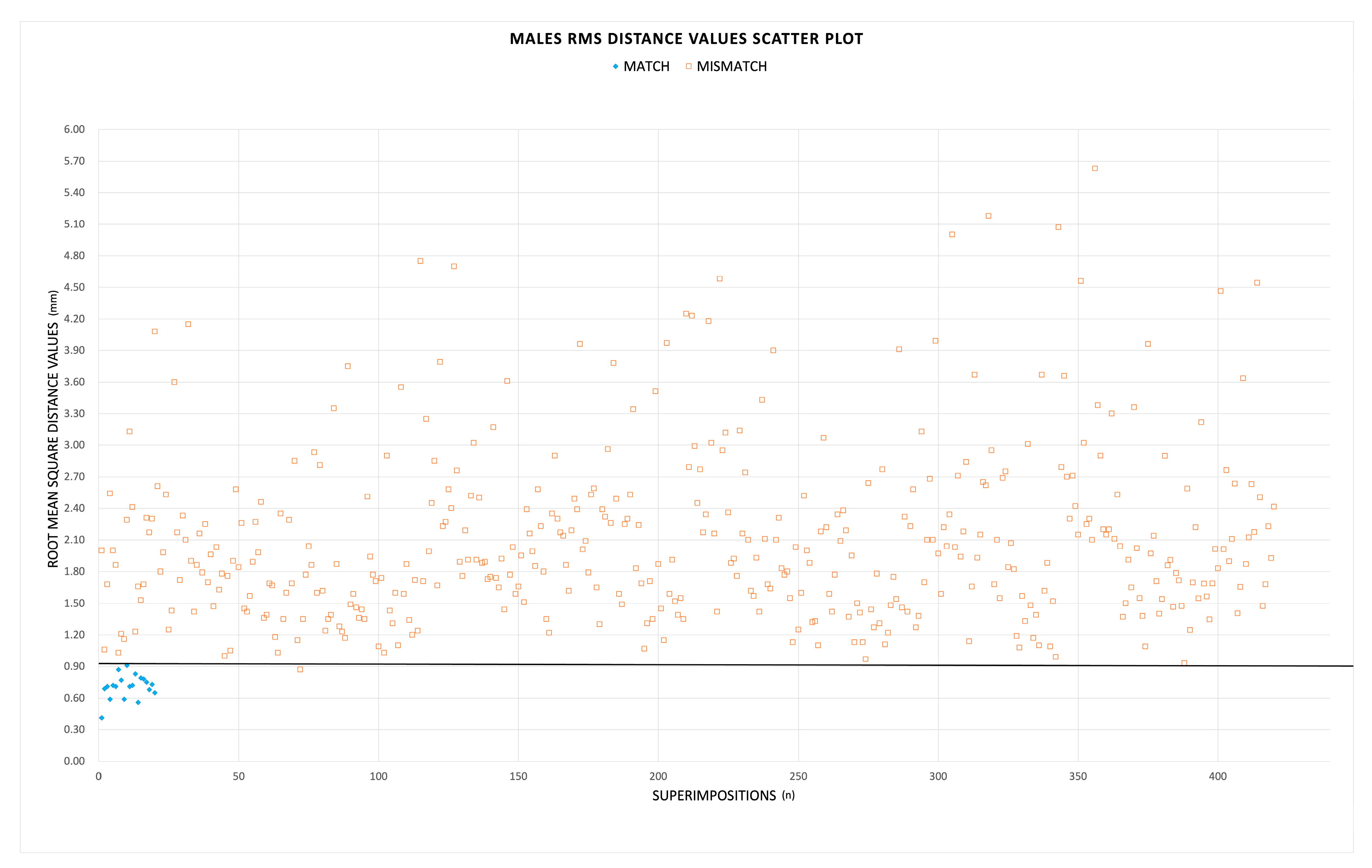
| Males | 0.91 mm | 20 | 1 | 419 | 0 | 100.0% | 99.8% |
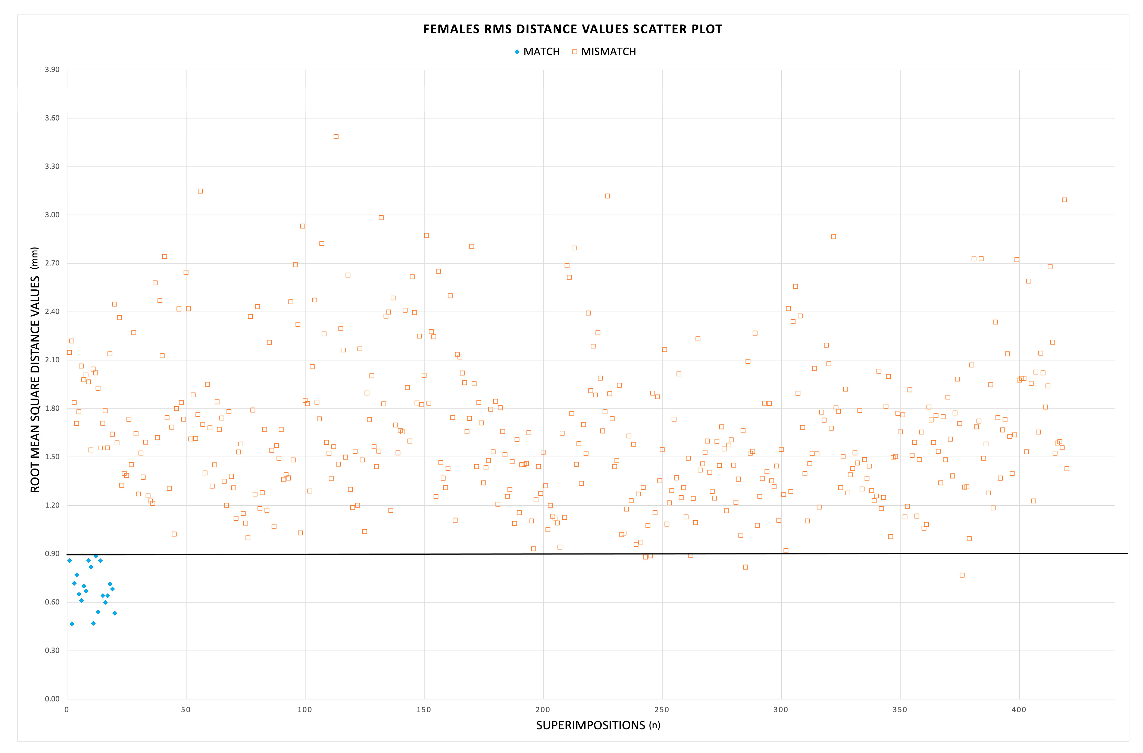
| Females | 0.89 mm | 20 | 5 | 415 | 0 | 100.0% | 98.8% |
| Combined | 0.91 mm | 40 | 6 | 834 | 0 | 100.0% | 99.3% |
Disclaimer/Publisher’s Note: The statements, opinions and data contained in all publications are solely those of the individual author(s) and contributor(s) and not of MDPI and/or the editor(s). MDPI and/or the editor(s) disclaim responsibility for any injury to people or property resulting from any ideas, methods, instructions or products referred to in the content. |
© 2022 by the authors. Licensee MDPI, Basel, Switzerland. This article is an open access article distributed under the terms and conditions of the Creative Commons Attribution (CC BY) license (https://creativecommons.org/licenses/by/4.0/).
Share and Cite
Palamenghi, A.; Cappella, A.; Cellina, M.; Mazzarelli, D.; De Angelis, D.; Sforza, C.; Cattaneo, C.; Gibelli, D. 3D-3D Superimposition of Pubic Bones: Expanding the Anthropological Toolkit for the Pair-Matching of Commingled Skeletal Remains. Biology 2023, 12, 30. https://doi.org/10.3390/biology12010030
Palamenghi A, Cappella A, Cellina M, Mazzarelli D, De Angelis D, Sforza C, Cattaneo C, Gibelli D. 3D-3D Superimposition of Pubic Bones: Expanding the Anthropological Toolkit for the Pair-Matching of Commingled Skeletal Remains. Biology. 2023; 12(1):30. https://doi.org/10.3390/biology12010030
Chicago/Turabian StylePalamenghi, Andrea, Annalisa Cappella, Michaela Cellina, Debora Mazzarelli, Danilo De Angelis, Chiarella Sforza, Cristina Cattaneo, and Daniele Gibelli. 2023. "3D-3D Superimposition of Pubic Bones: Expanding the Anthropological Toolkit for the Pair-Matching of Commingled Skeletal Remains" Biology 12, no. 1: 30. https://doi.org/10.3390/biology12010030