Article

Morphological and Molecular Characterization, and Demonstration of a Definitive Host, for Sarcocystis masoni from an Alpaca (Vicugna pacos) in China

1
Yunnan Key Laboratory for Plateau Mountain Ecology, Restoration of Degraded Environments, School of Ecology and Environmental Sciences, Yunnan University, Kunming 650091, China
2
Electron Microscope Laboratory, Kunming Medical University, Kunming 650500, China
3
School of Biological Sciences, Yunnan University, Kunming 650091, China
4
College of Veterinary Medicine, Henan Agricultural University, Zhengzhou 450064, China
5
College of Veterinary Medicine, Yangzhou University, Yangzhou 225009, China
*
Authors to whom correspondence should be addressed.
Biology 2022, 11(7), 1016; https://doi.org/10.3390/biology11071016
Submission received: 7 June 2022 / Revised: 28 June 2022 / Accepted: 4 July 2022 / Published: 6 July 2022
(This article belongs to the Section Zoology)

Abstract

:

Simple Summary

Sarcocystis spp. are cyst-forming intracellular protozoan parasites characterized by a two-host prey–predator life cycle. The alpaca (Vicugna pacos) is one of the South American camelids (SACs), and in recent years, this animal was introduced to China to be raised for its meat, skin, and wool and to be kept as tourist attractions and as pets. There is considerable confusion regarding the classification and nomenclature of the species of Sarcocystis in SACs. Two Sarcocystis species, named S. auchenia and S. masoni, are currently regarded as valid in SACs based on sarcocyst morphology and 18S rDNA sequences. However, the definitive host of S. masoni remains unknown. Here, S. masoni sarcocysts in an alpaca were morphologically described and molecularly characterized. Furthermore, the life cycle of S. masoni was completed via experimental animal infection. The present analysis showed that S. masoni has a close relationship with S. cameli in the dromedary camel (Camelus dromedaries), and the relationship between the two parasites needs to be clarified in the future.

Abstract

Only 18S rDNA sequences of Sarcocystis spp. in South American camelids (SACs) are deposited in GenBank as references, and the definitive host of S. masoni in SACs is still unclear. Here, S. masoni sarcocysts detected in an alpaca (Vicugna pacos) in China were investigated with the aid of light (LM) and transmission electron (TEM) microscopy, and characterized using four genetic markers, i.e., 18S rDNA, 28S rDNA and ITS, and the mitochondrial cox1. Additionally, the life cycle of the parasite was completed via experimental animal infection. Under LM, S. masoni sarcocysts exhibited numerous 1.3–2.1 μm conical protrusions. Under TEM, the sarcocyst wall contained conical, cylindrical, or irregular-shaped villar protrusions, similar to type 9j. Two dogs (Canis familiaris) fed S. masoni sarcocysts shed sporocysts with a prepatent period of 8–9 days. The newly obtained 18S rDNA sequences showed 98.4–100% identity with those of S. masoni in SACs previously deposited in GenBank. Interestingly, the newly obtained sequences of 18S rDNA and mitochondrial cox1 shared 99.6–100% and 98.2–98.5% identity, respectively, with those of S. cameli in dromedary camels (Camelus dromedaries). Phylogenetic analysis based on sequences of 18S rDNA, 28S rDNA, or mitochondrial cox1 revealed that S. masoni has a close relationship with Sarcocystis spp. in ruminants. The relationship between S. masoni and S. cameli deserves to be further clarified in the future.

1. Introduction

Sarcocystis spp. are cyst-forming intracellular protozoan parasites with an obligate two-host life cycle, with predators as definitive hosts and their prey animals as intermediate hosts. Collectively, these species have considerable veterinary, economic, and public health importance [1]. The alpaca (Vicugna pacos, formerly Lama pacos) is one of the South American camelids (SACs). This animal is a domesticated farm animal and is mainly distributed in Peru, where it plays a crucial role in local socioeconomics [2]. During the last two decades, this animal has been introduced to China to be raised for its meat, skin, and wool and to be kept as tourist attractions and as pets [3].
SACs, including the alpaca, llama (Lama glama), vicuna (Vicugna vicugna), and guanaco (Lama guanicoe), serve as important intermediate hosts for Sarcocystis spp., which usually cause subclinical infections in SACs, although fatal cases have also been reported [4,5]. Two kinds of sarcocysts, macroscopic cysts (up to 8 mm long) and microscopic cysts (up to 800 µm long), have been observed and described in SACs. However, there is considerable confusion regarding the classification and nomenclature of the species of Sarcocystis in SACs [1]. The history of the taxonomy of Sarcocystis spp. in SACs has been reviewed by different authors recently [1,6,7]. Currently, based on the sarcocysts’ ultrastructure, the macroscopic cysts in SACs are regarded as S. auchenia, which was originally found in a llama and named by Brumpt (1913) [8], and the microscopic cysts in SACs are regarded as S. masoni, named by Moré et al. (2016) [6]. The microscopic cysts have been called S. lamacenis/lamacanis in two review papers [9,10], but no explanation for these proposals was provided by the authors.
Traditionally, sarcocyst structure and life cycle are the two major criteria for naming a new species of Sarcocystis in a given intermediate host [1]. The definitive host of S. aucheniae in guanaco and llama has been experimentally demonstrated to be dogs (Canis familaris) [11,12]. However, the life cycle of S. masoni remains unknown. At present, PCR assays and sequencing procedures are considered much more practical, accurate, and reliable for the delineation and identification of Sarcocystis species than traditional methods based on the morphological characteristics [13,14]. Only the 18S rDNA sequences of S. masoni and S. auchenia presently deposited in GenBank serve as references for species identification. However, the discriminatory power of the gene has been shown to be unsuitable for the differentiation of closely related lineages of Sarcocystis in ruminants due to its highly conserved nature [13,15]
Therefore, the aims of the present study were: (1) to investigate the morphology of sarcocysts in an alpaca from China using light (LM) and transmission electron (TEM) microscopy; (2) to sequence and analyze the near-full or full length of the three nuclear DNA regions 18S rDNA, 28S rDNA, and ITS (ITS1-5.8S-ITS2) and the partial mitochondrial cox1 of the sarcocysts to augment the species description; and (3) to elucidate the S. masoni life cycle in the definitive host via experimental infection.

2. Materials and Methods

2.1. Examination for Sarcocysts in Alpaca

In December 2021, one captive-bred alpaca died from an illness at a zoo in Zhengzhou, the capital city of Henan province, China. The veterinarian conducted a pathological dissection, and the myocardium of the animal was shipped on ice to the zoological laboratory at the School of Ecology and Environmental Sciences, Yunnan University, for diagnosis of sarcocystosis. In the laboratory, 1–3 mm pieces were pressed between 2 glass slides and examined for sarcocysts via stereomicroscopy. Sarcocysts were extracted from muscular fibers and processed for LM and TEM, experimental infection, and DNA analysis. For TEM, six sarcocysts were fixed in 2.5% glutaraldehyde in cacodylate buffer (0.1 M, pH 7.4) and examined using a JEM100-CX TEM (JEOL Ltd., Tokyo, Japan) at 80 kV. For DNA isolation, individual sarcocysts were stored in sterile water at −20 °C prior to processing.

2.2. Experimental Infection of Potential Definitive Hosts

Three dogs and three cats (Felis catus) were used for the experimental infection. These animals were 1 to 2 months old and were purchased from a commercial source, housed separately in steel cages, and fed dog or cat pellets and water ad libitum. The feces of these animals were examined for four successive days with the use of centrifugal flotation and a sucrose solution (density 1.20 at 20 °C) before infection to prove that they were coccidian free.
Pieces of muscle from the alpaca were fed to two dogs and two cats. The remaining animals were kept as controls. Before inoculation, it was confirmed by extensive microscopic examination that the muscles of the alpaca contained only microcysts. The animals were each fed muscle pieces containing approximately 600 sarcocysts. Fecal samples from the cats and dogs were tested daily for at least 20 days post infection (PI) to determine the presence of sporocysts or oocysts. All animals were euthanized at 20 days PI. The small intestine of each animal was removed, and a mucosa sample from the small intestine, approximately 2.5 cm in length in each part, was examined for the presence of oocysts or sporocysts and via DNA extraction.

2.3. Molecular Characterization

Genomic DNA was extracted from six individual sarcocysts isolated from the alpaca and six mucosa samples each from the small intestine of the experimental animals using a TIANamp Genomic DNA Kit (Tiangen Biotech Ltd., Beijing, China) according to the manufacturer’s instructions. Four genetic markers—18S rDNA, 28S rDNA, ITS, and mitochondrial cox1—were amplified from the sarcocysts using the primer pairs shown in Table 1.
To further verify the results of experimental infection, mitochondrial cox1 of the parasite in the definitive hosts was amplified and sequenced using the same primers SF1/SR9 for sequence analysis and polymerase chain reaction–restriction fragment length polymorphism (PCR-RFLP). PCR products were gel-purified, cloned and sequenced, and assembled using the methods detailed in a previous paper [20]. For PCR-RFLP, PCR products of mitochondrial cox1 obtained from sarcocysts and oocysts/sporocysts were digested separately with restriction enzymes ClaI and AvaII (New England BioLabs).
Phylogenetic analyses were conducted separately on the nucleotide sequences of the 18S rDNA, 28S rDNA, and mitochondrial cox1 sequences using MEGAX software [21]. The maximum likelihood (ML) trees of 18S rDNA, 28S rDNA, and mitochondrial cox1 were generated using the Tamura 3-parameter, Hasegawa–Kishino–Yano, and Kimura 2-parameter models, respectively, according to the Find Best DNA/Protein Models program integrated into MEGAX. All sites were used. The reliability of the maximum likelihood phylograms was tested via the bootstrap method using 1000 replications.
The 18S rDNA, 28S rDNA, and mitochondrial cox1 sequences of Sarcocystis spp. from different hosts were downloaded from GenBank and aligned using the ClustalW program implemented in MEGAX, using a gap opening penalty of 10/10 and a gap extension penalty of 0.1/0.2 as pairwise and multiple alignment parameters, respectively. The alignments were subsequently checked visually; some sequences were slightly truncated at both ends, so that all sequences started and ended at the same nucleotide positions. The final alignment of the 18S rDNA sequences consisted of 33 nucleotide sequences and 1592 aligned positions including gaps from 24 taxa. Cystoisospora ohioensis (GU292304), Besnoitia besnoiti (DQ227418), and Toxoplasma gondii (U03070) were chosen as outgroups. The final alignment of the 28S rDNA sequences consisted of 26 nucleotide sequences and 3839 aligned positions including gaps from 24 taxa. Hammondia heydorni (AF159240), B. besnoiti (AF076900), and T. gondii (AF076901) were chosen as outgroups. The final alignment of mitochondrial cox1 sequences consisted of a total of 37 nucleotide sequences and 995 aligned positions with no gaps from 29 taxa. T. gondii (JX473253), H. triffittae (JX473247), and H. heydorni (JX473251) were used as outgroup species to root the tree.

3. Results

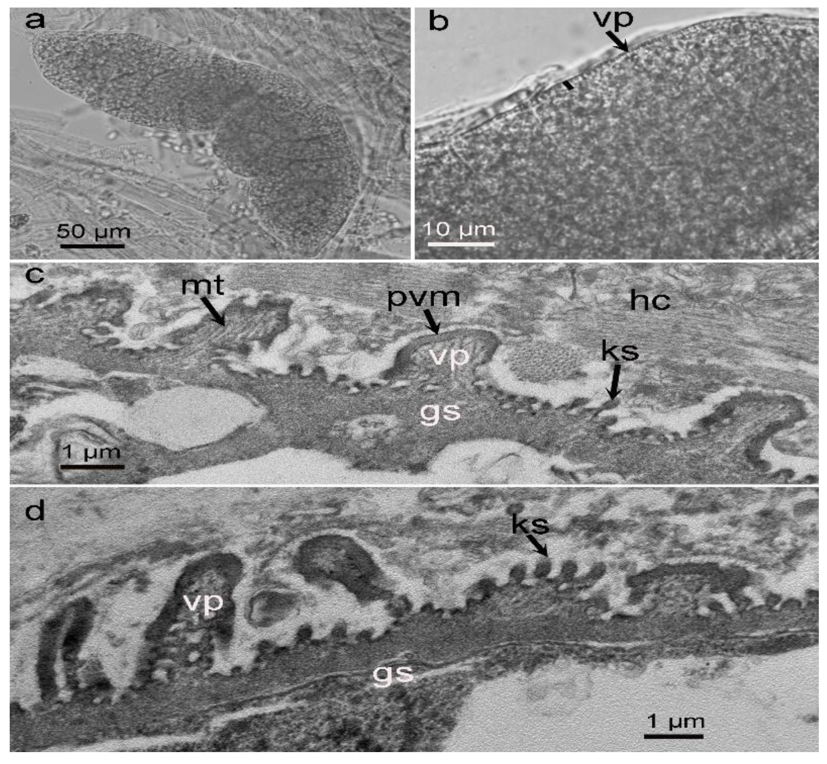
3.1. Morphological Observation of Sarcocysts in the Alpaca

Only sarcocysts resembling those of S. masoni were found in the alpaca. LM examination revealed that the sarcocysts were microscopic, measuring 342–550 × 88–102 μm (n = 20) in size, and exhibited numerous 1.3–2.1 μm conical villar protrusions (vp) (Figure 1a,b). They were septate and contained bradyzoites that were 10.2–12.3 × 2.8–5.5 μm (n = 20) in size.
Four sarcocysts from the alpaca were examined via TEM, all of which appeared to have walls that were ultrastructurally similar and that closely resembled “type 9j” (Figure 1c,d). The sarcocyst walls contained conical, cylindrical, or irregular-shaped villar protrusions (vp) depending on the plane of the section. The vp were at irregular distances and lined by a 60–80 nm thick electron-dense parasitophorous vacuolar membrane (pvm), with knob-like structures (ks) on the pvm. Each vp had scattered microtubules, which extended from the tip of the villus into the middle of the ground substance (gs). The gs, measuring 0.6–1.1 μm in thickness, was located immediately beneath the sarcocyst wall.

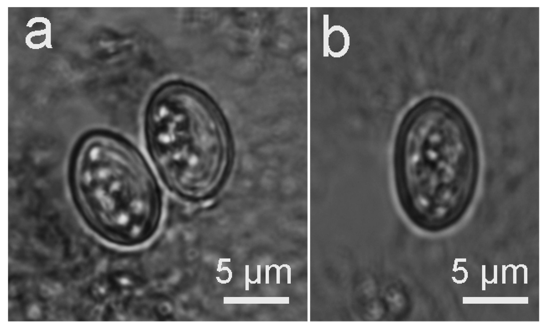
3.2. Infection of the Definitive Hosts

The two dogs that were fed with muscle tissues containing S. masoni sarcocysts from the alpaca both excreted sporulated oocysts and sporocysts (Figure 2a,b) beginning at Days 8 and 9 PI. The oocysts measured 12.5–15.8 × 9.8–11.2 μm (n = 10) with two elliptical sporocysts that measured 10.2–11.6 × 7.3–8.2 μm. Upon the death of the dogs and examination of their intestinal mucosa, the oocysts and sporocysts were found to be mostly concentrated toward the tips of the villi, but some were located deep in the villi as well. However, the intestinal mucosa was not appreciably altered, and infected dogs did not show any clinical signs due to sarcosporidian infection. No oocysts or sporocysts were found in the three cats or in the control dog.

3.3. Molecular Analysis

The four selected genes (18S rDNA, 28S rDNA, ITS, and mitochondrial cox1) and mitochondrial cox1 alone were successfully amplified, sequenced, and assembled from six individual sarcocysts in the alpaca and from oocysts in the intestines of the two infected dogs, respectively. The six 18S rDNA sequences (ON533528–ON533533) were 1848 and 1849 bp in length and shared 99.2–99.8% identity (average 99.5%). The most similar sequences in GenBank to the newly obtained 18S rDNA sequences were those of S. cameli (OM462704 and OM462705) obtained from one-humped camels (Camelus dromedaries) (99.6–100% identity, average 99.8%) and S. masoni obtained from SACs, including 99.4–100% identity (average 99.6%) in alpacas (MW481703, MW481704, KU527112 and KU527113), 98.4–99.6% identity (average 99.0%) in guanacos (KU527107–KU527109), and 99.5–99.7% identity (average 99.6%) in llamas (KU527110 and KU527111), followed by those of S. bovini (93.5–94.0% identity, average 93.7%) in cattle (Bos taurus) (KT901139–KT9011). Nevertheless, the identity with those of S. aucheniae was only 87.1–91.8% (average 90.5%) in SACs (KU527114–KU527124).
The six 28S rDNA sequences (ON533536–ON533541) were 3404–3414 bp in length and shared 98.7–99.6% identity (average 99.1%). The most similar sequences in GenBank to the newly obtained 28S rDNA sequences were those of Sarcocystis spp. obtained from domestic ruminants, including S. gigantea (U85706) in sheep (Ovis aries) (91.9–92.2% identity, average 92.1%), S. capracanis (AF012885, KU820978, KU820979) in goat (Capra hircus) (91.5–91.6% identity, average 91.6%), and S. cruzi (AF076903) in cattle (91.5–91.6% identity, average 91.6%).
The six ITS sequences (ON540302–ON540307) were 1547–1566 bp in length and shared 92.7–98.4% identity (average 94.8%). BLAST searches indicated that no sequences in GenBank shared significant similarities with the newly obtained ITS sequences.
The six mitochondrial cox1 sequences obtained from the sarcocysts were 1085 bp in length and shared 99.4–100% identity (average 99.6%). Therefore, only five sequences (ON564410–ON564414) were deposited in GenBank. The four mitochondrial cox1 sequences obtained from oocysts in the two dogs were 1085 bp in length and shared 99.7–100% identity (average 99.8%). Therefore, only three sequences (ON564415–ON564417) were deposited in GenBank. The similarity between sarcocysts and oocysts was 99.0–100% (average 99.5%). The most similar sequence in GenBank was that of S. cameli (MW651858) in the one-humped camel (98.2–98.5% identity, average 98.4%), followed by those of S. rommeli (KY10286–KY10292) in cattle (79.5–80.5% identity, average 80.3%).

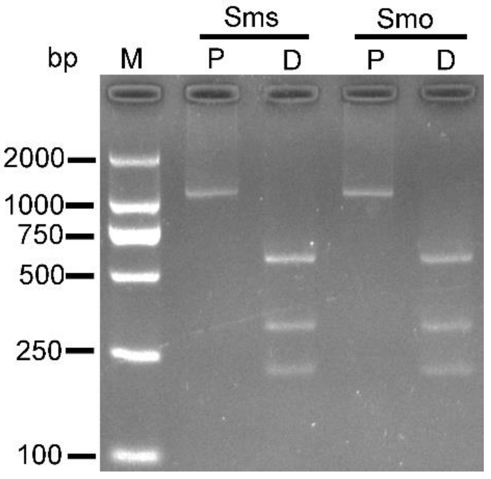
3.4. PCR-RFLP Based on Mitochondrial cox1 Obtained from S. masoni Sarcocysts and Oocysts

The PCR-amplified products (1085 bp) of mitochondrial cox1 from S. masoni sarcocysts and oocysts were successfully digested by ClaI and AvaII and produced three fragments (218, 316, and 551 bp) (Figure 3).

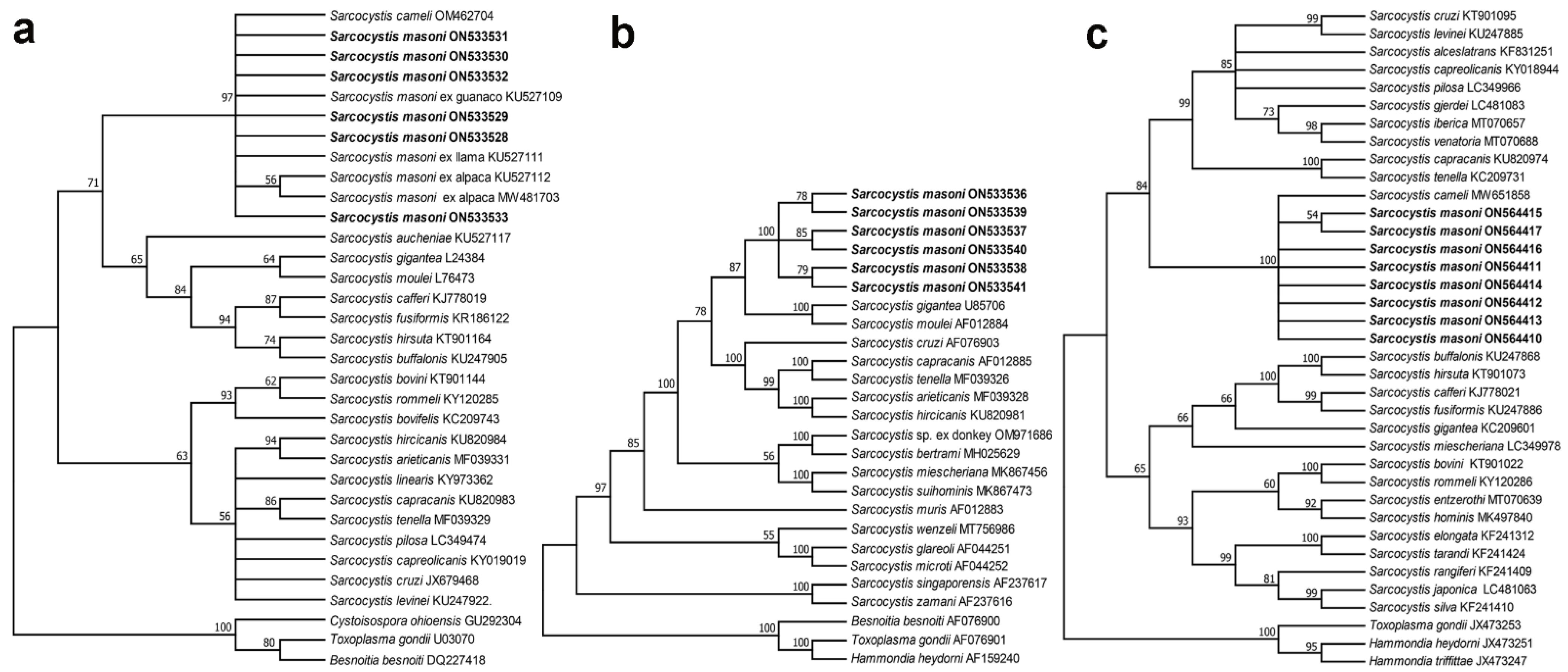
3.5. Phylogenetic Analysis

Phylogenetic analysis based on the 18S rDNA, 28S rDNA, and mitochondrial cox1 sequences of S. masoni confirmed their association with Sarcocystis species (Figure 4). In the tree inferred from 18S rDNA sequences (Figure 4a), S. masoni in SACs and S. cameli in the one-humped camel formed an individual clade within a group comprising Sarcocystis spp. in ruminants with felids or canids as known or suspected definitive hosts. Based on 28S rDNA sequences, the phylogenetic tree (Figure 4b) showed that the newly obtained S. masoni formed an individual clade within a group comprising Sarcocystis spp. in ruminants with felids or canids as known or suspected definitive hosts. The phylogenetic tree inferred from mitochondrial cox1 (Figure 4c) showed that the newly obtained S. masoni formed an individual clade with S. cameli basal to a group comprising Sarcocystis spp. in ruminants with canids as known or suspected definitive hosts.

4. Discussion

Sarcocystis infection is common in many species of animal worldwide, including in China. Sarcocystosis of alpacas has mostly been reported in South America and Australia [1], but recently, the disease was also diagnosed in alpacas from China [22]. Sarcocysts are common in asymptomatic alpacas and other similar Camelidae, usually causing subclinical infections in SACs [4]. An extensive survey in Southern Bolivia indicated that carcass downgrades caused by Sarcocystis infection in llamas resulted in 13–20% economic loss to the local farmers [23].
The ultrastructure of the sarcocyst wall is a useful indicator used to distinguish Sarcocystis spp. within a given host. A recent review indicated that there are more than 200 Sarcocystis species with at least 42 types and several subtypes of sarcocyst wall [1]. Ultrastructurally, the sarcocysts in SACs can be divided into two types. The macroscopic sarcocysts have a cyst wall with cauliflower-like protrusions, similar to TEM wall type 21, known as S. aucheniae [6,12,24]. The microscopic sarcocysts have a cyst wall with conical to cylindrical vp, similar to TEM wall type 9j, known as S. masoni [6]. In our materials, only microscopic sarcocysts were observed in the alpaca, and they were identified as S. masoni due to their high similarity to the original morphological descriptions of the parasite [6]. Recently, morphologically similar microcysts were also diagnosed in alpacas from Peru but were named Sarcocsytis sp. based on the slight difference in the sizes of sarcocysts and vp compared with those of S. masoni [25].
The definitive host of S. aucheniae in guanaco and llama has been experimentally demonstrated to be dogs [11,12]. In the present study, the life cycle of S. masoni was completed for the first time, and the dog was proven to be the definitive host of the parasite based on experimental infection and PCR-RFLP. The prepatent period of 8–9 days for S. masoni is shorter than that of 9–16 days for S. aucheniae [11,12]. It is worth noting that the molecular data on S. aucheniae oocysts/sporocysts need to be supplemented in the future to address the possibility of mixed Sarcocystis infection in SACs.
Nucleotide sequence analysis has proven to be a useful tool for delineating or identifying species of Sarcocystis from the same or different hosts, and different genetic markers have shown different levels of intra- or inter-specific sequence diversity; mitochondrial cox1 seems to perform better than nuclear genes for distinguishing Sarcocystis spp. in ruminants [13,14,15,20]. To date, there are only 18S rDNA sequences of S. aucheniae and S. masoni deposited in GenBank as references. Here, four genetic markers (18S rDNA, 28S rDNA, ITS, and mitochondrial cox1) of S. masoni were sequenced and analyzed. Comparing the newly obtained sequences with those deposited in GenBank, the 18S rDNA sequences showed 99.4–100% identity with those of S. masoni obtained from SACs. However, at this locus, they only showed 87.1–91.8% identity with those of S. aucheniae. Therefore, the 18S rDNA was suitable for distinguishing S. masoni from S. aucheniae. However, the newly obtained sequences of 18S rDNA and mitochondrial cox1 exhibited high similarity with those of S. cameli from one-humped camels, i.e., 99.6–100% and 98.2–98.5% identity, respectively. Interestingly, S. cameli (synonym S. camelicanis) sarcocysts and S. masoni sarcocysts present similar morphological characteristics [1,26,27], and the definitive hosts of S. cameli are also dogs with a prepatent period of 11 days [26]. Phylogenetic analysis based on the 18S rDNA or mitochondrial cox1 sequences demonstrated that the close relationship between S. masoni and S. cameli for both species formed an individual clade in the phylogenetic trees. Therefore, the plausible explanation is that they probably represent the same species of Sarcocystis in different hosts. However, this must be proven using more molecular markers of the two parasites and cross-transmission of Sarcocystis between SACs and the one-humped camel in the future.

5. Conclusions

In summary, based on LM and TEM morphological analysis, only S. masoni was identified in the alpaca. Four genetic markers (18S rDNA, 28S rDNA, ITS, and mitochondrial cox1) of S. masoni were sequenced and characterized. Among them, the sequences of 28S rDNA, ITS, and mitochondrial cox1 constituted the first records of the parasite in GenBank. For the first time, the life cycle of S. masoni was completed, and dogs are its definitive hosts as proven by experimental animal infection and molecular data. In our analysis, the 18S rDNA and mitochondrial cox1 sequences could not effectively distinguish S. masoni from S. cameli. The two parasites have a similar LM and TEM morphology and life cycle. Therefore, the relationship between the two species should be further clarified in the future.

Author Contributions

Conceptualization, J.H. and Y.Y.; methodology, Z.W., J.S. (Jun Sun) and J.S. (Jingling Song); software, S.D.; formal analysis, J.H.; investigation, Z.W., J.S. (Jun Sun), J.H. and J.S. (Jingling Song); resources, Y.Y. and N.Z.; writing—original draft preparation, J.H.; writing—review and editing, J.H. and J.T. All authors have read and agreed to the published version of the manuscript.

Funding

This research was funded by the National Key R&D Program of China (grant 2017YFD0500400) and the Natural Sciences Foundation of China (Grant 31460557).

Institutional Review Board Statement

The study was approved by the Animal Ethics Committee of Yunnan University (permission number AECYU2018004).

Informed Consent Statement

Not applicable.

Data Availability Statement

Not applicable.

Conflicts of Interest

The authors declare no conflict of interest.

References

  1. Dubey, J.P.; Calero-Bernal, R.; Rosenthal, B.M.; Speer, C.A.; Fayer, R. Sarcocystosis of Animals and Humans, 2nd ed.; CRC Press: Boca Raton, FL, USA, 2016; pp. 257–267. [Google Scholar]
  2. Reyna, J. Alpaca breeding in Peru and perspectives for the future. N. Z. Alpacas. 2005, 18, 21. [Google Scholar]
  3. She, Q.; Zhang, H.; Lou, P.; Ji, X.; Zhao, Z. Current situation and development countermeasures of alpaca Breeding. Agric. Sci. Technol. 2011, 12, 733–736. [Google Scholar]
  4. La Perle, K.M.; Silveria, F.; Anderson, D.E.; Blomme, E.A. Dalmeny disease in an alpaca (Lama pacos): Sarcocystosis, eosinophilic myositis and abortion. J. Comp. Pathol. 1999, 121, 287–293. [Google Scholar] [CrossRef] [PubMed]
  5. Chávez, V.; Leyva, V.; Panez, L.; Ticona, S.; García, V.; Pezo, C. Sarcocystiosis and productive efficiency of the alpaca. Rev. Investig. Vet. del Perú. 2008, 19, 160–167. [Google Scholar]
  6. Moré, G.; Regensburger, C.; Gos, M.L.; Pardini, L.; Verma, S.K.; Ctibor, J.; Serrano-Martínez, M.E.; Dubey, J.P.; Venturini, M.C. Sarcocystis masoni, n. sp. (Apicomplexa: Sarcocystidae), and redescription of Sarcocystis aucheniae from llama (Lama glama), guanaco (Lama guanicoe) and alpaca (Vicugna pacos). Parasitology 2016, 143, 617–626. [Google Scholar] [CrossRef]
  7. Saeed, M.A.; Rashid, M.H.; Vaughan, J.; Jabbar, A. Sarcocystosis in South American camelids: The state of play revisited. Parasit. Vectors 2018, 11, 146. [Google Scholar] [CrossRef] [Green Version]
  8. Brumpt, E. Précis de Parasitologie, 2nd ed.; Masson et Cie: Paris, France, 1913. [Google Scholar]
  9. Leguía, G. The epidemiology and economic impact of llama parasites. Parasitol. Today 1991, 7, 54–56. [Google Scholar] [CrossRef]
  10. Lucas, J.R. Sarcocystis spp. in Peru. Peruv. J. Parasitol. 2012, 20, e64e73. [Google Scholar]
  11. Gorman, T.R.; Alcaíno, H.A.; Muñoz, H.; Cunazza, C. Sarcocystis sp. in guanaco (Lama guanicoe) and effect of temperature on its viability. Vet. Parasitol. 1984, 15, 95–101. [Google Scholar] [CrossRef]
  12. Schnieder, T.; Kaup, F.J.; Drommer, W.; Thiel, W.; Rommel, M. Fine structure and development of Sarcocystis aucheniae in llamas. Z. Parasitenkd. 1984, 70, 451–458. [Google Scholar] [CrossRef]
  13. Gjerde, B. Phylogenetic relationships among Sarcocystis species in cervids, cattle and sheep inferred from the mitochondrial cytochrome c oxidase subunit I gene. Int. J. Parasitol. 2013, 43, 579–591. [Google Scholar] [CrossRef] [PubMed]
  14. Gjerde, B. Sarcocystis species in red deer revisited: With a re-description of two known species as Sarcocystis elongata n. sp. and Sarcocystis truncata n. Sp. based on mitochondrial cox1 sequences. Parasitology 2014, 141, 441–452. [Google Scholar] [CrossRef] [PubMed]
  15. Hu, J.J.; Huang, S.; Wen, T.; Esch, G.W.; Liang, Y.; Li, H.L. Sarcocystis spp. in domestic sheep in Kunming City, China: Prevalence, morphology, and molecular characteristics. Parasite 2017, 24, 30. [Google Scholar] [CrossRef] [PubMed] [Green Version]
  16. Barta, J.R.; Martin, D.S.; Liberator, P.A.; Dashkevicz, M.; Anderson, J.W.; Feighner, S.D.; Elbrecht, A.; Perkins-Barrow, A.; Jenkins, M.C.; Danforth, H.D.; et al. Phylogenetic relationships among eight Eimeria species infecting domestic fowl inferred using complete small subunit ribosomal DNA sequences. J. Parasitol. 1997, 83, 262–271. [Google Scholar] [CrossRef]
  17. Fischer, S.; Odening, K. Characterization of bovine Sarcocystis species by analysis of their 18S ribosomal DNA sequences. J. Parasitol. 1998, 84, 50–54. [Google Scholar] [CrossRef]
  18. Fenger, C.K.; Granstrom, D.E.; Langemeier, J.L.; Stamper, S.; Donahue, J.M.; Patterson, J.S.; Gajadhar, A.A.; Marteniuk, J.V.; Xiaomin, Z.; Dubey, J.P. Identification of opossums (Didelphis virginiana) as the putative definitive host of Sarcocystis neurona. J. Parasitol. 1995, 81, 916–919. [Google Scholar] [CrossRef]
  19. Mugridge, N.B.; Morrison, D.A.; Johnson, A.M.; Luton, K.; Dubey, J.P.; Votýpka, J.; Tenter, A.M. Phylogenetic relationships of the genus Frenkelia: A review of its history and new knowledge gained from comparison of large subunit ribosomal ribonucleic acid gene sequences. Int. J. Parasitol. 1999, 29, 957–972. [Google Scholar] [CrossRef]
  20. Hu, J.J.; Liu, T.T.; Liu, Q.; Esch, G.W.; Chen, J.Q.; Huang, S.; Wen, T. Prevalence, morphology, and molecular characteristics of Sarcocystis spp. in domestic goats (Capra hircus) from Kunming, China. Parasitol. Res. 2016, 115, 3973–3981. [Google Scholar] [CrossRef]
  21. Kumar, S.; Stecher, G.; Li, M.; Knyaz, C.; Tamura, K. MEGA X: Molecular evolutionary genetics analysis across computing platforms. Mol. Biol. Evol. 2018, 35, 1547–1549. [Google Scholar] [CrossRef]
  22. Jiang, N.; Xin, S.; Zhu, N.; Yang, L.; Huang, W.; Hu, J.; Zhu, X.; Yang, Y. First report of Sarcocystis masoni in a captive alpaca (Vicugna pacos) from China. Front. Vet. Sci. 2021, 8, 759252. [Google Scholar] [CrossRef]
  23. Rooney, A.L.; Limon, G.; Vides, H.; Cortez, A.; Guitian, J. Sarcocystis spp. in llamas (Lama glama) in Southern Bolivia: A cross sectional study of the prevalence, risk factors and loss in income caused by carcass downgrades. Prev. Vet. Med. 2014, 116, 296–304. [Google Scholar] [CrossRef] [PubMed]
  24. Regensburger, C.; Gos, M.L.; Ctibor, J.; Moré, G. Morphological and molecular characteristics of Sarcocystis aucheniae isolated from meat of guanaco (Lama guanicoe). J. Food Qual. Hazards Control 2015, 2, 118–121. [Google Scholar]
  25. Lucas, J.R.; Barrios-Arpi, M.; Rodríguez, J.; Balcázarnakamatsu, S.; Zarria, J.; Namiyama, G.; Taniwaki, N.; Gonzales-Viera, O. Ultrastructural description of Sarcocystis sp. in cardiac muscle of naturally infected alpacas (Vicugna pacos). Iran. J. Parasitol. 2019, 14, 174–179. [Google Scholar] [PubMed]
  26. Abdel-Ghaffar, F.; Mehlhorn, H.; Bashtar, A.R.; Al-Rasheid, K.; Sakran, T.; El-Fayoumi, H. Life cycle of Sarcocystis camelicanis infecting the camel (Camelus dromedarius) and the dog (Canis familiaris), light and electron microscopic study. Parasitol. Res. 2009, 106, 189–195. [Google Scholar] [CrossRef] [PubMed]
  27. Dubey, J.P.; Hilali, M.; Van Wilpe, E.; Calero-Bernal, R.; Verma, S.K.; Abbas, I.E. A review of sarcocystosis in camels and redescription of Sarcocystis cameli and Sarcocystis ippeni sarcocysts from the one-humped camel (Camelus dromedarius). Parasitology 2015, 142, 1481–1492. [Google Scholar] [CrossRef] [Green Version]
Figure 1. Morphological characteristics of Sarcocystis masoni sarcocysts isolated from the myocardium of an alpaca. (a) Overview of a sarcocyst (unstained, light microscopy, LM). (b) Sarcocyst bounded by short conical villar protrusions (vp) (unstained, LM). (c,d) Diagonal section of a sarcocyst (under transmission electron microscopy, TEM). Sarcocyst surrounded by host cell (hc). The sarcocyst wall exhibits numerous villar protrusions (vp) lined by an electron-dense parasitophorous vacuolar membrane (pvm). Knob-like structures (ks) present on the surface of the pvm. Each vp contains scattered microtubules (mt) in its core, which extend from the tip of the vp into the ground substance (gs).
Biology 11 01016 g001
Figure 2. Light micrograph of Sarcocystis masoni sporulated oocyst and sporocysts in the feces of experimentally infected dogs (unstained). (a) Sporulated oocyst. (b) Sporocyst.
Biology 11 01016 g002
Figure 3. Results of PCR with primers SF1/SR9 and restriction enzyme digestion with ClaI and AvaII of sarcocyst and oocyst DNA from Sarcocystis masoni. M, molecular mass marker; Sms, S. masoni sarcocyst; Smo, S. masoni oocyst; P, PCR product; D, digestion of PCR product with ClaI and AvaII.
Biology 11 01016 g003
Figure 4. Phylogenetic trees of selected Sarcocystis species. The trees constructed based on (a) 18S rDNA sequences, (b) 28S rDNA sequences, and (c) mitochondrial cox1 sequences using maximum likelihood (ML) with the Tamura 3-parameter, Hasegawa–Kishino–Yano, and Kimura 2-parameter models, respectively. The values between the branches represent bootstrap values per 1000 replicates, and values below 50% are not shown. (a) The six newly obtained 18S rDNA sequences of S. masoni (ON533528–ON533533, shown in boldface) obtained from the alpaca formed an individual clade with those of S. cameli in the one-humped camel and S. masoni in the llama, alpaca, and guanaco. (b) The six new sequences of S. masoni (ON533536–ON533541, shown in boldface) obtained from an alpaca formed an individual clade within a group comprising Sarcocystis spp. in domestic ruminants. (c) The eight sequences of S. masoni (ON564410–ON564417, shown in boldface) obtained from the alpaca and two infected dogs formed an individual clade with S. cameli in the one-humped camel.
Biology 11 01016 g004
Table 1. Primers used for the amplification of four DNA regions.
DNA RegionPrimer NamePrimer Sequence (5′–3′)References
18S rDNAERIB1 aACCTGGTTGATCCTGCCAG[16]
S2 bCTGATCGTCTTCGAGCCCCTA[17]
S3 aTTGTTAAAGACGAACTACTGCG[17]
B bGATCCTTCTGCAGGTTCACCTAC[18]
28S rDNAKL1 aTACCCGCTGAACTTAAGC[19]
KL3 bCCACCAAGATCTGCACTAG[19]
KL6a aGGATTGGCTCTGAGGG[19]
KL2 bACTTAGAGGCGTTCAGTC[19]
KL4 aAGCAGGACGGTGGTCATG[19]
KL5b bCTCAAGCTCAACAGGGTC[19]
ITSITSF aGGAATGGAAAGTTTTGTGAThis study
ITSR bTTTCTTCTCCTCCGCTTAThis study
cox1SF1 aATGGCGTACAACAATCATAAAGAA[13]
SR9 bATATCCATACCRCCATTGCCCAT[14]
a Forward primer; b Reverse primer. The forward and reverse primers for ITS used in this study were separately designed using OLIGO 5.0 (National BioScience, Plymouth, MN, USA) based on the newly obtained sequences of 18S rDNA and 28S rDNA.
Publisher’s Note: MDPI stays neutral with regard to jurisdictional claims in published maps and institutional affiliations.

