Article

Semen Modulates Cell Proliferation and Differentiation-Related Transcripts in the Pig Peri-Ovulatory Endometrium

by
Jaume Gardela
1,2,†,
Mateo Ruiz-Conca
1,2,†,
Dominic Wright
3,
Manel López-Béjar
2,4,
Cristina A. Martínez
1,
Heriberto Rodríguez-Martínez
1,*,‡ and
Manuel Álvarez-Rodríguez
1,2,‡
1
Department of Biomedical & Clinical Sciences (BKV), BKH/Obstetrics & Gynecology, Faculty of Medicine and Health Sciences, Linköping University, 58185 Linköping, Sweden
2
Department of Animal Health and Anatomy, Veterinary Faculty, Universitat Autònoma de Barcelona, Bellaterra, 08193 Barcelona, Spain
3
Department of Physics, Chemistry and Biology, Faculty of Science and Engineering, Linköping University, 58185 Linköping, Sweden
4
College of Veterinary Medicine, Western University of Health Sciences, Pomona, CA 91766, USA
*
Author to whom correspondence should be addressed.
These authors contributed equally to this work.
Biology 2022, 11(4), 616; https://doi.org/10.3390/biology11040616
Submission received: 11 March 2022 / Revised: 6 April 2022 / Accepted: 16 April 2022 / Published: 18 April 2022

Abstract

:

Simple Summary

Homeostasis of the uterus after mating is crucial for the subsequent reproductive events. The post-mating inflammatory response is restricted to the uterus, but semen also modulates the expression of other genes involved in regulation along the female reproductive tract, including the oviduct. This study aims to determine if several ejaculate fractions of the pig may modulate cell proliferation and differentiation-related transcripts in different sections of the peri-ovulatory sow reproductive tract. Our data demonstrate that most of the mRNA expression changes of the 144 transcripts tested were induced by mating. Additionally, spermatozoa and seminal plasma also triggered differential expression of the transcripts tested. Finally, our data imply that spermatozoa, seminal plasma components, and the act of mating induce differential mechanisms in the peri-ovulatory female reproductive tract, which are essential for tissue repair.

Abstract

Uterine homeostasis is maintained after mating by eliminating pathogens, foreign cells, and proteins by a transient inflammation of the uterus. Such inflammation does not occur in the oviductal sperm reservoir (utero-tubal junction, UTJ), colonized by a population of potentially fertile spermatozoa before the inflammatory changes are triggered. Semen entry (spermatozoa and/or seminal plasma) modifies the expression of regulatory genes, including cell proliferation and differentiation-related transcripts. Considering pigs display a fractionated ejaculation, this study aims to determine whether different ejaculate fractions differentially modulate cell proliferation and differentiation-related transcripts in the sow reproductive tract during the peri-ovulatory stage. Using species-specific microarray analyses, the differential expression of 144 cell proliferation and differentiation-related transcripts was studied in specific segments: cervix (Cvx), distal and proximal uterus (DistUt, ProxUt), UTJ, isthmus (Isth), ampulla (Amp), and infundibulum (Inf) of the peri-ovulatory sow reproductive tract in response to semen and/or seminal plasma cervical deposition. Most mRNA expression changes were induced by mating. In addition, while mating upregulates the fibroblast growth factor 1 (FGF1, p-value DistUt = 0.0007; ProxUt = 0.0253) transcript in the endometrium, both its receptor, the fibroblast growth factor receptor 1 (FGFR1, p-value DistUt = 2.14 e−06; ProxUt = 0.0027; UTJ = 0.0458) transcript, and a potentiator of its biological effect, the fibroblast growth factor binding protein 1 (FGFBP1), were downregulated in the endometrium (p-value DistUt = 0.0068; ProxUt = 0.0011) and the UTJ (p-value UTJ = 0.0191). The FGFBP1 was downregulated in the whole oviduct after seminal depositions (p-value Isth = 0.0007; Amp = 0.0007; Inf = 6.87 e−05) and, interestingly, FGFR1 was downregulated in the endometrium in the absence of semen (p-value DistUt = 0.0097; ProxUt = 0.0456). In conclusion, the findings suggest that spermatozoa, seminal components, and the act of mating trigger, besides inflammation, differential mechanisms in the peri-ovulatory female reproductive tract, relevant for tissue repair.

1. Introduction

Infertility is a complex component of reproductive health [1]. With the limitations of bioethics on human research, animal models have become a crucial tool for developing such studies [2]. The porcine model presents several similarities with humans, including genetics, anatomy, and physiology [3]. Further research is needed to highlight the importance of the subjacent roles of the molecules involved in infertility in both human and animal models, such as the pig, for the study of reproduction [3,4].
Fertility depends upon a correct balance between pro-and anti-inflammatory components, warranting homeostasis [5,6]. Inflammation is a part of relevant steps of reproductive physiology, present during ovulation, mating, sperm tolerance, placentation, or parturition [7]. The role of semen and seminal plasma (SP) triggering temporary inflammation in the female reproductive tract has been demonstrated in most species [8], including human [9], mice [10], bovine [11], equine [12], porcine [13], and lagomorpha [14]. This temporal post-mating inflammation removes potential pathogens and exogenous SP-proteins, as well as the spermatozoa that do not reach the utero-tubal junction [15]. The endometrium shows essential, hormonally driven, plastic changes along the estrous cycle and embryo implantation [16], primarily influenced by estrogen and progesterone [17], but also influenced by conceptus-derived signals [18]. These endometrial changes involve apoptotic and cell proliferation and differentiation processes [19]. However, whether the semen components (spermatozoa and/or SP) influence expression changes of cell proliferation and differentiation transcripts in the peri-ovulatory reproductive tract is still unknown.
The inflammasomes are large multiprotein oligomers known for contributing to the sensing of pathogen- and damaged-associated molecular patterns triggered by a wide range of substances that emerged during infections, tissue damage, or metabolic imbalance [20,21]. For instance, the nucleotide-binding leucine-rich repeat receptor 3 (NLRP3), a sensor molecule for the formation of inflammasomes, has been pointed as critically involved in the induction of the inflammation [22,23,24], and responsible, at least in part, of preeclampsia in humans when upregulated [25]. The NLRP3-inflammasome interacts with well-characterized inflammation and immune reaction pathways, such as insulin-like growth factors (IGFs) and fibroblast growth factors (FGFs). The IGFs are single-chain polypeptides, structurally related to insulin, playing vital roles in different key processes, including the growth and proliferation of embryos, tissue differentiation, cell survival, and proliferation [26]. The biological effects of IGFs are mediated by a family of cell-surface receptors that includes insulin receptors and IGF receptors (IGFRs) [27]. Additionally, the biological actions of IGFs are modulated by the IGF binding proteins (IGFBPs) [27,28]. The IGF system is essential for preimplantation and placenta development [29]. Local production of IGFs has been reported in the uterus and placenta of several species [30,31,32], including the pig [33,34,35]. The FGFs integrate a large family of polypeptides growth factors with a large variety of functions in development, wound repair, angiogenesis, and metabolism [36,37]. They are sequentially named (FGF1-23) requiring heparan sulfate to activate one of four cell-surface FGF receptors [36]. The FGF family is a multifunctional regulator of differentiation, migration, and cell growth processes [36,37].
The overall aim is to study changes in cell proliferation and differentiation-related transcripts in the peri-ovulatory sow reproductive tract in response to natural mating (i), the insemination with either the first portion of the sperm rich fraction of the ejaculate (ii), the cervical deposition of the SP of the first fraction (iii), or the SP of the complete ejaculate (iv). The hypothesis tested is that semen components (spermatozoa and/or SP) act differentially on specific segments of the genital tract, comparing the tubal sperm reservoir, where fertile spermatozoa escape the post-mating inflammatory response, whereas in the endometrium, the inflammatory cleansing requires tissue repair prior to implantation.

2. Materials and Methods

2.1. Ethics Permit

Animal handling and experiments were carried out according to the European Community Directive 2010/63/EU, 22 September 2010, and Swedish legislation (SJVFS 2017:40). The study was accepted by the Regional Committee for Ethical Approval of Animal Experiments (Linköpings Djurförsöksetiska nämnd, Linköping, Sweden). Permits number 75-12 (10 February 2012), ID1400 (2 February 2018), and Dnr 03416-2020 (26 March 2020).

2.2. Animal Handling and Tissue Collection

Weaned sows (parity 1–3, n = 20) and matured boars (9–11 months of age, n = 5) of Swedish Landrace breed (Sus scrofa domestica) were held under temperature and light control in separate pens at the Translational Medicine Centre (TMC/CBR-3) of Linköping University. Animals were fed with commercial feedstuff, and water was provided ad libitum. Sows were exposed to snout-to-snout contact with adjacent boars while pro-estrus and estrus behavior signs were checked by experienced staff, applying back pressure to sows two times per day. When sows presented estrus reflex (first day of behavioral estrus), they were randomly assigned to different experimental groups.
Control females were cervically infused with protein-free Beltsville thawing solution (Control group, n = 4), while experimental females were cervically infused with pooled sperm rich-fraction of the ejaculate (Semen-AI group, n = 4), pools of sperm-free SP of the whole ejaculate (SP-Total group, n = 4), sperm-free SP harvested from pooled sperm-peak fractions (SP-AI group, n = 4), or mated with a single male per sow, using 4 different males in total (Natural Mating group, n = 4) as previously described [38,39]. After 24 h of each treatment, sows were subjected to general anesthesia. The mucosa of the tubular reproductive tract (right side) was exposed and collected at specific segments: cervix (Cvx), distal uterus (DistUt), proximal uterus (ProxUt), utero-tubal junction (UTJ), isthmus (Isth), ampulla (Amp), and infundibulum (Inf). Mucosal samples were directly plunged into liquid nitrogen and stored in cryovials at −80 °C for mRNA expression analyses.

2.3. Collection of Semen and Seminal Plasma

Semen and SP were collected following prior methods [38,39]. The gloved-hand approach was used to collect semen once a week. Sperm concentration and motility were assessed in all samples (QualiSperm; AKYmed, Cheseaux-sur-Lausanne, Switzerland). Ejaculates used for the study had more than 70% motility and 75% spermatozoa with normal morphology. After double centrifugation at 1500× g for 10 min and checking for the absence of sperm cells, SP was collected from the total ejaculate or the sperm-rich fraction.

2.4. Transcriptome Analysis and Bioinformatics
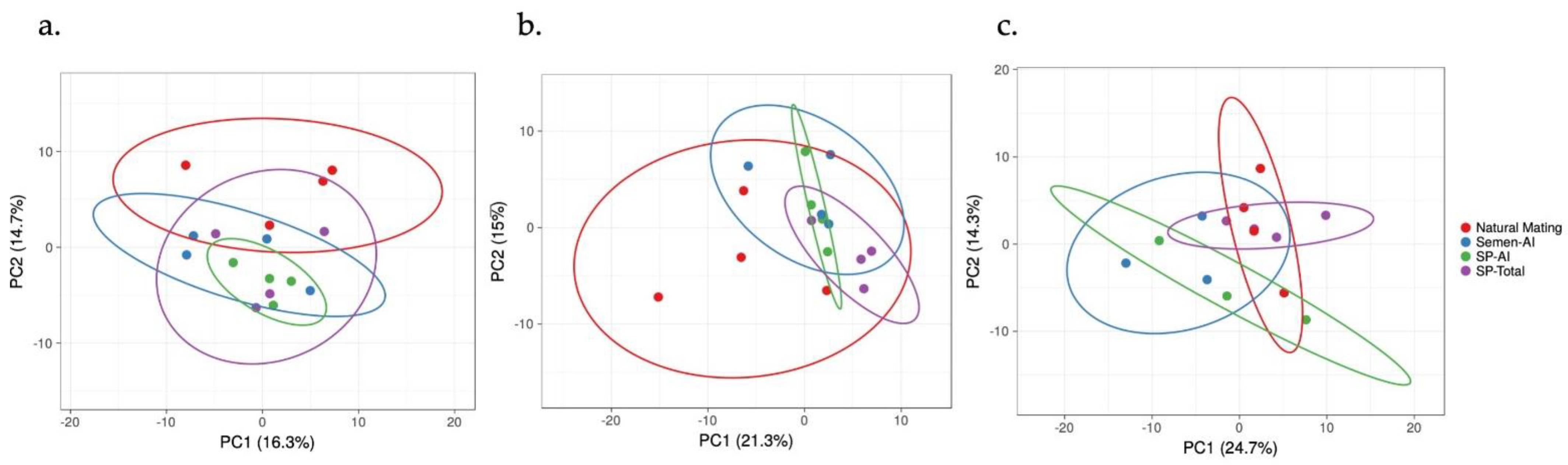
Total RNA from reproductive samples was extracted following a TRIzol modified protocol [38,39]. RNA concentration, integrity evaluation, cDNA synthesis, and microarray analyses (GeneChip® Porcine Gene 1.0 ST Array, Affymetrix Inc., 3420 Central Expressway, Santa Clara, CA, USA) were performed according to methods previously described [38,39]. Only samples with RNA integrity values larger than 9 were employed for microarray hybridization. The GeneChip®Whole Transcript Plus reagent kit (Affymetrix, Santa Clara, CA, USA) was used to synthesize cDNA (250 ng/reaction). An initial incubation of the hybridization cocktail at 99 °C for 5 min was done after a fall to 45 °C before loading the microarray (GeneChip® Porcine Gene 1.0 ST Array, Affymetrix Inc., 3420 Central Expressway, Santa Clara, CA, USA). The cocktail hybridization solution (130 μL) was then put into each microarray and incubated for 16 h at 45 °C under 60 rotations/min. The hybridized microarray chip was unloaded after incubation and washed and stained with the GeneChip® Fluidics Station 450 (Affymetrix, Santa Clara, CA, USA) before being scanned with the Affymetrix GeneChip® Scanner GCS3000 (Affymetrix, Santa Clara, CA, USA).
Data processing was previously described [38,39], by using the Transcriptome Analysis Console (TAC; version 4.0) from Affymetrix. Briefly, the array chip data were processed using robust multi-array average (RMA) normalization, computing average intensity values by background adjustment, quantile normalization among microarrays, and finally, log2 transformation for extracting the expression values of each transcript in the probe set. The normalized mRNA expression data of the 144 selected transcripts were analyzed, for validation, using the open-source R [40]. Differentially mRNA expression was calculated using a linear model and the empirical Bayes’ approach implemented in the package limma [41]. Control type-I errors were checked using a Benjamini–Hochberg false discovery rate (q < 0.05) and a principal component analysis-based p-value correction. Gene Ontology (GO) terms and pathways were analyzed by the PANTHER classification system (protein annotation through evolutionary relationship) [42] based on the Kyoto Encyclopedia of Genes and Genomes database (KEGG) [43]. Graphical illustration of overrepresented GO terms and pathways was produced with the Cytoscape v3.0.0 application ClueGO v2.0.3 [44] and principal component analysis (PCA) and heatmaps were performed by using the ClustVis web tool [45].

3. Results

The PCA analysis of the 144 transcripts depicted an appropriate division of the treatment groups (Figure 1 and Figure S1) and the grouping factor by heatmaps of the chosen genes was conserved across tissues analyzed (Figure S2).
The official names of the genes and the Sus scrofa annotation in the KEGG pathways database [43] was used to find the differentially expressed genes with different biological pathways and GO biological process subgroups (Figure 2).
The differential expression of the 144 selected transcripts showed the changes were in response to the natural mating, mostly downregulated (Figure 3a and Table S1). Moreover, some transcriptomic changes were found in the groups containing the first portion of the sperm-rich fraction (Semen-AI; Figure 4a) and only SP, either from the first fraction of the ejaculate (SP-AI; Figure 5a) or from the entire ejaculate (SP-Total; Figure 6a).

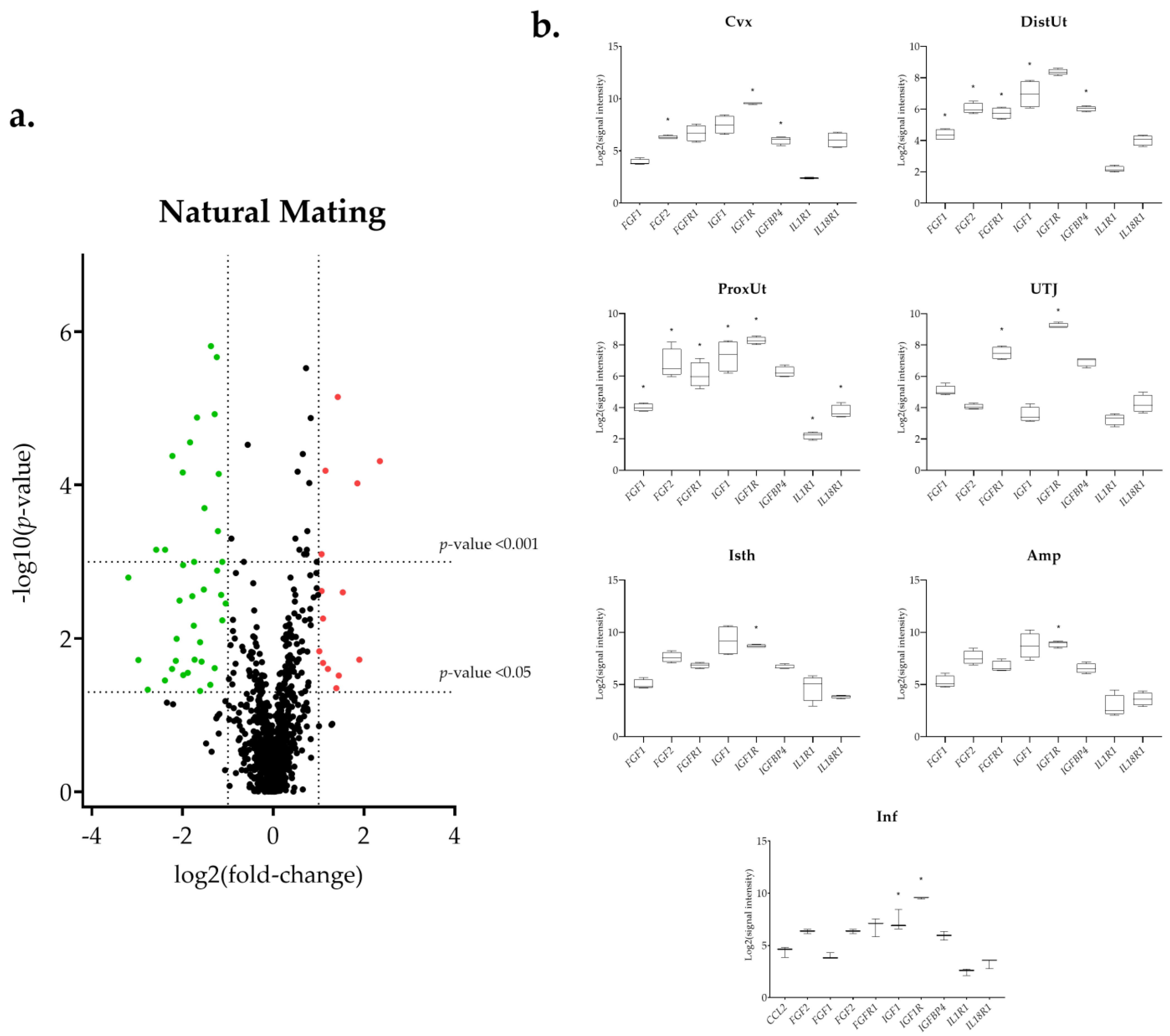
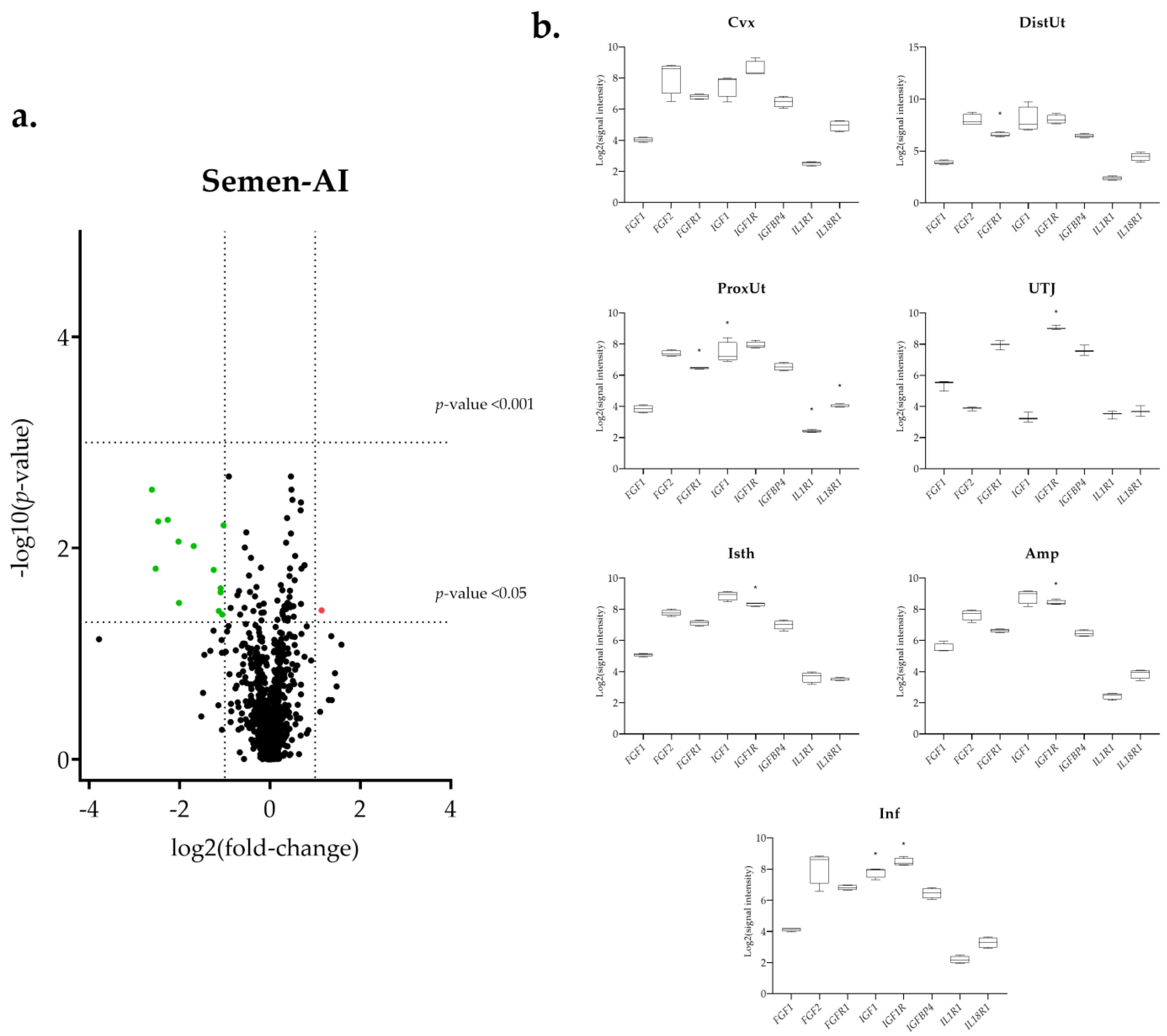
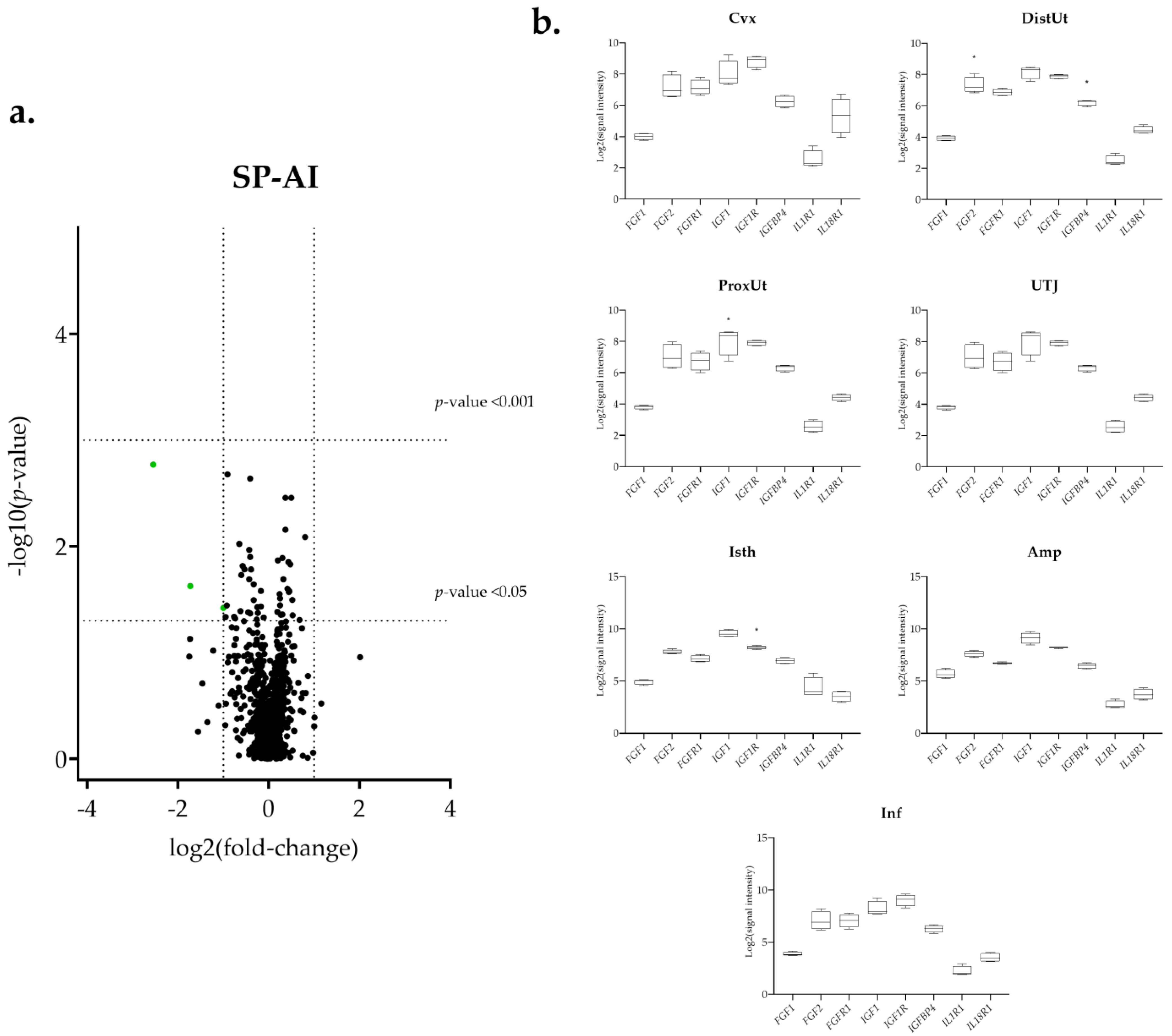
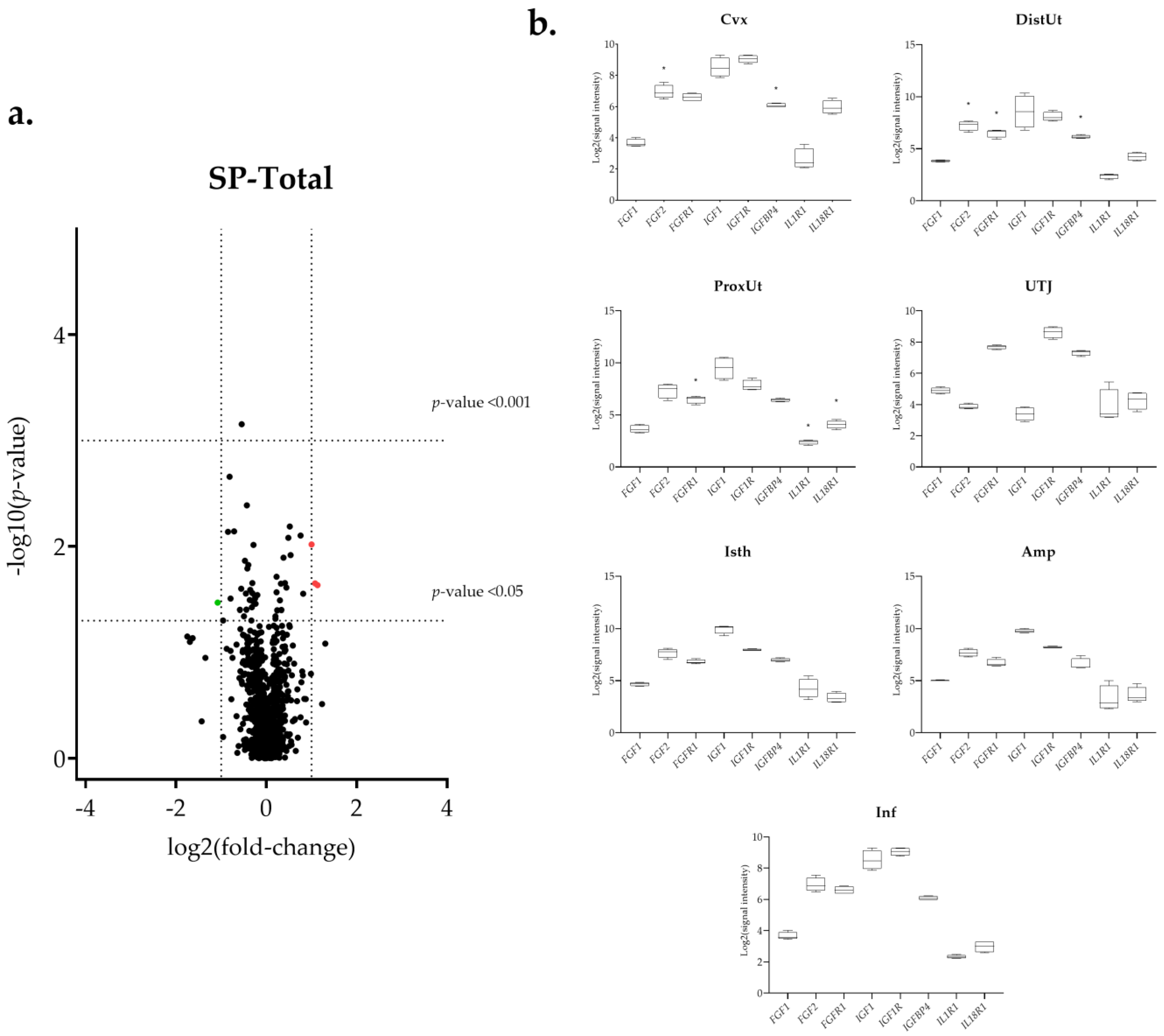
3.1. The IGF1 Transcript Downregulation Was Strongly Marked in Mating Compared to Semen-AI and SP-AI

The insulin-like growth factor 1 (IGF1) presented a common downregulation in the ProxUt for the mating, Semen-AI, and SP-AI groups (Figure 3b, Figure 4b, and Figure 5b). Additionally, it was also downregulated in the DistUt (natural mating group) and Inf (natural mating and Semen-AI groups; Figure 3b and Figure 4b). In contrast, its receptor IGF1R, especially in the natural mating group, was upregulated in all tissues but not in the DistUt (Table S1). The insulin-like growth factor receptor 1 (IGFR1) transcript was also upregulated in the UTJ, Isth, Amp, and Inf in the Semen-AI group, whereas it was only upregulated in Isth in the SP-AI group (Table S1).

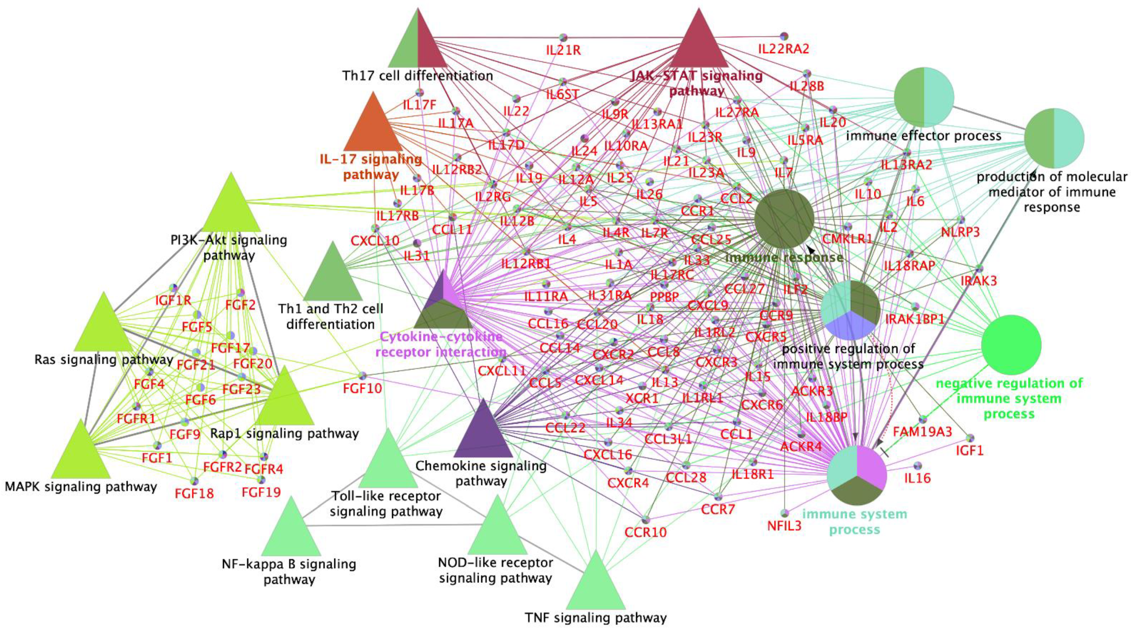
3.2. Interleukin Modulation along the Sow Reproductive Tract in Response to Semen and SP

The atypical chemokine receptor 3 (ACKR3) transcript expression was downregulated in the mating group (ProxUt, DistUt, and Isth, q < 0.05; Cvx and UTJ, p < 0.05), the SP-AI group (DistUt, p < 0,05), and the SP-Total group (Cvx, p < 0.05). The IL-10 transcript (anti-inflammatory) was upregulated in the UTJ, but only for the natural mating group (Figure 3b). The interleukin 1 alpha (IL-1A, pro-inflammatory) was downregulated in the natural mating group (Cvx, DistUt, and ProxUt; Table S1). One of its receptors, IL1RL1, was downregulated in the ProxUt for the natural mating, Semen-AI, and SP-Total groups, whereas the IL1R1 was upregulated, only for the natural mating group, in the UTJ, Isth, and Inf (Table S1). The nuclear factor interleukin 3 regulated (NFIL3, activates transcription from the interleukin-3 promoter in T-cells) was upregulated in the UTJ, and the entire oviduct (Isth, Amp, and Inf) in response to natural mating, whereas it was downregulated in the Cvx, ProxUt, and Inf in SP-Total (Table S1). In contrast to the downregulation of CXCL8 (pro-inflammatory) in the Cvx and ProxUt (natural mating group) and UTJ in Semen-AI, the receptor IL17RB, of its main inductor (IL17E), was upregulated in the DistUt, UTJ, Isth, Amp, and Inf (natural mating group) and only in the Isth (Semen-AI group) (Table S1). Interleukin 34 (IL-34, pro-inflammatory) was upregulated in the natural mating group in the DistUt, ProxUt, Amp, and Inf (Table S1). Interleukin 6 (IL-6, showing both pro- and anti-inflammatory actions) was downregulated in the Amp (natural mating and Semen-AI groups; Figure 3b and Figure 4b). IL-6 receptor alpha-like was downregulated, only for the mating group in the ProxUt, Amp, and Inf (Table S1). Interleukin 19 (IL-19, anti-inflammatory) was downregulated in the Cvx and DistUt for the natural mating group (Figure 3b), and in the DistUt (SP-Total group; Figure 6b), whereas it was upregulated in the Isth in both sperm-free groups (SP-AI and SP-Total groups; Figure 5b and Figure 6b).

3.3. Sperm-Free Treatments and Mating Triggered a Common Downregulation of FGF2 Transcript in the Distal Uterus

The FGF2 was downregulated in the DistUt of the mating, SP-AI, and SP-Total groups, and also in the cervix (Cvx) of the natural mating and SP-Total groups (Figure 3b, Figure 5b and Figure 6b). Additionally, the natural mating group also triggered downregulation in the ProxUt (Figure 3b). Its specific receptor, the fibroblast growth factor receptor 1 (FGFR1), was downregulated in the DistUt and ProxUt of the natural mating, Semen-AI, and SP-Total groups (Table S1). Moreover, it was also downregulated in the UTJ, but only for the natural mating group.

4. Discussion

Semen and SP can trigger significant gene expression changes in the female reproductive tract [8]. In the present study, we have explored the modulation of 144 mRNA transcripts related to cell proliferation and differentiation in the sow reproductive tract in triggered by semen and sperm-free SP. For this purpose, we analyzed the changes in response to the four experimental groups included in the study: natural mating (i), artificial insemination of the sperm rich fraction (ii), infusion of sperm-free SP of the sperm rich fraction (iii), and infusion of sperm-free SP of the entire ejaculate (iv), using commercial microarrays to assess gene expression [46]. Overall, the natural mating group induced most of the transcriptomic changes. However, some key differentially expressed transcripts were shared between experimental groups, suggesting a common underlying physiological mechanism at work across these treatments. It might be related, at least in part, to the signaling agents present in the SP that have been pointed out to promote molecular and cellular changes into the female reproductive tract of several species [8,47]. Our results confirm changes in the expression of cell proliferation and differentiation-related transcripts in the female reproductive tract in response to both sperm and SP. However, independent verification of such selected transcripts should be performed, especially for markers that have a less than 2-fold difference expression.
Several chemokines are produced throughout the estrous cycle and pregnancy, playing essential roles in regulating endometrial functions for estrous cyclicity and establishing and maintaining pregnancy [48,49,50]. The atypical chemokine receptors (ACKRs) are similar to traditional chemokine receptors but regulate chemokine activity by limiting their spatial availability or removing them from in vivo sites [51]. Because of their structural inability to couple to G proteins, ACKRs bind chemokines with high affinity but do not trigger cell migration [52]. Expression regulation of ACKR1, ACKR2, ACKR3, and ACKR4 transcripts has been demonstrated in the endometrium of pigs during the estrous cycle [53], maternal-fetal interface [54,55], and pregnancy [53]. Our results confirm that this modulation is also present during the male-female interaction along the female reproductive tract, suggesting that the ACKRs may play essential roles in regulating chemokine action in the endometrium and oviduct during the male-female interaction.
ACKR3 binds to CXCL12, the ligand of CXCR4, and CXCL11, one of the ligands of CXCR3 [52]. Our results show that the expression of ACKR3 mRNA was lower in the cervix, endometrium, utero-tubal junction, and isthmus after mating. Additionally, the expression of ACKR3 mRNA was lower in the distal uterus and cervix after infusion of sperm-free SP of the sperm rich fraction and the infusion of sperm-free SP of the entire ejaculate, respectively. The in vitro inhibition of ACKR3 reduces M2 macrophages receptor expression on monocytes, suggesting that ACKR3 may play a role in fine-tuning immunological tolerance at the fetal-maternal interface [56]. Moreover, because ACKR3 acts as a decoy receptor for CXCL11 and CXCL12 [52], the decrease in ACKR3 mRNA expression after mating is likely to allow CXCL11 and CXCL12 to perform their functions of immune cell recruitment during the post-mating inflammatory response. However, CXCL11 mRNA expression was lower in the cervix, proximal uterus, and ampulla after mating, and no changes in CXCL12 mRNA expression were reported.
CXCR4 is the most regularly expressed chemokine receptor and is involved in various physiological and pathological situations involving cell movement and cell proliferation [57]. ACKR3 modulates the CXCR4 expression and signaling activity by forming heterodimers with CXCR4, producing conformational rearrangements in G-protein complexes and partially to β-arrestin rather than classical G-protein coupled receptor signaling in response to CXCL12 binding [58]. After mating, we reported lower ACKR3 mRNA expression along the female reproductive tract, whereas CXCR4 mRNA expression was higher. This dual modulation of both receptors may regulate an appropriate response after mating. However, the factors regulating ACKR3 and CXCR4 in the female reproductive tract during the male-female interaction still need to be determined. Although more research is needed to understand the precise functions of the ACKRs and their ligands during the male-female interaction, our findings suggest that the expression and regulation of these chemokine receptors may be necessary in regulating endometrial and oviductal functions for the post-mating inflammatory response.
The IGFs play crucial roles in tissue differentiation, cell survival, proliferation [26], and myometrial steroidogenic activity [59]. The main focus on pig reproduction research has been on the IGFs expression in the uterus [33,34,35,59,60,61], although some research focused on oviductal IGFs expression [62,63]. Our data showed a consistent downregulation of the IGF1 expression in the proximal uterus in response to mating, the artificial insemination of the sperm-rich fraction, and the infusion of seminal plasma from the sperm-peak portion. In contrast, upregulation of IGF1R transcript in oviductal tissues in the mating and artificial insemination of the sperm-rich fraction was found. Previous research confirmed that IGF1 contributes to increase the intrauterine content of estrogen during the peri-implantation period, and the relative abundance of IGF-1R mRNA transcript and protein is altered in the porcine endometrium depending on the reproductive status of the sow [59]. Our results suggest an implication of the IGF system not only during the peri-implantation period but also in the peri-ovulatory stage. Additionally, these changes are present in the myometrium and the oviduct, and it seems that the sperm-containing treatments trigger the changes observed in the oviduct.
In conjunction with other growth factor systems, the FGF system appears to be involved in a paracrine network to effectively establish and maintain pregnancy in the pig [64]. Two of the most studied members of the FGF family (FGF1 and FGF2), and their receptors (FGFR1IIIc and FGFR2IIIc) are constitutively expressed in the porcine uterus and oviduct [64,65,66]. Our study detected downregulation of FGF2 expression in the uterus after mating and sperm-free SP treatments during the peri-ovulatory stage. In the same way, we observed a downregulation of FGFR1 expression in sperm-containing treatments and sperm-free SP of the whole ejaculate. Otherwise, sperm-containing treatments induced upregulation of FGFR2 and the isoform FGFR2IIIc expression in the uterus and oviduct during the peri-ovulatory stage. In addition, we observed an upregulation of FGF1 expression in the endometrium induced by mating. FGF receptors play different biological importance for a functional endometrium and are promoted differently by estradiol (FGFR1IIIc) and progesterone (FGFR2IIIc) [66]. The luteal phase of the estrous cycle also changes the expression of the FGF ligands in the bovine endometrium, and this modulation is linked with the expression of their receptors in the embryo [67]. Additionally, previous research demonstrated that SP upregulates the expression of FGF2 in the porcine oviduct [68]. Our results suggest that FGF1, FGFR2, and FGFR2IIIc isoform play more essential roles in the pre-/peri-ovulatory stage of the porcine endometrium and endosalpinx compared to FGFR1 and FGF2, which seems required in early stages of pregnancy [65].
FGF2 has been described as an adipokine that may exacerbate the inflammatory response in adipocytes through NLRP3 inflammasome activation [69]. NLRP3 is a critical component of the inflammasome activated by microbial pathogens and endogenous molecules [70]. The activation of NLRP3 requires extracellular inflammatory stimuli, which induces the transcription of NLRP3 and controls post-translational modifications that license receptor activation [71]. Once activated, the inflammasome leads to an induction of pro-IL-1β, and activation of caspase-1, promoting the secretion of the pro-inflammatory cytokines IL-1β and IL-18 [72]. The wide variety of stimuli activating NLRP3 inflammasome [70,71] and the immune regulatory modulation produced in the female reproductive tract after entry of seminal components [13] could be related to our results that showed an upregulation of the NLRP3 transcript in the distal uterus after mating. However, the expression of the IL-18 transcript was downregulated in the same tissue fragment, and no modulation of the IL-1β transcript was found. Additionally, previous results demonstrated a downregulation of the caspase-1 transcript in the endometrium of sows after 24 h of mating [73]. In addition, while the NLRP3 transcript between the mating and the sperm-free SP of the entire ejaculate infusion was upregulated, it was not present in the infusion of sperm-free SP-rich fraction group. Thus, our results suggest that the SP components from the entire ejaculate, not only from the rich sperm fraction, are responsible for the NLRP3 transcript modulation, similarly to the common upregulation of the NLRP3 transcript that was present in the previously described sperm reservoir of the sow reproductive tract, the utero-tubal junction [74,75].
Even though more experiments are required, is it therefore likely that NLRP3 inflammasome, including FGFs and IGFs-related transcripts, may be involved in sperm selection, facilitate tissue remodeling, or elimination of damaged endometrial cells after mating. An inflammatory response is induced in the uterus after mating in response to semen; however, this response facilitates the efficient removal of spermatozoa and induces transient immune tolerance to seminal antigens because of components present in the SP [76]. Additionally, it has been suggested that the post-mating inflammation and the immunological events that follow the beginning of inflammation have a role in establishing immune tolerance to embryonic antigens and subsequent embryonic development in domestic animals [76]. Similarly, the inflammasomes are essential in the maternal-fetal interface remodeling of endometrium during the early stage of implantation and are a defense against pathogen infections [77]. However, when overly activated, they can be highly pathological, inducing several reproductive diseases [77]. Overall, our results suggest a hypothetical involvement of cell proliferation and remodeling genes in the physiological interaction between the female reproductive tract and sperm, including the seminal fluid.

5. Conclusions

In conclusion, we have demonstrated that the seminal fluid, including the SP alone, triggers differential mRNA transcript expression in proliferation and differentiation-related pathways in the sow reproductive tract. Our data suggested that as early as the periovulatory stage, relevant factors present in sperm and/or SP produce changes in mRNA expression in the female genital tract, including the FGFs and the IGFs (and their specific receptors) as well as some interleukin-immune related genes, such as the interaction between IL-18 and NLRP3, and the ACKR3 and CXCR4. However, further studies are required to precisely characterize the mechanism by which the sperm (and/or SP) affects the transcript expression and whether this knowledge may enhance the fertility performance in assisted reproductive techniques.

Supplementary Materials

The following supporting information can be downloaded at: https://www.mdpi.com/article/10.3390/biology11040616/s1, Table S1: Fold change of the differential mRNA expression (p < 0.05, red: False Discovery Rate, q < 0.05) in the different segments of the sow reproductive tract (cervix to infundibulum), 24 h post-treatment. Figure S1 Principal Component Analysis of the genes included in this study of natural mating (red); Semen-AI (blue); SP-AI (green); and SP-Total (purple) treatment groups. a. Cvx: cervix; b. Isth: isthmus; c. Amp: ampulla; and d. Inf: infundibulum. Figure S2. Heatmap of the genes included in this study of natural mating (red); Semen-AI (blue); SP-AI (green); and SP-Total (purple) treatment groups. a. Cvx: cervix; b. DistUt: distal uterus; c. ProxUt: proximal uterus; d. UTJ: utero-tubal junction; e. Isth: isthmus; f. Amp: ampulla; and g. Inf: infundibulum.

Author Contributions

Conceptualization, M.Á.-R., H.R.-M. and D.W.; methodology, J.G., M.R.-C., C.A.M., M.Á.-R. and H.R.-M.; software, J.G., M.R.-C., C.A.M. and M.Á.-R.; validation, J.G., M.R.-C. and M.Á.-R.; formal analysis, J.G., M.R.-C. and M.Á.-R.; investigation, J.G., M.R.-C., C.A.M. and M.Á.-R.; resources, H.R.-M. and M.Á.-R.; data curation, J.G. and M.Á.-R.; writing—original draft preparation, J.G.; writing—review and editing, M.R.-C., M.Á.-R., H.R.-M., D.W. and M.L.-B.; visualization, J.G., M.R.-C. and C.A.M.; supervision, M.Á.-R., H.R.-M. and M.L.-B.; project administration, M.Á.-R. and H.R.-M.; funding acquisition, H.R.-M. and M.Á.-R. All authors have read and agreed to the published version of the manuscript.

Funding

This research was funded by the Research Council FORMAS, Stockholm (Project 2017-00946 and Project 2019-00288), and by the Grant PID2019-108320RJ-I00 and IJCI-2015-24380 funded by MCIN/AEI /10.13039/501100011033 (Spain) and FEDER funds (EU), the Government of Catalonia-AGAUR and the European Social Fund (2018 FI_B 00236), and the Government of Spain-Ministry of Education, Culture and Sports (Training for Academic Staff FPU15/06029).

Institutional Review Board Statement

Animal handling and experiments were carried out according to the European Community Directive 2010/63/EU, 22 September 2010, and Swedish legislation (SJVFS 2017:40). The study was accepted by the Regional Committee for Ethical Approval of Animal Experiments (Linköpings Djurförsöksetiska nämnd, Linköping, Sweden). Permits number 75-12 (10 February 2012), ID1400 (2 February 2018), and Dnr 03416-2020 (26 March 2020).

Informed Consent Statement

Not applicable.

Data Availability Statement

Not applicable.

Acknowledgments

The authors of this manuscript thank Annette Molbaek and Åsa Schippert, from the Genomics Core Facility at LiU for expert assistance with transcriptomic analysis. To QualiSperm (AKYmed, Cheseaux-sur-Lausanne, Switzerland) for their support with the computerized sperm motility and kinetics assessment.

Conflicts of Interest

The authors declare no conflict of interest. The funders had no role in the design of the study; in the collection, analyses, or interpretation of data; in the writing of the manuscript, or in the decision to publish the results.

References

  1. Cui, W. Mother or nothing: The agony of infertility. Bull. World Health Organ. 2010, 88, 881–882. [Google Scholar] [CrossRef] [PubMed]
  2. Perleberg, C.; Kind, A.; Schnieke, A. Genetically engineered pigs as models for human disease. DMM Dis. Model. Mech. 2018, 11, dmm030783. [Google Scholar] [CrossRef] [PubMed] [Green Version]
  3. Mordhorst, B.R.; Prather, R.S. Pig Models of Reproduction. In Animal Models and Human Reproduction; John Wiley & Sons, Inc.: Hoboken, NJ, USA, 2017; pp. 213–234. [Google Scholar]
  4. Abedal-Majed, M.A.; Cupp, A.S. Livestock animals to study infertility in women. Anim. Front. 2019, 9, 28–33. [Google Scholar] [CrossRef] [PubMed]
  5. Dahm-Kähler, P.; Ghahremani, M.; Lind, A.K.; Sundfeldt, K.; Brännström, M. Monocyte chemotactic protein-1 (MCP-1), its receptor, and macrophages in the perifollicular stroma during the human ovulatory process. Fertil. Steril. 2009, 91, 231–239. [Google Scholar] [CrossRef] [PubMed]
  6. Cicchese, J.M.; Evans, S.; Hult, C.; Joslyn, L.R.; Wessler, T.; Millar, J.A.; Marino, S.; Cilfone, N.A.; Mattila, J.T.; Linderman, J.J.; et al. Dynamic balance of pro- and anti-inflammatory signals controls disease and limits pathology. Immunol. Rev. 2018, 285, 147–167. [Google Scholar] [CrossRef]
  7. Jabbour, H.N.; Sales, K.J.; Catalano, R.D.; Norman, J.E. Inflammatory pathways in female reproductive health and disease. Reproduction 2009, 138, 903–919. [Google Scholar] [CrossRef]
  8. Schjenken, J.E.; Robertson, S.A. The female response to seminal fluid. Physiol. Rev. 2020. [Google Scholar] [CrossRef]
  9. Bromfield, J.J. Seminal fluid and reproduction: Much more than previously thought. J. Assist. Reprod. Genet. 2014, 31, 627–636. [Google Scholar] [CrossRef] [Green Version]
  10. Song, Z.H.; Li, Z.Y.; Li, D.D.; Fang, W.N.; Liu, H.Y.; Yang, D.D.; Meng, C.Y.; Yang, Y.; Peng, J.P. Seminal plasma induces inflammation in the uterus through the γδT/IL-17 pathway. Sci. Rep. 2016, 6, 1–8. [Google Scholar] [CrossRef] [Green Version]
  11. Ibrahim, L.A.; Rizo, J.A.; Fontes, P.L.P.; Lamb, G.C.; Bromfield, J.J. Seminal plasma modulates expression of endometrial inflammatory meditators in the bovine. Biol. Reprod. 2019, 100, 660–671. [Google Scholar] [CrossRef]
  12. Fedorka, C.E.; Scoggin, K.E.; Woodward, E.M.; Squires, E.L.; Ball, B.A.; Troedsson, M.H.T. The effect of select seminal plasma proteins on endometrial mRNA cytokine expression in mares susceptible to persistent mating-induced endometritis. Reprod. Domest. Anim. 2017, 52, 89–96. [Google Scholar] [CrossRef] [PubMed]
  13. Alvarez-Rodriguez, M.; Atikuzzaman, M.; Venhoranta, H.; Wright, D.; Rodriguez-Martinez, H. Expression of Immune Regulatory Genes in the Porcine Internal Genital Tract Is Differentially Triggered by Spermatozoa and Seminal Plasma. Int. J. Mol. Sci. 2019, 20, 513. [Google Scholar] [CrossRef] [PubMed] [Green Version]
  14. Gardela, J.; Jauregi-Miguel, A.; Martinez, C.A.; Rodriguez-Martinez, H.; Lopez-Bejar, M.; Alvarez-Rodriguez, M. Semen Modulates Inflammation and Angiogenesis in the Reproductive Tract of Female Rabbits. Animals 2020, 10, 2207. [Google Scholar] [CrossRef]
  15. Rodriguez-Martinez, H.; Nicander, L.; Viring, S.; Einarsson, S.; Larsson, K. Ultrastructure of the Uterotubal Junction in Preovulatory Pigs. Anat. Histol. Embryol. 1990, 19, 16–36. [Google Scholar] [CrossRef]
  16. Sternberg, A.K.; Buck, V.U.; Classen-Linke, I.; Leube, R.E. How mechanical forces change the human endometrium during the menstrual cycle in preparation for embryo implantation. Cells 2021, 10, 2008. [Google Scholar] [CrossRef] [PubMed]
  17. Krzymowski, T.; Stefańczyk-Krzymowska, S. The oestrous cycle and early pregnancy—A new concept of local endocrine regulation. Vet. J. 2004, 168, 285–296. [Google Scholar] [CrossRef]
  18. Bazer, F.W.; Johnson, G.A. Pig blastocyst-uterine interactions. Differentiation 2014, 87, 52–65. [Google Scholar] [CrossRef]
  19. Jung, W.; Yoo, I.; Han, J.; Kim, M.; Lee, S.; Cheon, Y.; Hong, M.; Jeon, B.Y.; Ka, H. Expression of Caspases in the Pig Endometrium Throughout the Estrous Cycle and at the Maternal-Conceptus Interface During Pregnancy and Regulation by Steroid Hormones and Cytokines. Front. Vet. Sci. 2021, 8, 1–13. [Google Scholar] [CrossRef]
  20. Guo, H.; Callaway, J.B.; Ting, J.P.Y. Inflammasomes: Mechanism of action, role in disease, and therapeutics. Nat. Med. 2015, 21, 677–687. [Google Scholar] [CrossRef] [Green Version]
  21. Zheng, D.; Liwinski, T.; Elinav, E. Inflammasome activation and regulation: Toward a better understanding of complex mechanisms. Cell Discov. 2020, 6, 1–22. [Google Scholar] [CrossRef]
  22. Kelley, N.; Jeltema, D.; Duan, Y.; He, Y. The NLRP3 inflammasome: An overview of mechanisms of activation and regulation. Int. J. Mol. Sci. 2019, 20, 3328. [Google Scholar] [CrossRef] [Green Version]
  23. Wang, L.; Hauenstein, A.V. The NLRP3 inflammasome: Mechanism of action, role in disease and therapies. Mol. Aspects Med. 2020, 76, 100889. [Google Scholar] [CrossRef] [PubMed]
  24. Yang, Y.; Wang, H.; Kouadir, M.; Song, H.; Shi, F. Recent advances in the mechanisms of NLRP3 inflammasome activation and its inhibitors. Cell Death Dis. 2019, 10, 1–11. [Google Scholar] [CrossRef] [PubMed] [Green Version]
  25. de Rivero Vaccari, J.P. The Inflammasome in Reproductive Biology: A Promising Target for Novel Therapies. Front. Endocrinol. 2020, 11, 8. [Google Scholar] [CrossRef] [PubMed] [Green Version]
  26. Stewart, C.E.H.; Rotwein, P. Growth, differentiation, and survival: Multiple physiological functions for insulin-like growth factors. Physiol. Rev. 1996, 76, 1005–1026. [Google Scholar] [CrossRef]
  27. Duan, C.; Xu, Q. Roles of insulin-like growth factor (IGF) binding proteins in regulating IGF actions. Gen. Comp. Endocrinol. 2005, 142, 44–52. [Google Scholar] [CrossRef]
  28. Bach, L.A. 40 years of IGF1: IGF-binding proteins. J. Mol. Endocrinol. 2018, 61, T11–T28. [Google Scholar] [CrossRef] [Green Version]
  29. Watson, A.J.; Westhusin, M.E.; Winger, Q.A. IGF paracrine and autocrine interactions between conceptus and oviduct. J. Reprod. Fertil. Suppl. 1999, 54, 303–315. [Google Scholar] [CrossRef]
  30. Tang, X.-M.; Rossi, M.J.; Masterson, B.J.; Chegini, N. Insulin-Like Growth Factor I (IGF-I), IGF-I Receptors, and IGF Binding Proteins 1–4 in Human Uterine Tissue: Tissue Localization and IGF-I Action in Endometrial Stromal and Myometrial Smooth Muscle Cells in Vitro. Biol. Reprod. 1994, 50, 1113–1125. [Google Scholar] [CrossRef] [Green Version]
  31. Cerro, J.A.; Pintar, J.E. Insulin-like Growth Factor Binding Protein Gene Expression in the Pregnant Rat Uterus and Placenta. Dev. Biol. 1997, 184, 278–295. [Google Scholar] [CrossRef] [Green Version]
  32. Wathes, D.C.; Reynolds, T.S.; Robinson, R.S.; Stevenson, K.R. Role of the Insulin-Like Growth Factor System in Uterine Function and Placental Development in Ruminants. J. Dairy Sci. 1998, 81, 1778–1789. [Google Scholar] [CrossRef]
  33. Hofig, A.; Michel, F.; Simmen, F.A.; Simmen, R.C.M. Constitutive expression of uterine receptors for isulin-like growth factor-I during the peri-implantation period in the pig. Biol. Reprod. 1991, 45, 533–539. [Google Scholar] [CrossRef] [PubMed]
  34. Simmen, F.A.; Simmen, R.C.M.; Geisert, R.D.; Martinat-Botte, F.; Bazer, F.W.; Terqui, M. Differential expression, during the estrous cycle and pre- and postimplantation conceptus development, of messenger ribonucleic acids encoding components of the pig uterine insulin-like growth factor system. Endocrinology 1992, 130, 1547–1556. [Google Scholar] [CrossRef] [PubMed] [Green Version]
  35. Persson, E.; Sahlin, L.; Masironi, B.; Dantzer, V.; Eriksson, H.; Rodriguez-Martinez, H. Insulin-like growth factor-I in the porcine endometrium and placenta: Localization and concentration in relation to steroid influence during early pregnancy. Anim. Reprod. Sci. 1997, 46, 261–281. [Google Scholar] [CrossRef]
  36. Ornitz, D.M.; Itoh, N. Fibroblast growth factors. Genome Biol. 2001, 2, 1–12. [Google Scholar] [CrossRef] [Green Version]
  37. Itoh, N.; Ornitz, D.M. Evolution of the Fgf and Fgfr gene families. Trends Genet. 2004, 20, 563–569. [Google Scholar] [CrossRef]
  38. Gardela, J.; Ruiz-Conca, M.; Martinez, C.A.; Wright, D.; López-Béjar, M.; Rodriguez-Martinez, H.; Alvarez-Rodriguez, M. The Expression of Cold-Inducible RNA-Binding Protein mRNA in Sow Genital Tract Is Modulated by Natural Mating, But Not by Seminal Plasma. Int. J. Mol. Sci. 2020, 21, 5333. [Google Scholar] [CrossRef]
  39. Ruiz-Conca, M.; Gardela, J.; Martínez, C.A.; Wright, D.; López-Bejar, M.; Rodríguez-Martínez, H.; Álvarez-Rodríguez, M. Natural Mating Differentially Triggers Expression of Glucocorticoid Receptor (NR3C1)-Related Genes in the Preovulatory Porcine Female Reproductive Tract. Int. J. Mol. Sci. 2020, 21, 4437. [Google Scholar] [CrossRef]
  40. R Core Team. R: A Language and Environment for Statistical Computing. R Foundation for Statistical Computing. Available online: https://www.r-project.org/ (accessed on 2 March 2022).
  41. Phipson, B.; Lee, S.; Majewski, I.J.; Alexander, W.S.; Smyth, G.K. Robust hyperparameter estimation protects against hypervariable genes and improves power to detect differential expression. Ann. Appl. Stat. 2016, 10, 946–963. [Google Scholar] [CrossRef]
  42. Mi, H.; Muruganujan, A.; Casagrande, J.T.; Thomas, P.D. Large-scale gene function analysis with the panther classification system. Nat. Protoc. 2013, 8, 1551–1566. [Google Scholar] [CrossRef]
  43. Kanehisa, M.; Goto, S. KEGG: Kyoto Encyclopedia of Genes and Genomes. Nucleic Acids Res. 2000, 28, 27–30. [Google Scholar] [CrossRef] [PubMed]
  44. Bindea, G.; Mlecnik, B.; Hackl, H.; Charoentong, P.; Tosolini, M.; Kirilovsky, A.; Fridman, W.H.; Pagès, F.; Trajanoski, Z.; Galon, J. ClueGO: A Cytoscape plug-in to decipher functionally grouped gene ontology and pathway annotation networks. Bioinformatics 2009, 25, 1091–1093. [Google Scholar] [CrossRef] [PubMed] [Green Version]
  45. Metsalu, T.; Vilo, J. ClustVis: A web tool for visualizing clustering of multivariate data using Principal Component Analysis and heatmap. Nucleic Acids Res. 2015, 43, W566–W570. [Google Scholar] [CrossRef] [PubMed]
  46. Mantione, K.J.; Kream, R.M.; Kuzelova, H.; Ptacek, R.; Raboch, J.; Samuel, J.M.; Stefano, G.B. Comparing bioinformatic gene expression profiling methods: Microarray and RNA-Seq. Med. Sci. Monit. Basic Res. 2014, 20, 138–142. [Google Scholar] [CrossRef] [PubMed] [Green Version]
  47. Morgan, H.L.; Watkins, A.J. The influence of seminal plasma on offspring development and health. Semin. Cell Dev. Biol. 2020, 97, 131–137. [Google Scholar] [CrossRef] [PubMed]
  48. Hannan, N.J.; Salamonsen, L.A. Role of chemokines in the endometrium and in embryo implantation. Curr. Opin. Obstet. Gynecol. 2007, 19, 266–272. [Google Scholar] [CrossRef]
  49. Du, M.R.; Wang, S.C.; Li, D.J. The integrative roles of chemokines at the maternal-fetal interface in early pregnancy. Cell. Mol. Immunol. 2014, 11, 438–448. [Google Scholar] [CrossRef]
  50. Choi, Y.; Seo, H.; Han, J.; Yoo, I.; Kim, J.; Ka, H. Chemokine (C-C Motif) ligand 28 and its receptor CCR10: Expression and function at the maternal-conceptus interface in pigs. Biol. Reprod. 2016, 95, 1–10. [Google Scholar] [CrossRef]
  51. Nibbs, R.J.B.; Graham, G.J. Immune regulation by atypical chemokine receptors. Nat. Rev. Immunol. 2013, 13, 815–829. [Google Scholar] [CrossRef]
  52. Bonecchi, R.; Graham, G.J. Atypical chemokine receptors and their roles in the resolution of the inflammatory response. Front. Immunol. 2016, 7, 1–7. [Google Scholar] [CrossRef] [Green Version]
  53. Han, J.; Yoo, I.; Lee, S.; Jung, W.; Kim, H.J.; Hyun, S.-H.; Lee, E.; Ka, H. Atypical chemokine receptors 1, 2, 3 and 4: Expression and regulation in the endometrium during the estrous cycle and pregnancy and with somatic cell nucleus transfer–cloned embryos in pigs. Theriogenology 2019, 129, 121–129. [Google Scholar] [CrossRef] [PubMed]
  54. Wessels, J.M.; Linton, N.F.; Van Den Heuvel, M.J.; Cnossen, S.A.; Edwards, A.K.; Croy, B.A.; Tayade, C. Expression of chemokine decoy receptors and their ligands at the porcine maternal-fetal interface. Immunol. Cell Biol. 2011, 89, 304–313. [Google Scholar] [CrossRef] [PubMed]
  55. Lim, W.; Bae, H.; Bazer, F.W.; Song, G. Cell-Specific Expression and Signal Transduction of C-C Motif Chemokine Ligand 2 and Atypical Chemokine Receptors in the Porcine Endometrium during Early Pregnancy; Elsevier Ltd.: Amsterdam, The Netherlands, 2018; Volume 81, ISBN 8223290499. [Google Scholar]
  56. Infantino, S.; Moepps, B.; Thelen, M. Expression and Regulation of the Orphan Receptor RDC1 and Its Putative Ligand in Human Dendritic and B Cells. J. Immunol. 2006, 176, 2197–2207. [Google Scholar] [CrossRef] [PubMed] [Green Version]
  57. Bianchi, M.E.; Mezzapelle, R. The Chemokine Receptor CXCR4 in Cell Proliferation and Tissue Regeneration. Front. Immunol. 2020, 11, 1–8. [Google Scholar] [CrossRef] [PubMed]
  58. Quinn, K.E.; Mackie, D.I.; Caron, K.M. Emerging roles of atypical chemokine receptor 3 (ACKR3) in normal development and physiology. Cytokine 2018, 109, 17–23. [Google Scholar] [CrossRef]
  59. Waszkiewicz, E.M.; Kozlowska, W.; Zmijewska, A.; Franczak, A. Expression of insulin-like growth factor 1 (IGF-1) and epidermal growth factor (EGF) receptors and the effect of IGF-1 and EGF on androgen and estrogen release in the myometrium of pigs—In vitro study. Animals 2020, 10, 915. [Google Scholar] [CrossRef]
  60. Sahlin, L.; Rodriguez-Martinez, H.; Stanchev, P.; Dalin, A.; Norstedt, G.; Eriksson, H. Regulation of the Uterine Expression of Messenger Ribonucleic Acids Encoding the Oestrogen Receptor and IGF–I Peptides in the Pig Uterus. J. Vet. Med. Ser. A 1990, 37, 795–800. [Google Scholar] [CrossRef]
  61. Simmen, R.C.M.; Simmen, F.A.; Hofig, A.; Farmer, S.J.; Bazer, F.W. Hormonal regulation of insulin-like growth factor gene expression in pig uterus. Endocrinology 1990, 127, 2166–2174. [Google Scholar] [CrossRef]
  62. Wiseman, D.L.; Henricks, D.M.; Eberhardt, D.M.; Bridges, W.C. Identification and Content of Insulin-like Growth Factors in Porcine Oviductal Fluid. Biol. Reprod. 1992, 47, 126–132. [Google Scholar] [CrossRef]
  63. Novak, S.; Treacy, B.K.; Almeida, F.R.C.L.; Mao, J.; Buhi, W.C.; Dixon, W.T.; Foxcroft, G.R. Regulation of IGF-I and porcine oviductal secretory protein (pOSP) secretion into the pig oviduct in the peri-ovulatory period, and effects of previous nutrition. Reprod. Nutr. Dev. 2002, 42, 355–372. [Google Scholar] [CrossRef] [Green Version]
  64. Wollenhaupt, K.; Welter, H.; Einspanier, R.; Manabe, N.; Brüssow, K.P. Expression of epidermal growth factor receptor (EGF-R), vascular endothelial growth factor receptor (VEGF-R) and fibroblast growth factor receptor (FGF-R) systems in porcine oviduct and endometrium during the time of implantation. J. Reprod. Dev. 2004, 50, 269–278. [Google Scholar] [CrossRef] [PubMed] [Green Version]
  65. Katsahambas, S.; Hearn, M.T.W. Localization of basic fibroblast growth factor mRNA (FGF-2 mRNA) in the uterus of mated and unmated gilts. J. Histochem. Cytochem. 1996, 44, 1289–1301. [Google Scholar] [CrossRef] [PubMed] [Green Version]
  66. Welter, H.; Wollenhaupt, K.; Einspanier, R. Developmental and hormonal regulated gene expression of fibroblast growth factor 2 (FGF-2) and its receptors in porcine endometrium. J. Steroid Biochem. Mol. Biol. 2004, 88, 295–304. [Google Scholar] [CrossRef] [PubMed]
  67. Okumu, L.A.; Forde, N.; Mamo, S.; McGettigan, P.; Mehta, J.P.; Roche, J.F.; Lonergan, P. Temporal regulation of fibroblast growth factors and their receptors in the endometrium and conceptus during the pre-implantation period of pregnancy in cattle. Reproduction 2014, 147, 825–834. [Google Scholar] [CrossRef] [Green Version]
  68. Krawczynski, K.; Kaczmarek, M.M. Does seminal plasma affect angiogenesis in the porcine oviduct? Reprod. Biol. 2012, 12, 347–354. [Google Scholar] [CrossRef]
  69. ZhuGe, D.-L.; Javaid, H.M.A.; Sahar, N.E.; Zhao, Y.-Z.; Huh, J.Y. Fibroblast growth factor 2 exacerbates inflammation in adipocytes through NLRP3 inflammasome activation. Arch. Pharm. Res. 2020, 43, 1311–1324. [Google Scholar] [CrossRef]
  70. Latz, E.; Xiao, T.S.; Stutz, A. Activation and regulation of the inflammasomes. Nat. Rev. Immunol. 2013, 13, 397–411. [Google Scholar] [CrossRef]
  71. Broz, P.; Dixit, V.M. Inflammasomes: Mechanism of assembly, regulation and signalling. Nat. Rev. Immunol. 2016, 16, 407–420. [Google Scholar] [CrossRef]
  72. Franchi, L.; Eigenbrod, T.; Muñoz-Planillo, R.; Nuñez, G. The inflammasome: A caspase-1-activation platform that regulates immune responses and disease pathogenesis. Nat. Immunol. 2009, 10, 241–247. [Google Scholar] [CrossRef]
  73. Gardela, J.; Ruiz-Conca, M.; Martinez, C.A.; Wright, D.; López-Béjar, M.; Rodriguez-Martinez, H.; Alvarez-Rodriguez, M. Natural mating represses pro-inflammatory responses in the pre-ovulatory porcine endometrium and endosalpinx (ampulla) by down-regulation of caspase-1 (CASP1) and caspase-12 (CASP12). Anim. Reprod. 2020, 17, 15. [Google Scholar]
  74. Suarez, S.S. Regulation of sperm storage and movement in the mammalian oviduct. Int. J. Dev. Biol. 2008, 52, 455–462. [Google Scholar] [CrossRef] [PubMed] [Green Version]
  75. Rodríguez-Martínez, H.; Saravia, F.; Wallgren, M.; Tienthai, P.; Johannisson, A.; Vázquez, J.M.; Martínez, E.; Roca, J.; Sanz, L.; Calvete, J.J. Boar spermatozoa in the oviduct. Theriogenology 2005, 63, 514–535. [Google Scholar] [CrossRef] [PubMed]
  76. Katila, T. Post-mating Inflammatory Responses of the Uterus. Reprod. Domest. Anim. 2012, 47, 31–41. [Google Scholar] [CrossRef] [PubMed]
  77. Yu, S.-Y.; Li, X.-L. Pyroptosis and inflammasomes in obstetrical and gynecological diseases. Gynecol. Endocrinol. 2021, 37, 385–391. [Google Scholar] [CrossRef]
Figure 1. Principal component analysis of the genes included in this study of natural mating, Semen-AI, SP-AI; and SP-Total groups. (a). DistUt: distal uterus; (b). ProxUt: proximal uterus; (c). UTJ: utero-tubal junction.
Biology 11 00616 g001
Figure 2. Schematic representation of altered transcripts of interest among all tissues and treatments. The over-representation of functional categories was carried out using the Cytoscape v3.0.0 (ClueGo v2.0.3). Gene Ontology (GO) subgroups biological process are depicted as circles. Terms are grouped based on common genes function (kappa score) and discriminated by colors. The size of the nodes shows the degree of significance (low to high significance, respectively). ClueGo parameters of biological process; GO tree levels, 2–4 (from 0); minimum number of genes, 1; minimum percentage of genes, 1.2; GO term fusion; GO term connection restriction (kappa score), 0.4; GO term grouping, initial group size of 2 and 50% for group merge.
Biology 11 00616 g002
Figure 3. Summary of the transcriptomic analysis in the natural mating group. (a) Volcano plot representation of differential expression analysis of transcripts in the natural mating versus negative control comparison. Red dots represent upregulated transcripts and green dots downregulated transcripts. Black dots represent all transcripts that showed an altered expression profile. The x-axis shows the log2 fold-changes in expression and the y-axis the statistical significance (−log10 p-value). (b) Box-plot representation of the log2 signal intensity of the selected transcripts by tissue: Cvx: cervix; DistUt: distal uterus; ProxUt: proximal uterus; UTJ: utero-tubal junction; Isth: isthmus; Amp: ampulla; and Inf: infundibulum. * p < 0.05 relative to negative control.
Biology 11 00616 g003
Figure 4. Summary of the transcriptomic analysis in the Semen-AI group. (a) Volcano plot representation of differential expression analysis of transcripts in the Semen-AI versus negative control comparison. Red dots represent upregulated transcripts and green dots downregulated transcripts. Black dots represent all transcripts that showed an altered expression profile. The x-axis shows the log2 fold-changes in expression and the y-axis the statistical significance (−log10 p-value). (b) Box-plot representation of the log2 signal intensity of the selected transcripts by tissue: Cvx: cervix; DistUt: distal uterus; ProxUt: proximal uterus; UTJ: utero-tubal junction; Isth: isthmus; Amp: ampulla; and Inf: infundibulum. * p < 0.05 relative to negative control.
Biology 11 00616 g004
Figure 5. Summary of the transcriptomic analysis in the SP-AI group. (a) Volcano plot representation of differential expression analysis of transcripts in the SP-AI versus negative control comparison. Green dots represent downregulated transcripts. Black dots represent all transcripts that showed an altered expression profile. The x-axis shows the log2 fold-changes in expression and the y-axis the statistical significance (−log10 p-value). (b) Box-plot representation of the log2 signal intensity of the selected transcripts by tissue: Cvx: cervix; DistUt: distal uterus; ProxUt: proximal uterus; UTJ: utero-tubal junction; Isth: isthmus; Amp: ampulla; and Inf: infundibulum. * p < 0.05 relative to negative control.
Biology 11 00616 g005
Figure 6. Summary of the transcriptomic analysis in the SP-Total group. (a) Volcano plot representation of differential expression analysis of transcripts in the SP-Total versus negative control comparison. Red dots represent upregulated transcripts and green dots downregulated transcripts. Black dots represent all transcripts that showed an altered expression profile. The x-axis shows the log2 fold-changes in expression and the y-axis the statistical significance (−log10 p-value). (b) Box-plot representation of the log2 signal intensity of the selected transcripts by tissue: Cvx: cervix; DistUt: distal uterus; ProxUt: proximal uterus; UTJ: utero-tubal junction; Isth: isthmus; Amp: ampulla; and Inf: infundibulum. * p < 0.05 relative to negative control.
Biology 11 00616 g006
Publisher’s Note: MDPI stays neutral with regard to jurisdictional claims in published maps and institutional affiliations.

