Article

Sustainable Approach to Development of Antimicrobial Textile Pads for Sweat Absorption

by
Daiva Mikucioniene
1,
Jurga Andreja Kazlauskaite
2,3,
Inga Matulyte
2,3,
Brigita Petkuviene
1,
Ginta Laureckiene
1,
Mindaugas Marksa
4 and
Jurga Bernatoniene
2,3,*
1
Department of Production Engineering, Faculty of Mechanical Engineering and Design, Kaunas University of Technology, Studentų 56, LT-51424 Kaunas, Lithuania
2
Department of Drug Technology and Social Pharmacy, Faculty of Pharmacy, Lithuanian University of Health Sciences, Sukileliu pr. 13, LT-50161 Kaunas, Lithuania
3
Institute of Pharmaceutical Technologies, Medical Academy, Lithuanian University of Health Sciences, Sukileliu pr. 13, LT-50161 Kaunas, Lithuania
4
Department of Analytical and Toxicological Chemistry, Medical Academy, Lithuanian University of Health Sciences, LT-50161 Kaunas, Lithuania
*
Author to whom correspondence should be addressed.
Fibers 2024, 12(3), 20; https://doi.org/10.3390/fib12030020
Submission received: 12 December 2023 / Revised: 29 January 2024 / Accepted: 30 January 2024 / Published: 23 February 2024

Abstract

:
Double-layered textile sweat-absorbing underarm pads with a natural antimicrobial treatment can be used to solve the problem of the wetness sensation in the case of increased physical activity or hyperhidrosis. In addition, changeable antimicrobial active underarm pads help to decrease the number of clothing washings, i.e., reducing water consumption and pollution. Another aspect of sustainability is that the underarm pads can be produced from clothing production waste. The moisture absorption capability of six hydrophilic cellulose-based knitted fabrics and two hydrophobic synthetic woven fabrics was investigated. It was found that the best result for next-to-skin moisture absorption and next-to-clothing protection against moisture penetration was achieved by using a double-layered underarm pad composed of a cotton-based fleece knitted structure in the next-to-skin layer and a very thin and tight 100% PA woven fabric in the outer layer. Four samples of impregnated liquid with herbal extracts and essential oils were prepared, and antimicrobial activity was evaluated using the discus method. Textile impregnated with tea tree essential oil, nutmeg, and birch extracts had the highest antimicrobial activity against Gram-positive bacteria—Staphylococcus aureus, Staphylococcus epidermidis, and Bacillus cereus.

1. Introduction

Clothing comfort is an important component of human satisfaction. Clothing comfort has two major aspects related to subjective perception: thermo-physiological and sensory comfort. Thermo-physiological comfort is related to the way clothing protects against external effects. It dissipates metabolic heat and moisture, while sensorial comfort is related to the interaction of clothing with the wearer [1]. Thermo-physiological comfort is mainly determined by the transport of heat and moisture from the human body to the outer layers of clothing or the environment and is related to the fibres and yarn characteristics, fabric construction, and fabric finish. The wetness sensation humans experience while wearing clothing is one of the most critical factors contributing to physiological and psychological comfort during wear. The level of wetness of textile materials can be defined as the combined effect of the amount of liquid present (for example, sweat rate) and the ability of the fabric to absorb moisture, that is, the hygroscopicity of the fabric [2]. The rate and amount of moisture absorption depend on the fibre composition (specific fibre properties) and the textile structure, and both factors must be analysed in a complex manner. Many research works have been published on how textile parameters and properties affect moisture absorption and wear comfort in recent years [3,4,5,6].
The wetness sensation, in many cases, depends on the level of sweating and the amount of sweat excreted. The evaporation of sweat is crucial to human thermoregulatory function, as it provides cooling where body heat losses would otherwise not be able to match metabolic heat generation. Moisture may be absorbed by textile fibres, condensed in outer layers, ventilated from the microclimate of clothing, or diffused through the outer layer of clothing [7,8].
The liquid sweat that gathers on the skin’s surface tends to accumulate within the inner layer of clothing and/or is transferred to the outer layers or the surrounding environment. When the concentration of sweat in a clothing system surpasses the saturation level for the local temperature, condensation occurs across the layers of the clothing system, resulting in an uncomfortable sensation [9]. If the continuous release of sweat exceeds the evaporation rate, visible sweat stains may form, particularly in the armpits. Moreover, the buildup of sweat beneath the armpits fosters bacterial growth, ultimately leading to the development of armpit malodour [10,11]. In normal circumstances, sweating is inconspicuous, and the scent of sweat from a healthy individual is nearly imperceptible.
As was mentioned before, the accumulation of sweat below the armpit, which naturally is almost entirely odourless, contributes to the growth of bacteria. Microbiota on the skin metabolise secretions (sweat) that produce malodorous byproducts, which cause body odour [12]. In the occluded axillary region environment, nutrients are readily available, allowing dense bacterial colonisation reaching up to 106 cells per cm2 [13]. Human microbiota consists mainly of Gram-positive bacteria of the genera Staphylococcus, Micrococcus, Corynebacterium, and Propionibacterium [12,13].
Various plant oils and extracts, such as chamomile, arnica, palmarosa, calendula, tea tree, and others, can naturally inhibit bacterial growth. Chamomile (Matricaria chamomilla L.) essential oil effectively decreases Staphylococcus aureus growth [14,15]. Furthermore, its extract and essential oil have anti-inflammatory and wound-healing effects [16]. Usually, chamomile tea is used for mouth mucosa regeneration [17]. Calendula (Calendula officinalis L.) flower extracts are effective against fungi and Gram-negative and Gram-positive bacteria, including Staphylococcus cultures [18,19]. Various plant extracts and essential oils inhibit the unpleasant smell of sweat, reduce the growth of bacteria, have antioxidant capacity, and can even be used as skin-lightening agents. Plants can reduce sweating; for example, sage leaves (Salvia officinalis L.) have tannic acid, which constricts sweat glands and reduces perspiration [20,21]. Tea tree (Melaleuca alternifolia) oil is already used in the preparation of cotton fabrics for its antimicrobial properties [22,23]. Tea tree essential oil is widely used in cosmetic products (deodorants, cleansers, sprays) as an odour-eliminating agent. High concentrations of tea tree oil may cause irritation. Therefore, the chosen optimal concentration should undergo dermatological testing, even though many studies suggest that concentrations less than 10% should be safe to use [23,24]. Mentha herb (Mentha arvensis L.) essential oil is also known for its antibacterial properties and is used in many products. Its essential oil possesses antioxidant, antibacterial, cytotoxic, and analgesic activities [25,26]. Ethanolic birch extract (Betula pendula Roth.) leaves have antibacterial activity against Staphylococcus and Bacillus bacterial cultures [27]. Nutmeg (Myristica fragrans Houtt.) extracts can be used to treat skin infections and irritation [28]. Pine (Pinus sylvestris L.) essential oil has an anti-inflammatory effect, is used for skin infections, and deodorises well [29]. Rosemary (Rosmarinus officinalis L.) extracts have high antimicrobial activity. Grapefruit peel (Citrus paradisi L.) essential oil has a pleasant odour and antibacterial and antioxidant activities [27,30,31]. Oregano (Origanum vulgare L.) extract has antibacterial and anti-inflammatory properties [32]. Lavender (Lavandula angustifolia L.) essential oil has antifungal properties, reduces inflammation, promotes wound healing, has a broad antibacterial spectrum, and inhibits the growth of odour-causing bacteria, and lemongrass/citronella grass (Cymbopogon nardus L.) essential oil has an excellent deodorising effect [28,33,34,35].
Using sweat pads to address sweat-related issues, particularly in the underarm region, is an effective solution. Underarm sweat pads serve as a practical tool to absorb excess sweat, preventing it from permeating into clothing and minimising the occurrence of unsightly stains. These pads can be incorporated into daily routines, especially for individuals who experience heavy sweating or anticipate elevated perspiration levels during activities such as exercise or important meetings in the office. Understanding the physiological aspects of sweating reveals its significance as a powerful autonomic thermo-effector, with sweat evaporation playing a crucial role in heat loss when the air temperature surpasses skin temperature [11]. With maximal human sweat rates varying among individuals, excess sweating, known as hyperhidrosis, affects nearly 3% of the population, impacting their quality of life. The underarm region, housing a higher concentration of sweat glands, serves as the primary defence mechanism for expelling excess heat [29,30,36,37,38]. Utilising sweat pads emerges as a practical and accessible strategy to manage sweat-related concerns, contributing to enhanced comfort and confidence in various situations.
By absorbing moisture, these sweat pads create an environment that is less conducive to the proliferation of bacteria in the underarm region [4,39]. The moisture-absorbing capacity of textile materials first depends on the hygroscopicity of the fibres. The most common absorbent fibres are natural cellulose-based, such as cotton, linen, hemp, ramie, etc., or viscose-based fibres. On the other hand, hydrophobic textile material can be used as a barrier between the absorbent and cloth. Therefore, the double-layered structure combining highly moisture-absorbing and fully hydrophobic materials into one packet would be the best solution for developing sweat-absorbing pads.
The main aim of this research was to develop sustainable double-layered textile sweat-absorbing underarm pads with natural antimicrobial treatment to prevent malodour. For the manufacture of underarm sweat pads, it can be used to cut waste, which is produced in large quantities during the clothing production process. It is a sustainable approach to reuse textile waste for the development of new products.

2. Materials and Methods

2.1. Plant Material and Reagents

The dried birch, rosemary, and sage leaves, marigold and chamomile flower, and oregano and thyme herbs were purchased from LSMU pharmacy (Kaunas, Lithuania), as well as nutmeg seeds (Spaisvilė, Pašaltuonys, Lithuania). The grinding of plant material was performed at 4025 g using a 0.5 mm trapezoid hole sieve. The milled plant material was then used for extractions. Ethanol 96% (Vilniaus degtinė, Vilnius, Lithuania) and its solutions were used for extraction. The emulsifier was polysorbate 80 (Tween® 80, Roth, Germany). All the essential oils were purchased from Sigma Aldrich (Steinheim, Germany).
In this experiment, purified water was prepared with GFL2004 (GFL, Burgwedelis, Germany). Deionised water was prepared with Millipore, SimPak 1 (Merck, Darmstadt, Germany). The following reagents were used: Mueller–Hinton Agar (BBL, Baltimore, MD, USA).

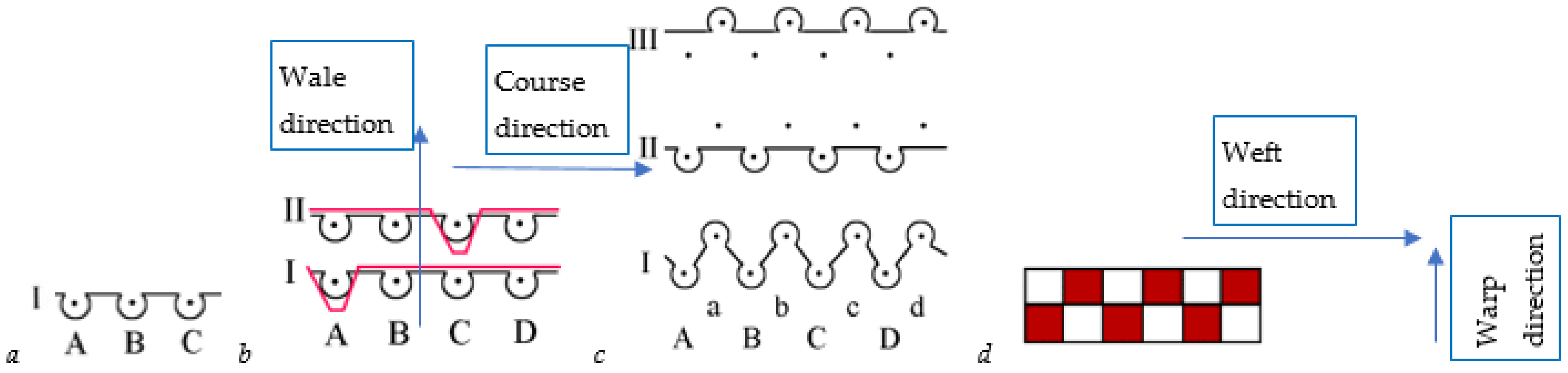
2.2. Tested Textile Fabrics

For this research, six hydrophilic knitted and two hydrophobic woven fabrics, which differed in structure, raw material, or surface density, were produced. The main characteristics of the investigated textile fabrics are presented in Table 1, and the structures of fabric patterns are shown in Figure 1.

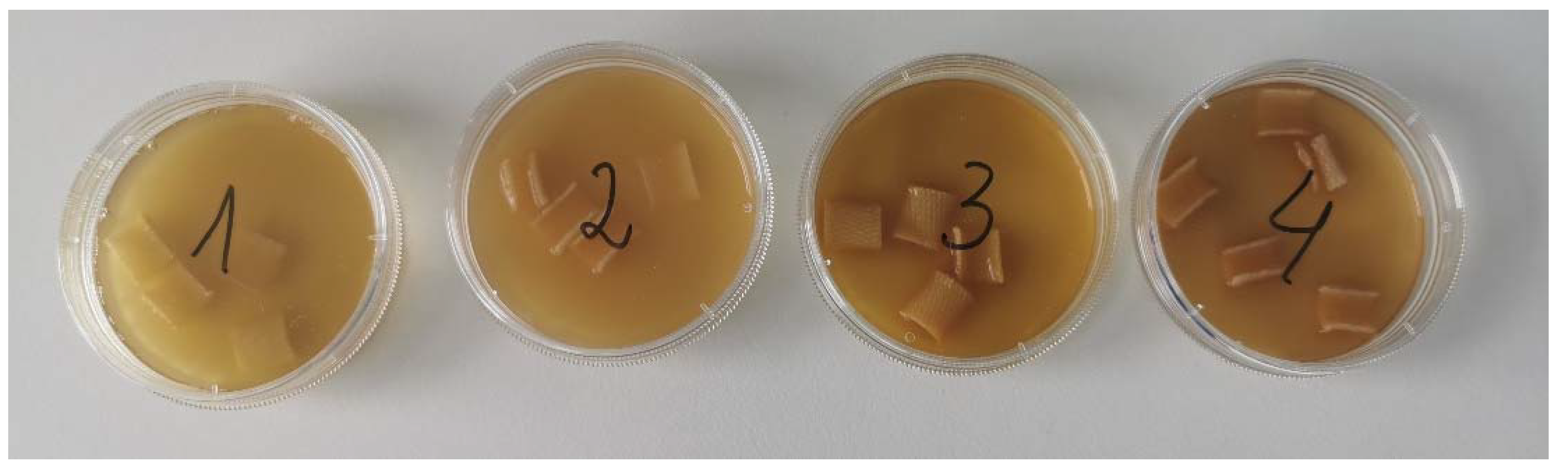
2.3. Impregnating Liquid Formulations and Preparation

Four different formulations with plant extracts and essential oils were prepared. The compositions are given in Table 2, Table 3, Table 4 and Table 5.
Extracts were prepared using ultrasound-assisted extraction from raw materials (herbs, leaves, flowers, seeds). Extractant was 40% ethanol. The crushed raw material was poured with ethanol and macerated in an ultrasonic bath at 40 °C for 30 min. After maceration, the solution was filtered through filter paper. Each extract was weighed according to each composition and mixed with the polysorbate 80. After that, the essential oil was added to the mixture. The solution was stirred with a stirrer for 15 min (100 rpm). Prepared solutions were used for textile impregnation.

2.4. Determination of Composition of Impregnating Liquid Using GC-MS Qualitative Analysis

The Gas Chromatography–Mass Spectrometry (GC-MS) analysis was conducted as described in Jurga Andreja Kazlauskaite et al.’s (2022) research [40]. The research was carried out utilising the GC-MS-QP2010 system (Shimadzu, Tokyo, Japan). A volume of 20 µL from the sample (either extract or essential oil) was diluted to 1 mL with hexane (99%, Sigma Aldrich, Schnelldorf, Germany). The primary GC-MS parameters included a 30 m × 0.25 i.d. × 0.25 µL film thickness RTX-5MS column, with helium (99.999%, AGA, Vilnius, Lithuania) as the carrier gas at a flow rate of 1.23 mL/min. Post-injection, the temperature was maintained at 40 °C for 2 min and then programmed from 3 °C/min to 210 °C. The split ratio was set at 1:10, and the mass detector electron ionisation was 70 eV.
Identification of volatile compounds was accomplished through a mass spectral library search (NIST 14), with comparisons made to mass spectral data available in the literature.

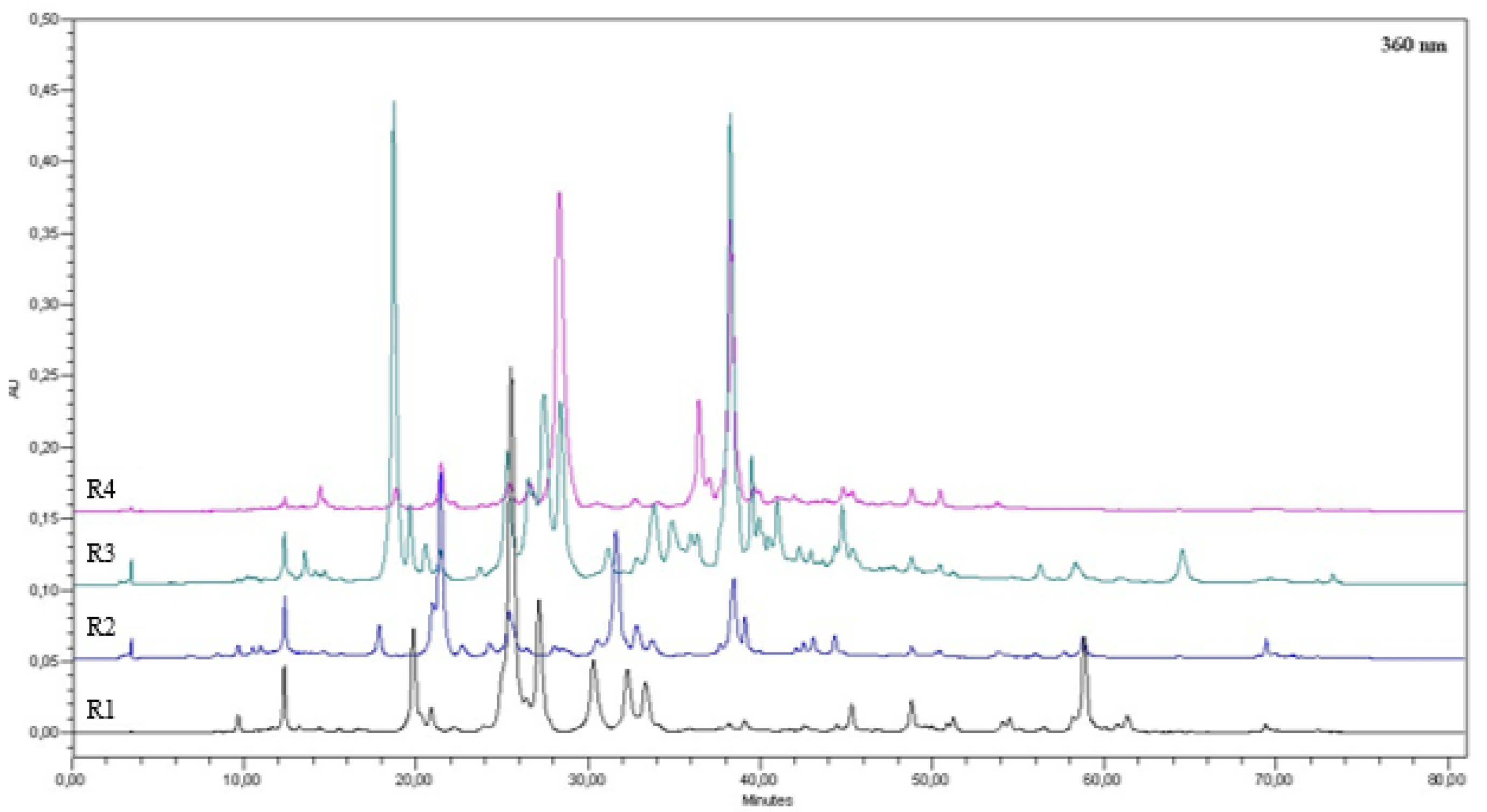
2.5. Determination of Total Amount of Phenolic Compounds in an Impregnating Liquid Using HPLC Analysis

The HPLC analyses were conducted using the Shimadzu Nexera X2 LC-30AD HPLC system (Shimadzu, Tokyo, Japan). The system includes a quaternary pump, an online de-gasser, a column temperature controller, the SIL-30AC autosampler (Shimadzu, Tokyo, Japan) with the CTO-20AC thermostat (Shimadzu, Tokyo, Japan), and the SPD-M20A diode array detector (DAD) (Shimadzu, Tokyo, Japan). To determine polyphenols, we employed an ACE 5 C18 250 × 4.6 mm column (Advanced Chromatography Technologies, Aberdeen, UK).
The eluents comprised 99.9% acetonitrile and 0.1% trifluoroacetic acid. Prior to analysis, samples underwent filtration through 0.45 μm syringe filters, and the results were expressed as μg/g dry weight (dw). This study utilised a method developed in our previous research by Stanciauskaite et al. (2021) [41], obtaining quantitative outcomes through a comparative analysis of peak area ratios between standards and samples.

2.6. Antimicrobial Treatment

The antimicrobial activity was assessed using the agar diffusion method on solid nutrient media, specifically Mueller-Hinton Agar (Mueller-Hinton II Agar, BBL, Cockeysville, MD, USA). The impregnated textile was treated with solutions containing extracts from four distinct plant sources to evaluate their antimicrobial efficacy.
The method was carried out as described in our previous study by Kazlauskaite et al. (2023) [42].
Standard cultures of non-spore bacteria, including Staphylococcus aureus, Staphylococcus epidermidis, Enterococcus faecalis, Escherichia coli, Klebsiella pneumoniae, Pseudomonas aeruginosa, and Proteus vulgaris, were cultivated for 20–24 h at 35–37 °C on Mueller-Hinton Agar. The bacterial suspension was prepared from these cultures using sterile physiological sodium chloride (0.9%) solution and standardised with a McFarland standard indicator. The standardisation criterion was set at an indicator value of 0.5, indicating that 1 mL of bacterial suspension contained 1.5 × 10⁸ cells of the respective microorganism.
For spore bacteria cultures of Bacillus cereus, 7-day cultivation on Mueller-Hinton Agar at 35–37 °C was performed. After culturing, the spore bacteria were washed off the medium surface using a sterile physiological solution. The resulting suspension underwent a 30-min heat treatment at 70 °C, followed by dilution with physiological saline to achieve a spore concentration in 1 mL ranging from 10 × 106 to 100 × 106.
The standard culture of the fungus Candida albicans was grown for 20 to 24 h at 30 °C for 72 h on Sabouraud agar. The fungal suspension was prepared from cultivated fungal cultures in physiological saline and standardised using a McFarland standard indicator.
A 0.5 McFarland turbidity suspension of the standard bacteria was then prepared. Subsequently, 1 mL of this bacterial suspension, which included Staphylococcus aureus, Staphylococcus epidermidis, Enterococcus faecalis, Escherichia coli, Klebsiella pneumoniae, Pseudomonas aeruginosa, Proteus vulgaris, Bacillus cereus, and Candida albicans, was mixed with 9 mL of liquid Mueller-Hinton agar at 45 °C. The entire solution was poured into a sterile Petri dish. After solidification of the Mueller-Hinton agar, 1 × 1 cm tissue pieces soaked in a solution of plant extracts (prepared as described in the Section 2.3) were placed on the agar surface. The Petri dishes with loaded samples were cultured in a thermostat for 20–24 h at 35 °C and subsequently stored at room temperature for an additional 24 h.
Interpretation of results involved observing the growth of the microorganism around the impregnated textile: “+” indicated growth, and “−” indicated no growth.
The experiment was done trice and average values of non growth zones was calculated. Only M1 samples were impregnated and tested for further research.

2.7. Static Water Absorption

The static water absorption was measured according to method BV S1008′ of the Bureau Veritas Consumer Products Service. The samples were conditioned according to the standard ISO 139:2005 [43], cut into pieces (100 × 100) mm, and weighed using the balance KERN EW 150-3M (Eschenlohe, Germany) with 0.001 g accuracy to determine the dry mass of the sample. After that, the samples were kept for 1 min in distilled water. After being removed from the water, they were hung for 3 min to remove excess water, and the weight of the wet samples was measured. The static water absorption Sw in % was calculated by Equation (1):
S w = m w m d m d · 100 %
where Sw is the static water absorption in %; md is the mass of the dry sample in g; mw is the mass of the wet sample in g.
The static water absorption of the studied textile fabrics was evaluated using an average value of five measurements.

2.8. Dynamic Water Absorption

Dynamic water absorption was investigated using an SMZ 800 Nikon Stereoscopic Microscope and Coolpix 4500 Digital Camera (Nikon, Minato City, Tokyo, Japan). The 7.0 PE-Live software was used for the analysis of video records. The research was carried out applying a water drop to the fabric’s outer side and monitoring the dynamic change process in the liquid spot. The water droplet’s absorption process was observed from the start moment, when 1 µL drop was put on the fabric surface, within a period of 180 s. The dropper was established at 1 cm above the farbic to ensure that the drop did not touch the dropper and the fabric surface simultaneously.
The area of the liquid spot was measured after every 5 s of the process, and changes in the spot’s area over time were calculated. Tests were carried out following ISO 8655-6:2022 [44] for a piston-stroke pipette with an air cushion, using a fine balance with a moisture trap approved by the standardisation authorities. An average value of 5 elementary measurements was used to evaluate the dynamic water absorption of the tested knitted and woven textile fabrics.

3. Results and Discussion

3.1. Selection of Impregnating Liquid Composition

Four different compositions of impregnating liquids with various herbal ingredients were modelled. In all liquids, 40% ethanol was used as extractant and 1% (w/w) polysorbate 80 as an emulsifier. In all the liquid samples, two herbal extracts and one or two essential oils with an amount of up to 1% (w/w) were used (compositions in Table 2, Table 3, Table 4 and Table 5).
The compositions were modelled by experimental methods and the scientific literature data about the antibacterial, anti-inflammatory, and wound-healing effects of the plants.
Plants selected for the composition of the fourth impregnating liquid (their extracts and essential oil) included mint (Mentha arvensis L.) essential oil, the components of which inhibit Gram-positive (including Staphylococcus cultures) and Gram-negative bacteria; this essential oil is used in the production of deodorants and antiperspirants. Sage (Salvia officinalis L.) leaf extract was also selected; it has strong anti-inflammatory properties and promotes wound healing. Lastly, thyme (Thymus vulgaris L.) extract has anti-inflammatory, antioxidant, and antibacterial effects, and it also suppresses bad odour [45,46,47].
Prepared impregnant liquids had specific odours and colours caused by different herbal extracts and essential oils. The colours of different liquids are presented in Figure 2.

3.2. Composition of Liquids

Using the GC-MS method, it was determined that 16 volatile compounds were detected in the first recipe, of which 11 were identified. The compounds are given in Table 6, formulation R1.
In the second formulation, five volatile compounds were found, of which four were identified (Table 6, formulation R2). Eleven volatile compounds were detected, of which eight were identified in the third recipe (Table 6, formulation R3). In the fourth recipe, nine volatile compounds were detected, of which seven were identified; the identified compounds are listed in Table 6, formulation R4.
Using high-performance liquid chromatography (HPLC), the concentration of phenolic compounds in the first formulation was determined to be 1382.561 µg/mL, the second 849.024 µg/mL, the third 2298.986 µg/mL, and the fourth 1491.874 µg/mL (see Figure 3).

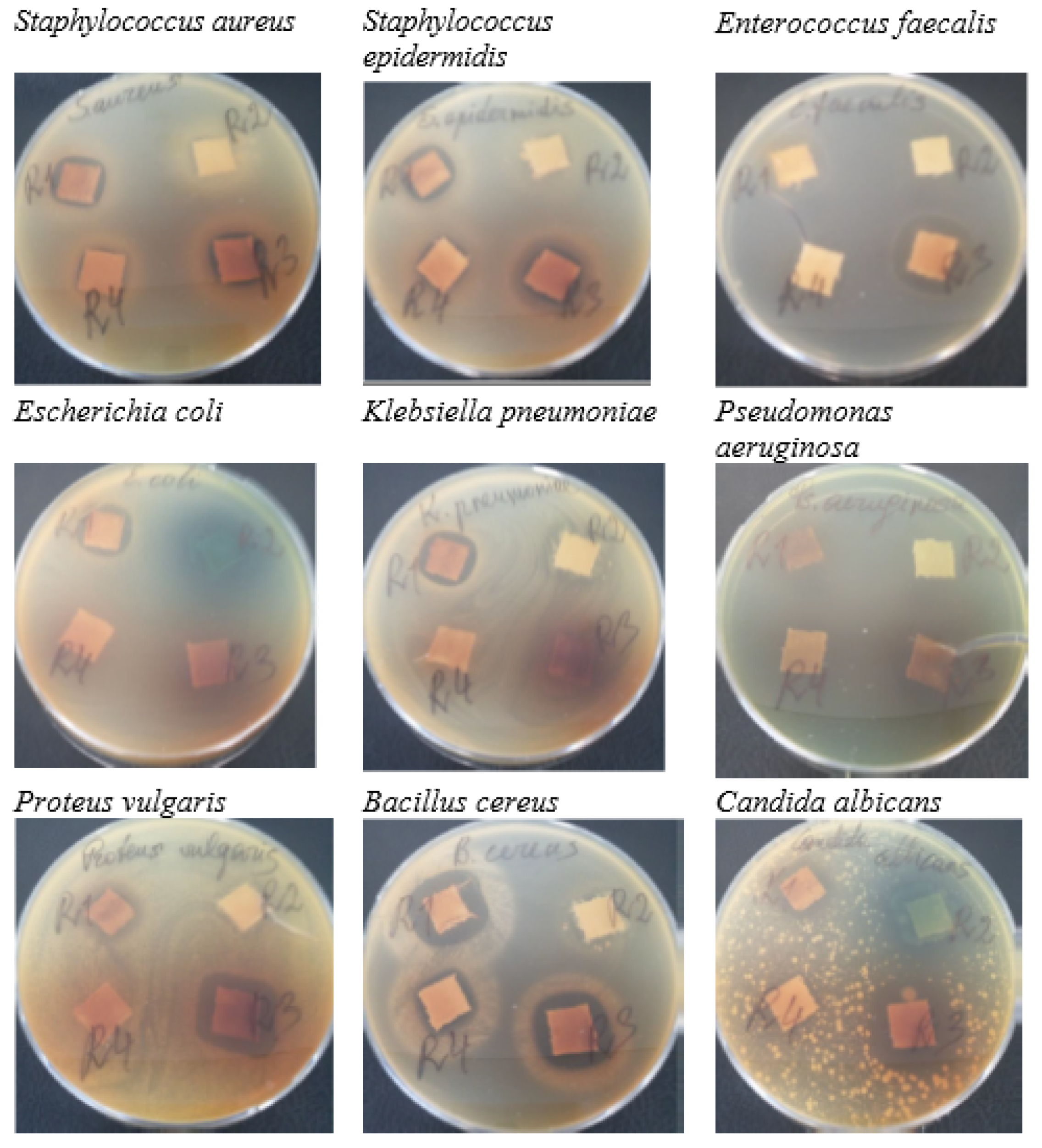
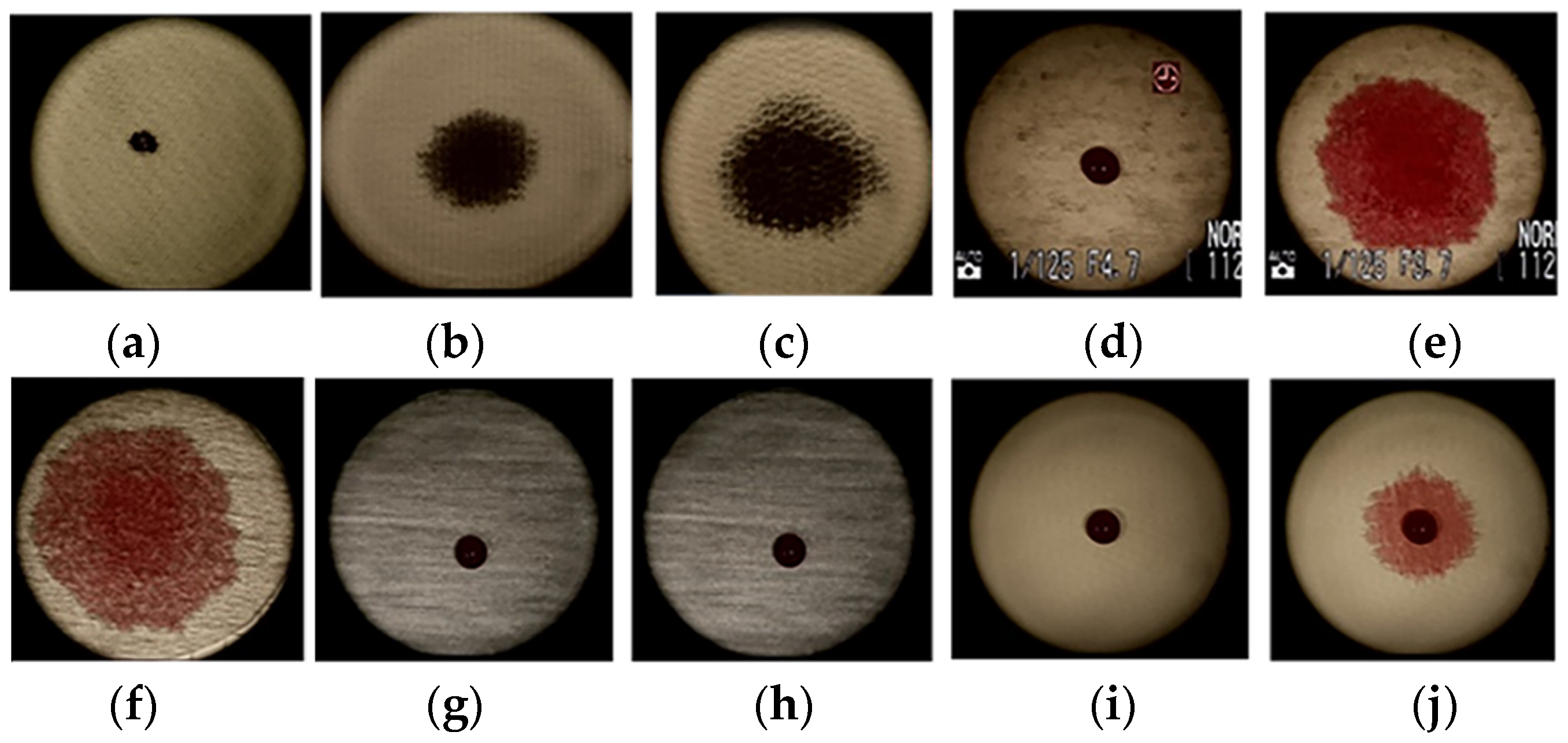
3.3. Antimicrobial Activity

Staphylococcus epidermidis is a microbiota of the skin, and Staphylococcus aureus is also a common skin coloniser. These bacteria are quite resistant to environmental factors and quickly acquire antibiotic resistance [48,49]. The antimicrobial activity of impregnating liquids has also been estimated for other pathogens. If hygiene requirements are not followed, the skin can be contaminated with bacteria from other cultures. Reference cultures of microorganisms are presented in Table 7 and Figure 4.
Plant formulations prepared from essential oils and extracts have shown antimicrobial properties against several Gram-negative and Gram-positive bacteria as well as Candida albicans yeast. It was determined that all four formulations (R1–4) had antimicrobial activity against all tested Gram-positive bacteria. However, not all formulations had the same effect on the Gram-negative bacteria.
It was found that the R1 (formulation consisting of nutmeg, birch extracts, and tea tree essential oil) solution-impregnated textile had antimicrobial activity against eight out of nine reference microorganisms; this fabric does not inhibit the growth of Gram-negative bacteria Pseudomonas aeruginosa. In the literature, it was found that tea tree essential oil has an antibacterial effect on Pseudomonas aeruginosa, but this can be due to different origin strains of Pseudomonas aeruginosa. To reach a decision on the antimicrobial activities of essential oils, extracts, or in this study’s case, formulation mixture, it is crucial to use strains from different origins in order to simulate a more realistic situation instead of just using reference strains that may not reflect the actual behaviour of the strains that can be found in nature [50,51]. Nevertheless, the R1 formulation showed great antibacterial activity compared with other formulations (R2–4).
Formulations R2 and R3 inhibited six out of nine microorganisms. Neither formulation, unlike R1, inhibited E. coli bacteria. Bigger inhibition zones were found using the R3 sample. The R4 (salvia and thyme extracts, mint essential oil) sample was the least antimicrobially active; the sample did not inhibit the growth of Gram-negative bacteria. Two different essential oils in composition (R2 and R3) did not increase the antimicrobial efficiency more than the tea tree essential oil in the R1 liquid-impregnated textile.
Only R1 and R2 liquids inhibited pathogenic yeast Candida albicans. The antimicrobial activity of the tea tree oil in in vitro conditions, found in the R1 formulation, is well-documented. It is characterised by high activity against Candida albicans in the literature [49]. This essential oil is the main ingredient of formulation R1, which increases antimicrobial activity. In the literature, it was found that Citrus x paradisi essential oil, as well as Calendula officinalis ethanolic extract, possesses antifungal activity against C. albicans [52,53,54].
The results of the antibacterial tests indicate that impregnation of the textile samples with prepared plant oils and extract formulations successfully reduced bacterial viability and effectively masked odour. These findings validate that impregnation is a highly efficient method for tackling bacterial growth and unpleasant odours.

3.4. Static Water Absorption

The best result of sweat-absorbing underarm pads can be achieved when hydrophilic and hydrophobic textile layers are used together [8]. The next-to-skin hydrophilic layer absorbs the excreted sweat, while the outer hydrophobic layer protects clothing from moisture penetration; it locks the moisture inside the underarm pad.
Static absorption shows the maximum amount of moisture a textile material can absorb. Thus, for the inner (next-to-skin) textile layer, the static absorption must be the highest possible and, on the contrary, for the outer textile layer, it must be the lowest possible. The results of the static water absorption are presented in Table 8.
As can be seen from the results presented in Table 8, the highest static water absorption (276.6%) was found for the pure cotton sample M1 knitted in the fleece pattern, where both the ground and the fleece yarns are cotton yarns, and they are arranged in the fabric in two layers, which highly increases the capability of the fabric to absorb the water. The high static water absorption (more than 200%) is characteristic of all tested fleece structures due to their inner structure. However, it is obvious that the raw material composition in the fabric also has an evident influence on the water absorption capability; even a small amount (5% in M2, M3, and M5 samples) of the hydrophobic elastomeric yarns causes a decrease in the static water absorption. The static water absorption of sample M4, knitted in a double Milano rib pattern, also reached a value of more than 200%. This structure has knitted loops arranged in two layers and, additionally, a tight structure, which helps to absorb more water. Almost twice as low static water absorption is characteristic of single-knitted structures M5 and M6. In the single jersey pattern, the yarn is situated in one layer. Therefore, such a structure has a significantly lower capability to absorb water. The presented results demonstrate that every cellulose-based knitted textile fabric can absorb more than 100% water. However, the most suitable for the inner layer of the underarm sweat pads are the cotton-based knitted fabrics with double-layered structures. Comparing the results of textile fabrics intended for the outer layer of underarm sweat pads shows that sample A1 is completely hydrophobic, i.e., did not absorb moisture at all. The degree of absorption of sample A2 is also not high (42%), but it shows that over time, moisture can be absorbed from the inner layer to the outer layers, i.e., some amount of the moisture may be transferred to the clothing, to which the underarm pad is attached.

3.5. Dynamic Water Absorption

When evaluating the sorption properties of textile fabrics, the dynamics of moisture absorption are particularly important. This shows how quickly moisture is absorbed into the fabric and how widely the moisture stain spreads across the surface of the fabric. For better comfort sense, the moisture absorption must be quick with a minimal moisture stain on the fabric surface. It is known that moisture absorption and water vapour permeability depend on the fibre properties as well as on the textile fabric pattern and structure [3,4,8].
The results of dynamic absorption are presented in Table 9 and Figure 5. The coefficient of variation of the liquid spot area measurements varied in the range of 2.6–9.8%.
The obtained results clearly demonstrate that the water drop put on the surface of cellulose-based knitted fabrics quickly penetrates into the fabric, and the liquid spot spreads on both the face side and backside of the fabric. The most rapid changes in the dynamics of the liquid absorption occur in the first 30 s, and then this process slows down. After 60 s, the changes in the liquid spot area become insignificant (less than 2%). It was also observed that fleece knitted fabrics with a double-layered yarn system absorb liquid very fast, which penetrates from the face side to the backside, and the liquid spot area on the back side is obviously higher. It is a positive effect, as it means that the wet area on the next-to-skin side is as small as possible, and the wet sensation is also as low as possible. A slightly different situation was observed analysing water absorption dynamics of single jersey (one-layer) knitted fabrics. The absorption process is also very fast; however, as there is only one layer of knitted loops in the knitted structure, the liquid spot area on both the face side and the backside is almost twice as high as on the double-layered knitted fabrics. This leads to the conclusion that the best liquid absorption and comfort result can be achieved by using cellulose-based fleece knitted structures for the inner, i.e., next-to-skin layer of the underarm sweat-absorbing pad.
Analysis of the wet absorption dynamics of the hydrophobic synthetic woven fabrics showed that PA woven fabric A1 (very thin, light, and at the same time tight) is fully hydrophobic, i.e., the shape of the liquid drop did not change during the 180 s observation time (see Figure 5). The liquid will be “locked” inside the underarm pad and not transferred to the clothing. The shape of the liquid drop on the surface of the PES woven fabric A2 also remained after 180 s observation time. However, some amount of the liquid spread on the surface of the fabric. The static water absorption analysis also demonstrates that this fabric tends to absorb some water. Therefore, the A1 woven fabric can be recommended as a better option for the outer layer of the underarm pad.

4. Conclusions

The highest antimicrobial activity was found in textile fabric which was impregnated with the R1 formulation—nutmeg extract, birch extract, and tea tree essential oil. The textiles lowest in R4 did not inhibit Gram-negative bacteria. Using the R1 formulation to impregnate the textile would be appropriate to achieve the best result in masking sweat odour and eliminating bacteria.
Analysis of the static and dynamic water absorption of six cellulose-based hydrophilic knitted and two synthetic hydrophobic woven fabrics showed that the best liquid absorption and comfort result of the sweat-absorbing underarm pads could be achieved by using the cellulose-based double-layered knitted structures (such like fleece) for the inner (next-to-skin) layer and fully hydrophobic synthetic tight and thin woven fabrics for outer (next-to-clothing) layer. Thus, the best results can be achieved using M1–M4 knitted structures, impregnated by the R1 formulation, for the inner layer and A1 woven fabric for the outer layer of the sweat-absorbing and clothing-protecting underarm pads.
Even more, underarm pads can be produced from clothing production waste. It would help to reduce the amount of textile waste and, on the other hand, to reduce the number of clothing washings, i.e., the amount of water consumption and pollution during the lifecycle of clothing.

Author Contributions

Conceptualisation D.M., G.L., J.A.K., I.M. and J.B.; methodology, D.M., G.L., B.P., J.A.K., I.M., J.B. and M.M.; investigation, D.M., G.L., B.P., J.A.K., I.M. and M.M.; resources, J.B., D.M., G.L.; data curation, I.M. and J.A.K.; writing—original draft preparation, D.M., G.L. J.A.K., I.M. and J.B.; writing—review and editing, D.M., G.L., J.A.K., I.M., J.B. and M.M.; supervision, J.B. All authors have read and agreed to the published version of the manuscript.

Funding

The presented research was partially funded by the Erasmus+ programme of the European Union under the project reference number 2021-2-PL01-KA220-VET-000048919. Views and opinions expressed are, however, those of the author(s) only and do not necessarily reflect those of the European Union or the European Education and Culture Executive Agency (EACEA). Neither the European Union nor EACEA can be held responsible for them.

Institutional Review Board Statement

Not applicable.

Informed Consent Statement

Not applicable.

Data Availability Statement

The data that support the findings of this study are available from the corresponding author, J.B., upon reasonable request.

Acknowledgments

The authors would like to thank the Open Access Centre at Advanced Pharmaceutical and Health Technologies (Lithuanian University of Health Sciences) for providing the opportunity to use their research infrastructure and perform this research.

Conflicts of Interest

The authors declare no conflicts of interest.

References

  1. Mansoor, T.; Hes, L.; Bajzik, V.; Noman, M.T. Novel Method on Thermal Resistance Prediction and Thermo-Physiological Comfort of Socks in a Wet State. Text. Res. J. 2020, 90, 1987–2006. [Google Scholar] [CrossRef]
  2. Raccuglia, M.; Hodder, S.; Havenith, G. Human Wetness Perception in Relation to Textile Water Absorption Parameters under Static Skin Contact. Text. Res. J. 2017, 87, 2449–2463. [Google Scholar] [CrossRef]
  3. Chen, Q.; Shou, D.; Zheng, R.; Tang, K.P.M.; Fu, B.; Zhang, X.; Ma, P. Moisture and Thermal Transport Properties of Different Polyester Warp-Knitted Spacer Fabric for Protective Application. Autex Res. J. 2021, 21, 182–191. [Google Scholar] [CrossRef]
  4. Abramavičiute, J.; Mikučioniene, D.; Čiukas, R. Static Water Absorption of Knits from Natural and Textured Yarns. Fibres Text. East. Eur. 2011, 86, 60–63. [Google Scholar]
  5. Eryuruk, S.H. Analyzing Thermophysiological Comfort and Moisture Management Behavior of Cotton Denim Fabrics. Autex Res. J. 2020, 21, 248–254. [Google Scholar] [CrossRef]
  6. Bivainyte, A.; Mikučioniene, D. Influence of Shrinkage on Air and Water Vapour Permeability of Double-Layered Weft Knitted Fabrics. Medziagotyra 2012, 18, 271–274. [Google Scholar] [CrossRef]
  7. Marolleau, A.; Salaun, F.; Dupont, D.; Gidik, H.; Ducept, S. Influence of Textile Properties on Thermal Comfort. IOP Conf. Ser. Mater. Sci. Eng. 2017, 254, 182007. [Google Scholar] [CrossRef]
  8. Bivainyte, A.; Mikučioniene, D.; Milašiene, D. Influence of the Knitting Structure of Double-Layered Fabrics on the Heat Transfer Process. Fibres Text. East. Eur. 2012, 91, 40–43. [Google Scholar]
  9. Yoo, S.; Kim, E. Effects of Multilayer Clothing System Array on Water Vapor Transfer and Condensation in Cold Weather Clothing Ensemble. Text. Res. J. 2008, 78, 189–197. [Google Scholar] [CrossRef]
  10. Zhang, X.; Yang, J.; Borayek, R.; Qu, H.; Nandakumar, D.K.; Zhang, Q.; Ding, J.; Tan, S.C. Super-Hygroscopic Film for Wearables with Dual Functions of Expediting Sweat Evaporation and Energy Harvesting. Nano Energy 2020, 75, 104873. [Google Scholar] [CrossRef]
  11. Gerrett, N.; Griggs, K.; Redortier, B.; Voelcker, T.; Kondo, N.; Havenith, G. Sweat from Gland to Skin Surface: Production, Transport, and Skin Absorption. J. Appl. Physiol. 2019, 125, 459–469. [Google Scholar] [CrossRef]
  12. James, A.G.; Austin, C.J.; Cox, D.S.; Taylor, D.; Calvert, R. Microbiological and Biochemical Origins of Human Axillary Odour. FEMS Microbiol. Ecol. 2013, 83, 527–540. [Google Scholar] [CrossRef] [PubMed]
  13. Fredrich, E.; Barzantny, H.; Brune, I.; Tauch, A. Daily Battle against Body Odor: Towards the Activity of the Axillary Microbiota. Trends Microbiol. 2013, 21, 305–312. [Google Scholar] [CrossRef] [PubMed]
  14. Stanojevic, L.P.; Marjanovic-Balaban, Z.R.; Kalaba, V.D.; Stanojevic, J.S.; Cvetkovic, D.J. Chemical Composition, Antioxidant and Antimicrobial Activity of Chamomile Flowers Essential Oil (Matricaria chamomilla, L.). J. Essent. Oil-Bear. Plants 2016, 19, 2017–2028. [Google Scholar] [CrossRef]
  15. Miraj, S.; Alesaeidi, S. A Systematic Review Study of Therapeutic Effects of Matricaria recuitta Chamomile (Chamomile). Electron. Physician 2016, 8, 3024–3031. [Google Scholar] [CrossRef] [PubMed]
  16. El Joumaa, M.M.; Borjac, J.M. Matricaria Chamomilla: A Valuable Insight into Recent Advances in Medicinal Uses and Pharmacological Activities. Phytochem. Rev. 2022, 21, 1913–1940. [Google Scholar] [CrossRef]
  17. Srivastava, J.K.; Shankar, E.; Gupta, S. Chamomile: A Herbal Medicine of the Past with a Bright Future (Review). Mol. Med. Rep. 2010, 3, 895–901. [Google Scholar] [CrossRef] [PubMed]
  18. Faria, R.L.; Cardoso, L.M.L.; Akisue, G.; Pereira, C.A.; Junqueira, J.C.; Jorge, A.O.C.; Santos, P.V., Jr. Adherence of Microorganisms to Sutures after Extraction of Unerupted Third Molars. In Vivo 2011, 19, 476–482. [Google Scholar]
  19. Efstratiou, E.; Hussain, A.I.; Nigam, P.S.; Moore, J.E.; Ayub, M.A.; Rao, J.R. Antimicrobial Activity of Calendula officinalis Petal Extracts against Fungi, as Well as Gram-Negative and Gram-Positive Clinical Pathogens. Complement. Ther. Clin. Pract. 2012, 18, 173–176. [Google Scholar] [CrossRef]
  20. Zhu, W.; Gao, J. The Use of Botanical Extracts as Topical Skin-Lightening Agents for the Improvement of Skin Pigmentation Disorders. J. Investig. Dermatol. Symp. Proc. 2008, 13, 20–24. [Google Scholar] [CrossRef]
  21. Tober, C.; Schoop, R. Modulation of Neurological Pathways by Salvia officinalis and Its Dependence on Manufacturing Process and Plant Parts Used. BMC Complement. Altern. Med. 2019, 19, 128. [Google Scholar] [CrossRef]
  22. Flinčec Grgac, S.; Tesla, T.; Čorak, I.; Žuvela Bošnjak, F. Hydrothermal Synthesis of Chitosan and Tea Tree Oil on Plain and Satin Weave Cotton Fabrics. Materials 2022, 15, 5034. [Google Scholar] [CrossRef] [PubMed]
  23. Carson, C.F.; Hammer, K.A.; Riley, T.V. Melaleuca alternifolia (Tea Tree) Oil: A Review of Antimicrobial and Other Medicinal Properties. Clin. Microbiol. Rev. 2006, 19, 50–62. [Google Scholar] [CrossRef] [PubMed]
  24. Lee, C.J.; Chen, L.W.; Chen, L.G.; Chang, T.L.; Huang, C.W.; Huang, M.C.; Wang, C.C. Correlations of the Components of Tea Tree Oil with Its Antibacterial Effects and Skin Irritation. J. Food Drug Anal. 2013, 21, 169–176. [Google Scholar] [CrossRef]
  25. Biswas, N.N.; Saha, S.; Ali, M.K. Antioxidant, Antimicrobial, Cytotoxic and Analgesic Activities of Ethanolic Extract of Mentha arvensis L. Asian Pac. J. Trop. Biomed. 2014, 4, 792–797. [Google Scholar] [CrossRef]
  26. Grgac, S.F.; Jablan, J.; Inić, S.; Malinar, R.; Kovaček, I.; Čorak, I. The Effect of Ultrasonic Treatment on the Binding of the Inclusion Complex β-Cyclodextrin-Peppermint Oil with Cellulose Material. Materials 2022, 15, 470. [Google Scholar] [CrossRef]
  27. Rastogi, S.; Pandey, M.M.; Kumar Singh Rawat, A. Medicinal Plants of the Genus Betula—Traditional Uses and a Phytochemical–Pharmacological Review. J. Ethnopharmacol. 2015, 159, 62–83. [Google Scholar] [CrossRef] [PubMed]
  28. Cui, C.A.; Jin, D.Q.; Hwang, Y.K.; Lee, I.S.; Hwang, J.K.; Ha, I.; Han, J.S. Macelignan Attenuates LPS-Induced Inflammation and Reduces LPS-Induced Spatial Learning Impairments in Rats. Neurosci. Lett. 2008, 448, 110–114. [Google Scholar] [CrossRef] [PubMed]
  29. Dziedziński, M.; Kobus-cisowska, J.; Szymanowska-powałowska, D. Polyphenols Composition, Antioxidant and Antimicrobial Properties of Pinus sylvestris L. Shoots Extracts Depending on Different Drying Methods. Emir. J. Food Agric. 2020, 32, 229–237. [Google Scholar] [CrossRef]
  30. Macedo, L.M.; De Mendes, É.; Milit, L.; Tundisi, L.L.; Souto, E.B.; Mazzola, P.G. Rosemary (Rosmarinus officinalis L., syn Salvia rosmarinus Spenn.) and Its Topical Applications: A Review. Plants 2020, 9, 651–663. [Google Scholar] [CrossRef]
  31. Deng, W.; Liu, K.; Cao, S.; Sun, J.; Zhong, B.; Chun, J. Chemical Composition, Antimicrobial, Antioxidant, and Antiproliferative Properties of Grapefruit Essential Oil Prepared by Molecular Distillation. Molecules 2020, 25, 217. [Google Scholar] [CrossRef] [PubMed]
  32. Chahla, B.; Salih, B.M.; Adriana, B.; Viviana, M.; Guido, F.; Sergio, S.; Federica, C.; Rosaria, N.; Marina, P.; Abdelmounaim, K.; et al. Chemical Composition and Biological Activities of Oregano and Lavender Essential Oils. Appl. Sci. 2021, 11, 5688. [Google Scholar] [CrossRef]
  33. Sienkiewicz, M.; Denys, P.; Kowalczyk, E. Antibacterial and Immunostimulatory Effect of Essential Oils. Int. Rev. Allergol. Clin. Immunol. 2011, 17, 40–44. [Google Scholar]
  34. Cavanagh, H.M.A.; Wilkinson, J.M. Biological Activities of Lavender Essential Oil. Phyther. Res. 2002, 16, 301–308. [Google Scholar] [CrossRef]
  35. Kaur, H.; Bhardwaj, U.; Kaur, R. Cymbopogon Nardus Essential Oil: A Comprehensive Review on Its Chemistry and Bioactivity. J. Essent. Oil Res. 2021, 33, 205–220. [Google Scholar] [CrossRef]
  36. Kenny, G.P.; Journeay, W.S. Human Thermoregulation: Separating Thermal and Nonthermal Effects on Heat Loss. Front. Biosci. 2010, 15, 259–290. [Google Scholar] [CrossRef] [PubMed]
  37. Kondo, N.; Nishiyasu, T.; Inoue, Y.; Koga, S. Non-Thermal Modification of Heat-Loss Responses during Exercise in Humans. Eur. J. Appl. Physiol. 2010, 110, 447–458. [Google Scholar] [CrossRef] [PubMed]
  38. Mekjavic, I.B.; Eiken, O. Contribution of Thermal and Nonthermal Factors to the Regulation of Body Temperature in Humans. J. Appl. Physiol. 2006, 100, 2065–2072. [Google Scholar] [CrossRef]
  39. Shibasaki, M.; Wilson, T.E.; Crandall, C.G. Neural Control and Mechanisms of Eccrine Sweating during Heat Stress and Exercise. J. Appl. Physiol. 2006, 100, 1692–1701. [Google Scholar] [CrossRef]
  40. Kazlauskaite, J.A.; Ivanauskas, L.; Marksa, M.; Bernatoniene, J. The Effect of Traditional and Cyclodextrin-Assisted Extraction Methods on Trifolium pratense L. (Red Clover) Extracts Antioxidant Potential. Antioxidants 2022, 11, 435. [Google Scholar] [CrossRef]
  41. Stanciauskaite, M.; Marksa, M.; Liaudanskas, M.; Ivanauskas, L.; Ivaskiene, M.; Ramanauskiene, K. Extracts of Poplar Buds (Populus balsamifera L., Populus nigra L.) and Lithuanian Propolis: Comparison of Their Composition and Biological Activities. Plants 2021, 10, 828. [Google Scholar] [CrossRef]
  42. Kazlauskaite, J.A.; Matulyte, I.; Marksa, M.; Lelesius, R.; Pavilonis, A.; Bernatoniene, J. Application of Antiviral, Antioxidant and Antibacterial Glycyrrhiza glabra L., Trifolium pratense L. Extracts and Myristica fragrans Houtt. Essential Oil in Microcapsules. Pharmaceutics 2023, 15, 464. [Google Scholar] [CrossRef]
  43. LST EN ISO 139:2005; Textiles—Standard Atmospheres for Conditioning and Testing. ISO: London, UK, 2019.
  44. ISO 8655-6:2022; The High Art of Pippete Calibration. ISO: London, UK, 2022.
  45. Reichling, J.; Schnitzler, P.; Suschke, U.; Saller, R. Essential Oils of Aromatic Plants with Antibacterial, Antifungal, Antiviral, and Cytotoxic Properties—An Overview. Complement. Med. Res. 2009, 16, 79–90. [Google Scholar] [CrossRef] [PubMed]
  46. Karimzadeh, S.; Farahpour, M.R. Topical Application of Salvia officinalis Hydroethanolic Leaf Extract Improves Wound Healing Process. Indian J. Exp. Biol. 2017, 55, 98–106. [Google Scholar] [PubMed]
  47. Abdel-Fattah, A.; Aboelazab, Y.; Khallaf, M.; El-Kenany, Y. Antimicrobial Activity of Ethanolic Extracts of Clove and Thyme. Arab Univ. J. Agric. Sci. 2019, 27, 491–499. [Google Scholar] [CrossRef]
  48. Erin Chen, Y.; Fischbach, M.A.; Belkaid, Y. Skin Microbiota-Host Interactions. Nature 2018, 553, 427–436. [Google Scholar] [CrossRef] [PubMed]
  49. Older, C.E.; Diesel, A.; Patterson, A.P.; Meason-Smith, C.; Johnson, T.J.; Mansell, J.; Suchodolski, J.S.; Hoffmann, A.R. The Feline Skin Microbiota. PLoS ONE 2017, 12, e0178555. [Google Scholar] [CrossRef] [PubMed]
  50. Sakkas, H.; Gousia, P.; Economou, V.; Sakkas, V.; Petsios, S.; Papadopoulou, C. In Vitro Antimicrobial Activity of Five Essential Oils on Multidrug Resistant Gram-Negative Clinical Isolates. J. Intercult. Ethnopharmacol. 2016, 5, 212–218. [Google Scholar] [CrossRef] [PubMed]
  51. Kunicka-Styczyńska, A.; Sikora, M.; Kalemba, D. Antimicrobial Activity of Lavender, Tea Tree and Lemon Oils in Cosmetic Preservative Systems. J. Appl. Microbiol. 2009, 107, 1903–1911. [Google Scholar] [CrossRef] [PubMed]
  52. Schwiertz, A.; Duttke, C.; Hild, J.; Müller, H.J. In Vitro Activity of Essential Oils on Microorganisms Isolated from Vaginal Infections. Int. J. Aromather. 2006, 16, 169–174. [Google Scholar] [CrossRef]
  53. Delgado, A.J.M.; Velázquez, U.C.; González, J.G.B.; Montes, A.C.; Villarreal, S.M.L.; García, L.E.V.; Casas, R.M.S.; Luis, O.E.R. Evaluation of the Essential Oil of Citrus Paradisi; as an Alternative Treatment against Candida albicans. Open J. Stomatol. 2020, 10, 258–270. [Google Scholar] [CrossRef]
  54. Sepehri, Z.; Javadian, F.; Khammari, D.; Hassanshahian, M. Antifungal Effects of the Aqueous and Ethanolic Leaf Extracts of Echinophora Platyloba and Rosmarinus Officinalis. Curr. Med. Mycol. 2016, 2, 30–35. [Google Scholar] [CrossRef]
Figure 1. Structure of tested textile fabrics: (a)—single jersey (5M and 6M), (b)—fleece (1M, 2M, 3M), (c)—Milano rib (4M), (d)—plain weave (1A and 2A).
Fibers 12 00020 g001
Figure 2. Impregnating liquids with textile samples (Arabic numerals correspond to the sample number).
Fibers 12 00020 g002
Figure 3. Chromatogram of the total amount of phenolic compounds of impregnant liquids prepared with different recipes.
Fibers 12 00020 g003
Figure 4. Visualisation of samples’ antimicrobial activity.
Fibers 12 00020 g004
Figure 5. Visualisation of liquid spot area changes: (a)—M1 face side after 5 s; (b)—M1 face side after 180 s; (c)—M1 backside after 180 s; (d)—M6 face side after 5 s; (e)—M6 face side after 180 s; (f)—M6 backside after 180 s; (g)—A1 face side after 5 s; (h)—A1 face side after 180 s; (i)—A2 face side after 5 s; (j)—A2 face side after 180 s.
Fibers 12 00020 g005
Table 1. Main characteristics of tested textile fabrics.
Knitted Fabrics
Sample CodeRaw MaterialArea Density, g/m2Course Density, cm−1Wale Density, cm−1PatternLinear Density of Yarns, Tex
1M100% CO30022.015.5Fleece24 (CO)
2M70% MO + 25% CO + 5% EL23020.014.0Fleece22.5 (MO) + 21 (CO) + 4 (EL)
3M95% CO + 5% EL33021.015.0Fleece20 (CO) + 4 (EL)
4M95% CO + 5% EL31019.012.0Milano rib24 (CO) + 2.2 (EL)
5M94% VI + 6% EL22021.015.0Single jersey16.5 (VI) + 4 (EL)
6M100% CO19018.013.0Single jersey25.5 (CO)
Woven Fabrics
Sample CodeRaw MaterialArea Density, g/m2Weft Density, cm−1Warp Density, cm−1PatternYarn Linear Density, Tex
1A100% PA326037Plain weave10 (PA)
2A100% PES446634Plain weave12 (PES)
Note: CO—cotton yarn, MO—modal yarn, VI—viscose yarn, PES—polyester yarn, PA—polyamide yarn, EL—elastomeric polyurethane yarn.
Table 2. First composition (R1) of impregnating solution.
MaterialFunctionExtract Concentration *Amount (w/w%)
Nutmeg seeds (Myristica fragrans Houtt.)Extract1:2049
Birch leaves (Betula pendula Roth.)Extract1:549
Tea tree (Melaleuca alternifolia)Essential oil-1
Polysorbate 80Emulsifier-1
* Extractant was 40% ethanol.
Table 3. Second composition (R2) of impregnating solution.
MaterialFunctionExtract Concentration *Amount (w/w%)
Marigold flower (Calendula officinalis L.)Extract1:549
Rosemary leaves (Rosmarinus officinalis L.)Extract1:549
Pine needle (Pinus sylvestris L.)Essential oil-0.5
Grapefruit peel (Citrus x paradisi L.)Essential oil-0.5
Polysorbate 80Emulsifier-1
* Extractant was 40% ethanol.
Table 4. Third composition (R3) of impregnating solution.
MaterialFunctionExtract Concentration *Amount (w/w%)
Chamomile flower (Matricaria chamomilla L.)Extract1:549
Oregano herb (Origanum vulgare L.)Extract1:549
Lavender flower (Lavandula angustifolia L.)Essential oil-0.5
Lemongrass (Cymbopogon nardus L.)Essential oil 0.5
Polysorbate 80Emulsifier-1
* Extractant was 40% ethanol.
Table 5. Fourth composition (R4) of impregnating solution.
MaterialFunctionExtract Concentration *Amount (w/w%)
Sage leaves (Salvia officinalis L.)Extract1:549
Thyme herb (Thymus vulgaris L.)Extract1:549
Mentha herb (Mentha arvensis L.)Essential oil-1
Polysorbate 80Emulsifier-1
* Extractant was 40% ethanol.
Table 6. Detected volatile compounds in the impregnating solution formulations R1–4.
FormulationDetermined Compounds
R11,1-dicyclopropylethene; 6,6-dimethylspiro [3,4-diazabicyclo [3.1.0]hex-3-ene-2,1′-cyclopropane]; (E)-2,7-dimethyloct-3-en-5-yne; 1,4-methano-1H-cyclopenta[d]pyridazine, 4,4; 6-methyl-6-hepten-2-on; terpinen-4-ol; caryophyllene, humulene; 3,3,6,6,9,9-hexamethyl tetracyclo [6.1.0.02,4.05,7]nonane.
R21,1-dicyclopropylethene; 6,6-dimethylspiro [3,4-diazabicyclo [3.1.0]hex-3-ene-2,1′-cyclopropane]; 1,3,6-octatriene, 1-methyl-4-prop-1-en-2-ylcyclohexene (limonene)
R33-cyclohexylpent-4-en-2-one; 3-cyclohexyl-4-penten-2-one, (3R)-3,7-dimethylocta-1,6-dien-3-ol (linalool); 4-carvomenthenol; longipinenepoxide; linalyl acetate, trans-2-cis-6-nonadienal; [(2E,6Z)-nona-2,6-dienyl] acetate, caryophyllene.
R41-oxacyclopropyl-3,4-epoxycyclohexane; 3-(Allyloxy)-2-methyl-1-propene; tert-dodecylmercaptan; 2-Methylpent-2-en-1-ol; 1-undecyne; cis-1,7-octadien-3-yl acetate; caryophyllene
Table 7. Antimicrobial effect of the liquids (R1–4) on the growth of cultures of reference microorganisms.
MicroorganismR1R2R3R4
Diameter of Non-Growth Zones of Reference Microorganisms (mm)
Staphylococcus aureus
ATCC 25923
15.4 ± 0.410.4 ± 0.613.5 ± 0.511.0 ± 1.0
Staphylococcus epidermidis
ATCC 12228
17.6 ± 0.514.0 ± 0.115.7 ± 1.314.6 ± 1.1
Enterococcus faecalis
ATCC 29212
18.3 ± 0.6N22.0 ± 0.115.2 ± 0.4
Escherichia coli
ATCC 25922
16.4 ± 0.3NNN
Klebsiella pneumoniae
ATCC 13883
17.1 ± 0.613.0 ± 0.1NN
Pseudomonas aeruginosa
ATCC 27853
NN16.1 ± 0.1N
Proteus vulgaris
ATCC 8427
10.6 ± 0.514.1 ± 0.118.3 ± 0.5N
Bacillus cereus
ATCC 11778
21.7 ± 0.312.7 ± 0.418.2 ± 0.114.1 ± 0.2
Candida albicans
ATCC 10231
13.4 ± 0.618.0 ± 0.1NN
N—does not inhibit the growth of microorganisms.
Table 8. Static water absorption.
Sample CodeMass of Dry Sample, md, gMass of Wet Sample, mw, gStatic Water Absorption, Sw, %
M12.01 ± 0.087.57 ± 0.15276.6
M21.93 ± 0.106.24 ± 0.11223.3
M32.41 ± 0.068.85 ± 0.12267.2
M42.11 ± 0.046.52 ± 0.08209.0
M51.90 ± 0.033.99 ± 0.10110.0
M61.82 ± 0.044.64 ± 0.12154.9
A10.27 ± 0.020.27 ± 0.010.00
A20.26 ± 0.010.37 ± 0.0142.31
Table 9. Dynamics of liquid spot area after 1 s, 5 s, 20 s, and 60 s of observation.
Sample CodeLiquid Spot Area, mm2
1 s5 s30 s60 s180 s
M120.8 ± 0.4240.4 ± 0.4761.6 ± 1.8572.9 ± 2.3274.0 ± 2.65
M222.8 ± 0.5139.6 ± 0.3552.3 ± 1.7968.6 ± 2.2570.2 ± 2.42
M324.2 ± 0.6239.9 ± 0.3859.1 ± 1.6270.9 ± 2.4272.7 ± 2.59
M420.5 ± 0.3934.2 ± 0.46 50.0 ± 1.6362.6 ± 2.3969.2 ± 2.40
M528.0 ± 0.2557.9 ± 0.8698.1 ± 1.92119.8 ± 2.59120.2 ± 2.72
M635.9 ± 0.3465.2 ± 0.71119.8 ± 1.64130.6 ± 2.81132.0 ± 2.12
A1NNNNN
A221.4 ± 0.1221.4 ± 0.1822.9 ± 0.1523.1 ± 0.06 S23.2 ± 0.04 S
NOTE: N means that no changes in the liquid spot area were observed during the 180 s; S means that the shape of a liquid drop remains, but some spread of the moisture on the fabric surface appears.
Disclaimer/Publisher’s Note: The statements, opinions and data contained in all publications are solely those of the individual author(s) and contributor(s) and not of MDPI and/or the editor(s). MDPI and/or the editor(s) disclaim responsibility for any injury to people or property resulting from any ideas, methods, instructions or products referred to in the content.

