Inclusion of 5-Mercapto-1-Phenyl-Tetrazole into β-Cyclodextrin for Entrapment in Silane Coatings: An Improvement in Bronze Corrosion Protection

 , , and
Abstract
:1. Introduction
2. Materials and Methods
2.1. Chemicals, Aggressive Environment and Alloy
2.2. [β-CD–MPT] Complex Stability Analysis
2.2.1. Nuclear Magnetic Resonance (NMR) Measurements
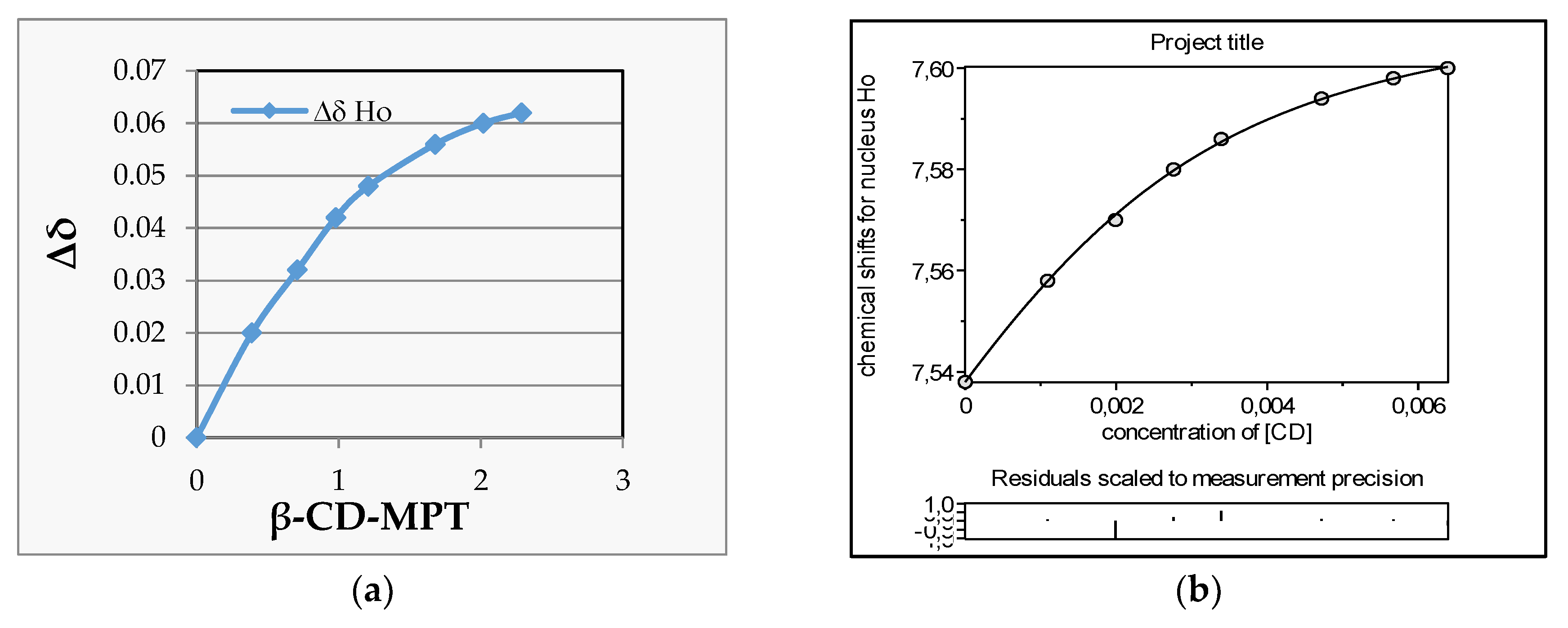
2.2.2. H NMR Titration
2.2.3. Electrospray Ionization (ESI) Mass Spectra
2.2.4. Fourier Transform Infra-Red (FTIR) Analysis
2.3. Silane Coating Production
2.4. Silane Coating Protectiveness
3. Results
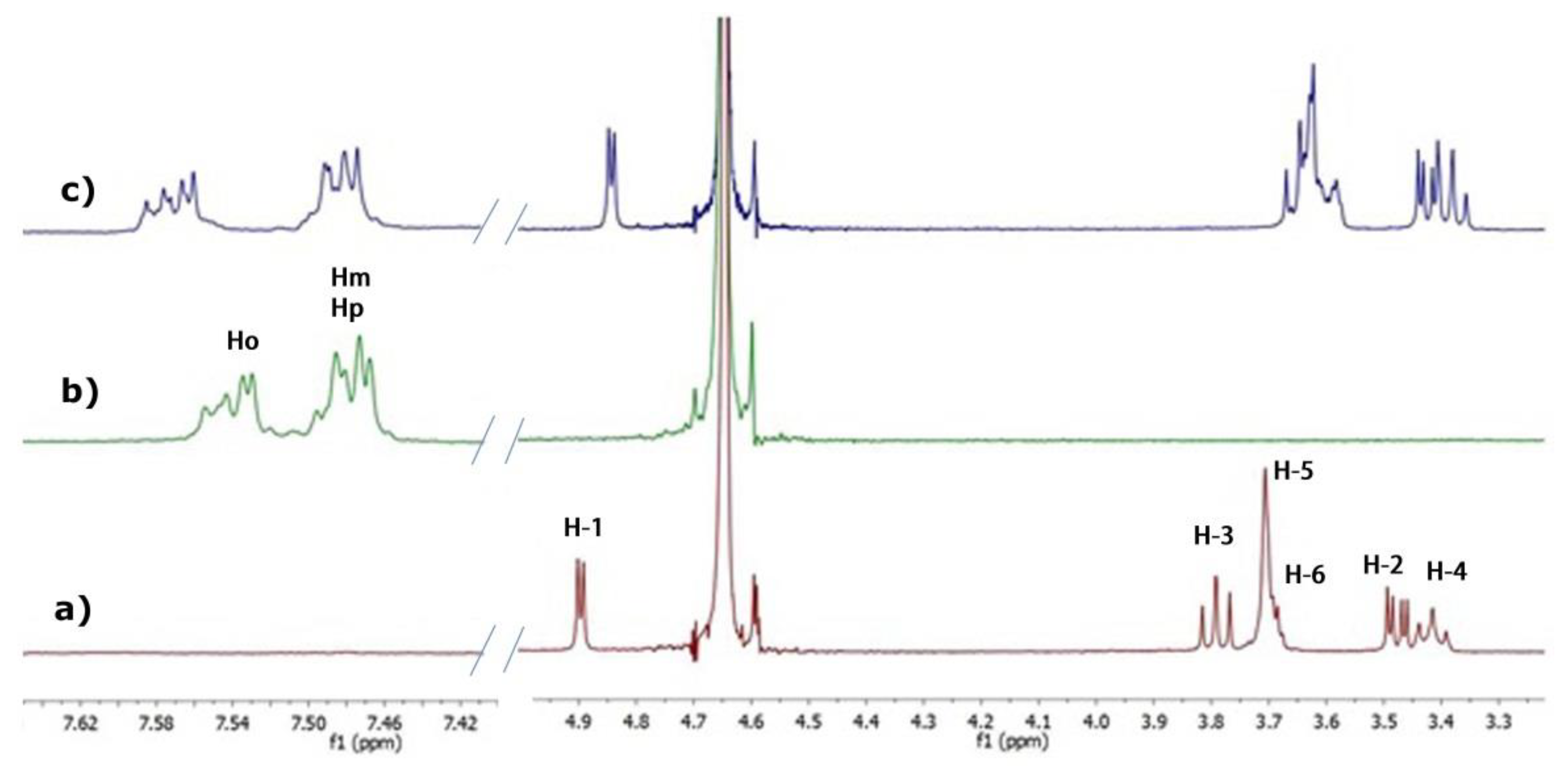
3.1. NMR Studies
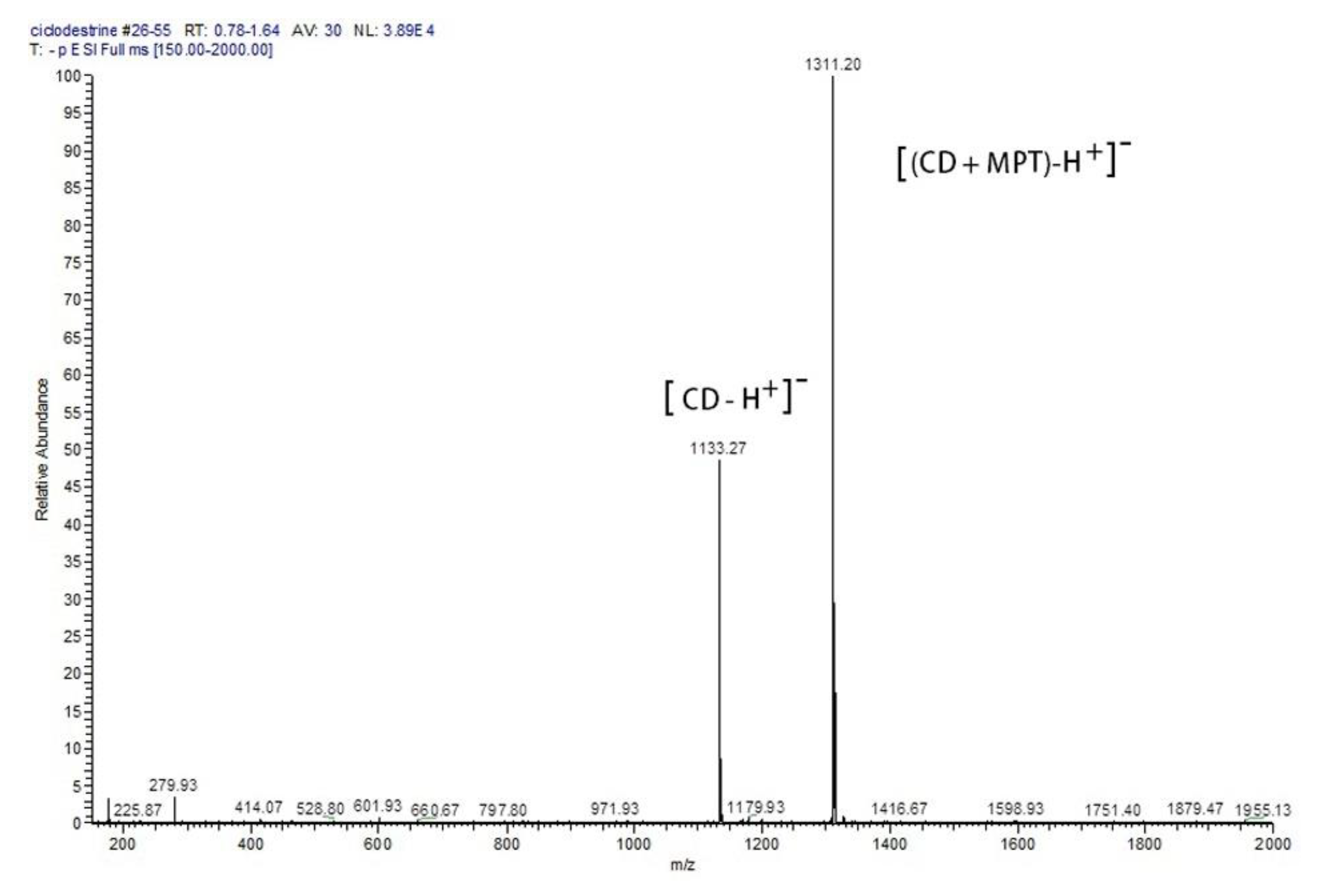
3.2. ESI Mass Spectra
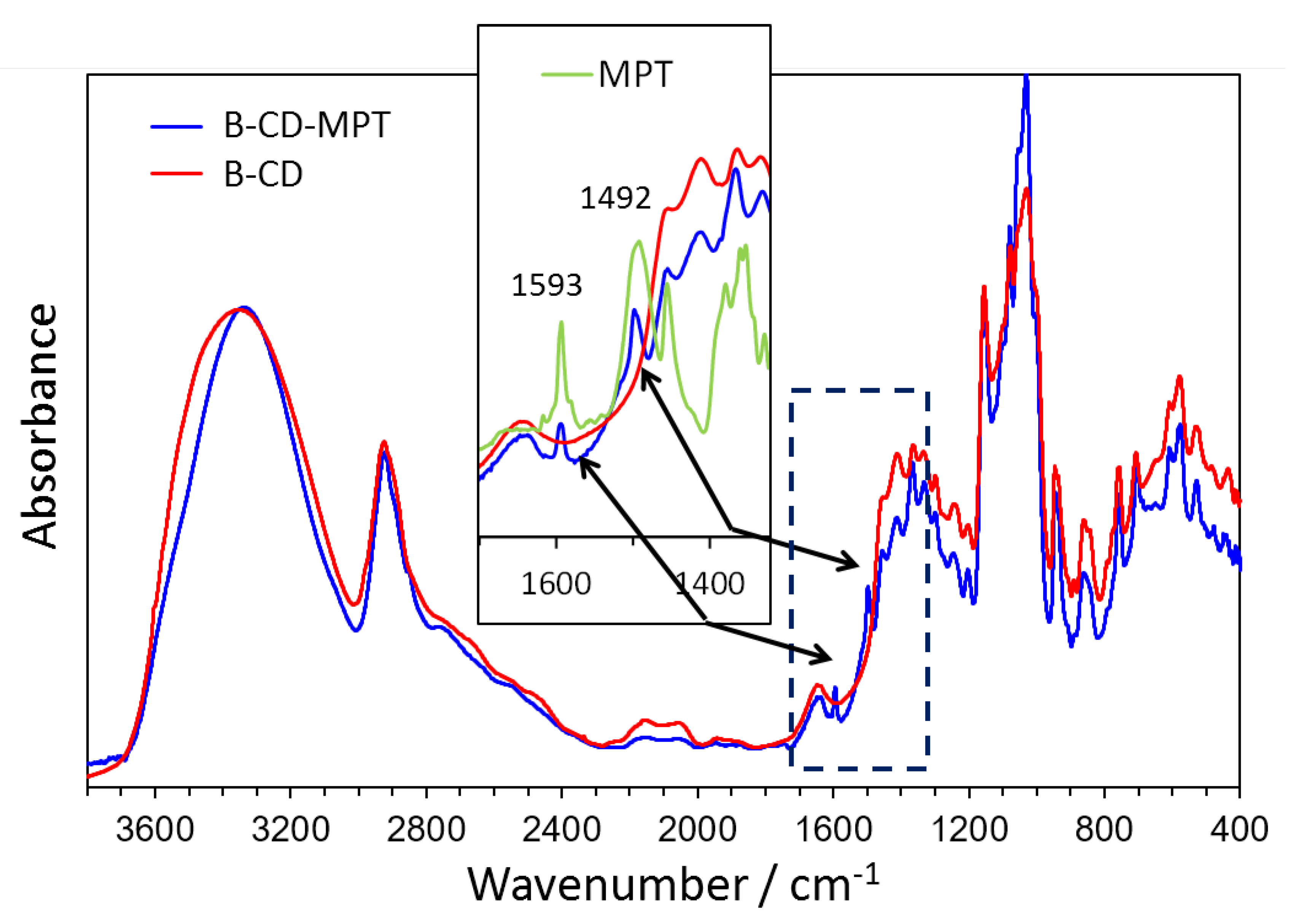
3.3. FTIR Spectra
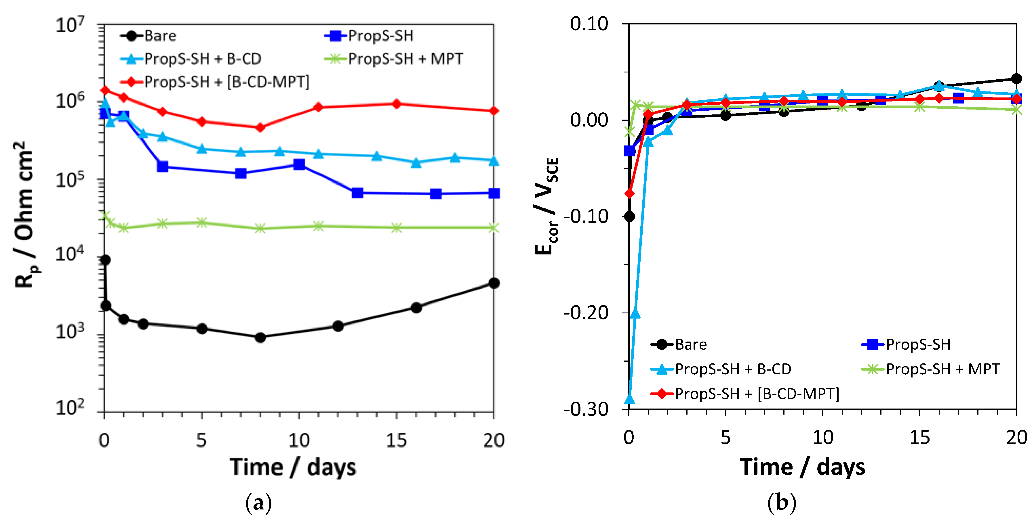
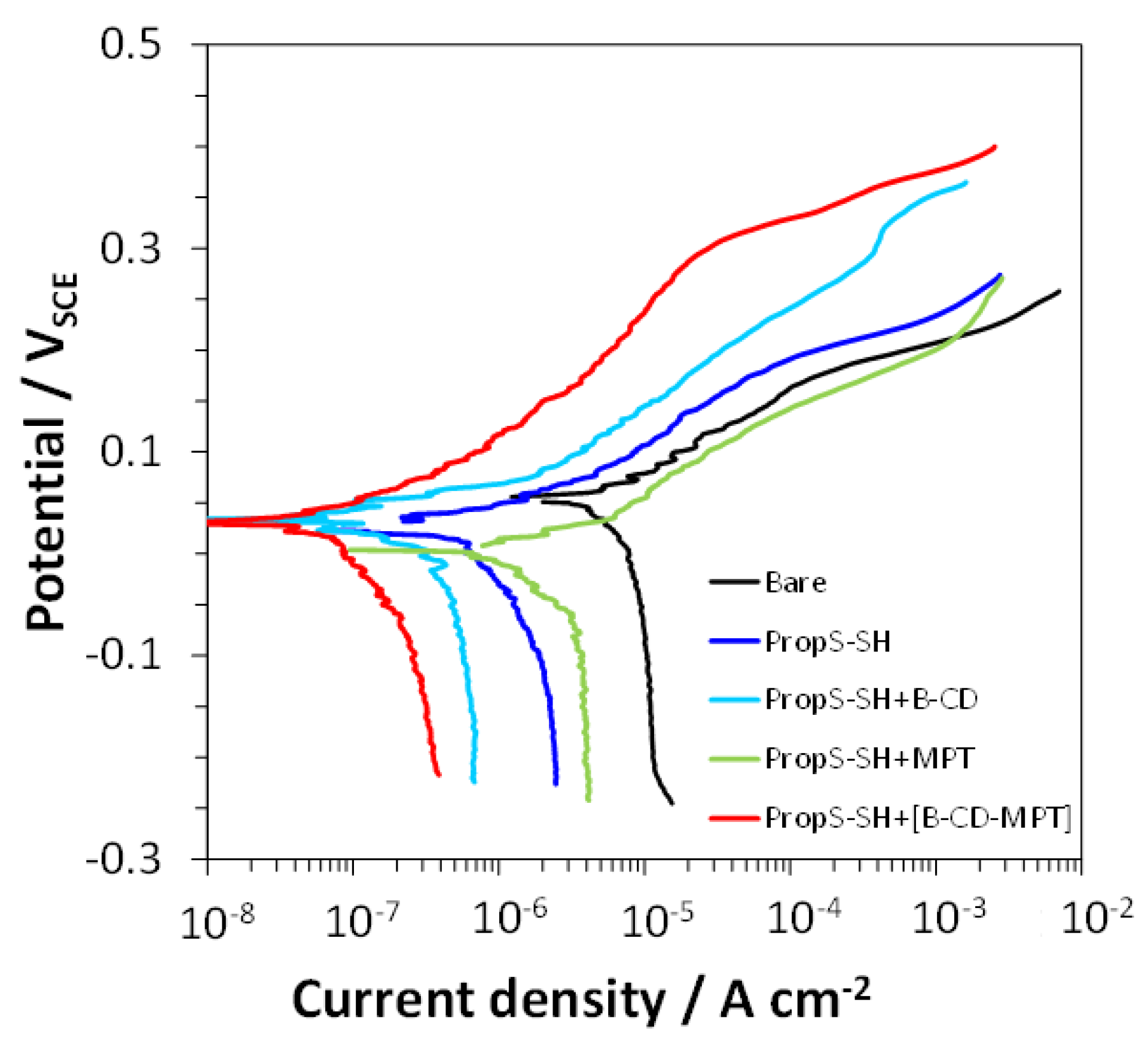
3.4. Electrochemical Tests
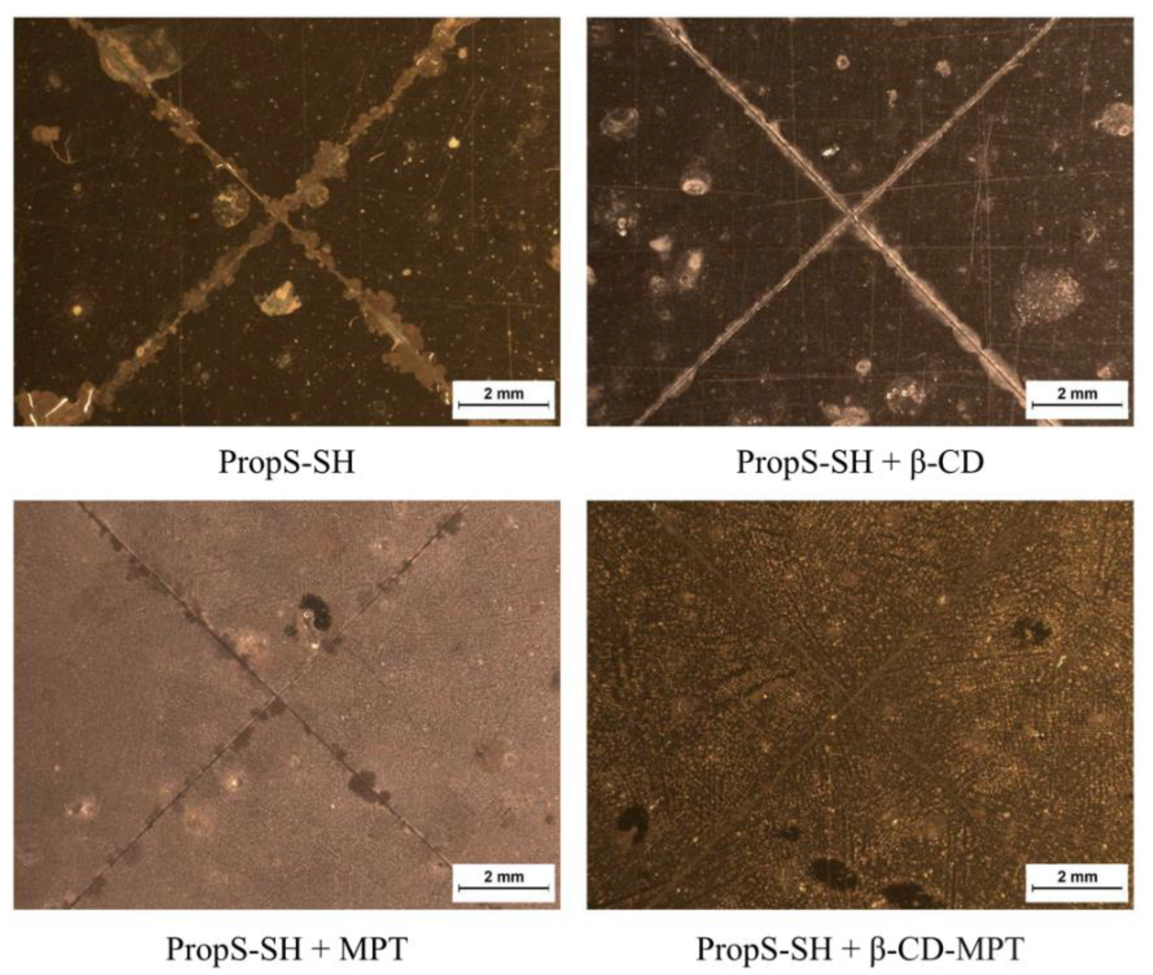
3.5. Cyclic AR Spray Test
4. Conclusions
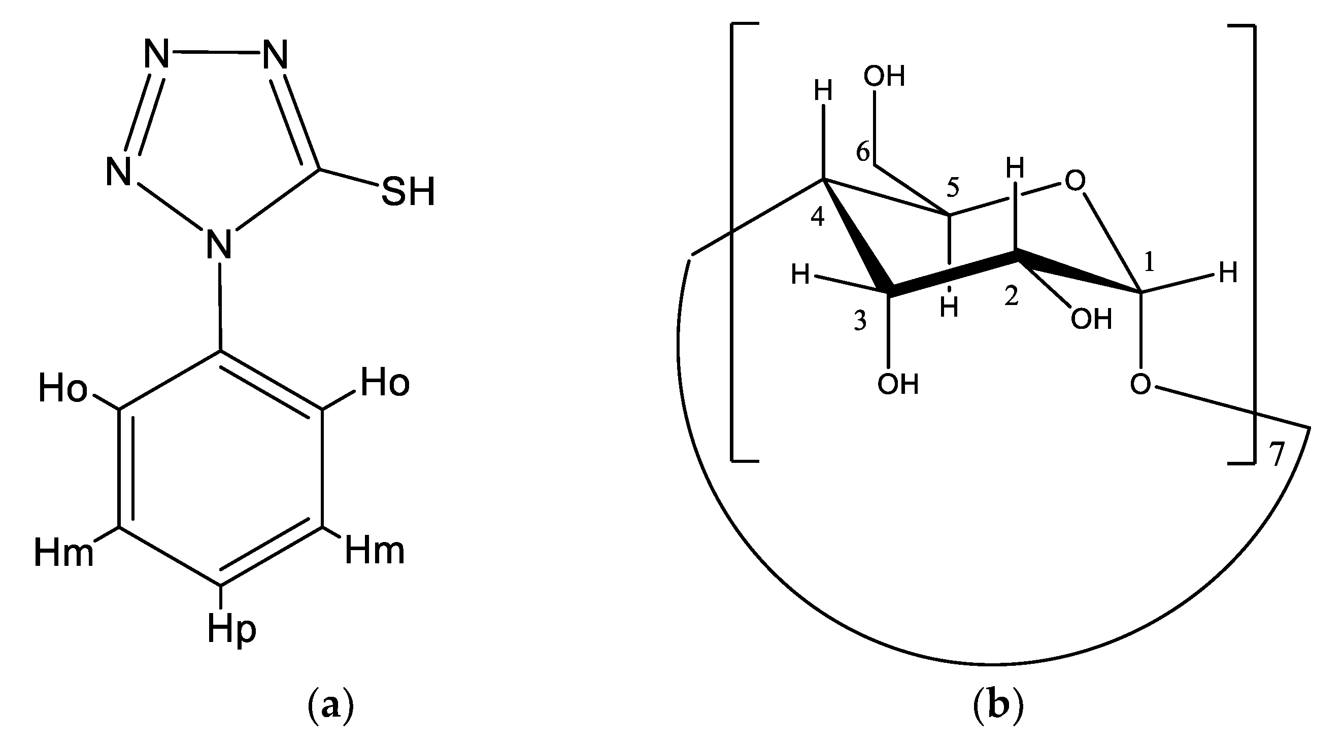
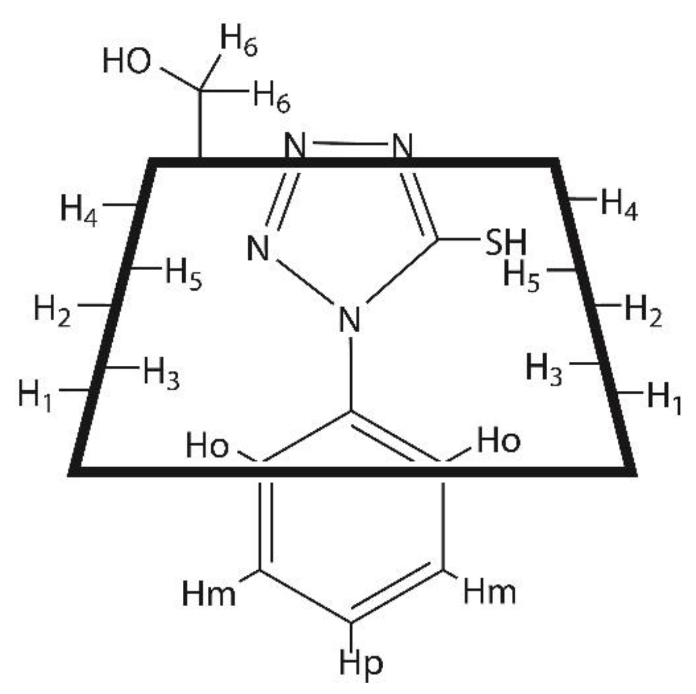
- The stability and the molecular structure of the host–guest β-CD–MPT complex was assessed by NMR, FTIR, and MS techniques.
- The analysis of the Complexation Induced Shifts suggested an inclusion structure of the complex, with partial insertion of the phenyl moiety of MPT at the wide rim of the hydrophobic β-CD cavity and partial protrusion of the tetrazole moiety of the inhibitor from the narrow rim of the host cavity.
- According to the DOSY experiments, the stability constant of the β-CD–MPT complex was 654 M−1, in good agreement with the value of 851 M−1 obtained by curve fitting the NMR binding isotherm.
- The PropS-SH coating with entrapped β-CD–MPT complex exhibited an improved protectiveness (η = 98%) against bronze corrosion in comparison to plain PropS-SH (η = 84%) or PropS-SH containing only MPT (η = 66%) or β-CD (η = 92%).
- The PropS-SH coating with entrapped β-CD–MPT complex exhibited self-healing properties on bronze during exposures to cyclic AR spray at 35 °C.
Supplementary Materials
Author Contributions
Funding
Acknowledgments
Conflicts of Interest
References
- Tasić, Ž.Z.; Petrović Mihajlović, M.B.; Radovanović, M.B.; Antonijević, M.M. New trends in corrosion protection of copper. Chem. Pap. 2019, 73, 2103–2132. [Google Scholar] [CrossRef]
- Ashassi-Sorkhabi, H.; Moradi-Alavian, S.; Esrafili, M.D.; Kazempour, A. Hybrid sol-gel coatings based on silanes-amino acids for corrosion protection of AZ91 magnesium alloy: Electrochemical and DFT insights. Prog. Org. Coat. 2019, 131, 191–202. [Google Scholar] [CrossRef]
- Zaferani, S.; Peikari, M.; Zaarei, D.; Danaee, I.; Fakhraei, J.; Mohammadi, M. Using silane films to produce an alternative for chromate conversion coatings. Corrosion 2013, 69, 372–387. [Google Scholar] [CrossRef]
- Bierwagen, G.; Shedlosky, T.J.; Stanek, K. Developing and testing a new generation of protective coatings for outdoor bronze sculpture. Prog. Org. Coat. 2003, 48, 289–296. [Google Scholar] [CrossRef]
- Lamaka, S.V.; Shchukin, D.G.; Andreeva, D.V.; Zheludkevich, M.L.; Möhwald, H.; Ferreira, M.G.S. Sol-gel/polyelectrolyte active corrosion protection system. Adv. Funct. Mater. 2008, 18, 3137–3147. [Google Scholar] [CrossRef]
- Zhang, F.; Ju, P.; Pan, M.; Zhang, D.; Huang, Y.; Li, G.; Li, X. Self-healing mechanisms in smart protective coatings: A review. Corros. Sci. 2018, 144, 74–88. [Google Scholar] [CrossRef]
- Montemor, M.F. Functional and smart coatings for corrosion protection: A review of recent advances. Surf. Coat. Technol. 2014, 258, 17–37. [Google Scholar] [CrossRef]
- Shchukin, D.G.; Zheludkevich, M.; Yasakau, K.; Lamaka, S.; Ferreira, M.G.S.; Möhwald, H.; Shchukin, D. Layer-by-layer assembled nanocontainers for self-healing corrosion protection. Adv. Mater. 2006, 18, 1672–1678. [Google Scholar] [CrossRef]
- Jafari, A.; Hosseini, S.; Jamalizadeh, E. Investigation of smart nanocapsules containing inhibitors for corrosion protection of copper. Electrochim. Acta 2010, 55, 9004–9009. [Google Scholar] [CrossRef]
- Mahmoudian, M.; Nozad, E.; Kochameshki, M.G.; Enayati, M. Preparation and investigation of hybrid self-healing coatings containing linseed oil loaded nanocapsules, potassium ethyl xanthate and benzotriazole on copper surface. Prog. Org. Coat. 2018, 120, 167–178. [Google Scholar] [CrossRef]
- Zheludkevich, M.L.; Poznyak, S.K.; Rodrigues, L.M.; Raps, D.; Hack, T.; Dick, L.F.; Nunes, T.; Ferreira, M.G.S. Active protection coatings with layered double hydroxide nanocontainers of corrosion inhibitor. Corros. Sci. 2010, 52, 602–611. [Google Scholar] [CrossRef]
- Szejtli, J. Introduction and general overview of cyclodextrin chemistry. Chem. Rev. 1998, 98, 1743–1754. [Google Scholar] [CrossRef]
- Hedges, A.R. Industrial applications of cyclodextrins. Chem. Rev. 1998, 98, 2035–2044. [Google Scholar] [CrossRef]
- Uekama, K.; Hirayama, F.; Irie, T. Cyclodextrin drug carrier systems. Chem. Rev. 1998, 98, 2045–2076. [Google Scholar] [CrossRef]
- Del Valle, E.M.M. Cyclodextrins and their uses: A review. Process Biochem. 2004, 39, 1033–1046. [Google Scholar] [CrossRef]
- Li, S.; Purdy, W.C. Cyclodextrins and their applications in analytical chemistry. Chem. Rev. 1992, 92, 1457–1470. [Google Scholar] [CrossRef]
- Takahashi, K. Organic reactions mediated by cyclodextrins. Chem. Rev. 1998, 98, 2013–2034. [Google Scholar] [CrossRef]
- De Souza, T.M.; Cordeiro, R.F.; Viana, G.M.; Aguiar, L.C.; De Senna, L.F.; Malta, L.F.B.; D’Elia, E.; D’Elia, E. Inclusion compounds of dibenzylthiourea with hydroxypropylated-cyclodextrins for corrosion protection of carbon steel in acidic medium. J. Mol. Struct. 2016, 1125, 331–339. [Google Scholar] [CrossRef]
- Casaletto, M.P.; Figà, V.; Privitera, A.; Mazzaglia, A.; Scala, A.; Zagami, R. Sustainable corrosion inhibition of Copper-based alloys by smart β-Cyclodextrin/Benzotriazole complexes. In Proceedings of the 5th European Cyclodextrin Conference, Lisbon, Portugal, 3–6 October 2017. [Google Scholar]
- Khramov, A.; Voevodin, N.; Balbyshev, V.; Donley, M. Hybrid organo-ceramic corrosion protection coatings with encapsulated organic corrosion inhibitors. Thin Solid Films 2004, 447, 549–557. [Google Scholar] [CrossRef]
- Khramov, A.N.; Voevodin, N.N.; Balbyshev, V.N.; Mantz, R.A. Sol–gel-derived corrosion-protective coatings with controllable release of incorporated organic corrosion inhibitors. Thin Solid Films 2006, 514, 174–181. [Google Scholar] [CrossRef]
- Amiri, S.; Rahimi, A. Anticorrosion behavior of cyclodextrins/inhibitor nanocapsule-based self-healing coatings. J. Coat. Technol. Res. 2016, 13, 1095–1102. [Google Scholar] [CrossRef]
- Altin, A.; Rohwerder, M.; Erbe, A. Cyclodextrins as carriers for organic corrosion inhibitors in organic coatings. J. Electrochem. Soc. 2017, 164, C128–C134. [Google Scholar] [CrossRef]
- Varvara, S.; Bostan, R.; Bobis, O.; Găină, L.; Popa, F.; Mena, V.; Souto, R.M. Propolis as a green corrosion inhibitor for bronze in weakly acidic solution. Appl. Surf. Sci. 2017, 426, 1100–1112. [Google Scholar] [CrossRef]
- Verma, C.; Ebenso, E.E.; Bahadur, I.; Quraishi, M. An overview on plant extracts as environmental sustainable and green corrosion inhibitors for metals and alloys in aggressive corrosive media. J. Mol. Liq. 2018, 266, 577–590. [Google Scholar] [CrossRef]
- Marzorati, S.; Verotta, L.; Trasatti, S.P. Green corrosion inhibitors from natural sources and biomass wastes. Molecules 2019, 24, 48. [Google Scholar] [CrossRef]
- Zucchi, F.; Trabanelli, G.; Fonsati, M. Tetrazole derivatives as corrosion inhibitors for copper in chloride solutions. Corros. Sci. 1996, 38, 2019–2029. [Google Scholar] [CrossRef]
- Mihit, M.; Salghi, R.; Bazzi, L.; Hammouti, B.; Kertit, S.; El Issami, S.; Addi, E.A. A study of tetrazoles derivatives as corrosion inhibitors of copper in nitric acid. Pigment Resin Technol. 2006, 35, 151–157. [Google Scholar] [CrossRef]
- Balbo, A.; Chiavari, C.; Martini, C.; Monticelli, C. Effectiveness of corrosion inhibitor films for the conservation of bronzes and gilded bronzes. Corros. Sci. 2012, 59, 204–212. [Google Scholar] [CrossRef]
- Wolfe, J.; Grayburn, R. A review of the development and testing of Incralac lacquer. J. Am. Inst. Conserv. 2017, 56, 225–244. [Google Scholar] [CrossRef]
- Aufray, M.; Balbo, A.; Benetti, F.; Bernardi, E.; Bignozzi, M.C.; Chiavari, C.; Esvan, J.; Gartner, N.; Grassi, V.; Josse, C.; et al. Protection of outdoor bronzes by eco-friendly and non-hazardous coatings based on silane and fluoropolymers: Results from the B-IMPACT project. In Proceedings of the Metal 2019, Neuchâtel, Switzerland, 2–6 September 2019; pp. 1–10. [Google Scholar]
- Chiavari, C.; Balbo, A.; Bernardi, E.; Martini, C.; Bignozzi, M.; Abbottoni, M.; Monticelli, C.; Bignozzi, M. Protective silane treatment for patinated bronze exposed to simulated natural environments. Mater. Chem. Phys. 2013, 141, 502–511. [Google Scholar] [CrossRef]
- Masi, G.; Balbo, A.; Esvan, J.; Monticelli, C.; Avila, J.; Robbiola, L.; Bernardi, E.; Bignozzi, M.; Asensio, M.; Martini, C.; et al. X-ray photoelectron spectroscopy as a tool to investigate silane-based coatings for the protection of outdoor bronze: The role of alloying elements. Appl. Surf. Sci. 2018, 433, 468–479. [Google Scholar] [CrossRef]
- Chiavari, C.; Bernardi, E.; Balbo, A.; Monticelli, C.; Raffo, S.; Bignozzi, M.; Martini, C.; Bignozzi, M. Atmospheric corrosion of fire-gilded bronze: Corrosion and corrosion protection during accelerated ageing tests. Corros. Sci. 2015, 100, 435–447. [Google Scholar] [CrossRef]
- Wu, D.H.; Chen, A.D.; Johnson, C.S., Jr. An improved diffusion-ordered spectroscopy experiment incorporating bipolar-gradient pulses. J. Magn. Reson. Ser. A 1995, 115, 260–264. [Google Scholar] [CrossRef]
- Fielding, L. Determination of association constants (Ka) from solution NMR data. Tetrahedron 2000, 56, 6151–6170. [Google Scholar] [CrossRef]
- Frassineti, C.; Ghelli, S.; Gans, P.; Sabatini, A.; Moruzzi, M.S.; Vacca, A. Nuclear magnetic resonance as a tool for determining protonation constants of natural polyprotic bases in solution. Anal. Biochem. 1995, 231, 374–382. [Google Scholar] [CrossRef]
- Zucchi, F.; Frignani, A.; Grassi, V.; Trabanelli, G.; DalColle, M. The formation of a protective layer of 3-mercapto-propyl-trimethoxy-silane on copper. Corros. Sci. 2007, 49, 1570–1583. [Google Scholar] [CrossRef]
- Chiavari, C.; Balbo, A.; Bernardi, E.; Martini, C.; Zanotto, F.; Vassura, I.; Bignozzi, M.; Monticelli, C.; Bignozzi, M. Organosilane coatings applied on bronze: Influence of UV radiation and thermal cycles on the protectiveness. Prog. Org. Coat. 2015, 82, 91–100. [Google Scholar] [CrossRef]
- Masi, G.; Josse, C.; Esvan, J.; Chiavari, C.; Bernardi, E.; Martini, C.; Bignozzi, M.C.; Monticelli, C.; Zanotto, F.; Balbo, A.; et al. Evaluation of the protectiveness of an organosilane coating on patinated Cu-Si-Mn bronze for contemporary art. Prog. Org. Coat. 2019, 127, 286–299. [Google Scholar] [CrossRef]
- Stern, M.; Geary, A.L. Electrochemical polarization I. A theoretical analysis of the shape of polarization curves. J. Electrochem. Soc. 1957, 104, 56–63. [Google Scholar] [CrossRef]
- Pessine, F.B.T.; Calderini, A.; Alexandrino, G.L. Review: Cyclodextrin inclusion complexes probed by NMR techniques. Magn. Reson. Spectr. 2012, 2012, 237–264. [Google Scholar]
- Kfoury, M.; Auezova, L.; Greige-Gerges, H.; Ruellan, S.; Fourmentin, S. Cyclodextrin, an efficient tool for trans-anethole encapsulation: Chromatographic, spectroscopic, thermal and structural studies. Food Chem. 2014, 164, 454–461. [Google Scholar] [CrossRef]
- Schneider, H.-J.; Hacket, F.; Rüdiger, V.; Ikeda, H. NMR studies of cyclodextrins and cyclodextrin complexes. Chem. Rev. 1998, 98, 1755–1786. [Google Scholar] [CrossRef]
- Bothner-By, A.A.; Stephens, R.L.; Lee, J.; Warren, C.D.; Jeanloz, R.W. ChemInform Abstract: Structure determination of a tetrasaccharide: transient nuclear overhauser effects in the rotating frame. Chem. Inf. 1984, 15, 811–813. [Google Scholar] [CrossRef]
- Holm, R.; Østergaard, J.; Schőnbeck, C.; Jensen, H.; Shi, W.; Peters, G.H.; Westh, P. Determination of stability constants of tauro- and glycoconjugated bile salts with the negatively charged sulfobutylether-β-cyclodextrin: Comparison of affinity capillary electrophoresis and isothermal titration calorimetry and thermodynamic analysisof the interaction. J. Incl. Phenom. Macrocycl. Chem. 2014, 78, 185–194. [Google Scholar] [CrossRef]
- Cameron, K.S.; Fielding, L. NMR diffusion coefficient study of steroid–cyclodextrin inclusion complexes. Magn. Reson. Chem. 2002, 40, S106–S109. [Google Scholar] [CrossRef]
- Rymdén, R.; Carlfors, J.; Stilbs, P. Substrate binding to cyclodextrins in aqueous solution: A multicomponent self-diffusion study. J. Incl. Phenom. Macrocycl. Chem. 1983, 1, 159–167. [Google Scholar]
- Mayzel, O.; Cohen, Y. Diffusion coefficients of macrocyclic complexes using the PGSE NMR technique: Determination of association constants. J. Chem. Soc. Chem. Commun. 1994, 16, 1901–1902. [Google Scholar] [CrossRef]
- Wimmer, R.; Aachmann, F.L.; Larsen, K.L.; Petersen, S.B. NMR diffusion as a novel tool for measuring the association constant between cyclodextrin and guest molecules. Carbohydr. Res. 2002, 337, 841–849. [Google Scholar] [CrossRef]
- Cameron, K.S.; Fielding, L. NMR diffusion spectroscopy as a measure of host−guest complex association constants and as a probe of complex size. J. Org. Chem. 2001, 66, 6891–6895. [Google Scholar] [CrossRef]
- Šmejkalová, D.; Piccolo, A. Host-guest interactions between 2,4-dichlorophenol and humic substances as evaluated by 1H NMR relaxation and diffusion ordered spectroscopy. Environ. Sci. Technol. 2008, 42, 8440–8445. [Google Scholar] [CrossRef]
- Ye, X.; Xin, X.; Zhu, J.; Xue, Z. Coordination compound films of 1-phenyl-5-mercaptotetrazole on copper surface. Appl. Surf. Sci. 1998, 135, 307–317. [Google Scholar] [CrossRef]
- Monticelli, C.; Balbo, A.; Esvan, J.; Chiavari, C.; Martini, C.; Zanotto, F.; Marvelli, L.; Robbiola, L. Evaluation of 2-(salicylideneimino) thiophenol and other Schiff bases as bronze corrosion inhibitors by electrochemical techniques and surface analysis. Corros. Sci. 2019, 148, 144–158. [Google Scholar] [CrossRef]


| β-CD | Protons | H-1 | H-2 | H-3 | H-4 | H-5 | H-6 |
| δ alone | 4.90 | 3.48 | 3.80 | 3.42 | 3.71 | 3.69 | |
| CIS | −0.06 | −0.06 | −0.15 | −0.04 | −0.09 | −1.1 | |
| MPT | Protons | H-o a | H-m a,b | H-p a,b | |||
| δ alone | 7.54 | 7.48 | 7.48 | ||||
| CIS | 0.04 | 0.01 | 0.01 |
| Chemical Species | Proton (ppm) | Da,b | Daverage | pbound | Ka (M−1) |
|---|---|---|---|---|---|
| MPT(free) | Ho (7.59) | 6.22 | 6.205 | 0 | − |
| Hm/Hp (7.51) c | 6.19 | − | − | − | |
| MPT(+ β -CD) | Ho (7.62) | 4.88 | 4.87 | 0.36 | 654 |
| Hm/Hp (7.52) c | 4.86 | − | − | − | |
| β-CD (+ MPT) | Hm/Hp (7.52) c | 2.256 | 2.56 | 0.36 | − |
| H3/H5/H6 (3.67) c | 2.57 | − | − | − | |
| H2/H4 (3.43) c | 2.55 | − | − | − |
| Coating Type | Ecor/VSCE | icor/µA·cm−2 | ba/V | η/% |
|---|---|---|---|---|
| - | 0.055 | 8.3 | 0.090 | − |
| PropS-SH | 0.024 | 1.3 | 0.092 | 84 |
| PropS-SH + β-CD | 0.030 | 0.70 | 0.088 | 92 |
| PropS-SH + MPT | 0.008 | 2.8 | 0.093 | 66 |
| PropS-SH + β-CD-MPT | 0.029 | 0.15 | 0.101 | 98 |
© 2019 by the authors. Licensee MDPI, Basel, Switzerland. This article is an open access article distributed under the terms and conditions of the Creative Commons Attribution (CC BY) license (http://creativecommons.org/licenses/by/4.0/).
Share and Cite
Monticelli, C.; Fantin, G.; Di Carmine, G.; Zanotto, F.; Balbo, A. Inclusion of 5-Mercapto-1-Phenyl-Tetrazole into β-Cyclodextrin for Entrapment in Silane Coatings: An Improvement in Bronze Corrosion Protection. Coatings 2019, 9, 508. https://doi.org/10.3390/coatings9080508
Monticelli C, Fantin G, Di Carmine G, Zanotto F, Balbo A. Inclusion of 5-Mercapto-1-Phenyl-Tetrazole into β-Cyclodextrin for Entrapment in Silane Coatings: An Improvement in Bronze Corrosion Protection. Coatings. 2019; 9(8):508. https://doi.org/10.3390/coatings9080508
Chicago/Turabian StyleMonticelli, Cecilia, Giancarlo Fantin, Graziano Di Carmine, Federica Zanotto, and Andrea Balbo. 2019. "Inclusion of 5-Mercapto-1-Phenyl-Tetrazole into β-Cyclodextrin for Entrapment in Silane Coatings: An Improvement in Bronze Corrosion Protection" Coatings 9, no. 8: 508. https://doi.org/10.3390/coatings9080508