Article

Shear Bond Strength of Nanohybrid Composite to Biodentine with Three Different Adhesives

by
Víctor Carretero
1,
Luís Giner-Tarrida
1,
Lissethe Peñate
2 and
María Arregui
1,*
1
Department of Dentistry, Faculty of Dentistry, Universitat Internacional de Catalunya, 08195 Sant Cugat del Vallès, Spain
2
Department of Restorative Dentistry, and Department of Patients with Special Needs and Geriatric Dentistry, Faculty of Dentistry, Universitat Internacional de Catalunya, 08195 Sant Cugat del Vallès, Spain
*
Author to whom correspondence should be addressed.
Coatings 2019, 9(12), 783; https://doi.org/10.3390/coatings9120783
Submission received: 27 September 2019 / Revised: 15 November 2019 / Accepted: 20 November 2019 / Published: 22 November 2019
(This article belongs to the Special Issue Bioactive Surfaces and Coatings for Bone Regeneration)

Abstract

:
Biodentine® is a bioactive dentin coating widely used for dental restoration; however, its adhesion to the substrate could limit its clinical success. The aim of this study was to evaluate the shear bond strength (SBS) between Biodentine® and a composite resin, using different types of adhesive. In total, 120 acrylic blocks with a central hole were prepared. They were fully filled with Biodentine®, and divided into two time groups: 12 min (n = 60) and 24 h (n = 60); each group was subdivided into four groups according to the adhesive: three-step etch and rinse (3-E&R) (n = 15), two-step etch and rinse (n = 15), and a universal adhesive subdivided into two groups, two-step etch and rinse (n = 15) and one-step self-etch adhesive system (n = 15). After adhesive application, the composite was applied and stored at 100% humidity, at 37 °C, for 24 h, before the SBS test. Data were analyzed with one-way ANOVA, Fisher post hoc test, and Kolmogorov–Smirnov test. The 12-min group showed statistically significant differences (p = 0.009), with the highest values of adhesion for 3-E&R. No statistically significant differences were observed for the 24-h group (p = 0.813) and between adhesive systems (p = 0.071) regardless of adhesion time. Higher adhesion values were found at 24 h. It is essential to consider the longest setting time for Biodentine®. In terms of adhesive, 3-E&R had the highest adhesion values.

Graphical Abstract

1. Introduction

Adhesion is one of the major goals of minimally invasive dentistry, the purpose of which is to achieve an intimate contact between two substrates [1]. Since the findings of Buonocore and Nakabayashi, researchers and manufacturers improved some properties, such as the sealing and bonding capabilities of dental adhesives, by looking for the ideal adhesive [2,3]. In spite of those improvements, the bond interface between tooth structure and biomaterials remains the weakest zone of a dental restoration [2]. Moreover, major drawbacks of adhesive systems such as the complexity and technique sensitivity still prevail [4].
Dental adhesive systems are solutions composed of monomers with hydrophilic and hydrophobic groups, which enhance wettability to the dental surface and allow interaction and co-polymerization, respectively. Furthermore, their composition includes curing initiators, solvent, inhibitors or stabilizers, and inorganic fillers [1].
Current adhesion depends on how adhesive systems interact with enamel or dentine, by using different strategies, such as dissolving and removing smear layer, or maintaining it and making it more permeable [1,2].
In consequence, dental adhesives are classified into two groups: etch and rinse (remove smear layer) and self-etch (maintain smear layer) [1,2,3,4]. The difference between the two approaches is the use of an etching agent for the etch-and-rinse technique [2]. Each bonding approach has different generations of bonding agents that can be applied together in a single bottle or separately depending on the adhesive system [2,3]. According to that, the etch-and-rinse adhesive can be done using three steps or two steps, and the self-etch system can be done using either two steps or one step [1,2,5].
Contemporary dentistry is based on minimally invasive treatments; for that reason, when removing a deep carious lesion close to the dental pulp, or having a little pulp exposure, direct pulp capping procedures are indicated [6,7,8,9,10]. For the success of this treatment, the bond strength between the biomaterial used as a pulp capping and the restorative material, usually a resin-based composite, is extremely important [11]. A great bonding strength can distribute the masticatory stress over the entire adhesion surface [11].
Another crucial factor is the pulp capping material used [12]. This can be calcium hydroxide (Ca(OH)2), mineral trioxide aggregate (MTA), glass ionomer, or tricalcium silicate cement (Biodentine®) [8,10,13].
Calcium hydroxide is not completely accepted as a pulp capping material due to its unpredictable results, towing to the poor quality of dentin bridges and the low sealing to dentine walls [11,14,15]. On the other hand, MTA has superior clinical performance for its ability to induce the formation of a regular dense mineralized tissue. The chemical composition of dentin bridges after pulp capping with MTA is similar to primary dentin [14]; however, it has some disadvantages such as a long setting time, poor handling properties, high material costs, and discoloration of dental tissue [16].
Biodentine® (Septodont, Saint Maur des Fossés, France) is a new tricalcium silicate-based cement, which has comparable mechanical properties to dentine, and can be used as a dentine substitute similar to MTA indications [11,13,16,17,18]. It is composed of a powder that contains tricalcium silicate, calcium carbonate, and zirconium dioxide, and a liquid consisting of calcium chloride [11,17,18,19,20]. Zircona is added as a contrast medium [21]. When compared with MTA, it has better sealing, a high compression resistance, a low setting time, no discoloration, and major biocompatibility and bioactivity [11,14,22,23,24]. Furthermore, Biodentine® induces transforming growth factor-β1 (TGF-β1), facilitates odontoblast differentiation, and stimulates reparative dentine formation, under the form of expressed markers of odontoblasts and osteodentine [23]. Biodentine® is able to penetrate through opened dentine tubules and has the ability to crystallize while interlocking with dentine, thus enhancing bonding [25,26]. Regarding compressive strength, it has compressive values comparable to natural dentine [25].
Unlike MTA, Biodentine® allows composite application after 12 min of mixing (when setting time starts), but it takes two weeks to achieve a complete setting [11]. Thus, the working time is shorter than for MTA, and treatment can be carried out in one visit [18,20]. However, waiting 12 min to set the adhesion can lead to complications [21]. The adhesion between restorative material and Biodentine® influences the quality and durability of the final restoration. Therefore, a higher level of Biodentine® setting is necessary before the restoration is done. Even if the manufacturers consider that restoration can be performed after the first 12 minutes of setting (one visit), it is recommended to wait more time (two visits) to obtain a higher setting and toughness of Biodentine®.
It was shown that, when Biodentine® is etched with 35% phosphoric acid for 20 s after 12 min of mixing, both structural and chemical changes can induce leakage [17]. Despite this fact, it was shown that etching time (5, 10, 15 s) does not affect the resin bond [27].
There is a lack of literature concerning the bond strength to Biodentine®, which remains unclear. Consequently, the aim of this study was to evaluate the bond strength between Biodentine® and composite, using different adhesion systems.

2. Materials and Methods

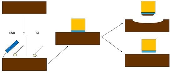
2.1. Specimen Preparation

In total, 120 specimens of Biodentine® were prepared using cylindrical acrylic blocks (Paladur, Heraeus Kulzer, Hanau, Germany) with a central hole measuring 5 mm in diameter and 2 mm in depth. Biodentine® was mixed according to the manufacturer’s instructions (Table 1) and placed in each acrylic block. Subsequently, all specimens were covered with wet gauze and stored at 37 °C with 100% humidity in an incubator to encourage setting time, and they were then divided into the two following groups:
  • Group 1 (n = 60): the adhesion process was done after setting time (12 min). To do so, the specimens were randomly divided into four subgroups of 15 specimens [18,24]:
    o
    Group 1a (n = 15): Optibond FL (Kerr Corp), three-step etch-and-rinse adhesive.
    o
    Group 1b (n = 15): Solobond M (VOCO), two-step etch-and-rinse adhesive.
    o
    Group 1c (n = 15): Scotchbond (3M), universal adhesive, used as a two-step etch-and-rinse adhesive.
    o
    Group 1d (n = 15): Scotchbond (3M), universal adhesive, used as a self-etch adhesive.
  • Group 2 (n = 60): the adhesion process was done after 24 h in the incubator. To do so, the specimens were randomly divided into four subgroups of 15 specimens. The distribution followed was the same as for Group 1.
After the adhesive protocol was performed (Table 1) the resin-based composite (Grandio, VOCO) was bonded onto the Biodentine® in all four groups. This composite block had a diameter of 2 ± 0.4 mm and a length of 3 ± 0.2 mm, and it was applied using a cylindrical-shaped matrix. Each composite block was polymerized using a light-emitting diode (LED) unit (Demi™, SDS Kerr Corp) with an intensity of 1200 mW/cm2 for 20 s. In order to verify the intensity of the LED unit, a radiometer was used (Bluephase® meter; Ivoclar Vivadent AG, Schaan, Liechtenstein). Then, the polymerized specimens were stored in 100% humidity at 37 °C for 24 h in an incubator.

2.2. Shear Bond Strength Measurement

The specimens were secured in a holder placed on the platform of the universal testing machine for shear bond strength (SBS) testing (Ultratester, Ultradent Products). A 2-mm-thick knife-edge blade was used to apply a vertical loading force at a cross-head speed of 0.5 mm/min until the failure of the bond between the Biodentine® and adhesive system. The shear bond strength in was noted in MPa.

2.3. Observation of Fractured Surfaces

After the shear bond test was performed, each fractured specimen was verified using a stereomicroscope (SteREO Discovery.V8, Zeiss, Oberkochen, Germany), and the failure mode was classified into adhesive, cohesive, and mixed failures. Adhesive failure only involves the adhesive surface keeping the Biodentine® and resin structure intact, cohesive failure involves failure within Biodentine®, and mixed failure involve both adhesive and cohesive failures simultaneously [19,24]. Photographs of the specimens were also taken.

2.4. Statistics

The Kolmogorov–Smirnov test was used to assess normal distribution of the data, and two-way ANOVA analysis was conducted to establish the significance level in the interaction between adhesive and time (12 min and 24 h) (α = 95%; p < 0.05). The Fisher post hoc test was used to show differences between groups. Within each group, one-way ANOVA was performed to establish the significance level between the different adhesive groups for each time. A chi-square test was used to establish the significance level in fracture failure. The statistical program used to perform all these tests was Statgraphics Centurion XV (StatPoint Technologies, Inc., Warrenton, VA, USA).

3. Results

3.1. Shear Bond Strength

All the data showed a normal distribution with p > 0.05 using the Kolmogorov–Smirnov test. The two-way ANOVA did not show statistically significant differences (p = 0.267) in the interaction between adhesive and time. When the data were analyzed, the two-way ANOVA did not show statistically significant differences among adhesives (p = 0.071), except for Optibond FL and one-step Scotchbond Universal. The results of SBS from the lowest to the highest were as follows: Scotchbond Universal one-step < Scotchbond Universal two-step < Solobond M < Optibond FL. The time analysis showed p = 0.056 between 12 min and 24 h, obtaining higher SBS values at 24 h (18.75 ± 6.44) and lower values at 12 min (16.65 ± 5.70).
However, when one-way ANOVA was conducted to analyze the data for each time period, a statistically significant difference was found among the adhesives at 12 min (p = 0.009), but not at 24 h (p = 0.813) (Table 2).

3.2. Fracture Failure

The failure between composite and Biodentine® can be observed in Figure 1. More cohesive failures in Biodentine® were shown in all of the 12-min groups, in contrast to the 24-h specimens having more adhesive failures (Figure 2). Optibond FL specimens showed 60% cohesive and 40% adhesive failures at 12 min; in contrast, at 24 h, only 13.33% of failures were cohesive. Similar results were found for Scotchbond. Solobond M samples resulted in 53.33% cohesive and 46.66% adhesive failures in Group 1, and 20% and 80%, respectively, in Group 2. The chi-square test showed statistically significant differences between cohesive and adhesive failures in Optibond FL (p = 0.008) and one-step Scotchbond Universal (p = 0.028).

4. Discussion

The current objective of restorative dentistry is to maintain pulp vitality. For this reason, pulp capping procedures are an efficient solution [12]. For the success of this treatment, it is really important to create a higher bond strength between the biomaterial used as pulp capping and the restorative material, which is usually a composite [11]. A perfect joint between each material is capable of spreading the stress of the occlusal forces [11]. Biodentine® is a dentine substitute with excellent biocompatibility, bioactivity, and biomineralization properties with great results when used in pulp capping procedures, as it as shown that this biomaterial induces TGF-β1 secretion from dental pulp cells [16,23,28,29,30]. Han and Okiji [31] demonstrated the biomineralization properties of Biodentine®, allowing the uptake of calcium and silicon in the adjacent dentine in contact with Biodentine®.
Adhesion studies revealed that a minimum bond strength to dentine and enamel of 17–20 MPa is needed to resist shrinkage and omit filtration and marginal gaps [18]. However, there are no studies that established optimum adhesion values between composite and Biodentine®.
There are many studies in the literature which studied the bond strength between restorative materials and MTA. Regarding adhesion on Biodentine®, only eight studies were published [11,18,19,21,24,25,30,31]. Furthermore, the SBS of composite to Biodentine® using four adhesive systems was investigated for the first time in the present study.
Biodentine, in pulp capping treatments is applied in a thin layer, and considering that is weaker than restorative material to be bonded, the SBS test can be used in these conditions to evaluate the adhesion strength. Further studies on other bond strength mode e.g., EM-SBS or TBS bond strength might give a better insight on adhesion values with this type of material.
It was suggested in all studies to make the samples with acrylic resin. However, Schmidt et al. [21] used cast models. In the present study, acrylic resin was used because it was an easy and fast way of standardizing the samples. Moreover, the studies differed in terms of the dimensions of the central hole. Altunsoy et al. [11] had a hole with a diameter of 3 mm and a depth of 1.5 mm. Cantekin et al. [18] used samples with a central hole of 5 mm × 2 mm, and Odabaş et al. [19] had a hole of 4 mm × 2 mm, coinciding with Çolak et al. [32]. After analyzing the studies, it was decided to use central holes of 5 mm × 2 mm, allowing better retention of the filling material. Regarding the composite block dimension, all studies coincided.
All studies stored the samples at 100% humidity and 37 °C, but they differed in the storing time. The Biodentine® manufacturer indicates that the setting time starts at 12 min, after which restorative treatment can be done. Thus, the samples of Group 1 were stored at 100% humidity and 37 °C for 12 min. Similarly to Odabaş et al. [19], the second group was stored for 24 h, in order to compare the bond strength when the restorative treatment was done in the same clinical visit or in another visit. It was shown in the data published that increasing the period of time did not have a significant effect on the mean SBS values. The evaluation of the shear bond strength between Biodentine® and composite was studied in eight studies. Those studies differed in the type of adhesive investigated. However, there were some adhesive systems that can be compared.
There were a few studies that thermocycled or aged samples through storage in water for long periods of time to mimic clinical conditions [33,34]. In both cases, the strength resistance was reduced, particularly when the samples were thermocycled [33]. Instead, Hashem et al. [34] in their study showed that the highest bond strength was obtained following 24 h of water storage, and it decreased at two weeks, one month, three months, and six months. However, they did not find differences between these time intervals in terms of maintaining an acceptable strength. Therefore, further studies of Biodentine aging are needed to obtain a more reliable simulation of oral environment.
The Scotchbond Universal is described as a universal adhesive that can be used as a self-etch adhesive and as an etch-and-rinse adhesive. In the present study, both techniques were investigated. Regardless of the adhesion approach, in a pulp capping procedure, the pulp remains vital and the surrounding tissue is dentine; thus, etching should be considered [7].
Biodentine® allows restorative treatment after 12 min of its mixing, which is when its setting time starts. Deepa et al. [35] studied a universal adhesive for self-etching and obtained a bond strength value of 5.66 MPa. In contrast, the results of this study found a mean value of 13.65 MPa for Group 1d (after 12 min of Biodentine® placement). This difference may be due to the different adhesive brand or operator variable.
Odabaş et al. [19] and Schmidt et al. [21] investigated a one-step self-etch adhesive and found mean values of 11.05 ± 3.85 and 6.25 ± 0.52 MPa, respectively, both having less bond strength compared to Scotchbond Universal used as a self-etch adhesive.
In this study, the two-step etch-and-rinse adhesive system was investigated with two adhesives, Scotchbond Universal and Solobond M, obtaining 15.63 ± 5.79 and 16.98 ± 3.56 MPa, respectively. Those values are in agreement with Cantekin et al. [18], who obtained a mean value of 17.7 ± 6.2 MPa using Allbond 2. In contrast, Odabaş et al. [19], who studied the Prime Bond NT, found a lower mean value of 9.12 ± 3.16 MPa at 12 min.
Apart from those systems, Optibond FL was also studied, finding a mean value of 20.34 ± 6.63 MPa. There are no data available that evaluated SBS using a three-step etch-and-rinse adhesive.
The results of the present study indicate that the three-step etch-and-rinse adhesive has a better bond strength than all other systems after 12 min of Biodentine® placement. Furthermore, the values obtained using this adhesive were superior to all other values obtained in previous studies with different adhesives at 12 min [18,19,21,35].
After 24 h of Biodentine® placement, the values obtained in the present study were higher than in a previous study [19]. Shafiei et al. [27], Cantekin et al. [18], and Tulumbaci et al. [24] studied a two-step etch-and-rinse adhesive founding significantly lower values compared to this study. In the same way, Schmidt et al. [21] and Altunsoy et al. [11] had lower results with a one-step self-etch adhesive. The poor results obtained in the Altunsoy et al. [11] study could be because they used a flow resin composite. Schmidt et al. [21] evaluated the SBS after 28 days of composite placement, in order to achieve Biodentine® setting; this could have interfered with the results, leading to lower values. The difference between the other studies may be due to the different adhesive brand or operator variable.
Optibond FL was not studied previously after 24 h, and it resulted in the highest bond strength value compared to the other systems. Moreover, there is a lack of studies using universal adhesives with Biodentine; therefore, further studies are needed to improve the adhesion of Biodentine to dental substrate and composite resins with this new group of adhesives.
The results of the present study showed a tendency of higher bond strength for the 24-h group, even though there was no significant difference with the 12-min groups. The better adhesion obtained for the 24-h group could be related to the setting time; thus, the Biodentine® achieved a better setting. Etching the Biodentine® seems to improve the adhesion after 12 min of mixing.
When comparing Scotchbond Universal as a self-etching agent regarding time, the 24-h group had significantly better results than after 12 min. Moreover, Optibond FL was demonstrated to be the best adhesive to achieve adhesion. However, the findings indicate that most of the adhesives showed an optimal SBS.
Some studies showed a surface roughness (Ra) range between 11.53 and 18.4 nm, depending on time (just after setting, at 45 min, and 24 h) or if the specimens were stored in a dry or wet environment before the test [36,37]. However, there were no studies published showing roughness measurement after surface treatment. In the present study, the surface roughness parameter following orthophosphoric acid etching was not analyzed with SEM images. Nonetheless, some studies reported that etching yields micromechanical interlocking due to the structural and chemical changes produced by Biodentine [34,38]. In the present study, this interlocking could have led to obtaining better results in the adhesives in which acid etching was used compared to the self-etch adhesives. The success of adhesion is related to the etching efficiency; for that reason, further studies are required to quantitatively measure Biodentine surface roughness after etching.
There is a lack of literature concerning the failure type of Biodentine®. Odabaş et al. [19] showed more cohesive fractures, and Deepa et al. [35] found 60% cohesive fractures and 40% adhesive fractures. Altunsoy et al. [11] did not have any adhesive fractures; however, Tulumbaci et al. [24] showed that most failures were because of adhesion. In the present study, more cohesive fractures were found in the 12-min group, and more adhesive fractures were found in the 24-h group. The difference among the data could be related to the operator variable, as well as the setting time of the Biodentine®, which probably resulted in more cohesive fractures after 12 min.
Only a few studies analyzed failure type between Biodentine® and composite [11,21,24,35]. Altunsoy et al. [11] applied the composite 72 h after Biodentine® placement and did not find any adhesive fractures, instead finding cohesive or mixed fractures. Those results are in contrast to those obtained in the present study. Deepa et al. [35] found 60% cohesive and 40% adhesive fractures. In their study, Tulumbaci et al. [24] showed that 12 of a total 15 specimens had adhesive fractures. These two studies followed the same setting time as Group 2. Furthermore, in the article of Schmidt et al. [21], 70% mixed fractures were obtained using the same protocol as Group 1.
When comparing these results with those obtained in our study, only Tulumbaci et al. [24] had similar results. Thus, there is no consensus in the literature regarding fracture failure type between the materials.
The limitations of this study were as follows: (1) the total setting time of Biodentine® was not considered; (2) the study design did not include adhesion to the tooth structure; (3) it was an SBS in vitro test. Future studies should evaluate the effect of allowing more time between the application of Biodentine® and the restorative material to observe the influence of the setting on the adhesion to the restorative material.

5. Conclusions

All adhesives obtained similar SBS results with Biodentine® following 12 min and 24 h of adhesion; however, but superior adhesion was observed at 24 h following Biodentine® placement. Hence, before the restorative treatment procedure, it is essential to consider the longest possible setting time for Biodentine®. Moreover, it should be taken into account that a three-step etch-and-rinse adhesive has superior adhesion values; however, most of the adhesion systems reached an optimal shear bond. In order to avoid dentine etching when using a self-etching agent, waiting 24 h seems to improve the bond strength.

Author Contributions

Conceptualization, V.C. and M.A.; methodology, V.C., M.A., and L.P.; investigation, V.C., M.A., and L.P.; resources, M.A.; data curation, V.C. and M.A.; writing—original draft preparation, V.C. and M.A.; writing—review and editing, M.A., L.P., and L.G.-T.; supervision, L.G.-T.

Funding

This research received no external funding.

Acknowledgments

The authors would like to thank VOCO for providing the materials for this study.

Conflicts of Interest

The authors declare no conflicts of interest.

References

  1. Sezinando, A. Looking for ideal adhesive—A review. Rev. Port. Estomatol. Med. Dent. Cir. Maxilofac. 2014, 55, 194–206. [Google Scholar] [CrossRef]
  2. Breschi, L.; Mazzoni, A.; Ruggeri, A.; Cadenaro, M.; Di Lenarda, R.; De Stefano Dorigo, E. Dental adhesion review: Aging and stability of the bonded interface. Dent. Mater. 2008, 24, 90–101. [Google Scholar] [CrossRef] [PubMed]
  3. Saiz, U.; Bock, T. Testing Adhesion of Direct Restoratives to Dental Hard Tissue—A Review. J. Adhes. Dent. 2010, 12, 343–371. [Google Scholar]
  4. De Munck, J.; Van Landuyt, K.; Peumans, M.; Potevin, A.; Labrechts, P.; Braem, M.; Van Meerbeek, B. A Critical Review of the Durability of Adhesion to Tooth Tissue: Methods and Results. J. Dent. Res. 2005, 84, 118–132. [Google Scholar] [CrossRef]
  5. Van Meerbeek, B.; De Munck, J.; Yoshida, Y.; Inoue, S.; Vargas, M.; Vijay, P.; Van Landuyt, K.; Lambrechts, P.; Vanherle, G. Buonocore memorial lecture. Adhesion to enamel and dentin: Current status and future challenges. Oper. Dent. 2003, 28, 215–235. [Google Scholar]
  6. Bjomdal, L.; Relt, C.; Bruun, G.; Markvart, M.; Kjaeldgaard, M.; Näsman, P.; Thordrup, M.; Dige, I.; Nyvad, B.; Fransson, H. Treatment of deep caries lesions in adults: Randomized clinical trials comparing stepwise vs direct complete excavation, and direct pulp capping vs partial pulpotomy. Eur. J. Oral Sci. 2010, 118, 290–297. [Google Scholar] [CrossRef]
  7. De Souza Costa, C.A.; Hebling, J.; Hanks, C.T. Current status of pulp capping with dentin adhesive systems: A review. Dent. Mater. 2000, 16, 188–197. [Google Scholar] [CrossRef]
  8. Akhlaghi, N.; Khademi, A. Outcomes of vital pulp therapy in permanent teeth with different medicaments based on review of the literature. J. Dent. Res. 2015, 12, 406–417. [Google Scholar]
  9. Hilton, T.J.; Ferracane, J.L.; Mancl, L. Comparison of CaOH with MTA for direct pulp capping. J. Dent. Res. 2013, 92, S16–S22. [Google Scholar] [CrossRef]
  10. Paula, A.B.; Laranjo, M.; Marto, C.M.; Paulo, S.; Abrantes, A.M.; Casalta-Lopes, J.; Marques-Ferreira, M.; Botelho, M.F.; Carrilho, E. Direct pulp capping: What is the most effective therapy? Systematic review and meta-analysis. J. Evid. Based Dent. Pract. 2018, 18, 298–314. [Google Scholar] [CrossRef]
  11. Altunsoy, M.; Tanriver, M.; Ok, E.; Kucukylmaz, E. Shear Bond Strength of a Self- adhering Flowable Composite and a Flowable Base Composite to Mineral Trioxide Aggregate, Calcium-enriched Mixture Cement, and Biodentine. J. Endod. 2015, 41, 1691–1695. [Google Scholar] [CrossRef] [PubMed]
  12. Schwendicke, F.; Brouwer, F.; Schwendicke, A.; Paris, S. Different materials for direct pulp capping: Systematic review and meta-analysis and trial sequential analysis. Clin. Oral Investig. 2016, 20, 1121–1332. [Google Scholar] [CrossRef] [PubMed]
  13. Kundzina, R.; Stangvaltaite, L.; Eriksen, H.M.; Kerosuo, E. Capping carious exposures in adults: A randomized controlled trial investigating mineral trioxide aggregate versus calcium hydroxide. Int. Endod. J. 2017, 50, 924–932. [Google Scholar] [CrossRef]
  14. Tran, X.V.; Salehi, H.; Truong, M.T.; Sandra, M.; Sadoine, J.; Jacquot, B.; Cuisinier, F.; Chaussain, C.; Boukpessi, T. Reparative mineralized tissue characterization after direct pulp capping with calcium-silicate-based cements. Materials. 2019, 12, 2102. [Google Scholar] [CrossRef] [PubMed]
  15. Tran, X.V.; Gorin, C.; Willing, C.; Baroukh, B.; Pellat, B.; Decup, F.; Opsahl Vital, S.; Chaussain, C.; Boukpessi, T. Effect of a calcium-silicate-based restorative cement on pulp repair. J. Dent. Res. 2012, 91, 1166–1171. [Google Scholar] [CrossRef] [PubMed]
  16. Nowicka, A.; Lipski, M.; Parafiniuk, M.; Sporniak-Tutak, K.; Lichota, D.; Kosierkiewicz, A.; Kaczmarek, W.; Buczkowska-Radlinska, J. Response of Human Dental Pulp Capped with Biodentine and mineral trioxide aggregate. J. Endod. 2013, 39, 743–747. [Google Scholar] [CrossRef]
  17. Camilleri, J. Investigation of biodentine as dentine replacement material. J. Dent. 2013, 41, 600–610. [Google Scholar] [CrossRef]
  18. Cantekin, K.; Avci, S. Evaluation of shear bond strength of two resin-based composites and glass ionomer cement to pure tricalcium silicate-based cement (Biodentine®). J. Appl. Oral Sci. 2014, 22, 302–306. [Google Scholar] [CrossRef]
  19. Odabaş, M.; Bani, M.; Tirali, R.E. Shear Bond Strengths of Different Adhesive Systems to Biodentine. Sci. World J. 2013, 10, 626103. [Google Scholar] [CrossRef]
  20. Malkondu, Ö.; Kazandag, M.K.; Kazazoglu, E. A Review on Biodentine, a Contemporary Dentine Replacement and Repair Material. BioMed Res. Int. 2014, 160951. [Google Scholar] [CrossRef]
  21. Schmidt, A.; Schäfer, E.; Dammaschke, T. Shear bond Strength of lining materials to calcium-silicate cements at different time intervals. J. Adhes. Dent. 2017, 19, 129–135. [Google Scholar] [PubMed]
  22. Koubi, G.; Colon, P.; Franquin, J.C.; Hartmann, A.; Richard, G.; Faure, M.O.; Lambert, G. Clinical evaluation of the performance and safety of a new dentine substitute, Biodentine, in the restoration of posterior teeth—A prospective study. Clin. Oral Invest. 2013, 17, 243–249. [Google Scholar] [CrossRef] [PubMed]
  23. Laurent, P.; Camps, J.; About, I. Biodentine(TM) induces TGF-beta1 release from human pulp cells and early dental pulp mineralization. Int. Endod. J. 2012, 45, 439–448. [Google Scholar] [CrossRef] [PubMed]
  24. Tulumbalaci, F.; Erkmen, M.; Arikan, V.; Safa, M. Shear Bond strength of different restorative materials to mineral trioxide aggregate and Biodentine. J. Conserv. Dent. 2017, 20, 292–296. [Google Scholar] [CrossRef]
  25. Kaur, M.; Singh, H.; Dhillon, J.S.; Batra, M.; Saini, M. MTA versus Biodentine: Review of literature with a comparative analysis. J. Clin. Diagn. Res. 2017, 11, ZG01–ZG05. [Google Scholar] [CrossRef]
  26. Uesrichai, N.; Nirunsittirat, A.; Chuveera, P.; Srisuwan, T.; Sastraruji, T.; Chompu-Inwai, P. Partial pulpotomy with two bioactive cements in permanent teeth of 6- to 18-year-old patients with signs and symptoms indicative of irreversible pulpitis: A noninferiority randomized controlled trial. Int. Endod. J. 2019, 52, 749–759. [Google Scholar] [CrossRef]
  27. Shafiei, F.; Doozandeh, M.; Gharibpour, F.; Adj, A. Effect of reducing acid-etching duration time on compressive strength and bonding of a universal adhesive to calcium silicate cements. Int. Endod. J. 2019, 52, 530–539. [Google Scholar] [CrossRef]
  28. Song, M.; Yu, B.; Kim, S.; Hayashi, M.; Smith, C.; Sohn, S.; Kim, E.; Stevenson, R.G.; Kim, R.H. Clinical and molecular perspectives of reparative dentin formation: Lessons learned from pulp-capping materials and the emerging roles of calcium. Dent. Clin. N. Am. 2017, 61, 93–110. [Google Scholar] [CrossRef]
  29. Karabucak, B.; Li, D.; Lim, J.; Iqbal, M. Vital pulp therapy with mineral trioxide aggregate. Dent. Traumatol. 2005, 21, 240–243. [Google Scholar] [CrossRef]
  30. Parirokh, M.; Torabinejad, M. Mineral trioxide aggregate: A comprehensive literature review-part1 1: Chemical, physical and antibacterial properties. J. Endod. 2010, 36, 16–27. [Google Scholar] [CrossRef]
  31. Han, L.; Okiji, T. Uptake of calcium and silicon released from calcium silicate-based endodontic materials into root canal dentine. Int. Endod. J. 2011, 44, 1081–1087. [Google Scholar] [CrossRef] [PubMed]
  32. Çolak, H.; Tokay, U.; Uzgur, R.; Uzgur, Z.; Ercan, E.; Hamidi, M.M. The effect of different adhesives and setting times on bond strength between Biodentine and composite. J. Appl. Biomater. Funct. Mat. 2016, 14, e217–e222. [Google Scholar]
  33. Meraji, N.; Camilleri, J. Bonding over dentin replacement materials. J. Endod. 2017, 43, 1343–1349. [Google Scholar] [CrossRef] [PubMed]
  34. Hashem, D.F.; Foxton, R.; Manoharan, A.; Watson, T.F.; Banerjee, A. The physical characteristics of resin composite-calcium silicate interface as part of layered/laminate adhesive restoration. Dent. Mater. 2014, 30, 343–349. [Google Scholar] [CrossRef] [PubMed]
  35. Deepa, V.L.; Dhamaraju, B.; Bollu, I.P.; Balaji, T.S. Shear bond strength evaluation of resin composite bonded to three different liners: TheraCal LC, Biodentine, and resin-modified glass ionomer cement using universal adhesive: An in vitro study. J. Conserv. Dent. 2016, 19, 166–170. [Google Scholar] [CrossRef]
  36. Attik, G.N.; Villat, C.; Hallay, F.; Pradelle-Plasse, N.; Bonnet, H.; Moreau, K.; Colon, P.; Grosgogeat, B. In vitro biocompatibility of a dentine substitute cement on human MG63 osteoblasts cells: Biodentine™ versus MTA®. Int. Endod. J. 2014, 47, 1133–1141. [Google Scholar] [CrossRef]
  37. Aksel, H.; Küçükkaya Eren, S.; Askerbeyli Órs, S.; Karaismailoğlu, E. Surface and vertical dimensional changes of mineral trioxide aggregate and biodentine in different environmental conditions. J. Appl. Oral Sci. 2019, 27, e20180093. [Google Scholar] [CrossRef]
  38. Cengiz, E.; Ulusoy, N. Microshear bond strength of tri-calcium silicate-based cement to different restorative materials. J. Adhes. Dent. 2016, 18, 231–237. [Google Scholar]
Figure 1. Failure mode in different experimental groups.
Coatings 09 00783 g001
Figure 2. Stereomicroscopic images of the failure mode in different experimental groups under 0.945× magnification (scale bar = 500 µm). (A) Cohesive failure at 12 min in Biodentine self-etch; (B) cohesive failure at 12 min in Biodentine total etch (two steps); (C) adhesive failure at 24 h in Biodentine self-etch; (D) adhesive failure at 24 h in Biodentine total etch (two steps).
Coatings 09 00783 g002
Table 1. Overview of the investigated materials.
MaterialManufacturerLot NumberClinical Procedure
Calcium silicate cementBiodentine®
(Septodont, Saint Maur des Fosses, France)
B22106Liquid and powder mixing at 4000–4200 rpm for 30 s
Nanohybrid compositeGrandio®
(VOCO GmbH, Cuxhaven, Germany)
1643371Light cure for 20 s
3-step etch-and-rinse adhesiveOptibond® FL
(Kerr Corp, Orange, CA, USA)
6605713
  • 37% phosphoric acid for 15 s
  • Water rinse for 15 s
  • Dry for 10 s
  • Apply primer for 15 s
  • Gentle air stream for 10 s
  • Apply adhesive
  • Gentle air stream
  • Light cure for 10 s
2-step etch-and-rinse adhesiveSolobond® M
(Voco GmbH, Cuxhaven, Germany)
1637105
  • 37% phosphoric acid for 15 s
  • Water rinse for 15 s
  • Dry for 10 s
  • Apply adhesive for 30 s
  • Gentle air stream for 5 s
  • Light cure for 20 s
Universal adhesiveScotchbond Universal® (3M ESPE, St. Paul, MN, USA)80702A
  • 37% phosphoric acid for 15 s as an etch and rinse
  • Apply adhesive 20 s
  • Gentle air stream for 5 s
  • Light cure for 10 s
Table 2. Mean shear bond strength (SBS) values (MPa) and standard deviation (SD) by adhesion time.
Adhesive12 min MPa (SD)24 h MPa (SD)
Scotchbond Universal 1-step13.65 (4.62) a19.16 (6.62) a
Scotchbond Universal 2-step15.63 (5.79) a17.73 (7.54) a
Solobond M16.98 (3.56) a,b18.24 (6.60) a
Optibond FL20.34 (6.63) b19.87 (5.24) a
Different letters in the same column show statistically significant differences (p < 0.05).

