
Wound Coating Collagen-Based Composites with Ag Nanoparticles: Synthesis, Structure and Biological Activity
 , ,
, ,  , , ,
, , ,
Abstract
:1. Introduction
2. Materials and Methods
2.1. Obtaining Collagen from Biological Raw Materials
2.2. Modification of Collagen by Silver Nanoparticles Obtained by Metal-Vapor Synthesis
2.3. Obtaining Fibroblast Cell Cultures to Assess the Cytotoxicity and Biocompatibility of Collagen
2.4. Determination of the Number of Living Cells on a Collagen Substrate by Staining with a Vital Dye in the MTT Assay
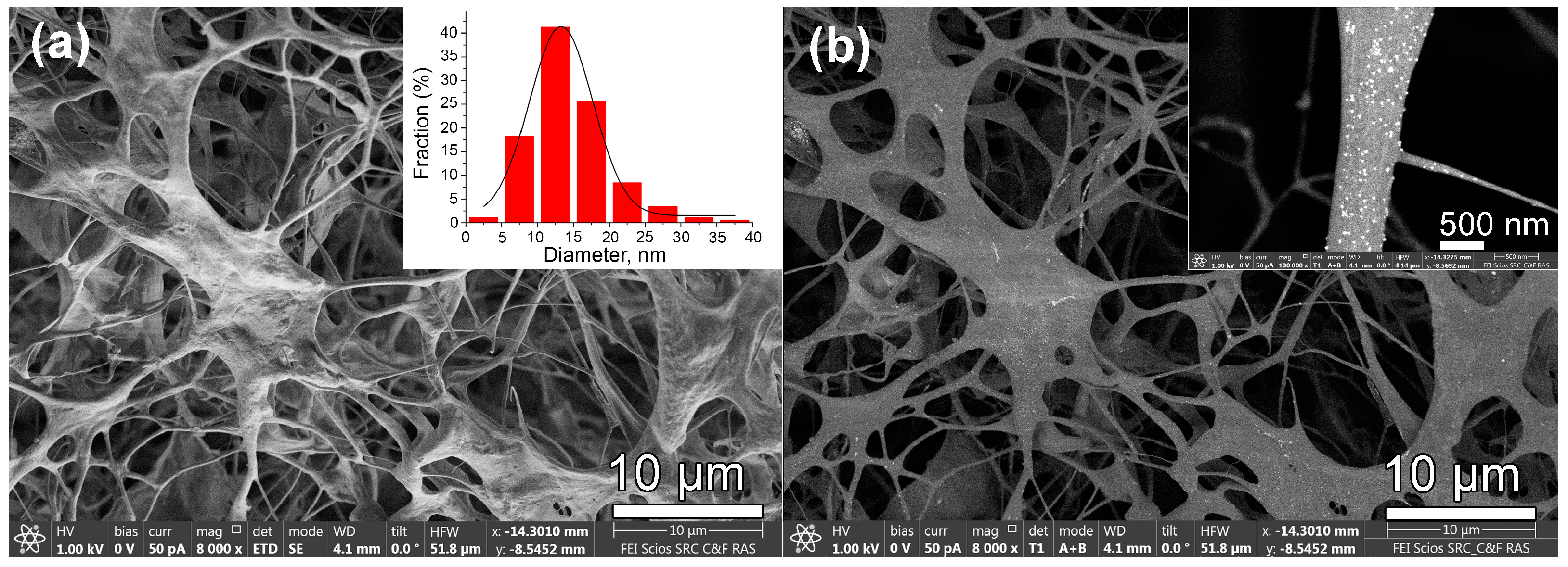
2.5. Scanning Electron Microscopy (SEM)
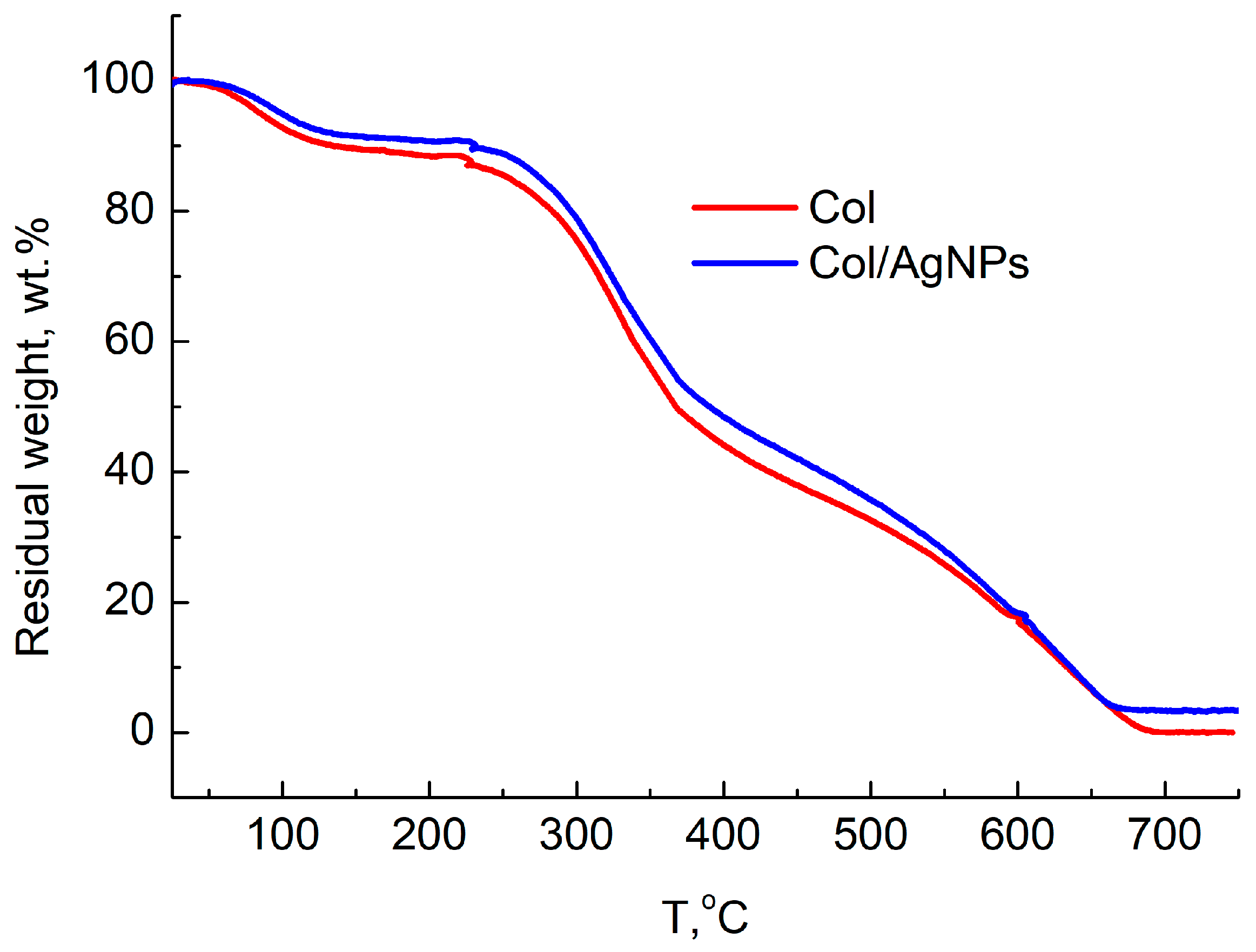
2.6. Thermogravimetric Analysis (TGA)
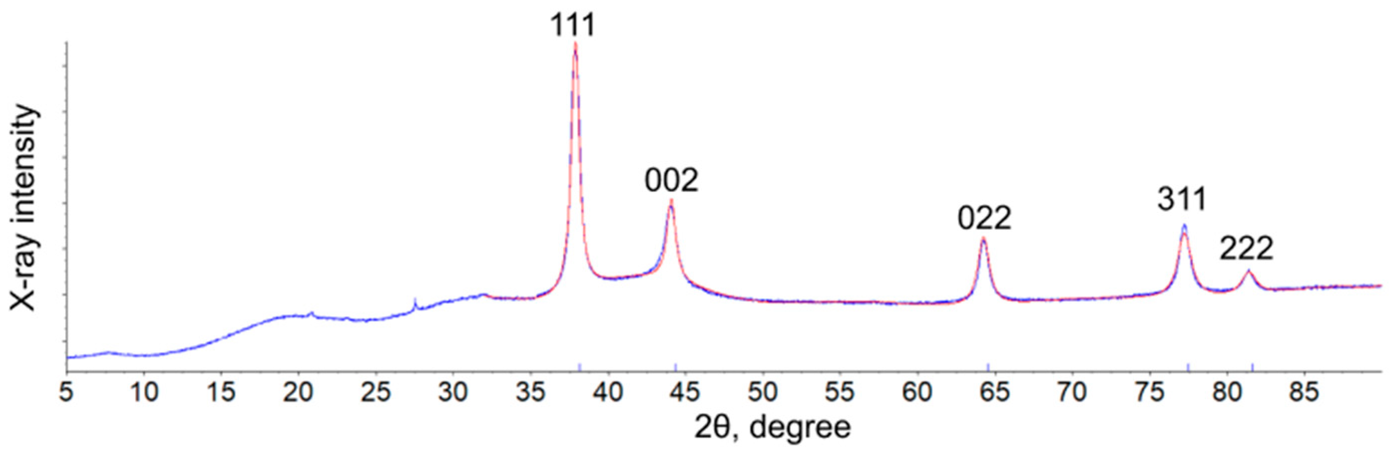
2.7. Powder X-ray Diffraction (PXRD)
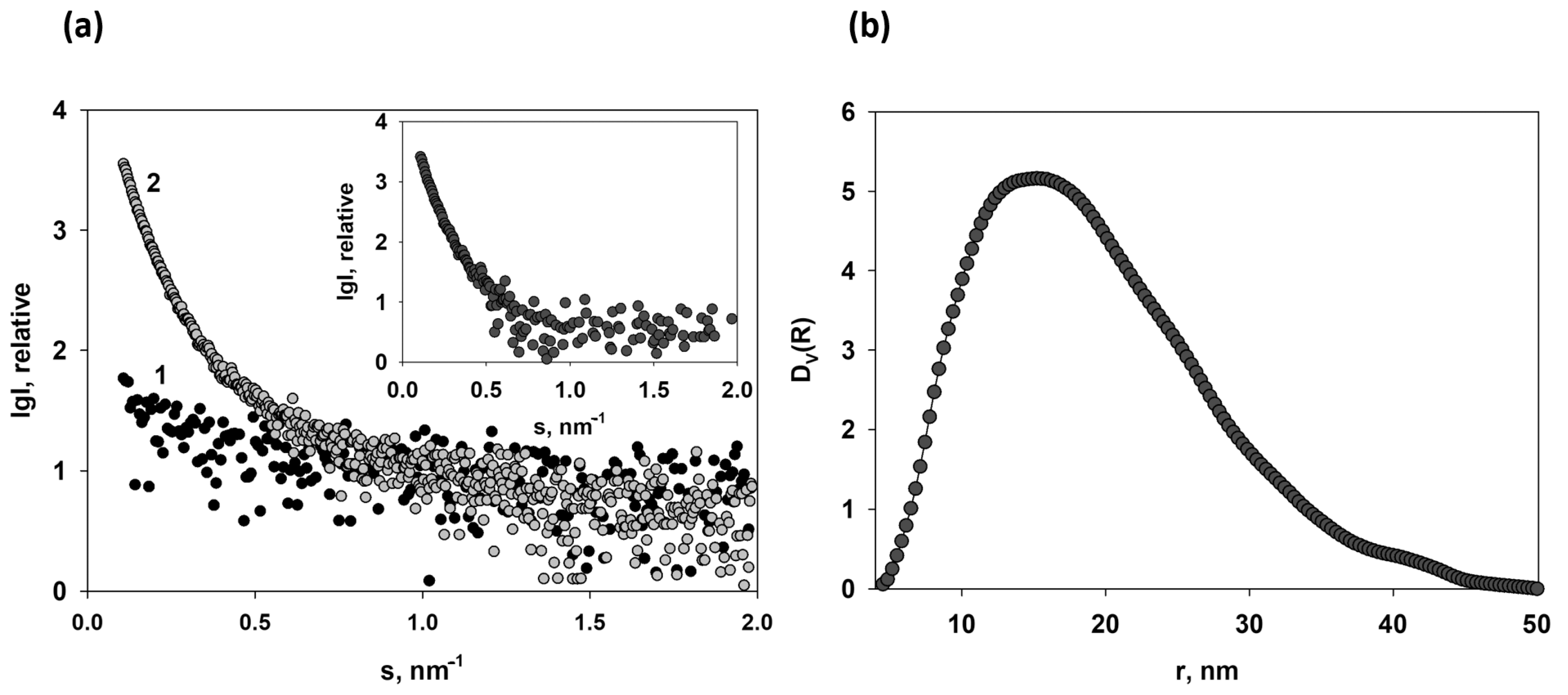
2.8. Small-Angle X-ray Scattering (SAXS)
2.9. X-ray Photoelectron Spectroscopy (XPS)
2.10. Antimicrobial Tests
3. Results and Discussion
4. Conclusions
Supplementary Materials
Author Contributions
Funding
Institutional Review Board Statement
Informed Consent Statement
Data Availability Statement
Acknowledgments
Conflicts of Interest
References
- Sabarees, G.; Velmurugan, V.; Tamilarasi, G.P.; Alagarsamy, V.; Solomon, V.R. Recent Advances in Silver Nanoparticles Containing Nanofibers for Chronic Wound Management. Polymers 2022, 14, 3994. [Google Scholar] [CrossRef]
- Norouzi, M.; Boroujeni, S.M.; Omidvarkordshouli, N.; Soleimani, M. Advances in Skin Regeneration: Application of Electrospun Scaffolds. Adv. Healthc. Mater. 2015, 4, 1114–1133. [Google Scholar] [CrossRef] [PubMed]
- Chang, H.; Feng, H.; Wang, R.; Zhang, X.; Wang, J.; Li, C.; Zhang, Y.; Li, L.; Ho, S.-H. Enhanced energy recovery from landfill leachate by linking light and dark bio-reactions: Underlying synergistic effects of dual microalgal interaction. Water Res. 2023, 231, 119578. [Google Scholar] [CrossRef] [PubMed]
- Pallaske, F.; Pallaske, A.; Herklotz, K.; Boese-Landgraf, J. The significance of collagen dressings in wound management: A review. J. Wound Care 2018, 27, 692–702. [Google Scholar] [CrossRef] [PubMed]
- Kempf, M.; Miyamura, Y.; Liu, P.Y.; Chen, A.C.H.; Nakamura, H.; Shimizu, H.; Tabata, Y.; Kimble, R.M.; McMillan, J.R. A denatured collagen microfiber scaffold seeded with human fibroblasts and keratinocytes for skin grafting. Biomaterials 2011, 32, 4782–4792. [Google Scholar] [CrossRef] [PubMed]
- Fuente, D.; Tornos, A.; Príncep, A.; Lorenzo, J.M.; Pateiro, M.; Berrada, H.; Barba, F.J.; Ruiz, M.-J.; Martí-Quijal, F.J. Scaling-up Processes: Patents and Commercial. Adv. Food Nutr. Res. 2020, 92, 187–223. [Google Scholar] [CrossRef]
- Shelah, O.; Wertheimer, S.; Haj-Ali, R.; Lesman, A. Coral-derived Collagen Fibers for Engineering Aligned Tissues. Tissue Eng. Part A 2021, 27, 187–200. [Google Scholar] [CrossRef]
- Claverie, M.; McReynolds, C.; Petitpas, A.; Thomas, M.; Fernandes, S.C.M. Marine-Derived Polymeric Materials and Biomimetics: An Overview. Polymers 2020, 12, 1002. [Google Scholar] [CrossRef]
- Nurilmala, M.; Hizbullah, H.H.; Karnia, E.; Kusumaningtyas, E.; Ochiai, Y. Characterization and Antioxidant Activity of Collagen, Gelatin, and the Derived Peptides From Yellowfin Tuna. Mar. Drugs 2020, 18, 98. [Google Scholar] [CrossRef]
- Maistrenko, L.; Iungin, O.; Pikus, P.; Pokholenko, I.; Gorbatiuk, O.; Moshynets, O.; Okhmat, O.; Kolesnyk, T.; Potters, G.; Mokrousova, O. Collagen Obtained from Leather Production Waste Provides Suitable Gels for Biomedical Applications. Polymers 2022, 14, 4749. [Google Scholar] [CrossRef]
- Zille, A.; Fernandes, M.M.; Francesko, A.; Tzanov, T.; Fernandes, M.; Oliveira, F.R.; Almeida, L.; Amorim, T.; Carneiro, N.; Esteves, M.F.; et al. Size and aging effects on antimicrobial efficiency of silver nanoparticles coated on polyamide fabrics activated by atmospheric DBD plasma. ACS Appl. Mater. Interfaces 2015, 7, 13731–13744. [Google Scholar] [CrossRef] [PubMed]
- Van Vugt, T.A.G.; Walraven, J.M.B.; Geurts, J.A.P.; Arts, J.J.C. Antibiotic-Loaded Collagen Sponges in Clinical Treatment of Chronic Osteomyelitis: A Systematic Review. J. Bone Jt. Surg. Am. 2018, 100, 2153–2161. [Google Scholar] [CrossRef] [PubMed]
- Yıldırım, M.A.; Çakır, M.; Fındık, S.; Kişi, Ö.; Şentürk, M. Comparison of the efficacy of growth factor collagen and antibiotic collagen on colon anastomosis in experimental animals with peritonitis. Indian J. Gastroenterol. 2021, 40, 309–315. [Google Scholar] [CrossRef] [PubMed]
- Berechet, M.D.; Gaidau, C.; Miletic, A.; Pilic, B.; Râpă, M.; Stanca, M.; Ditu, L.M.; Constantinescu, R.; Lazea-Stoyanova, A. Bioactive Properties of Nanofibres Based on Concentrated Collagen Hydrolysate Loaded with Thyme and Oregano Essential Oils. Materials 2020, 13, 1618. [Google Scholar] [CrossRef]
- Ying, H.; Zhou, J.; Wang, M.; Su, D.; Ma, Q.; Lv, G.; Chen, J. In situ formed collagen-hyaluronic acid hydrogel as biomimetic dressing for promoting spontaneous wound healing. Mater. Sci. Eng. C Mater. Biol. Appl. 2019, 101, 487–498. [Google Scholar] [CrossRef]
- Nogueira, L.F.B.; Cruz, M.A.E.; Aguilar, G.J.; Tapia-Blácido, D.R.; da Silva Ferreira, M.E.; Maniglia, B.C.; Bottini, M.; Ciancaglini, P.; Ramos, A.P. Synthesis of Antibacterial Hybrid Hydroxyapatite/Collagen/Polysaccharide Bioactive Membranes and Their Effect on Osteoblast Culture. Int. J. Mol. Sci. 2022, 23, 7277. [Google Scholar] [CrossRef]
- Vijayan, V.; Sreekumar, S.; Ahina, K.M.; Lakra, R.; Kiran, M.S. Lanthanum Oxide Nanoparticles Reinforced Collagen ƙ-Carrageenan Hydroxyapatite Biocomposite as Angio-Osteogenic Biomaterial for In Vivo Osseointegration and Bone Repair. Adv. Biol. 2023, e2300039. [Google Scholar] [CrossRef]
- Martins, E.; Diogo, G.S.; Pires, R.; Reis, R.L.; Silva, T.H. 3D Biocomposites Comprising Marine Collagen and Silica-Based Materials Inspired on the Composition of Marine Sponge Skeletons Envisaging Bone Tissue Regeneration. Mar. Drugs 2022, 20, 718. [Google Scholar] [CrossRef]
- Goodarzi, H.; Hashemi-Najafabadi, S.; Baheiraei, N.; Bagheri, F. Preparation and Characterization of Nanocomposite Scaffolds (Collagen/β-TCP/SrO) for Bone Tissue Engineering. Tissue Eng. Regen. Med. 2019, 16, 237–251. [Google Scholar] [CrossRef]
- Añazco, C.; Riedelsberger, J.; Vega-Montoto, L.; Rojas, A. Exploring the Interplay between Polyphenols and Lysyl Oxidase Enzymes for Maintaining Extracellular Matrix Homeostasis. Int. J. Mol. Sci. 2023, 24, 10985. [Google Scholar] [CrossRef]
- Tseomashko, N.Y.; Abidova, A.D.; Makhmudov, S.D.; Azimova, S.S.; Aripova, S.F. Evaluation of the biological activity of phyllalbin and rutin, and application in wound dressings. J. Hum. Univ. 2021, 48, 530–550. [Google Scholar]
- Tseomashko, N.E.; Abidova, A.D.; Makhmudov, S.D.; Rakhmanov, A.K. Evaluation of gastroprotective and regenerative properties of flavonoids isolated from buds of Saphora Japonica in collagen. J. Hum. Univ. 2021, 48, 551–561. [Google Scholar]
- Kadian, S.; Manik, G.; Das, N.; Nehra, P.; Chauhan, R.P.; Roy, P. Synthesis, characterization and investigation of synergistic antibacterial activity and cell viability of silver-sulfur doped graphene quantum dot (Ag@S-GQDs) nanocomposites. J. Mater. Chem. 2020, 8, 3028–3037. [Google Scholar] [CrossRef]
- Rai, M.; Yadav, A.; Gade, A. Silver nanoparticles as a new generation of antimicrobials. Biotechnol. Adv. 2009, 27, 76–83. [Google Scholar] [CrossRef]
- Shrivastava, S.; Bera, T.; Roy, A.; Singh, G.; Ramachandrarao, P.; Dash, D. Characterization of enhanced antibacterial effects of novel silver nanoparticles. Nanotechnology 2007, 18, 225103. [Google Scholar] [CrossRef]
- Kim, J.S.; Kuk, E.; Yu, K.N.; Kim, J.-H.; Park, S.J.; Lee, H.J.; Kim, S.H.; Park, Y.K.; Park, Y.H.; Hwang, C.-Y.; et al. Antimicrobial effects of silver nanoparticles. Nanomed. Nanotechnol. Biol. Med. 2007, 3, 95–101. [Google Scholar] [CrossRef] [PubMed]
- Amiri, N.; Ghaffari, S.; Hassanpour, I.; Chae, T.; Jalili, R.; Taghi Kilani, R.; Ko, F.; Ghahary, A.; Lange, D. Antibacterial Thermosensitive Silver–Hydrogel Nanocomposite Improves Wound Healing. Gels 2023, 9, 542. [Google Scholar] [CrossRef]
- Albu, M.G.; Vladkova, T.G.; Ivanova, I.A.; Shalaby, A.S.A.; Moskova-Doumanova, V.S.; Staneva, A.D.; Dimitriev, Y.B.; Kostadinova, A.S.; Topouzova-Hristova, T.I. Preparation and Biological Activity of New Collagen Composites, Part I: Collagen/Zinc Titanate Nanocomposites. Appl. Biochem. Biotechnol. 2016, 180, 177–193. [Google Scholar] [CrossRef] [PubMed]
- Park, D.; Osuji, C.O.; Kim, J.W. Multi-Compartmentalized Cellulose Macrobead Catalysts for In Situ Organic Reaction in Aqueous Media. Small Methods 2023, 7, 2201195. [Google Scholar] [CrossRef]
- Prucek, R.; Kvítek, L.; Panáček, A.; Vančurová, L.; Soukupová, J.; Jančík, D.; Zbořil, R. Polyacrylate-assisted synthesis of stable copper nanoparticles and copper(I) oxide nanocubes with high catalytic efficiency. J. Mater. Chem. 2009, 19, 8463–8469. [Google Scholar] [CrossRef]
- Jinga, S.I.; Isopencu, G.; Stoica-Guzun, A.; Stroescu, M.; Ferdes, M.; Ohreac, B. Silver green synthesis on bacterial cellulose membranes using tannic acid. Dig. J. Nanomater. Bios. 2013, 8, 1711–1717. [Google Scholar]
- Sarkandi, F.A.; Montazer, M.; Harifi, T.; Rad, M.M. Innovative preparation of bacterial cellulose/silver nanocomposite hydrogels: In situ green synthesis, characterization, and antibacterial properties. J. Appl. Polym. Sci. 2021, 138, 49824. [Google Scholar] [CrossRef]
- Kucińska-Lipka, J.; Gubanska, I.; Janik, H. Bacterial Cellulose in the Field of Wound Healing and Regenerative Medicine of Skin: Recent Trends and Future Prospectives. Polym. Bull. 2015, 72, 2399–2419. [Google Scholar] [CrossRef]
- Wei, D.; Sun, W.; Qian, W.; Ye, Y.; Ma, X. The synthesis of chitosan-based silver nanoparticles and their antibacterial activity. Carbohydr. Res. 2009, 344, 2375–2382. [Google Scholar] [CrossRef]
- Twu, Y.-K.; Chen, Y.-W.; Shih, C.-M. Preparation of silver nanoparticles using chitosan suspensions. Powder Technol. 2008, 185, 251–257. [Google Scholar] [CrossRef]
- Vasil’kov, A.Y.; Abd-Elsalam, K.A.; Olenin, A.Y. Biogenic Silver Nanoparticles: New Trends and Applications. In Green Synthesis of Silver Nanomaterials; Abd-Elsalam, K.A., Ed.; Elsevier: Amsterdam, The Netherlands, 2021; pp. 241–281. [Google Scholar] [CrossRef]
- Mikhailov, O.V.; Mikhailova, E.O. Elemental silver nanoparticles: Biosynthesis and bio applications. Materials 2019, 12, 3177. [Google Scholar] [CrossRef] [PubMed]
- Ahn, E.-Y.; Jin, H.; Park, Y. Assessing the antioxidant, cytotoxic, apoptotic and wound healing properties of silver nanoparticles green-synthesized by plant extracts. Mater. Sci. Eng. C 2019, 101, 204–216. [Google Scholar] [CrossRef]
- Hamedi, S.; Shojaosadati, S.A. Rapid and green synthesis of silver nanoparticles using Diospyros lotus extract: Evaluation of their biological and catalytic activities. Polyhedron 2019, 171, 172–180. [Google Scholar] [CrossRef]
- Hernández-Morales, L.; Espinoza-Gómez, H.; Flores-López, L.Z.; Sotelo-Barrera, E.L.; Núñez-Rivera, A.; Cadena-Nava, R.D.; Alonso-Núñez, G.; Espinoza, K.A. Study of the green synthesis of silver nanoparticles using a natural extract of dark or white Salvia hispanica L. seeds and their antibacterial application. Appl. Surf. Sci. 2019, 489, 952–961. [Google Scholar] [CrossRef]
- Küp, F.Ö.; Çoşkunçay, S.; Duman, F. Biosynthesis of silver nanoparticles using leaf extract of Aesculus hippocastanum (horse chestnut): Evaluation of their antibacterial, antioxidant and drug release system activities. Mater. Sci. Eng. C 2020, 107, 110207. [Google Scholar] [CrossRef]
- Tseomashko, N.E.; Rai, M.; Vasil’kov, A.Y. New hybrid materials for wound cover dressings. In Bio-Polymer-Based Nano Films; Rai, M., Dos Santos, C.A., Eds.; Elsevier Inc.: New York, NY, USA, 2021; pp. 203–246. [Google Scholar] [CrossRef]
- Barbaro, D.; Di Bari, L.; Gandin, V.; Evangelisti, C.; Vitulli, G.; Schiavi, E.; Marzano, C.; Ferretti, A.M.; Salvadori, P. Glucose-coated superparamagnetic iron oxide nanoparticles prepared by metal vapour synthesis are electively internalized in a pancreatic adenocarcinoma cell line expressing GLUT1 transporter. PLoS ONE 2015, 10, e0123159. [Google Scholar] [CrossRef] [PubMed]
- Cárdenas-Triviño, G.; Cruzat-Contreras, C. Study of Aggregation of Gold Nanoparticles in Chitosan. J. Clust. Sci. 2018, 29, 1081–1088. [Google Scholar] [CrossRef]
- Vasil’kov, A.; Voronova, A.; Batsalova, T.; Moten, D.; Naumkin, A.; Shtykova, E.; Volkov, V.; Teneva, I.; Dzhambazov, B. Evolution of gold and iron oxide nanoparticles in conjugates with methotrexate: Synthesis and anticancer effects. Materials 2023, 16, 3238. [Google Scholar] [CrossRef] [PubMed]
- Vasil’kov, A.; Butenko, I.; Naumkin, A.; Voronova, A.; Golub, A.; Buzin, M.; Shtykova, E.; Volkov, V.; Sadykova, V. Hybrid Silver-Containing Materials Based on Various Forms of Bacterial Cellulose: Synthesis, Structure, and Biological Activity. Int. J. Mol. Sci. 2023, 24, 7667. [Google Scholar] [CrossRef] [PubMed]
- Jose, D.; Jagirdar, B.R. Au@Pd core-shell nanoparticles through digestive ripening. J. Phys. Chem. C 2008, 112, 10089–10094. [Google Scholar] [CrossRef]
- Bhaskar, S.P.; Jagirdar, B.R. Digestive ripening: A synthetic method par excellence for core-shell, alloy, and composite nanostructured materials. J. Chem. Sci. 2012, 124, 1175–1180. [Google Scholar] [CrossRef]
- Balerna, A.; Evangelisti, C.; Psaro, R.; Fusini, G.; Carpita, A. Structural characterization of bimetallic Pd-Cu vapor derived catalysts. J. Phys. Conf. Ser. 2016, 712, 12057. [Google Scholar] [CrossRef]
- Abd-Elsalam, K.A.; Alghuthaymi, M.A.; Shami, A.; Rubina, M.S.; Abramchuk, S.S.; Shtykova, E.V.; Vasil’kov, A.Y. Copper-Chitosan Nanocomposite Hydrogels against Aflatoxigenic Aspergillus flavus from Dairy Cattle Feed. J. Fungi 2020, 6, 112. [Google Scholar] [CrossRef]
- Vasil’kov, A.Y.; Migulin, D.A.; Muzalevskiy, V.M.; Naumkin, A.V.; Pereyaslavtsev, A.Y.; Zubavichus, Y.V.; Nenajdenko, V.G.; Muzafarov, A.M. Copper-containing polymethylsilsesquioxane nanocomposites in catalytic olefination reaction. Mend. Commun. 2022, 32, 478–481. [Google Scholar] [CrossRef]
- Rubina, M.S.; Said-Galiev, E.E.; Naumkin, A.V.; Shulenina, A.V.; Belyakova, O.A.; Vasil’kov, A. Preparation and characterization of biomedical collagen–chitosan scaffolds with entrapped ibuprofen and silver nanoparticles. Polym. Eng. Sci. 2019, 59, 2479–2487. [Google Scholar] [CrossRef]
- Vasil’kov, A.; Migulin, D.; Naumkin, A.; Volkov, I.; Butenko, I.; Golub, A.; Sadykova, V.; Muzafarov, A. Hybrid Materials with Antimicrobial Properties Based on Hyperbranched Polyaminopropylalkoxysiloxanes Embedded with Ag Nanoparticles. Pharmaceutics 2023, 15, 809. [Google Scholar] [CrossRef] [PubMed]
- Rubina, M.S.; Elmanovich, I.V.; Shulenina, A.V.; Peters, G.S.; Svetogorov, R.D.; Egorov, A.A.; Vasil’kov, A.Y. Chitosan aerogel containing silver nanoparticles: From metal-chitosan powder to porous material. Polym. Test. 2020, 86, 106481. [Google Scholar] [CrossRef]
- Vasil’kov, A.; Rubina, M.; Naumkin, A.; Buzin, M.; Dorovatovskii, P.; Peters, G.; Zubavichus, Y. Cellulose-Basedm Hydrogels and Aerogels Embedded with Silver Nanoparticles: Preparation and Characterization. Gels 2021, 7, 82. [Google Scholar] [CrossRef]
- Vasil’kov, A.; Rubina, M.; Naumkin, A.; Belyakova, O.; Zubavichus, Y.; Maksimov, Y.; Imshennik, V. Metal-containing systems based on chitosan and collagen-chitosan composite. Russ. Chem. Bull. 2015, 64, 1663–1670. [Google Scholar] [CrossRef]
- Rubina, M.S.; Kamitov, E.; Zubavichus, Y.; Peters, G.; Naumkin, A.; Suzer, S.; Vasil’kov, A. Collagen-chitosan scaffold modified with Au and Ag nanoparticles: Synthesis and structure. Appl. Surf. Sci. 2016, 366, 365–371. [Google Scholar] [CrossRef]
- Kaulambaeva, M.Z.; Nurmukhambetova, A.B.; Aidarova, M.M.; Akhmetsadykov, N.N.; Khusainov, D.M. Method for Obtaining. Collagen. Patent (19) KZ (13) A4 (11) 30528, 2015. Available online: https://kzpatents.com/2-ip30528-sposob-polucheniya-kollagena.html (accessed on 30 June 2023).
- Konarev, P.V.; Volkov, V.V.; Sokolova, A.V.; Koch, M.H.J.; Svergun, D.I. PRIMUS—A Windows-PC based system for small-angle scattering data analysis. J. Appl. Cryst. 2003, 36, 1277–1282. [Google Scholar] [CrossRef]
- Manalastas-Cantos, K.; Konarev, P.V.; Hajizadeh, N.R.; Kikhney, A.G.; Petoukhov, M.V.; Molodenskiy, D.S.; Panjkovich, A.; Mertens, H.D.T.; Gruzinov, A.; Borges, C.; et al. ATSAS 3.0: Expanded functionality and new tools for small-angle scattering data analysis. J. Appl. Cryst. 2021, 54, 343–355. [Google Scholar] [CrossRef] [PubMed]
- Feigin, L.A.; Svergun, D.I. Structure Analysis by Small-Angle X-ray and Neutron Scattering; Plenum Press: New York, NY, USA, 1987; 176p. [Google Scholar]
- Svergun, D.I. Determination of the regularization parameter in indirect-transform methods using perceptual criteria. J. Appl. Cryst. 1992, 25, 495–503. [Google Scholar] [CrossRef]
- Beamson, G.; Briggs, D. High Resolution XPS of Organic Polymers: The Scienta ESCA300 Database; Wiley: Chichester, UK, 1992. [Google Scholar]
- Abidova, A.D.; Tseomashko, N.E.; Aripova, S.F. Obtaining components for wound coverings and assessing their biological activity. Univers. Chem. Biol. Electron. Sci. J. 2019, 11, 65. Available online: http://7universum.com/ru/nature/archive/item/7904 (accessed on 30 June 2023).
- Vidal, A.R.; Duarte, L.P.; Schmidt, M.M.; Cansian, R.L.; Fernandes, I.A.; de Oliveira, M.R.; Demiate, I.M.; Dornelles, R.C.P. Extraction and characterization of collagen from sheep slaughter by-products. Waste Manag. 2020, 102, 838–846. [Google Scholar] [CrossRef]
- Wu, W.; Wang, R.; Chang, H.; Zhong, N.; Zhang, T.; Wang, K.; Ren, N.; Ho, S.-H. Rational electron tunning of magnetic biochar via N, S co-doping for intense tetracycline degradation: Efficiency improvement and toxicity alleviation. Chem. Eng. J. 2023, 458, 141470. [Google Scholar] [CrossRef]
- Cucos, A.; Budrugeac, P. Simultaneous TG/DTG–DSC–FTIR characterization of collagen in inert and oxidative atmospheres. J. Therm. Anal. Calorim. 2014, 115, 2079–2087. [Google Scholar] [CrossRef]
- Owczarzy, A.; Kurasiński, R.; Kulig, K.; Rogóż, W.; Szkudlarek, A.; Maciążek-Jurczyk, M. Collagen—Structure, properties and application. Eng. Biomater. 2020, 156, 17–23. [Google Scholar] [CrossRef]
- Gentile, P.; McColgan-Bannon, K.; Gianone, N.C.; Sefat, F.; Dalgarno, K.; Ferreira, A.M. Biosynthetic PCL-graft-Collagen Bulk Material for Tissue Engineering Applications. Materials 2017, 10, 693. [Google Scholar] [CrossRef] [PubMed]
- Sionkowska, A.; Wisniewski, M.; Kaczmarek, H.; Skopinska, J.; Chevallier, P.; Mantovani, D.; Lazare, S.; Tokarev, V. The influence of UV irradiation on surface composition of collagen/PVP blended films. Appl. Surf. Sci. 2006, 253, 1970–1977. [Google Scholar] [CrossRef]
- Gengenbach, T.R.; Major, G.H.; Linford, M.R.; Easton, C.D. Practical guides for X-ray photoelectron spectroscopy (XPS): Interpreting the carbon 1 s spectrum. J. Vac. Sci. Technol. A 2021, 39, 13204. [Google Scholar] [CrossRef]
- Zambonin, G.; Losito, I.; Triffitt, J.T.; Zambonin, C.G. Detection of collagen synthesis by human osteoblasts on a tricalcium phosphate hydroxyapatite: An X-ray photoelectron spectroscopy investigation. J. Biomed. Mater. Res. 2000, 49, 120–126. [Google Scholar] [CrossRef]
- Boronin, A.I.; Koscheev, S.V.; Zhidomirov, G.M. XPS and UPS study of oxygen states on silver. J. Electron Spectrosc. Relat. Phenom. 1998, 96, 43–51. [Google Scholar] [CrossRef]
- Hoflund, G.B.; Weaver, J.F.; Epling, W.S. Ag Foil by XPS. Surf. Sci. Spectra 1994, 3, 151–156. [Google Scholar] [CrossRef]
- Hoflund, G.B.; Weaver, J.F.; Epling, W.S. Ag2O XPS Spectra. Surf. Sci. Spectra 1994, 3, 157–162. [Google Scholar] [CrossRef]
- Hoflund, G.B.; Weaver, J.F.; Epling, W.S. AgO XPS Spectra. Surf. Sci. Spectra 1994, 3, 163–168. [Google Scholar] [CrossRef]
- Kaushik, V.K. XPS core level spectra and Auger parameters for some silver compounds. J. Electron. Spectrosc. Relat. Phenom. 1991, 56, 273–277. [Google Scholar] [CrossRef]


| Sample | Number of Living Cells, % | ||
|---|---|---|---|
| 24 h | 48 h | 72 h | |
| Col | 97.6 ± 0.3 | 106.2 ± 0.5 | 110.5 ± 0.4 |
| NeuSkin-F | 98.3 ± 0.2 | 108.1 ± 0.3 | 119.0 ± 0.6 |
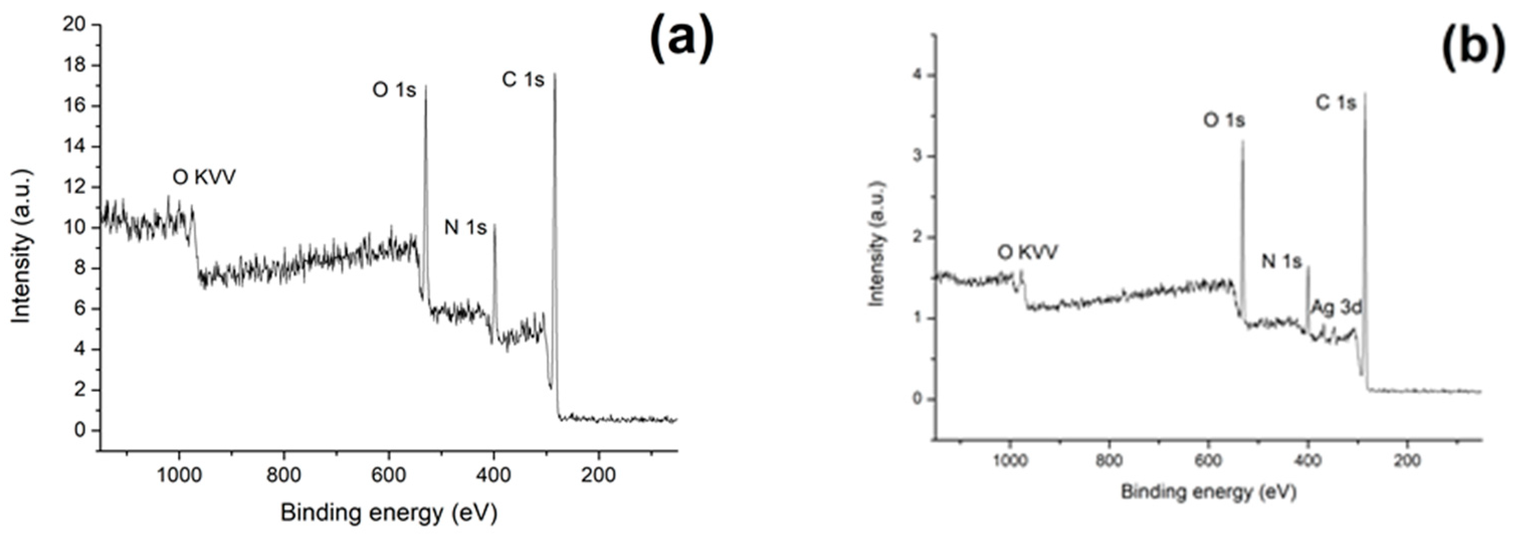
| Sample | C | N | O | Ag |
|---|---|---|---|---|
| Col | 52 | 15 | 32 | – |
| Col/Ag NPs | 74 | 9 | 17 | 0.1 |
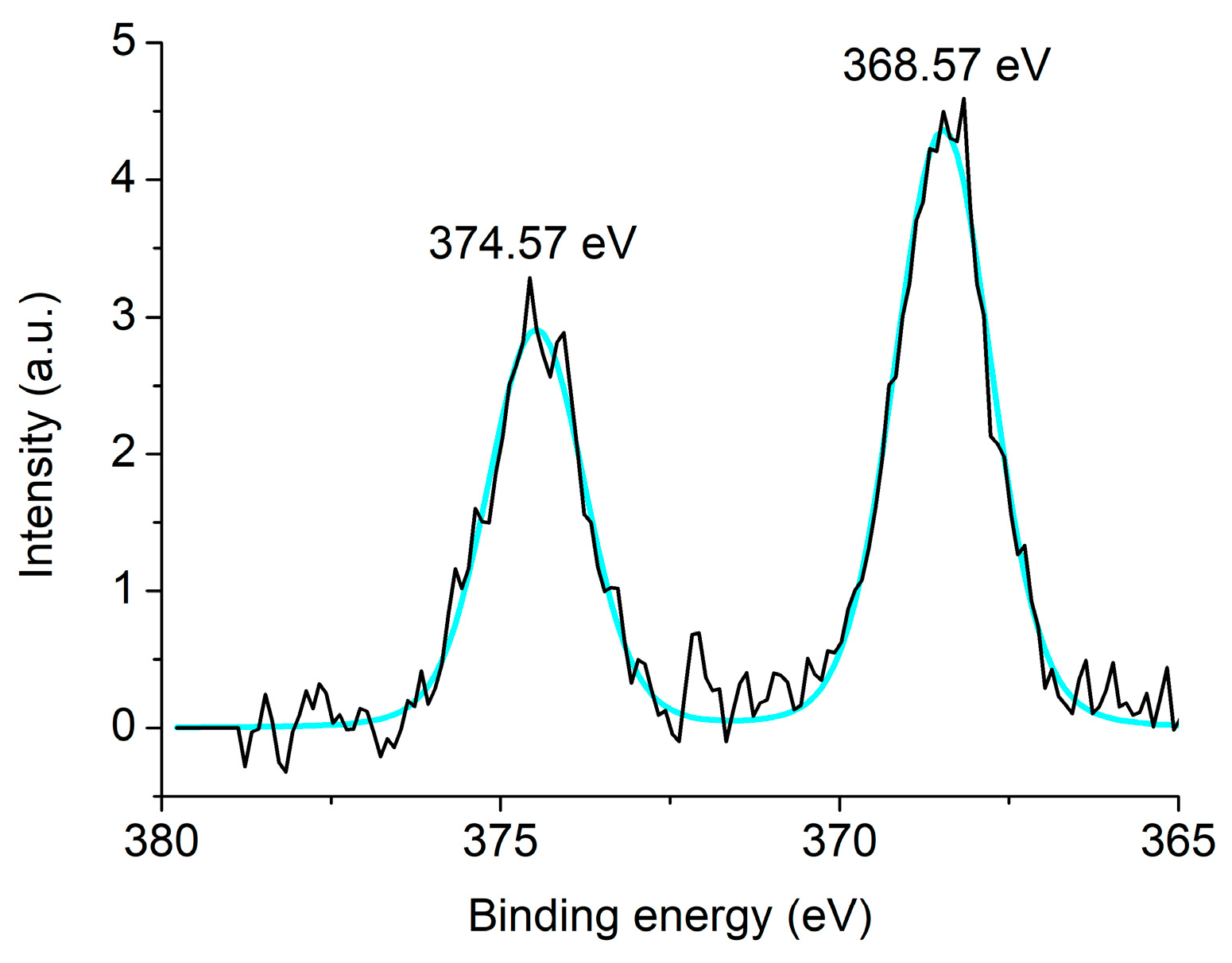
| Sample | Eb Ag 3d5/2,eV | Eb Ag 3d3/2, eV | SOS, eV | State |
|---|---|---|---|---|
| Col/Ag NPs | 368.57 | 374.57 | 6.0 | Ag0 |
| Sample | Zone of Inhibition, mm | ||
|---|---|---|---|
| B. subtilis TCC 6633 | E. coli ATCC 25922 | A. niger INA 00760 | |
| Col/Ag NPs | 6.5 ± 0.4 | 11.3 ± 0.7 | 6.5 ± 0.4 |
| ampicillin | 30.7 ± 1.0 | 27.0 ± 1.7 | * nt |
| amphotericin B | * nt | * nt | 15.3 ± 0.3 |
Disclaimer/Publisher’s Note: The statements, opinions and data contained in all publications are solely those of the individual author(s) and contributor(s) and not of MDPI and/or the editor(s). MDPI and/or the editor(s) disclaim responsibility for any injury to people or property resulting from any ideas, methods, instructions or products referred to in the content. |
© 2023 by the authors. Licensee MDPI, Basel, Switzerland. This article is an open access article distributed under the terms and conditions of the Creative Commons Attribution (CC BY) license (https://creativecommons.org/licenses/by/4.0/).
Share and Cite
Vasil’kov, A.; Tseomashko, N.; Tretyakova, A.; Abidova, A.; Butenko, I.; Pereyaslavtsev, A.; Arkharova, N.; Volkov, V.; Shtykova, E. Wound Coating Collagen-Based Composites with Ag Nanoparticles: Synthesis, Structure and Biological Activity. Coatings 2023, 13, 1315. https://doi.org/10.3390/coatings13081315
Vasil’kov A, Tseomashko N, Tretyakova A, Abidova A, Butenko I, Pereyaslavtsev A, Arkharova N, Volkov V, Shtykova E. Wound Coating Collagen-Based Composites with Ag Nanoparticles: Synthesis, Structure and Biological Activity. Coatings. 2023; 13(8):1315. https://doi.org/10.3390/coatings13081315
Chicago/Turabian StyleVasil’kov, Alexander, Natalya Tseomashko, Anastasia Tretyakova, Aziza Abidova, Ivan Butenko, Alexander Pereyaslavtsev, Natalia Arkharova, Vladimir Volkov, and Eleonora Shtykova. 2023. "Wound Coating Collagen-Based Composites with Ag Nanoparticles: Synthesis, Structure and Biological Activity" Coatings 13, no. 8: 1315. https://doi.org/10.3390/coatings13081315