
Physico-Mechanical Properties of a Newly Developed Base Material Containing Mineral Trioxide Aggregate
 ,
,
Abstract
:1. Introduction
2. Materials and Methods
2.1. Materials Used in This Study
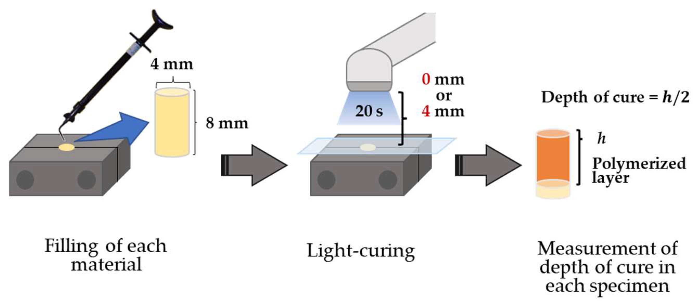
2.2. Depth of Cure
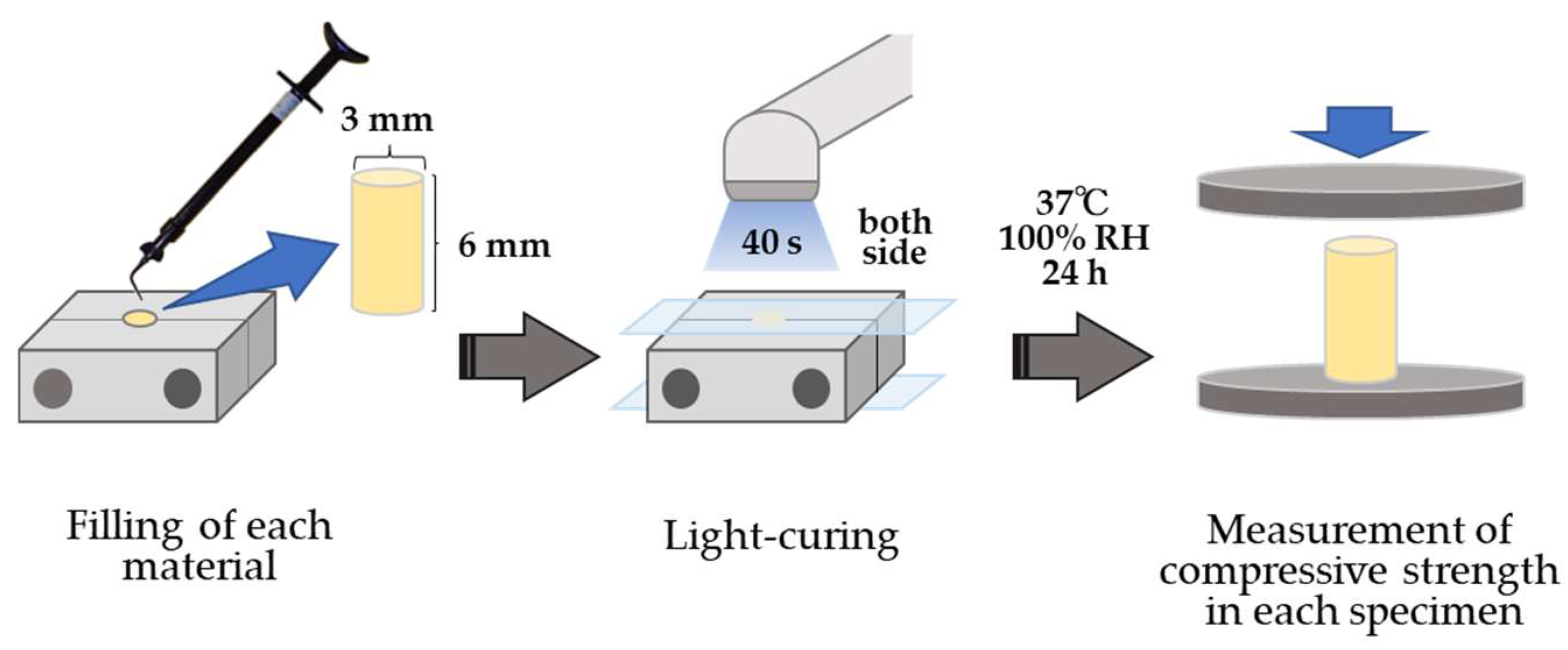
2.3. Compressive Strength
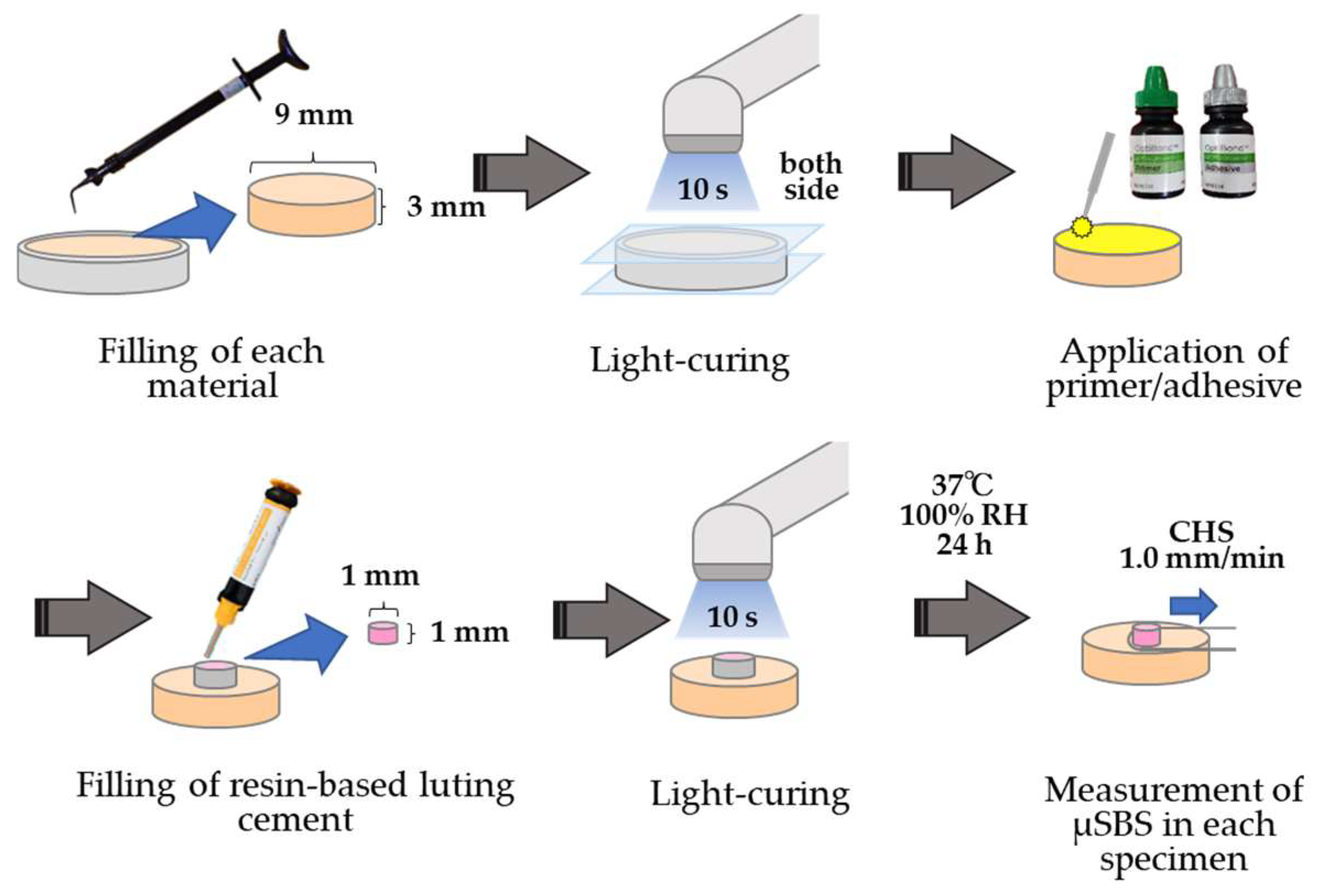
2.4. μSBS with a Resin-Based Composite Luting Cement
2.5. Statistical Analyses
3. Results
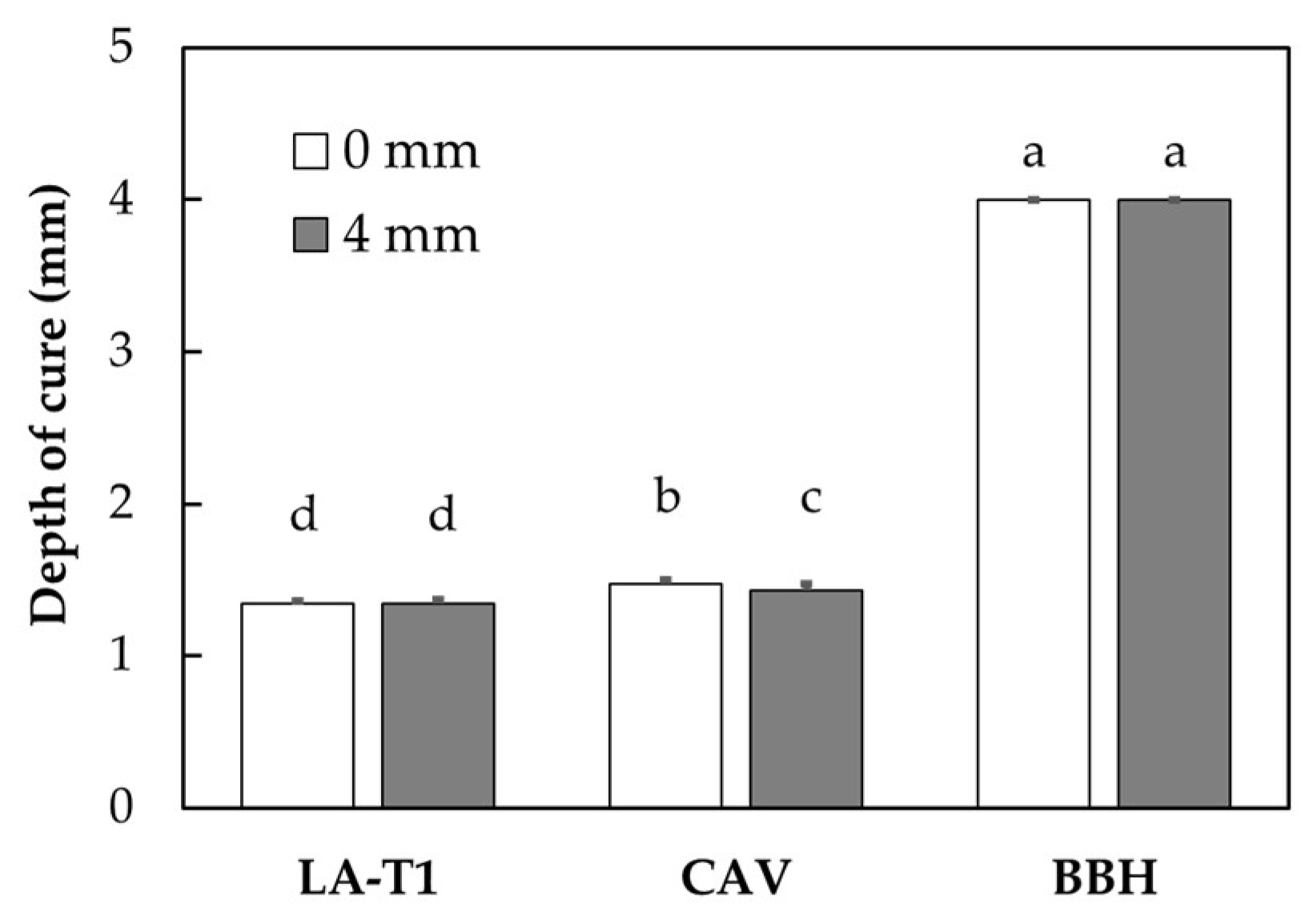
3.1. Depth of Cure
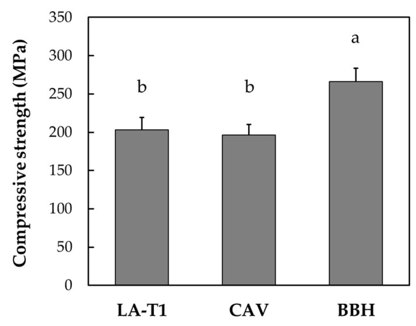
3.2. Compressive Strength
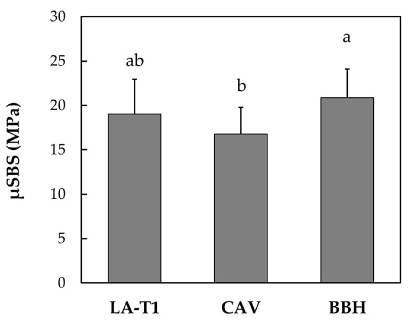
3.3. µSBS
4. Discussion
5. Conclusions
- The depth of cure of the base material LA-T1 containing MTA was significantly but slightly lower than that of the existing product Cavios containing α-TCP, irrespective of the light irradiation distance, 0 mm (light irradiated adjacent to the topside of the mold) and 4 mm (from the topside of the mold).
- The bulk-fill resin-based composite base material Bulk Base Hard had a significantly higher depth of cure than Cavios and LA-T1, irrespective of the light irradiation distance of 0 and 4 mm.
- The compressive strength of LA-T1 was not significantly different from that of Cavios.
- The microshear bond strength of LA-T1 bonded to the resin-based composite luting cement was similar to those of Cavios and Bulk Base Hard bonded to the same luting cement.
Author Contributions
Funding
Institutional Review Board Statement
Informed Consent Statement
Data Availability Statement
Acknowledgments
Conflicts of Interest
References
- Perdigão, J.; Araujo, E.; Ramos, R.Q.; Gomes, G.; Pizzolotto, L. Adhesive dentistry: Current concepts and clinical considerations. J. Esthet. Restor. Dent. 2021, 33, 51–68. [Google Scholar] [CrossRef] [PubMed]
- Dressano, D.; Salvador, M.V.; Oliveira, M.T.; Marchi, G.M.; Fronza, B.M.; Hadis, M.; Palin, W.M.; Lima, A.F. Chemistry of novel and contemporary resin-based dental adhesives. J. Mech. Behav. Biomed. Mater. 2020, 110, 103875. [Google Scholar] [CrossRef]
- German, M.J. Developments in resin-based composites. Br. Dent. J. 2022, 232, 638–643. [Google Scholar] [CrossRef] [PubMed]
- Kameyama, A.; Haruyama, A.; Asami, M.; Takahashi, T. Effect of emitted wavelength and light guide type on irradiance discrepancies in hand-held dental curing radiometers. Sci. World J. 2013, 2013, 647941. [Google Scholar] [CrossRef] [Green Version]
- Price, R.B.; Ferracane, J.L.; Hickel, R.; Sullivan, B. The light-curing unit: An essential piece of dental equipment. Int. Dent. J. 2020, 70, 407–417. [Google Scholar] [CrossRef] [PubMed]
- Price, R.B.; Ferracane, J.L.; Shortall, A.C. Light-curing units: A review of what we need to know. J. Dent. Res. 2015, 94, 1179–1186. [Google Scholar] [CrossRef]
- Unemori, M.; Matsuya, Y.; Akashi, A.; Goto, Y.; Akamine, A. Self-etching adhesives and postoperative sensitivity. Am. J. Dent. 2004, 17, 191–195. [Google Scholar]
- Unemori, M.; Matsuya, Y.; Hyakutake, H.; Matsuya, S.; Goto, Y.; Akamine, A. Long-term follow-up of composite resin restorations with self-etching adhesives. J. Dent. 2007, 35, 535–540. [Google Scholar] [CrossRef] [PubMed]
- Moazzami, S.M.; Sarabi, N.; Hajizadeh, H.; Majidinia, S.; Li, Y.; Meharry, M.R.; Shahrokh, H. Efficacy of four lining materials in sandwich technique to reduce microleakage in class II composite resin restorations. Oper. Dent. 2014, 39, 256–263. [Google Scholar] [CrossRef]
- Azeem, R.A.; Sureshbabu, N.M. Clinical performance of direct versus indirect composite restorations in posterior teeth: A systematic review. J. Conserv. Dent. 2018, 21, 2–9. [Google Scholar] [CrossRef]
- Vetromilla, B.M.; Opdam, N.J.; Leida, F.L.; Sarkis-Onofre, R.; Demarco, F.F.; van der Loo, M.P.J.; Cenci, M.S.; Pereira-Cenci, T. Treatment options for large posterior restorations: A systematic review and network meta-analysis. J. Am. Dent. Assoc. 2020, 151, 614–624. [Google Scholar] [CrossRef] [PubMed]
- Aoki, S.; Takase, Y.; Ishikawa, T. Evaluation of lining materials and a new concept for lining: Alpha-TCP cement or visible light curing calcium hydroxide composition in composite resin restoration. Bull. Tokyo Dent. Coll. 1991, 32, 171–181. [Google Scholar] [PubMed]
- Seino, E. Evaluation of α-TCP cement for dentin-pulp complex. Jpn. J. Conserv. Dent. 1992, 35, 1374–1411. (In Japanese) [Google Scholar]
- Seino, E. Study of crystal growth from α-TCP cement in the cavity of the human vital dentin—Feature of ultrastructure and component analysis. Jpn. J. Conserv. Dent. 1993, 36, 355–371. (In Japanese) [Google Scholar]
- Seino, E.; Nakazawa, Y.; Hirai, Y.; Ishikawa, T.; Toda, T.; Hata, G.; Baba, T. A study on light-cured lining materials—Part 1: Clinical evaluation of “Cavios®”. Jpn. J. Conserv. Dent. 1998, 41, 933–939. (In Japanese) [Google Scholar]
- Seino, E.; Amagai, T.; Takizawa, M.; Nakazawa, Y.; Hirai, Y.; Ishikawa, T. A study on light-cured lining materials—Part 4: Histopathological evaluation of “Cavios®”. Jpn. J. Conserv. Dent. 1999, 42, 116–129. (In Japanese) [Google Scholar]
- Nakazawa, Y.; Seino, E.; Takase, Y.; Makiishi, T.; Hirai, Y.; Ishikawa, T.; Kawada, E.; Oda, Y. A study on light-cured lining materials “Cavios®”—Part 3: Marginal sealing. Jpn. J. Conserv. Dent. 1998, 41, 1080–1084. (In Japanese) [Google Scholar]
- Rode, K.M.; Kawano, Y.; Turbino, M.L. Evaluation of curing light distance on resin composite microhardness and polymerization. Oper. Dent. 2007, 32, 571–578. [Google Scholar] [CrossRef]
- Cervino, G.; Laino, L.; D’Amico, C.; Russo, D.; Nucci, L.; Amoroso, G.; Gorassini, F.; Tepedino, M.; Terranova, A.; Gambino, D.; et al. Mineral trioxide aggregate applications in endodontics: A review. Eur. J. Dent. 2020, 14, 683–691. [Google Scholar] [CrossRef]
- Willershausen, I.; Wolf, T.; Kasaj, A.; Weyer, V.; Willershausen, B.; Marroquin, B.B. Influence of a bioceramic root end material and mineral trioxide aggregates on fibroblasts and osteoblasts. Arch. Oral Biol. 2013, 58, 1232–1237. [Google Scholar] [CrossRef]
- Solanki, N.P.; Venkappa, K.K.; Shah, N.C. Biocompatibility and sealing ability of mineral trioxide aggregate and biodentine as root-end filling material: A systematic review. J. Conserv. Dent. 2018, 21, 10–15. [Google Scholar] [CrossRef] [PubMed]
- Morita, M.; Kitagawa, H.; Nakayama, K.; Kitagawa, R.; Yamaguchi, S.; Imazato, S. Antibacterial activities and mineral induction abilities of proprietary MTA cements. Dent. Mater. J. 2021, 40, 297–303. [Google Scholar] [CrossRef] [PubMed]
- Bartols, A.; Roussa, E.; Walther, W.; Dörfer, C.E. First evidence for regeneration of the periodontium to mineral trioxide aggregate in human teeth. J. Endod. 2017, 43, 715–722. [Google Scholar] [CrossRef]
- Zhu, C.; Ju, B.; Ni, R. Clinical outcome of direct pulp capping with MTA or calcium hydroxide: A systematic review and meta-analysis. Int. J. Clin. Exp. Med. 2015, 8, 17055–17060. [Google Scholar] [PubMed]
- Cushley, S.; Duncan, H.F.; Lappin, M.J.; Chua, P.; Elamin, A.D.; Clarke, M.; El-Karim, I.A. Efficacy of direct pulp capping for management of cariously exposed pulps in permanent teeth: A systematic review and meta-analysis. Int. Endod. J. 2021, 54, 556–571. [Google Scholar] [CrossRef]
- Tsai, Y.-L.; Lan, W.-H.; Jeng, J.-H. Treatment of pulp floor and stripping perforation by mineral trioxide aggregate. J. Formos. Med. Assoc. 2006, 105, 522–526. [Google Scholar] [CrossRef] [Green Version]
- Yildirim, G.; Dalci, K. Treatment of lateral root perforation with mineral trioxide aggregate: A case report. Oral Surg. Oral Med. Oral Pathol. Oral Radiol. Endod. 2006, 102, e55–e58. [Google Scholar] [CrossRef]
- Favieri, A.; Campos, L.C.; Burity, V.H.; Santa Cecília, M.; Abad Eda, C. Use of biomaterials in periradicular surgery: A case report. J. Endod. 2008, 34, 490–494. [Google Scholar] [CrossRef]
- Timmerman, A.; Parashos, P. Delayed root development by displaced mineral trioxide aggregate after regenerative endodontics: A case report. J. Endod. 2017, 43, 252–256. [Google Scholar] [CrossRef]
- Sheykhrezae, M.S.; Meraji, N.; Ghanbari, F.; Nekoofar, M.H.; Bolhari, B.; Dummer, P.M.H. Effect of blood contamination on the compressive strength of three calcium silicate-based cements. Aust. Endod. J. 2018, 44, 255–259. [Google Scholar] [CrossRef]
- Salamoni Sinhori, B.; Vieira, L.C.C.; Baratieri, L.N. Influence of preparation reconstruction on the compressive strength of CAD/CAM ceramic inlays. Int, J. Biomater. 2019, 2019, 7307649. [Google Scholar] [CrossRef]
- Furukawa, K.; Inai, N.; Tagami, J. The effects of luting resin bond to dentin on the strength of dentin supported by indirect resin composite. Dent. Mater. 2002, 18, 136–142. [Google Scholar] [CrossRef] [PubMed]
- ISO 4049:2000; Dentistry—Polymer-Based Filling Restorative and Luting Materials. International Organization for Standardization: Geneva, Switzerland, 2000.
- Sakaguchi, R.L.; Douglas, W.H.; Peters, M.C. Curing light performance and polymerization of composite restorative materials. J. Dent. 1992, 20, 183–188. [Google Scholar] [CrossRef]
- Pilo, R.; Oelgiesser, D.; Cardash, H.S. A survey of output intensity and potential for depth of cure among light-curing units in clinical use. J. Dent. 1999, 27, 235–241. [Google Scholar] [CrossRef] [PubMed]
- Rizzante, F.A.P.; Duque, J.A.; Duarte, M.A.H.; Mondelli, R.F.L.; Mendonça, G.; Ishikiriama, S.K. Polymerization shrinkage, microhardness and depth of cure of bulk fill resin composites. Dent. Mater. J. 2019, 38, 403–410. [Google Scholar] [CrossRef] [PubMed] [Green Version]
- Ludovichetti, F.S.; Lucchi, P.; Zambon, G.; Pezzato, L.; Bertolini, R.; Zerman, N.; Stellini, E.; Mazzoleni, S. Depth of cure, hardness, roughness and filler dimension of bulk-fill flowable, conventional flowable and high-strength universal injectable composites: An in vitro study. Nanomaterials 2022, 12, 1951. [Google Scholar] [CrossRef]
- Rocha, M.G.; Maucoski, C.; Roulet, J.F.; Price, R.B. Depth of cure of 10 resin-based composites light-activated using a laser diode, multi-peak, and single-peak light-emitting diode curing lights. J. Dent. 2022, 122, 104141. [Google Scholar] [CrossRef] [PubMed]
- Fujita, K.; Nishiyama, N.; Nemoto, K.; Okada, T.; Ikemi, T. Effect of base monomer’s refractive index on curing depth and polymerization conversion of photo-cured resin composites. Dent. Mater. J. 2005, 24, 403–408. [Google Scholar] [CrossRef] [Green Version]
- Par, M.; Spanovic, N.; Mohn, D.; Attin, T.; Tauböck, T.T.; Tarle, Z. Curing potential of experimental resin composites filled with bioactive glass: A comparison between Bis-EMA and UDMA based resin systems. Dent. Mater. 2020, 36, 711–723. [Google Scholar] [CrossRef]
- Pereira, L.D.E.; Couto Neto, M.P.; Pereira, R.G.; Schneider, L.F.J. Influence of resin matrix on the rheology, translucency, and curing potential of experimental flowable composites for bulk-fill applications. Dent. Mater. 2021, 37, 1046–1053. [Google Scholar] [CrossRef]
- Knezevic, A.; Zeljezic, D.; Kopjar, N.; Tarle, Z. Cytotoxicity of composite materials polymerized with LED curing units. Oper. Dent. 2008, 33, 23–30. [Google Scholar] [CrossRef] [PubMed] [Green Version]
- Al-Zain, A.O.; Platt, J.A. Effect of light-curing distance and curing time on composite microflexural strength. Dent. Mater. J. 2021, 40, 202–208. [Google Scholar] [CrossRef] [PubMed]
- Watts, D.C.; EL Mowafy, O.M.; Grant, A.A. Temperature-dependence of compressive properties of human dentin. J. Dent. Res. 1987, 66, 29–32. [Google Scholar] [CrossRef] [PubMed]
- Morita, S. Mechanical properties of newly developed calcium-phosphate cement for base cement. J. Kyushu Dent. Soc. 1996, 50, 504–514. (In Japanese) [Google Scholar] [CrossRef]
- Shariff, K.A.; Tsuru, K.; Ishikawa, K. Fabrication of interconnected pore forming α-tricalcium phosphate foam granules cement. J. Biomater. Appl. 2016, 30, 838–845. [Google Scholar] [CrossRef]
- Tanomaru-Filho, M.; Morales, V.; da Silva, G.F.; Bosso, R.; Reis, J.M.; Duarte, M.A.; Guerreiro-Tanomaru, J.M. Compressive strength and setting time of MTA and Portland cement associated with different radiopacifying agents. ISRN Dent. 2012, 2012, 898051. [Google Scholar] [CrossRef] [Green Version]
- Kosar, M.A.; Basturk, F.B.; Turkaydin, D.; Nekoofar, M.H. The effect of operator-induced variability on the physical properties of ProRoot MTA. Niger. J. Clin. Pract. 2020, 23, 1068–1072. [Google Scholar] [CrossRef]
- Korkut, E.; Torlak, E.; Altunsoy, M. Antimicrobial and mechanical properties of dental resin composite containing bioactive glass. J. Appl. Biomater. Funct. Mater. 2016, 14, e296–e301. [Google Scholar] [CrossRef]
- Burgess, J.O.; Barghi, N.; Chan, D.C.; Hummert, T. A comparative study of three glass ionomer base materials. Am. J. Dent. 1993, 6, 137–141. [Google Scholar]
- Peskersoy, C.; Recen, D.; Kemaloğlu, H. The effect of composite placement technique on the internal adaptation, gap formation and microshear bond strength. Eur. Oral Res. 2022, 56, 10–16. [Google Scholar] [CrossRef]
- Akturk, E.; Bektas, O.O.; Ozkanoglu, S.; Akin, E.G.G. Do ozonated water and boric acid affect the bond strength to dentin in different adhesive systems? Niger. J. Clin. Pract. 2019, 22, 1758–1764. [Google Scholar] [CrossRef] [PubMed]
- Nakazawa, Y.; Seino, E.; Takase, Y.; Makiishi, T.; Hirai, Y.; Ishikawa, T.; Kawada, E.; Oda, Y. A study on light-cured lining materials “Cavios®”—Part 2: Adhesive properties. Jpn. J. Conserv. Dent. 1998, 41, 1073–1079. (In Japanese) [Google Scholar]


| Material | Manufacturer | Composition | Batch No. |
|---|---|---|---|
| [Base material] | |||
| LA-T1 | Neo Dental Chemical Products (Tokyo, Japan) | UDMA, mineral trioxide aggregate, barium sulfate, CQ, other | LA-T1 |
| Cavios® | Neo Dental Chemical Products (Tokyo, Japan) | UDMA, α-tricalcium phosphate, barium sulfate, CQ, other | B0A1 |
| Bulk Base Hard® (Medium Flow, Blue) | Sun Medical (Moriyama, Shiga, Japan) | methacrylic acid esters (Bis-MPEPP, other), acrylic acid esters (urethane acrylate), barium silica glass, strontium silica glass, aromatic amines, other | TT1 |
| [Dental adhesive] | |||
| OptiBond™ eXTRa Universal | Kerr (Orange, CA, USA) | Primer: GPDM, HEMA, acetone, ethanol, purified water, other Adhesive: GPDM, HEMA, Bis-GMA, glycerol dimethacrylate, ethanol, barium glass, sodium hexafluorosilicate, CQ, other | Primer: 7941589 Adhesive: 7945265 |
| [Resin-based luting cement] | |||
| Nexus™ Universal | Kerr (Orange, CA, USA) | Bis-GMA, TEGDMA, UDMA, HEMA, glycerol dimethacrylate, aluminoborosilicate glass, ytterbium fluoride, other | 7509656 |
Disclaimer/Publisher’s Note: The statements, opinions and data contained in all publications are solely those of the individual author(s) and contributor(s) and not of MDPI and/or the editor(s). MDPI and/or the editor(s) disclaim responsibility for any injury to people or property resulting from any ideas, methods, instructions or products referred to in the content. |
© 2023 by the authors. Licensee MDPI, Basel, Switzerland. This article is an open access article distributed under the terms and conditions of the Creative Commons Attribution (CC BY) license (https://creativecommons.org/licenses/by/4.0/).
Share and Cite
Nakamura, K.; Horasawa, N.; Okuse, T.; Uchikawa, R.; Shimada, K.; Kuroiwa, A.; Murakami, S.; Hasegawa, H.; Kameyama, A. Physico-Mechanical Properties of a Newly Developed Base Material Containing Mineral Trioxide Aggregate. Coatings 2023, 13, 597. https://doi.org/10.3390/coatings13030597
Nakamura K, Horasawa N, Okuse T, Uchikawa R, Shimada K, Kuroiwa A, Murakami S, Hasegawa H, Kameyama A. Physico-Mechanical Properties of a Newly Developed Base Material Containing Mineral Trioxide Aggregate. Coatings. 2023; 13(3):597. https://doi.org/10.3390/coatings13030597
Chicago/Turabian StyleNakamura, Keigo, Noriko Horasawa, Toshiyuki Okuse, Ryutaro Uchikawa, Katsumitsu Shimada, Akihiro Kuroiwa, Satoshi Murakami, Hiromasa Hasegawa, and Atsushi Kameyama. 2023. "Physico-Mechanical Properties of a Newly Developed Base Material Containing Mineral Trioxide Aggregate" Coatings 13, no. 3: 597. https://doi.org/10.3390/coatings13030597