Communication

Antimicrobial Activity and Transparency of Polyvinyl Butyral Paint Containing Heated Scallop-Shell Powder

1
Department of Nutrition and Life Science, Faculty of Health and Medical Sciences, Kanagawa Institute of Technology, 1030 Shimo-Ogino, Atsugi 243-0292, Kanagawa, Japan
2
Faculty of Applied Biosciences, Kanagawa Institute of Technology, 1030 Shimo-Ogino, Atsugi 243-0292, Kanagawa, Japan
3
No Corrosion & Slip Technical Research, 1-10-16 Tenjin-Cho, Fujisawa 252-0814, Kanagawa, Japan
*
Author to whom correspondence should be addressed.
Coatings 2023, 13(2), 364; https://doi.org/10.3390/coatings13020364
Submission received: 24 December 2022 / Revised: 27 January 2023 / Accepted: 3 February 2023 / Published: 5 February 2023
(This article belongs to the Special Issue Advances in Antibacterial Coatings: From Materials to Applications)

Abstract

:
The main component of seashells is calcium carbonate (CaCO3). When seashells are calcined at high temperatures, CaCO3 becomes calcium oxide (CaO), and this CaO exhibits antimicrobial activity. In this study, we attempted to develop a transparent coating that retains antimicrobial activity for a long time by mixing polyvinyl butyral (PVB), which has excellent adhesive and binding properties, with heated shell powder (HSP). The scallop HSPs used in this study were nanoparticles with a particle diameter of approximately several hundred nm, and the prepared paint showed high transparency. Elemental analysis showed that scallop HSP particles existed in the paint as Ca(OH)2. The antimicrobial activity of the surface applied with scallop HSP-containing PVB (HSSP-PVB) paint was then evaluated using JIS Z 2801 and ISO 21702: 2019. The HSSP-PVB paint-applied surfaces showed high antibacterial and antiviral activity. The antimicrobial activity of the scallop HSP-PVB paint-applied surface was attributed to the creation of a strongly alkaline environment due to surface hydration, and the strongly alkaline environment was maintained for a long period of time. It was suggested that the PVB covered the surface of the scalloped HSP particles, which significantly prevented the HSP from contacting CO2 and H2O molecules in the air.

1. Introduction

The main component of shells is calcium carbonate (CaCO3). When shells are calcined at high temperatures, the main component, CaCO3, becomes CaO, which exhibits antimicrobial activity [1]. Some scallop shells are reused as food additives, but most are disposed of as industrial waste. In the producing areas, the odor from the abandoned shells becomes a pollution problem [2,3]. Similarly, oyster shells [4,5], clams and roll shells [6], mussel shells [7], and blood cockle shells [8] have also been reported to exhibit antimicrobial activity after calcination treatment. Using heated shell power (HSP) for the environment and food products as an antimicrobial agent can be expected to control micro-organisms and improve preservation. In addition, shells, which are mostly treated as waste, can be used as a practical resource, reducing pollution problems.
Scallop HSP produced by heat treatment at 1000 °C had antimicrobial activity almost equal to CaO [1]. Scallop HSP has also been reported to be effective in the disinfection and sanitization of bacteria [1,9,10], fungi [11,12], heat-resistant spores [13], viruses [10,14,15], and biofilms [16,17,18,19,20]. There have been increasing reports on the application of HSP in food products, such as fresh fruits and vegetables [21,22,23,24,25,26,27], sausages [28], chicken wings [29], frozen meat [30], fresh fish [31], and sprout seeds [32,33], for improving the shelf life of foods and disinfecting them.
The addition of HSPs to plastic moldings, polyethylene films, nonwoven fabrics, and paper to impart antimicrobial activity has also been reported [34]. Other studies on surface water disinfection [35] and antimicrobial polymer nanocomposite materials containing HSP [34,36] are underway. In Japan and abroad, interior paints for residential walls containing HSP are marketed as coatings with antibacterial, deodorizing, and humidity-control functions [37,38]. However, the paints containing HSP are not transparent, and their applications are limited. If paints containing transparent HSP can be developed, the range of applications in indoor spaces can be significantly expanded.
Therefore, we attempted to develop a transparent antimicrobial coating containing HSP. This study focused on polyvinyl butyral (PVB), a polymer also used as an adhesive and a binder between powders. The properties of PVB are characterized by the presence of butyral, hydroxyl, and acetyl groups, and it has excellent optical transparency. PVB is insoluble in water and soluble in various organic solvents, including alcohols, esters, ethanol, phenylacetyl, and ketones, such as cyclohexanone. In addition to its weather and water resistance, it is safe, odorless, and noncorrosive [39,40]. Due to its strong adhesion and excellent resistance to UV degradation, it is used in automotive windshield interlayers [41]. The development of coatings with high transparency and long-lasting antimicrobial activity by mixing PVB, which has excellent paint properties, with scallop HSP, which has a broad antimicrobial spectrum, will lead to further expansion of HSP applications. It has also been found that nanoparticulation of HSPs significantly increases their antimicrobial activity [42,43]. The nanosizing of HSPs also contributes to increasing the transparency of paints. In this study, we prepared transparent paints containing nanosized HSPs using this PVB and investigated their properties and antimicrobial activity.

2. Materials and Methods

2.1. Preparation of Paint Containing Scallop HSP

Scallop HSP (Natural Japan Co., Abashiri, Hokkaido, Japan), calcined at 1200 °C with an average particle diameter of 14.7 µm, was mixed with an ethanol solvent (Eta Cohol 7: ethanol 80%–90%; n-propylalcohol < 10% by weight, i-propylalcohol < 5% by weight) (Sankyo Chemical Co., Kusumoto Chemicals, Ltd., Chiyoda, Tokyo, Japan) and a dispersing agent (Disoaron DA-234, Kusumoto Chemicals, Ltd., Chiyoda, Tokyo, Japan) at the weight ratios shown in Table 1. The mixed slurry was processed by bead mill grinding (LMZ015, Ashizawa Finetech Ltd., Narashino, Chiba, Japan) at room temperature. After 10 min of processing, PVB (Mowtial B30H, Kuraray Co., Ltd., Tokyo, Japan) was added, and the mixture was milled for another 5 min to yield paint containing scallop HSP (scallop HSP–PVB paint).

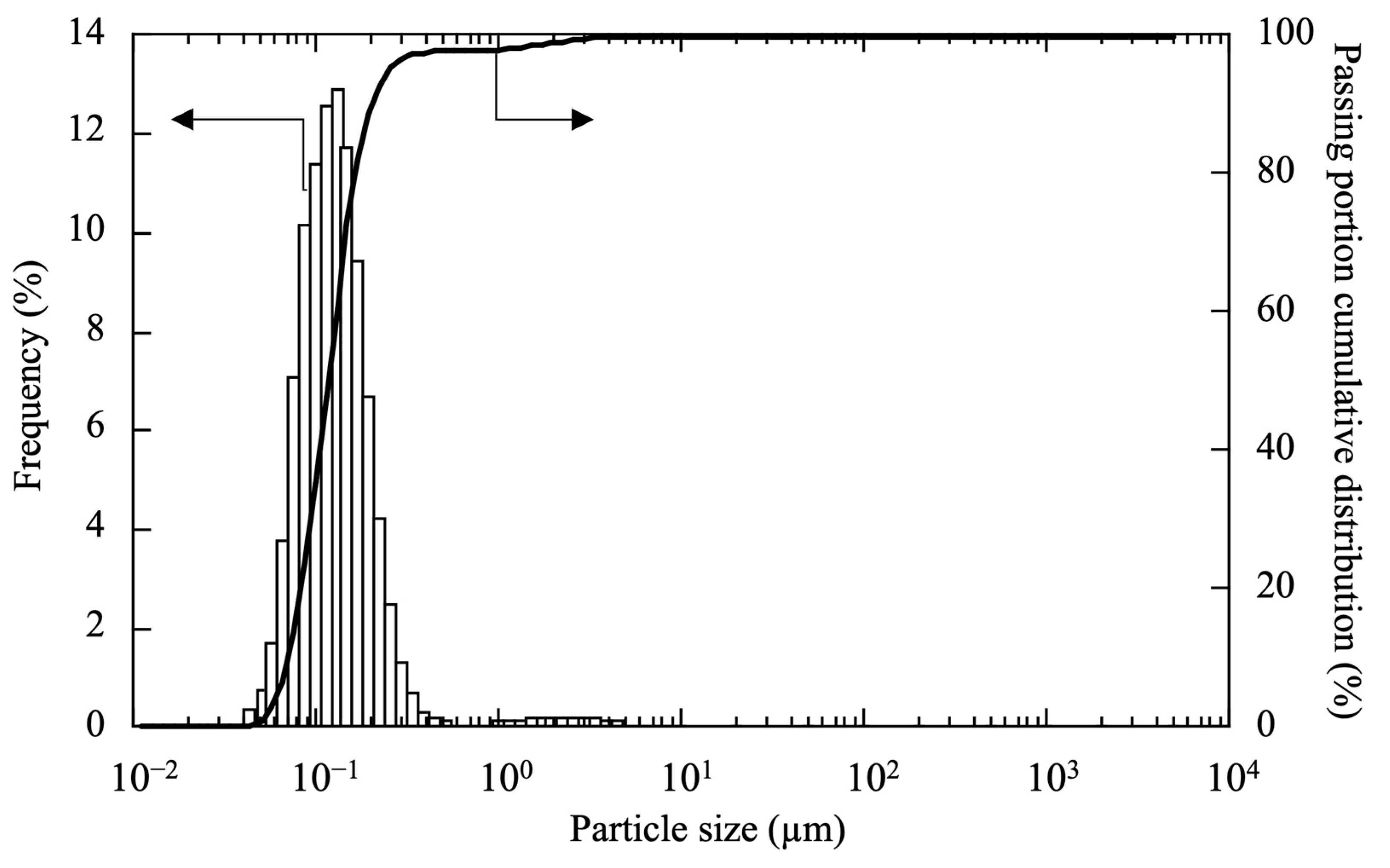
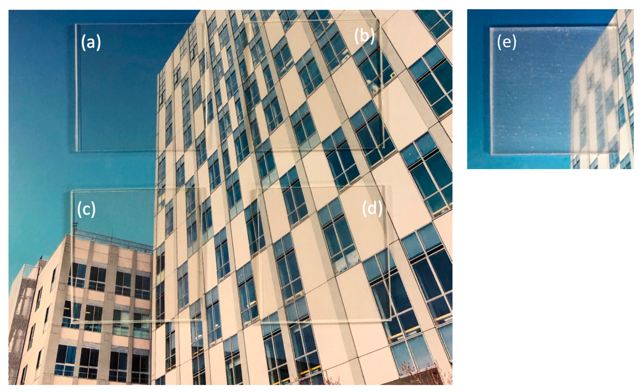
2.2. Characterization of Scallop HSP–PVB Paint

The HSP–PVB paint was applied to a 5 × 5 cm (2-mm-thick) acrylic plate (Mitsubishi Chemical Co., Chiyoda, Tokyo, Japan) using a paintbrush. One coat was applied in one direction. The plate was placed on a clean bench, checked for solidification, and stored in a sterile petri dish in a desiccator. This was used as the test specimen.
The surfaces of the painted specimens were observed using a scanning electron microscope (SEM; Phenom G2Pro, Thermo Fisher Scientific, Waltham, MA, USA). The acceleration voltage and working distance were 5 kV and 3.7 mm, respectively.
Elemental analysis of the painted surfaces was performed by field emission SEM (JSM-7800F Prime, JASCO Co., Hachioji, Tokyo, Japan) and energy-dispersive X-ray spectroscopy (EDX) (X-MaxN80, Oxford Inst., High Wycombe, UK).
The amount of HSP-PVB paint coated on the acrylic board was calculated from the weight change before and after the application.
Water contact angle measurement was performed to evaluate the degree of surface of acrylic plates coated with scallop HSP–PVB paints 1–3 by a contact angle meter (ST-1, Hagitec Co. Ltd., Chiba, Japan). Ten microliter of water was dropped onto the surface coated with scallop HSP–PVB paints or PVB using a micropipette. Each contact angle was measured five times and the average value obtained.
The hardness of the coating surfaces of HSP-PVB paints was determined according to JIS K 5600-5-4:1999 (pencil method) [44].
The transmittance of HSP-PVB paints in the visible light range was measured in the 200–100 nm range using a UV-VIS spectrophotometer (U-5100, Hitachi High-Tech Co., Tokyo, Japan).

2.3. Antibacterial and Antiviral Efficacies of Scallop HSP–PVB Paint

The antibacterial efficacy of the specimens was evaluated according to JIS Z 2801 [45]. Briefly, gram-negative Escherichia coli NBRC 3301 and gram-positive Staphylococcus aureus NBRC 13276, used as test bacteria, were cultured in nutrient broth (Eiken Chemicals, Tokyo, Japan) at 37 °C for 24 h with shaking (110 strokes/min). The culture was centrifuged (3000 rpm, 10 min), and the pellet was resuspended in 1/500 nutrient broth to yield a suspension of 106 colony-forming units per milliliter. An aliquot (0.1 mL) of the suspension was placed on a specimen, covered with a polyethylene (PE) film (4 cm × 4 cm, ASONE, Osaka, Japan), disinfected by 70% ethanol, and incubated at 35 °C with a relative humidity of 90% or higher for 5 min–24 h. The experiment was conducted in triplicates (n = 3). After incubation, the PE film and specimens were placed in sterile stomacher bags (bioMérieux Japan, Tokyo, Japan), to which 10 mL of soybean-casein digest broth with lecithin and polysorbate 80 (SCDLP) broth (Eiken Chemicals) was added, and the bacteria were washed out by stomaching for 60 s. The washed-out bacterial solution was diluted with phosphate-buffered saline and cultured (35 °C, 48 h) on standard plate count agar (Eiken Chemicals) to count the number of colonies.
The antiviral efficacy of the specimens was tested according to ISO 21702: 2019 [46]. Briefly, the enveloped influenza A virus (H3N2; ATCC VR-1679) and the nonenveloped feline calicivirus (ATCC VR-782), an alternative strain of norovirus, were used as test viruses, and MDCK (ATCC CCL-34) and CRFK cells (ATCC CCL-94) were used as their hosts, respectively. The viruses were collected and suspended in purified water at 1–5 × 10⁷ plaque-forming units per milliliter. An aliquot (0.4 mL) of the virus suspension was placed on a specimen coated with scallop HSP–PVB paint 2, covered with a disinfected PE film (4 × 4 cm), and incubated at 25 °C for 24 h at a relative humidity of 90% or higher. The experiment was conducted in triplicates (n = 3). After incubation, the virus was washed out of the specimen with 10 mL of SCDLP broth, and the viral titer was determined by plaque assay.
The antibacterial or antiviral activity R was determined using the following formula:
R = (UtU0) − (AtU0) = UtAt
where
U0: mean logarithm of the number of viable bacteria or plaques immediately after inoculation on the noncoated acrylic plates.
Ut: mean logarithm of the number of viable bacteria or plaques at a given time on the noncoated acrylic plates.
At: mean logarithm of the number of viable bacteria or plaques at a given time on the acrylic plates coated with scallop HSP–PVB paint.
The paint was considered to have antibacterial or antiviral activity when R was ≧2.

2.4. pH Measurement of Scallop HSP–PVB Paint

The specimens were placed in the laboratory at room temperature (25 °C ± 3 °C, relative humidity of 40%–65%). The pH was measured once a week for up to 1 month and every other month after that for up to 17 months, using the following procedure: 100 µL of deionized water was dropped onto a portion of the acrylic plate surface coated with scallop HSP–PVB paints; the solution was collected with a pipette 1 min later, and its pH was measured with a pH meter (pH-33, Horiba Ltd., Minami, Kyoto, Japan).

3. Results and Discussion

3.1. Properties of Scallop HSP–PVB Paint

Figure 1 shows the particle size distribution of scallop HSP after bead milling. The frequency peaks between 100 and 200 nm indicate that scallop HSP was nanoparticulated by bead milling. The means, medians, and modes of particle diameters were 169, 117, and 122 nm, respectively.
Figure 2 shows SEM photographs of the surface of acrylic plates coated with scallop HSP–PVB paints 1–3 (Table 1). Scallop HSPs were found all over the acrylic plates. The particles were mostly nanosized, and some were microsized. The particles were not spherical but distorted, and some were needle-shaped. For scallop HSP–PVB paint 1, gaps were observed between some of the particles, but as the concentration of HSP particles increased (paints 2 and 3), the gaps almost disappeared.
Figure 3 shows the elemental analysis of surfaces coated with HSP–PVB paint 2, as determined by energy-dispersive X-ray spectroscopy. Carbon (C), oxygen (O), and calcium (Ca) peaks were detected. Palladium (Pd) peaks were also observed, but these were derived from the coating for SEM imaging. Considering the composition of PVB, (C8H14O2)n, the elemental ratio of Ca to O, excluding O in PVB, was approximately 1:2, indicating that scallop HSP particles existed as Ca(OH)2 in the applied layer.
We measured the transmittance of the HSP-PVB paints in the visible light range before and after coating (Figure 4). The transmittance of the HSP-PVB paints in the visible light range (380–780 nm) was almost 90% for the acrylic plate and the acrylic plate coated with PVB only. The transmittance for HSP-PVB-1 and HSP-PVB-2 was approximately 60%~85%, and that of HSP-PVB-3 was 40%~60%. HSP-PVB-1 and 2 were found to exhibit high transmittance. Nanoparticles significantly affect light scattering and reflection, depending on their size. In the case of oxides and other materials with a low refractive index, it has been found that when the particle size is smaller than the visible light wavelength, the scattering becomes very small, resulting in increased transparency [47].
Table 2 shows the characteristics of the coated surface of HSP-PVB paints. First, the coating amount increased as the scallop HSP content percentage increased. Next, the change in hydrophilicity of the surface was confirmed by measuring the contact angle. First, the acrylic plate had a hydrophobic surface of 53.7 ± 3.2°. When PVB was applied, the contact angle was 25.3°, less than 30°, and the surface became hydrophilic. As the HSP content was increased, the contact angle became smaller and more hydrophilic. Therefore, it can be assumed that when water droplets containing bacteria or virus particles come into contact with the coated surface, they quickly spread, and the bacteria or virus particles are likely to come into contact with the HSP.
The hardness of the coating film was also determined (Table 2). The HSP-PVB coating film is soft, and increasing its strength is a future challenge. However, considering the excellent adhesion of PVB, it can be applied to surfaces rich in flexibility.
The coated acrylic plates were placed on a booklet cover (Figure 5). Compared with the noncoated plate, those coated with scallop HSP–PVB paints 1–3 were highly transparent, as evidenced by the adequate visibility of the photo on the booklet cover. Figure 5e shows a photograph of HSP-PVB-2 prepared with microparticles before milling. The transparency of the paint was significantly reduced when microparticles were added. As shown in Figure 2, though the scallop HSPs covered the entire surface of the acrylic plate, a highly transparent paint that allows sufficient visible light transmission was realized by nanosizing the HSP.

3.2. Antibacterial and Antiviral Activities

Table 3 summarizes the antibacterial and antiviral activities of the paint-coated acrylic specimens. R-values with inequality signs signify that the viable bacterial and active viral counts were below the detection limit in all the triplicates. PVB itself showed antimicrobial activity against the gram-negative bacterium E. coli. However, its antibacterial activity value was R < 2. On the other hand, the paint showed no antimicrobial activity against S. aureus, a gram-positive bacterium. All the R-values of the HSP-PVB paints at 24 h were greater than or equal to two, indicating that the HSP–PVB paints exhibited significant antibacterial and antiviral activities. These results indicated that HSP dramatically increased the antimicrobial activity of the paint.
The R-values at 5 min and 1 h reveal that E. coli began to die earlier than S. aureus, which is consistent with the bacterial susceptibility to HSP alone [1]. In addition, scallop HSP–PVB paint inactivated both influenza virus and feline calicivirus. Thammakarn et al. [14,15] have reported that treatment with scallop HSP for 60 s reduces the titer of both enveloped and nonenveloped viruses to below the detection limit.
Figure 6 shows the pH measurement of a 100-µL drop of deionized water dropped on the coated acrylic plates using pH test papers. The color of the test paper indicates that the pH was strongly alkaline (above pH 12). Moreover, the highly alkaline environment was maintained for a long time. The pH was regularly measured using a pH meter for 17 months, and it never fell below 12. Although the pH measurements had to be stopped at this point due to the coronavirus pandemic, we can reasonably assume that a highly alkaline environment can be maintained for an extended period of time.
Figure 7 depicts a schematic diagram of the antibacterial and antiviral mechanisms of scallop HSP–PVB paint. Water droplets or splashes containing bacterial cells and virus particles adhere to the coated acrylic surface. Water molecules in these droplets react with HSPs in the painted layer, and the hydrated HSPs partially dissolve and release OH into the droplets. Consequently, the environment around the bacterial cells and virus particles becomes strongly alkaline, causing the destruction of cell membranes and the inactivation of enzymes, ultimately killing the bacteria and inactivating the viruses. The antibacterial activities of CaO and HSP have been shown to be considerably greater than simple alkaline treatment (NaOH), even at the same pH [1,17,18,19,20,48]. Although strong alkali is the main driver of the antimicrobial mechanism of CaO-based HSP, reactive oxygen species (ROS), especially superoxide (O2), have been confirmed to be generated from CaO and scallop HSP slurries [17,49]. Moreover, the antibiotic susceptibility of CaO powder slurry-treated E. coli was more similar to that of O2-treated E. coli than alkali-treated E. coli [50]. A large amount of ROS has also been shown to be generated from the solid solution of alumina and CaO [51], and ROS are another antimicrobial factor in CaO-based HSP as well.
CaO, the main component of HSP, absorbs H2O and CO2 from the air to convert into CaCO3, the main component of uncalcined seashells without antimicrobial activity. Ishihara et al. [52] reported that the pH of the supernatant of HSP slurry (pH 12 or higher) decreased to pH 10 in approximately 1 day. PVB is insoluble in water but has -OH groups. Some interaction between PVB and HSP blocks the reaction sites of CaO and Ca(OH)2 with H2O and CO2, significantly inhibiting their absorption of CO2 and H2O from the air, consequently preventing the drop in pH of the paint. Therefore, PVB contributes to the long-term retention of the antimicrobial activity of HSP–PVB paint.

4. Conclusions

In this study, we mixed PVB, which has excellent adhesive and binder properties, with nanoparticulate scallop HSP to create a highly transparent paint that exhibits high antibacterial and antiviral activity. Surfaces coated with this paint could create and maintain a strongly alkaline environment over a long period of time.
In addition to shells, eggshells [53], and dolomite [54] are mainly composed of CaCO3. Naturally, their composition can be changed to CaO by calcination to confer antimicrobial activity upon them. We can also use such materials in this antimicrobial PVB paint. Due to the wide applicability and excellent properties of PVB, the paint proposed in this study can be applied to various surfaces, including glass, metal, wood, paper, and plastic. However, this paint, which contains alkaline calcined shells, is not suitable for outdoor use where it may come in contact with acid rain. At present, there are limitations to the use of this paint. We are also investigating the paint composition, stability, preservation properties, and antifungal activity of HSP-PVB coatings in detail to develop a realistic paint design for spray-coating applications of this paint.

Author Contributions

Conceptualization, J.S. and Y.N.; methodology, J.S. and Y.N.; validation, J.S., A.O. and R.W.; investigation, R.A., Y.N. and J.S.; writing—original draft preparation, R.A. and J.S.; writing review and editing, J.S., A.O., R.W. and Y.N. All authors have read and agreed to the published version of the manuscript.

Funding

This work was partially supported by the Kanagawa Institute of Technology’s strategic priority research funds.

Institutional Review Board Statement

Not applicable.

Informed Consent Statement

Not applicable.

Data Availability Statement

Not applicable.

Acknowledgments

The authors would like to thank the Boken Quality Evaluation Institute (Osaka, Japan), the Kanagawa Institute of Industrial Science and Technology, Japan Paint Inspection, and the Testing Association for technical assistance regarding the antivirus activity analysis, the SEM and SEM-EDX analyses, and the scratch hardness evaluation of the coatings, respectively.

Conflicts of Interest

The authors declare no conflict of interest.

Abbreviations

HSPHeated shell powder
PVBPolyvinyl butyral
SEMScanning electron microscope
ISOInternational Organization for Standardization
JISJapanese Industrial Standard
SCDLPSoybean-casein digest broth with lecithin and polysorbate 80
ROSReactive oxygen species

References

  1. Sawai, J.; Shiga, H.; Kojima, H. Kinetic analysis of the bactericidal action of heated scallop-shell powder. Int. J. Food Microbiol. 2001, 71, 211–218. [Google Scholar] [CrossRef] [PubMed]
  2. Ghimire, K.N.; Kai, H.; Inoue, K.; Ohto, K.; Kawakita, H.; Harada, H.; Morita, M. Heavy metal removal from contaminated scallop waste for feed and fertilizer application. Bioresour. Technol. 2008, 99, 2436–2441. [Google Scholar] [CrossRef] [PubMed]
  3. Morris, J.P.; Backeljau, T.; Chapelle, G. Shells from aquaculture: A valuable biomaterial, not a nuisance waste product. Rev. Aquacult. 2019, 11, 42–57. [Google Scholar] [CrossRef]
  4. Tongwanichniyom, S.; Kitjaruwankul, S.; Phornphisutthimas, S. Production of biomaterials from seafood waste for application as vegetable wash disinfectant. Heliyon 2022, 8, e09357. [Google Scholar] [CrossRef] [PubMed]
  5. Widakdo, J.; Chen, T.M.; Lin, M.C.; Wu, J.H.; Lin, T.L.; Yu, P.J.; Hung, W.S.; Lee, K.R. Evaluation of the antibacterial activity of eco-friendly hybrid composites on the base of oyster shell powder modified by metal Ions and LLDPE. Polymers 2022, 14, 3001. [Google Scholar] [CrossRef] [PubMed]
  6. Oikawa, K.; Asada, T.; Yamamoto, K.; Wakabayashi, H.; Sasaki, M.; Sato, M.; Matsuda, J. Antibacterial activity of calcined shell calcium prepared from wild surf clam. J. Health Sci. 2000, 46, 98–103. [Google Scholar] [CrossRef]
  7. Li, M.; Yao, Z.T.; Chen, T.; Lou, Z.H.; Xia, M. The antibacterial activity and mechanism of mussel shell waste derived material. Powder Technol. 2014, 264, 577–582. [Google Scholar] [CrossRef]
  8. Rusdaryanti, A.F.; Amalia, U.; Suharto, S. Antibacterial activity of CaO from blood cockle shells (Anadara granosa) calcination against Escherichia coli. Biodiversitas 2020, 21, 2827–2831. [Google Scholar]
  9. Bae, D.H.; Yeon, J.H.; Park, S.Y.; Lee, D.H.; Ha, S.D. Bactericidal effects of CaO (scallop-shell powder) on foodborne pathogenic bacteria. Arch. Pharm. Res. 2006, 29, 298–301. [Google Scholar] [CrossRef]
  10. Nakamura, S.; Ishihara, M.; Sato, Y.; Takayama, T.; Hiruma, S.; Ando, N.; Fukuda, K.; Murakami, K.; Yokoe, H. Concentrated bioshell calcium oxide (BiSCaO) water kills pathogenic microbes: Characterization and activity. Molecules 2020, 25, 3001. [Google Scholar] [CrossRef]
  11. Sawai, J.; Shiga, H. Kinetic analysis of antifungal activity of heated scallop-shell powder against Trichophyton and its possible application to the treatment of dermatophytosis. Biocontrol Sci. 2006, 11, 125–128. [Google Scholar] [CrossRef] [PubMed] [Green Version]
  12. Xing, R.; Qin, Y.; Guan, X.; Liu, S.; Yu, H.; Li, P. Comparison of antifungal activities of scallop shell, oyster shell and their pyrolyzed products. Egypt. J. Aquat. Res. 2013, 39, 83–90. [Google Scholar] [CrossRef]
  13. Sawai, J.; Miyoshi, H.; Kojima, H. Sporicidal kinetics of Bacillus subtilis spores by heated scallop shell powder. J. Food Prot. 2003, 66, 1482–1485. [Google Scholar] [CrossRef] [PubMed]
  14. Thammakarn, C.; Satoh, K.; Suguro, A.; Hakim, H.; Ruenphet, S.; Takehara, K. Inactivation of avian influenza virus, Newcastle disease virus and goose parvovirus using solution of nano-sized scallop shell powder. J. Vet. Med. Sci. 2014, 76, 1277–1280. [Google Scholar] [CrossRef]
  15. Thammakarn, C.; Tsujimura, M.; Satoh, K.; Hasegawa, T.; Tamura, M.; Kawamura, A.; Ishida, Y.; Suguro, A.; Hakim, H.; Ruenphet, S.; et al. Efficacy of scallop shell powders and slaked lime for inactivating avian influenza virus under harsh conditions. Arch. Virol. 2015, 160, 2577–2581. [Google Scholar] [CrossRef]
  16. Bodur, T.; Cagri-Mehmetoglu, A. Removal of Listeria monocytogenes, Staphylococcus aureus and Escherichia coli O157:H7 biofilms on stainless steel using scallop shell powder. Food Control 2012, 25, 1–9. [Google Scholar] [CrossRef]
  17. Kubo, M.; Ohshima, Y.; Irie, F.; Kikuchi, M.; Sawai, J. Disinfection treatment of heated scallop-shell powder on biofilm of Escherichia coli ATCC 25922 surrogated for E. coli O157:H7. J. Biomater. Nanobiotechnol. 2013, 4, 10–19. [Google Scholar] [CrossRef]
  18. Shimamura, N.; Irie, F.; Yamakawa, T.; Kikuchi, M.; Sawai, J. Heated scallop-shell powder treatment for deactivation and removal of Listeria sp. biofilm formed at a low temperature. Biocontrol Sci. 2015, 20, 153–157. [Google Scholar] [CrossRef]
  19. Tsukuda, H.; Akimoto, T.; Fukikoshi, N.; Wada, R.; Sawai, J. Antibiofilm effects of heated scallop shell powder on Campylobacter jejuni biofilms. Membranes 2021, 12, 43. [Google Scholar] [CrossRef]
  20. Sawai, J.; Nagasawa, K.; Kikuchi, M. Ability of heated scallop-shell powder to disinfect Staphylococcus aureus biofilm. Food Sci. Technol. Res. 2013, 19, 561–568. [Google Scholar] [CrossRef]
  21. Bari, M.L.; Inatsu, Y.; Kawasaki, S.; Nazuka, E.; Isshiki, K. Calcinated calcium killing of Escherichia coli O157: H7, Salmonella, and Listeria monocytogenes on the surface of tomatoes. J. Food Prot. 2002, 65, 1706–1711. [Google Scholar] [CrossRef]
  22. Chen, X.; Tango, C.N.; Daliri, E.B.M.; Oh, S.Y.; Oh, D.H. Disinfection efficacy of slightly acidic electrolyzed water combined with chemical treatments on fresh fruits at the industrial scale. Foods 2019, 8, 497. [Google Scholar] [CrossRef]
  23. Kim, J.G.; Nimitkeatkai, H.; Choi, J.W.; Cheong, S.R. Calcinated calcium and mild heat treatment on storage quality and microbial populations of fresh-cut iceberg lettuce. Hortic. Environ. Biotechnol. 2011, 52, 408–412. [Google Scholar] [CrossRef]
  24. Mamun, A.A.; Simul, H.A.; Rahman, A.; Gazi, N.N.; Bari, M.L. Prevalence of foodborne pathogens and effectiveness of washing or cooking in reducing micro- biological risk of contaminated red amaranth. Agric. Food Anal. Bacteriol. 2012, 2, 222–231. [Google Scholar]
  25. Nomoto, Y.; Sawada, S.; Abe, S.; Wakazawa, J.; Kikuchi, M.; Sawai, J. Sorbitol minimizes calcium carbonate scale generation while maintaining the disinfection effect of heated scallop-shell powder for fresh produce. Biocontrol Sci. 2018, 23, 157–165. [Google Scholar] [CrossRef]
  26. Sawai, J.; Satoh, M.; Horikawa, M.; Shiga, H.; Kojima, H. Heated scallop-shell powder slurry treatment of shredded cabbage. J. Food Prot. 2001, 64, 1579–1583. [Google Scholar] [CrossRef]
  27. Tsuruma, A.; Nomoto, Y.; Nishio, M.; Ishikawa, M.; Sawai, J. Efficacy of sorbitol-coated heated scallop-shell powder for the antimicrobial treatment of fresh vegetables. Food Control 2020, 110, 106972. [Google Scholar] [CrossRef]
  28. Bodur, T.; Yaldirak, G.; Kola, O.; Çağri-mehmetoğlu, A. Inhibition of Listeria monocytogenes and Escherichia coli O157: H7 on frankfurters using scallop-shell powder. J. Food Saf. 2010, 30, 740–752. [Google Scholar] [CrossRef]
  29. Cagri-Mehmetoglu, A. Inhibition of Listeria monocytogenes and Salmonella enteritidis on chicken wings using scallop-shell powder. Poult. Sci. 2011, 90, 2600–2605. [Google Scholar] [CrossRef] [PubMed]
  30. Ro, E.Y.; Ko, Y.M.; Yoon, K.S. Survival of pathogenic enterohemorrhagic Escherichia coli (EHEC) and control with calcium oxide in frozen meat products. Food Microbiol. 2015, 49, 203–210. [Google Scholar] [CrossRef]
  31. Ahmed, S.; Akand, N.R.; Islam, M.T.; Mamun, A.; Bari, M.L. Effectiveness of scallop powder ice in reducing bacterial load on fresh whole fish and in the melted ice water. LWT Food Sci. Technol. 2015, 64, 270–274. [Google Scholar] [CrossRef]
  32. Fransisca, L.; Zhou, B.; Park, H.; Feng, H. The effect of calcinated calcium and chlorine treatments on Escherichia coli O157:H7 87-23 population reduction in radish sprouts. J. Food Sci. 2011, 76, M404–M412. [Google Scholar] [CrossRef] [PubMed]
  33. Gandhi, M.; Matthews, K.R. Efficacy of chlorine and calcinated calcium treatment of alfalfa seeds and sprouts to eliminate Salmonella. Int. J. Food Microbiol. 2003, 87, 301–306. [Google Scholar] [CrossRef] [PubMed]
  34. Kao, C.Y.; Huang, Y.C.; Chiu, S.Y.; Kuo, K.L.; Hwang, P.A. Bacteriostatic effect of a calcined waste clamshell-activated plastic film for food packaging. Materials 2018, 11, 1370. [Google Scholar] [CrossRef] [PubMed]
  35. Zaman, S.; Begum, A.; Rabbani, K.S.; Bari, L. Low cost and sustainable surface water purification methods using Moringa seeds and scallop powder followed by bio-sand filtration. Water Sci. Technol. Water Supply 2017, 17, 125–137. [Google Scholar] [CrossRef]
  36. Loyo, C.; Moreno-Serna, V.; Fuentes, J.; Amigo, N.; Sepúlveda, F.A.; Ortiz, J.A.; Rivas, L.M.; Ulloa, M.T.; Benavente, R.; Zapata, P.A. PLA/CaO nanocomposites with antimicrobial and photodegradation properties. Polym. Degrad. Stab. 2022, 197, 109865. [Google Scholar] [CrossRef]
  37. Lim, H.; Chae, D.; Son, S.; Ha, J.; Lee, H. CaCO3 micro particle-based radiative cooling device without metal reflector for entire day. Mater. Today Commun. 2022, 32, 103990. [Google Scholar] [CrossRef]
  38. Egerić, M.; Smičiklas, I.; Mraković, A.; Jović, M.; Šljivić-Ivanović, M.; Antanasijević, D.; Ristić, M. Experimental and theoretical consideration of the factors influencing cationic pollutants retention by seashell waste. J. Chem. Technol. Biotechnol. 2018, 93, 1477–1487. [Google Scholar] [CrossRef]
  39. Yalcinkaya, F. Experimental study on electrospun polyvinyl butyral nanofibers using a non-solvent system. Fibers Polym. 2015, 16, 2544–2551. [Google Scholar] [CrossRef]
  40. Baştürk, E.; Çakmakçi, E.; Madakbaş, S.; Kahraman, M.V. Surface and proton conductivity properties of electrospun poly(vinyl butyral)/polyaniline nanofibers. Adv. Polym. Technol. 2018, 37, 1774–1781. [Google Scholar] [CrossRef]
  41. Swain, B.; Park, J.R.; Lee, C.G. Industrial recycling of end-of-life vehicle windshield glass by mechanical beneficiation and complete recovery of polyvinyl butyral. J. Clean. Prod. 2022, 334, 130192. [Google Scholar] [CrossRef]
  42. Watanabe, T.; Fujimoto, R.; Kikuchi, M.; Sawai, J.; Yahata, S.; Satoh, T. Antibacterial characteristics of heated scallop-shell nano-particles. Biocontrol Sci. 2014, 19, 93–97. [Google Scholar] [CrossRef] [Green Version]
  43. Jeong, M.S.; Park, J.S.; Song, S.H.; Jang, S.B. Characterization of antibacterial nanoparticles from the scallop, Ptinopecten yessoensis. Biosci. Biotechnol. Biochem. 2007, 71, 2242–2247. [Google Scholar] [CrossRef]
  44. JIS K 5600-5-4:1999; Testing Methods for Paints—Part 5: Mechanical Property of Film—Section 4: Scratch Hardness (Pencil Method). Japanese Standards Association: Tokyo, Japan, 1999.
  45. JIS Z2801; Antimicrobial Products-Test for Antimicrobial Activity and Efficacy. Japanese Standards Association: Tokyo, Japan, 2001.
  46. ISO 21702: 2019; Measurement of Antiviral Activity on the Plastics and Other Nonporous Surfaces. International Standard Organization: London, UK, 2019.
  47. Druffel, T.; Buazza, O.; Lattis, M.; Farmer, S.; Spencer, M.; Mandzy, N.; Grulke, E.A. The role of nanoparticles in visible transparent nanocomposites. In Nanophotonic Materials V; SPIE: Bellingham, WA, USA, 2008; p. 70300F. [Google Scholar] [CrossRef]
  48. Isshiki, K.; Suhara, H.; Mizuuchi, K.; Tokuoka, K. Effectiveness of calcium preparation to control microbial growth in food. Nippon. Shokuhin Kogyo Gakkaishi 1994, 41, 135–140. (In Japanese) [Google Scholar] [CrossRef]
  49. Sawai, J.; Kawada, E.; Kanou, F.; Igarashi, H.; Hashimoto, A.; Kokugan, T.; Shimizu, M. Detection of active oxygen generated from ceramic powders having antibacterial activity. J. Chem. Eng. Jpn. 1996, 29, 627–633. [Google Scholar] [CrossRef]
  50. Sawai, J. Antimicrobial characteristics of heated scallop shell powder and its application. Biocontrol Sci. 2011, 16, 95–102. [Google Scholar] [CrossRef]
  51. Hayashi, K.; Hirano, M.; Matsuishi, S.; Hosono, H. Microporous crystal 12CaO·7Al2O3 encaging abundant O radicals. J. Am. Chem. Soc. 2002, 124, 738–739. [Google Scholar] [CrossRef] [PubMed]
  52. Ishihara, M.; Hata, Y.; Hiruma, S.; Takayama, T.; Nakamura, S.; Sato, Y.; Ando, N.; Fukuda, K.; Murakami, K.; Yokoe, H. Safety of concentrated bioshell calcium oxide water application for surface and skin disinfections against pathogenic microbes. Molecules 2020, 25, 4502. [Google Scholar] [CrossRef] [PubMed]
  53. Ohshima, Y.; Takada, D.; Namai, S.; Sawai, J.; Kikuchi, M.; Hotta, M. Antimicrobial characteristics of heated eggshell powder. Biocontrol Sci. 2015, 20, 239–246. [Google Scholar] [CrossRef]
  54. Yasue, S.; Sawai, J.; Kikuchi, M.; Nakakuki, T.; Sano, K.; Kikuchi, T. Sporicidal characteristics of heated dolomite powder against Bacillus subtilis spores. Biocontrol Sci. 2014, 19, 113–119. [Google Scholar] [CrossRef] [Green Version]
Figure 1. Particle size distribution of scallop heated shell powder (HSP) after bead milling. Bars and lines indicate the frequencies and passing portion cumulative distributions, respectively.
Coatings 13 00364 g001
Figure 2. Scanning electron micrographs (SEM) of acrylic surfaces coated with (a) scallop HSP–PVB-1, (b) scallop HSP–PVB-2, and (c) scallop HSP–PVB-3. The scale bar shows 10 µm. HSP, heated shell powder; PVB, polyvinyl butyral.
Coatings 13 00364 g002
Figure 3. SEM–energy-dispersive X-ray spectroscopic analyses of surfaces coated with scallop HSP–PVB-2 paint. HSP, heated shell powder; PVB, polyvinyl butyral.
Coatings 13 00364 g003
Figure 4. Transmittance of HSP-PVB paints.
Coatings 13 00364 g004
Figure 5. Transparency of scallop HSP–PVB paint. (a) noncoated acrylic plate; acrylic plate coated with (b) HSP–PVB-1; (c) HSP–PVB-2; (d) HSP–PVB-3; and (e) HSP–PVB-2 prepared with micro HSP particles before milling. HSP, heated shell powder; PVB, polyvinyl butyral.
Coatings 13 00364 g005
Figure 6. Development of a highly alkaline environment on surfaces coated with scallop HSP–PVB paints. (a) HSP–PVB-3; (b) HSP–PVB-2; and (c) HSP–PVB-1. HSP, heated shell powder; PVB, polyvinyl butyral.
Coatings 13 00364 g006
Figure 7. Schematic diagram of the inactivation mechanism of bacteria and viruses on surfaces coated with scallop HSP–PVB paint.
Coatings 13 00364 g007
Table 1. Composition of the different scallop HSP–PVB paints prepared (weight %).
Test SampleScallop HSPEthanol SolventDispersing AgentPVBTotal
HSP-PVB-17.878.12.311.7100
HSP-PVB-28.181.32.48.1100
HSP-PVB-38.584.72.54.2100
Table 2. Properties of surface of acrylic plates coated with scallop HSP–PVB paints 1–3.
Test SampleAmount of Coating (g/m2)Contact Angel (°) *Hardness
PVB4.4 ± 0.625.3 ± 0.6HB
HSP-PVB-16.4 ± 1.625.3 ± 1.5<6B
HSP-PVB-29.6 ± 0.320.0 ± 2.62B
HSP-PVB-311.8 ±0.315.3 ± 2.54B
* Contact angle of acrylic plate: 53.7 ± 3.2°; HSP, heated shell powder; PVB, polyvinyl butyral.
Table 3. Antibacterial and antiviral activity of surfaces coated with scallop HSP–PVB paints.
Bacteria/VirusTest Sample *R Values
5 min1 h3 h24 h
Escherichia coli NBRC 3301PVB---1.8
HSP-PVB-13.0-->5.1
HSP-PVB-23.0-->5.1
HSP-PVB-33.13.4>3.8>4.5
Staphylococcus aureus NBRC 13276PVB---0
HSP-PVB-10.5-->3.1
HSP-PVB-21.4-->4.2
HSP-PVB-32.22.8>3.1>4.4
Influenza A virus (H3N2) ATCC VR-1679HSP-PVB-2--->4.4
Feline calicivirus ATCC VR-782HSP-PVB-2--->4.0
* The sample details are summarized in Table 1. -: Not done; HSP, heated shell powder; PVB, polyvinyl butyral.
Disclaimer/Publisher’s Note: The statements, opinions and data contained in all publications are solely those of the individual author(s) and contributor(s) and not of MDPI and/or the editor(s). MDPI and/or the editor(s) disclaim responsibility for any injury to people or property resulting from any ideas, methods, instructions or products referred to in the content.

