1. Introduction
Chitin is a natural polysaccharide that abundantly occurs in nature, composed of β(1→4)-linked
N-acetyl-
d-glucosamine residues [
1,
2,
3]. Despite its huge annual biosynthesis, chitin exhibits quite poor solubility and processability owing to its highly crystalline structure and extended fibrous chain packing, and consequently, it is mostly unutilized as a biomass resource. Nanostructuration from native chitin resources has been identified as one of the most powerful methods to obtain functional chitin-based materials [
4,
5,
6,
7]. For example, chitin nanofibers (ChNFs) show some exceptional properties as useful biobased functional materials, such as low thermal expansion coefficient, high tensile strength, lightweight character, nanosheet formability, and biocompatibility [
4,
5]. Previously, two types of approach for fabrication of nanochitins have been developed, namely the top-down approach, where native chitin resources break down to the nanoscale [
8], and the bottom-up approach, where chitin molecules self-assemble regeneratively at the nanoscale [
9]. We have reported an efficient bottom-up process to prepare self-assembled ChNFs by regeneration of an ion gel [
10,
11], which is prepared by heating a mixture of chitin with an ionic liquid, 1-allyl-3-methylimidazolium bromide (AMIMBr), using methanol [
12]. Filtration from the obtained ChNF/methanol dispersion gave rise to the formation of a ChNF (nanochitin) film, comprising a highly entangled ChNF morphology. However, the resulting film showed poor mechanical properties and a quite brittle nature, leading to the limited applications as soft materials. We have successfully fabricated ChNF-based soft materials by modification of mono- and polysaccharides or biodegradable and biocompatible polymers on nanochitin films [
13,
14,
15,
16,
17].
One of the reasons for such poor mechanical property of the abovementioned nanochitin film is probably owing to the highly crystalline nature of ChNFs. To reduce crystallinity in the ChNF film, in this study, oligochitin chains were grafted on the nanofibers by a chemical reaction. Oligochitin dihexanoate, which was prepared by partial depolymerization of a parent chitin dihexanoate under acidic conditions according to the procedure in the literature (
Figure S1) [
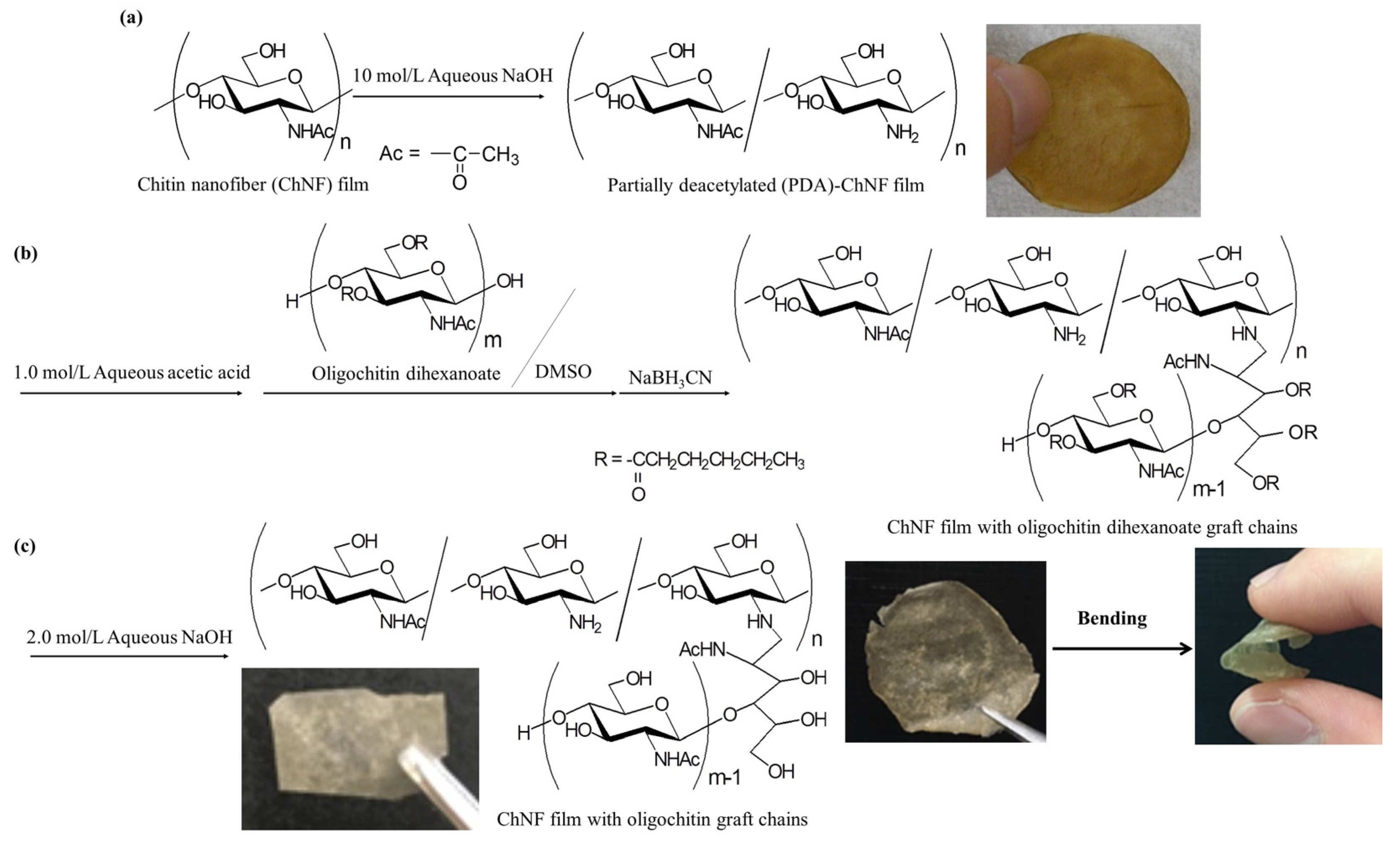
18], was employed as the graft chains to efficiently reduce the crystallinity in the ChNF film owing to such a relative bulky oligochitin derivative. Grafting of oligochitin dihexanoate on ChNFs was carried out by reductive amination of its hemiacetalic reducing end with amino groups present in the partially deacetylated (PDA)-ChNF film using a reducing agent; the amino groups were generated by partial deacetylation of chitin in ChNFs (
Figure 1a,b). Reductive amination is the useful reaction for amino derivation at the hemiacetalic-reducing end of saccharides using amines via an imine intermediate [
19]. We have previously achieved modification of monosaccharide and oligosaccharide moieties on ChNFs by reductive amination [
15,
17,
20]. The present ChNF film with the oligochitin dihexanoate graft chains exhibited a more flexible nature than the parent ChNF film owing to reduction of the crystallinity of chitin. We also confirmed reformation of the crystalline structure in the ChNF film with the oligochitin graft chains after dehexanoylation under alkaline conditions (
Figure 1c). In this study, accordingly, we developed the method for fabrication of nanochitin-based graft and soft materials entirely composed of chitin moieties.
2. Materials and Methods
2.1. Materials
Chitin powder from the crab shells was purchased from FUJIFILM Wako Pure Chemical Corporation (Osaka, Japan). The weight-average molecular weight was estimated by viscometric analysis according to the literature method to be 7 × 10
5 [
21]. The ionic liquid, AMIMBr, was synthesized by quaternization of 1-methylimidazole with 3-bromo-1-propene according to the method adapted from the procedure in the literature [
22]. Chitin dihexanoate was provided by acylation of chitin powder with hexanoyl chloride in methanesulfonic acid at 0 °C for 5 h according to the procedure in the literature [
23];
1H NMR (DMSO-
d6): δ 0.8, 1.2, 1.4–1.6, 2.1–2.4 (br, CH
3(CH
2)
2CH
2CH
2C=O), 1.7 (br s, CH
3C(=O)N-), 3.4–5.0 (br m, sugar protons). The degree of hexanoylation (DH) value was estimated from the integrated ratio of the methyl signals of hexanoyl groups to the sugar signals to be 1.96. Other solvents and reagents were used as commercially received.
2.2. Preparation of Oligochitn Dihexanoate
A mixture of chitin dihexanoate (14.0 g) with conc. hydrochloric acid (5.0 mL)/acetic acid (50 mL) was stirred for 13 h at room temperature to occur partial depolymerization according to the procedure described in the literature [
18]. The reaction mixture was neutralized using aqueous ammonia. The product was isolated by filtration and then dried under reduced pressure to give oligochitin dihexanoate (8.4 g);
1H NMR (DMSO-
d6,
Figure S2): δ 0.8, 1.2, 1.4–1.6, 2.1–2.4 (br, CH
3(CH
2)
2CH
2CH
2C=O), 1.7 (br s, CH
3C(=O)N-), 3.4–5.0 (br m, sugar protons). The DH value was evaluated from the integrated ratio of the methyl signals of hexanoyl groups to the sugar signals to be 1.92.
2.3. Reaction of Terminal Hydroxy Groups in Oligochitin Dihexanoate with Phenyl Isocyanate
Oligochitin dihexanoate (0.020 g) was dissolved in DMF (0.45 mL) by stirring for 1 h at room temperature under argon. After phenyl isocyanate (0.025 mL) was added to the solution, the mixture was attired for 1 h at 80 °C. The reaction mixture was poured into a large amount of diethyl ether to produce the precipitate. The precipitate was isolated by filtration and then dried under reduced pressure to obtain phenyl carbamoyl-terminated oligochitin dihexanoate (0.0173 g).
1H NMR (DMSO-
d6,
Figure S3): δ 6.9–7.4 (br m, aromatics). The molecular weight of oligochitin dihexanoate was evaluated by the integrated ratio of aromatic signals to the sugar signals to be 1870.
2.4. Preparation of PDA-ChNF Film [24]
A mixture of chitin (0.120 g, 0.59 mmol) with AMIMBr (1.00 g, 4.92 mmol) was left stand for 24 h at room temperature and subsequently heated with stirring for 48 h at 90 °C to produce a chitin ion gel (10 wt%). The gel was immersed in methanol (200 mL) for 72 h at room temperature for regeneration and subsequently ultrasonicated (Branson 1510 (42 kHz, 70 W)) to produce a self-assembled ChNF dispersion with methanol. The dispersion was subjected to filtration to isolate ChNFs, which were washed with methanol and then dried under reduced pressure to yield a ChNF film (0.100 g).
A mixture of the resulting film (0.100 g, 0.49 mmol) with 10 mol/L aqueous NaOH (30 mL) was heated for 24 h at 80 °C. The film was separated by filtration, immersed in water (40 mL) for 3 h, filtered, and washed with water. After this procedure was additionally repeated 4 times, the obtained film was dried under reduced pressure to yield a PDA-ChNF film (0.085 g); 1H NMR (DCl/D2O) δ 2.1–2.3 (br s, CH3), 3.0–4.0 (br m, H2–6), and 4.5–5.2 (br m, H1). The degree of deacetylation (DDA) value of the product was evaluated by the integrated ratio of the methyl signal to the sugar signal to be 25.3% for the total repeating units.
2.5. Grafting of Oligochitin Dihexanoate on PDA-ChNF Film
The PDA-ChNF film (0.0907 g) was first dispersed in 1.0 mol/L aqueous acetic acid (9.0 mL) by ultrasonication (Branson 1510 (42 kHz, 70 W)). After a solution of oligochitin dihexanoate (7.9 g, 50 equiv. with amino groups of PDA-chitin) in DMSO was mixed to the dispersion, the mixture was stirred for 1 h at 70 °C. The reducing agent, NaBH3CN (1.5 g, 50 equiv. with amino groups of PDA-chitin), was then added to the mixture, which was stirred for 24 h at 70 °C. The product was isolated by filtration and then dried under reduced pressure to yield ChNFs with oligochitin dihexanoate graft chains (0.0928 g); 1H NMR (DCl/D2O) δ 0.6, 1.1, 1.3, 2.2 (br, CH3(CH2)2CH2CH2C=O), 2.3 (br s, CH3C(=O)N-), 3.3–4.0 (br m, H2–6), 4.5–5.2 (br m, H1). The degree of grafting (DG) value of the product was estimated by the integrated ratio of the methyl signal of the hexanoyl groups to the anomeric (H1) signals to be 3.9% for the total of repeating units.
A suspension of the above product (0.0907 g) in 1.0 mol/L aqueous acetic acid was subjected to filtration. The filtrate was dried under reduced pressure to form a film (0.082 g).
2.6. Dehexanoylation of Graft Chains on ChNF Film
A mixture of the resulting film (0.0353 g) with 2.0 mol/L aqueous NaOH (10 mL) was stirred for 1 h at 40 °C. The film was washed with a large amount of water and then dried under reduced pressure to give a ChNF film with free oligochitin graft chains (0.0263 g).
2.7. Measurements
1H NMR spectra were recorded on a JEOL ECX400 spectrometer (JEOL, Akishima, Tokyo, Japan). Powder X-ray diffraction (XRD) measurements were performed using a Rigaku Geigerflex RADIIB diffractometer (PANalytical B.V., EA Almelo, The Netherlands) with Ni-filtered CuKα radiation (λ = 0.15418 nm). Scanning electron microscopy (SEM) images were obtained using the Hitachi S-4100H electron microscope (Hitachi High-Technologies Corporation, Tokyo, Japan) applying a 5 kV accelerating voltage. IR spectra were recorded on a PerkinElmer Spectrum Two spectrometer (PerkinElmer Japan Co., Ltd., Yokohama, Japan). The stress–strain curves were measured using rectangular strips (10 mm × 5 mm) of ca. 0.2 mm thickness on a tensile tester (Little Senstar LSC-1/30, Tokyo Testing Machine, Tokyo, Japan).
3. Results and Discussion
In this study, we employed oligochitin dihexanoate as a graft chain, modified on ChNFs, because we already developed its preparation procedure from chitin powder [
18]. Therefore, chitin was first hexanoylated using hexanoyl chloride in methanesulfonic acid to obtain chitin dihexanoate (
Figure S1) [
23]. From the
1H NMR analysis in DMSO-
d6, the DH value was calculated to be 1.96, suggesting quantitative hexanoylation of hydroxy groups in the parent chitin. Partial depolymerization of the product was then performed by treatment with conc. hydrochloric acid/acetic acid to produce oligochitin dihexanoate (
Figure S2). The DH value of the produced oligochitin derivative, estimated by
1H NMR analysis in DMOS-
d6 (
Figure S2), was intact (1.92), indicating that obvious dehexanoylation did not occur during the depolymerization experiment. The molecular weight of the product was evaluated by terminal quantification (
1H NMR,
Figure S3) after reaction of the terminal hydroxy groups with phenyl isocyanate, according to the method in the literature, to be 1870 [
18]. According to the procedure in the literature, previously reported by us [
24], on the other hand, the PDA-ChNF film (degree of deacetylation = 25.3%, estimated by
1H NMR analysis after acid hydrolysis in DCl/D
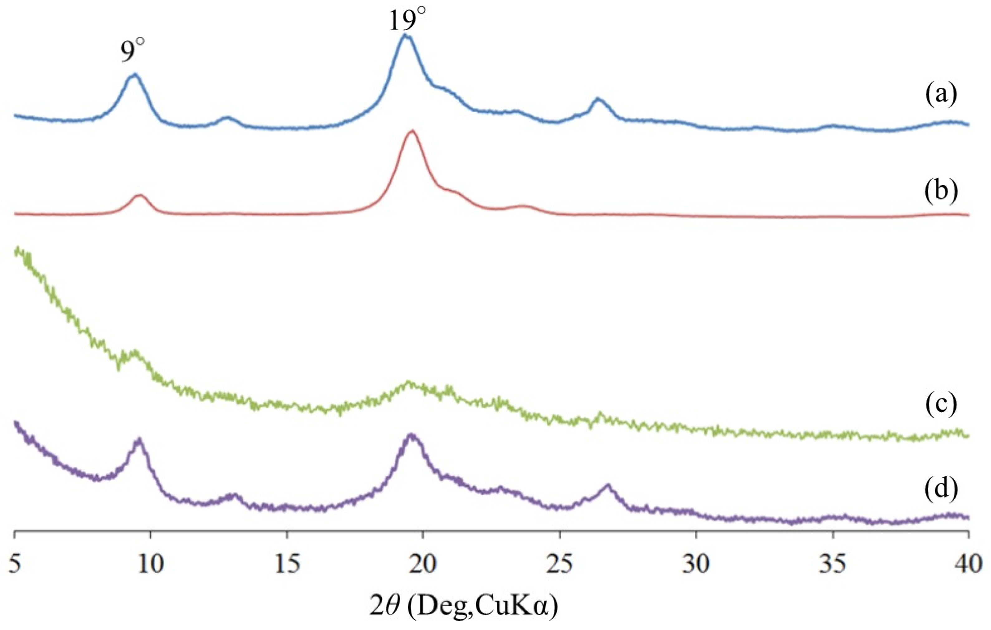
2O) was yielded through self-assembled regeneration of a chitin/AMIMBr ion gel using methanol and filtration, followed by partial deacetylation for 24 h at 80 °C in 10 wt% aqueous NaOH. The XRD profile of the obtained film (
Figure 2b) detected two characteristic diffraction peaks at 9° and 19°, whose pattern was the same as that of the XRD profile of chitin powder (
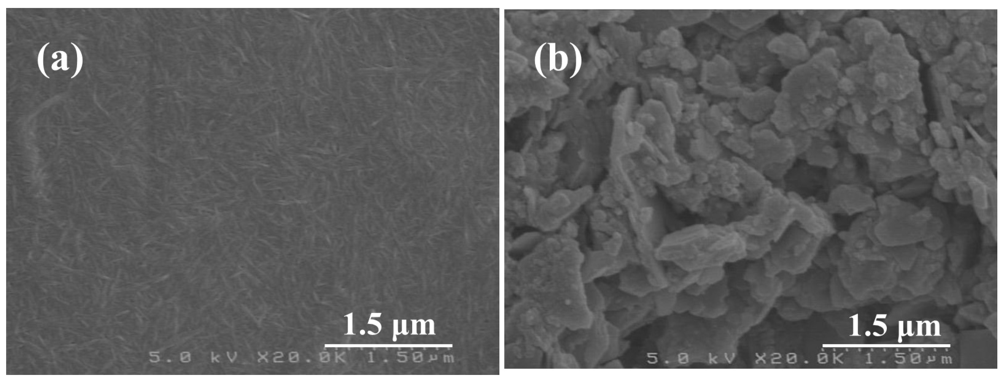
Figure 2a). Furthermore, the remaining nanofiber morphology in the PDA-ChNF film was confirmed by the SEM image (
Figure 3a). These results suggested that partial deacetylation did not predominantly affect the crystalline structure and nano-morphology of the ChNF film.
Grafting of the oligochitin dihexanoate chains on ChNFs was conducted by reductive amination using NaBH
3CN as a reducing agent. After the PDA-ChNF film (DDA = 25.3%) was dispersed in 1.0 mol/L aqueous acetic acid, a solution of oligochitin dihexanoate (50 equiv. for amino groups) in DMSO was mixed. The mixture was stirred with NaBH
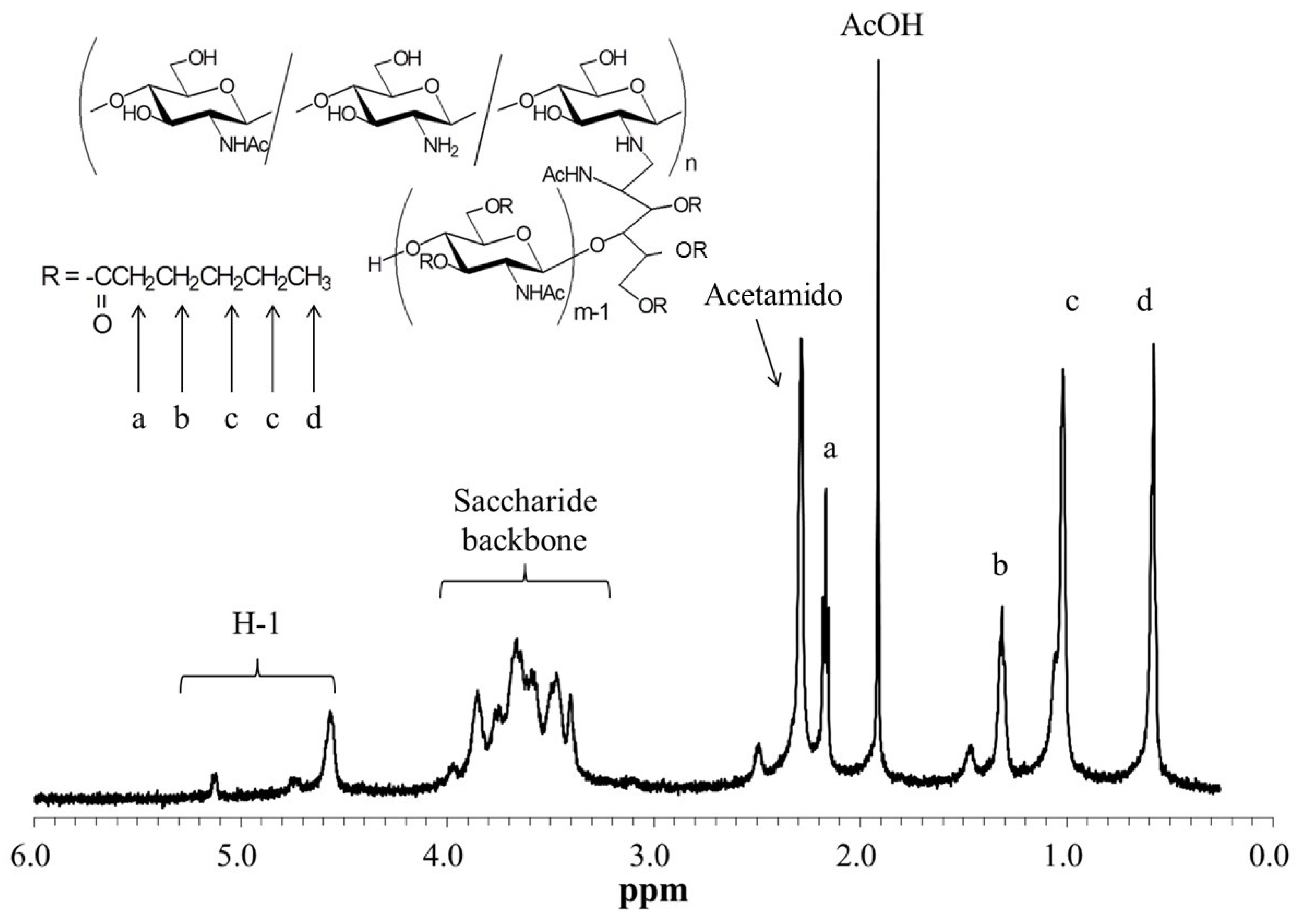
3CN (50 equiv. for amino groups) for 24 h at 70 °C for occurrence of reductive amination. The
1H NMR spectrum of the sample after acid hydrolysis and dissolution of the product in DCl/D
2O (
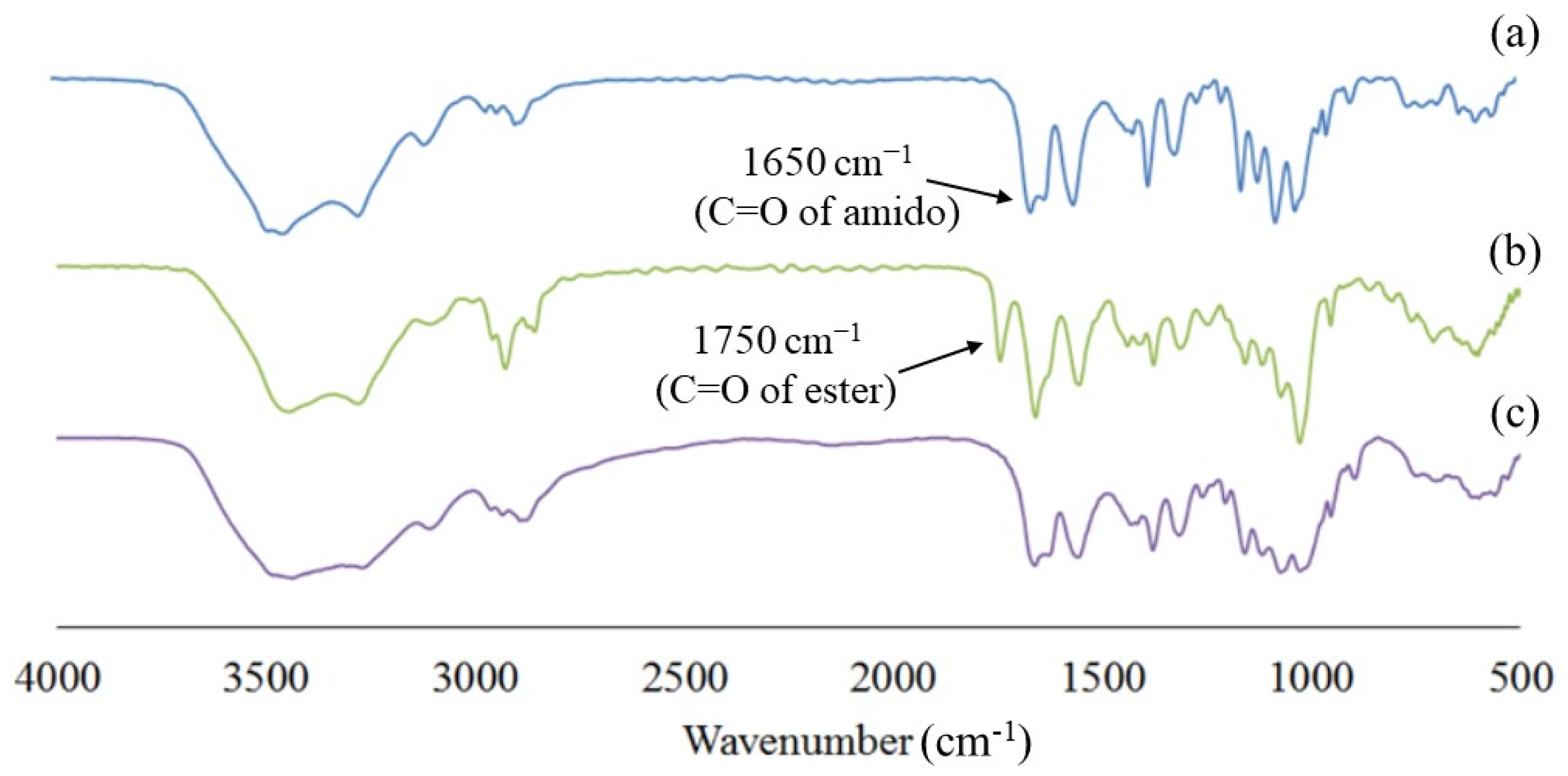
Figure 4) newly observed the signals derived from the hexanoyl groups in addition to the chitin signals, which strongly supported the grafting of the oligochitin dihexanoate chains on ChNFs. From the integrated ratio of the methyl signal of hexanoyl groups to the anomeric (H1) signals in the chitin main chain, the DG value of the graft chains was estimated to be 3.9% for the total repeating units. The IR spectrum of the product (
Figure 5b) also suggested the grafting of the oligochitin dihexanoate chains on ChNFs, because of the detection of a carbonyl absorption that could be assigned to hexanoate groups at 1750 cm
−1, in addition to that which could be assigned to the acetamido group at 1650 cm
−1, as observed in the IR spectrum of chitin powder (
Figure 5a).
When a suspension of the grafted product in 1.0 mol/L aqueous acetic acid was filtered and subsequently dried under reduced pressure, a film was obtained. The XRD profile of the film (
Figure 2c) did not largely observe the diffraction peaks at 9° and 19° ascribable to the crystalline structure of chitin. This result suggested that even grafting of 3.9% of the oligochitin dihexanoate chains for the total number of repeating units strongly contributed to disrupting the regular crystalline structure of chitin molecules. In fact, the SEM image of the film (
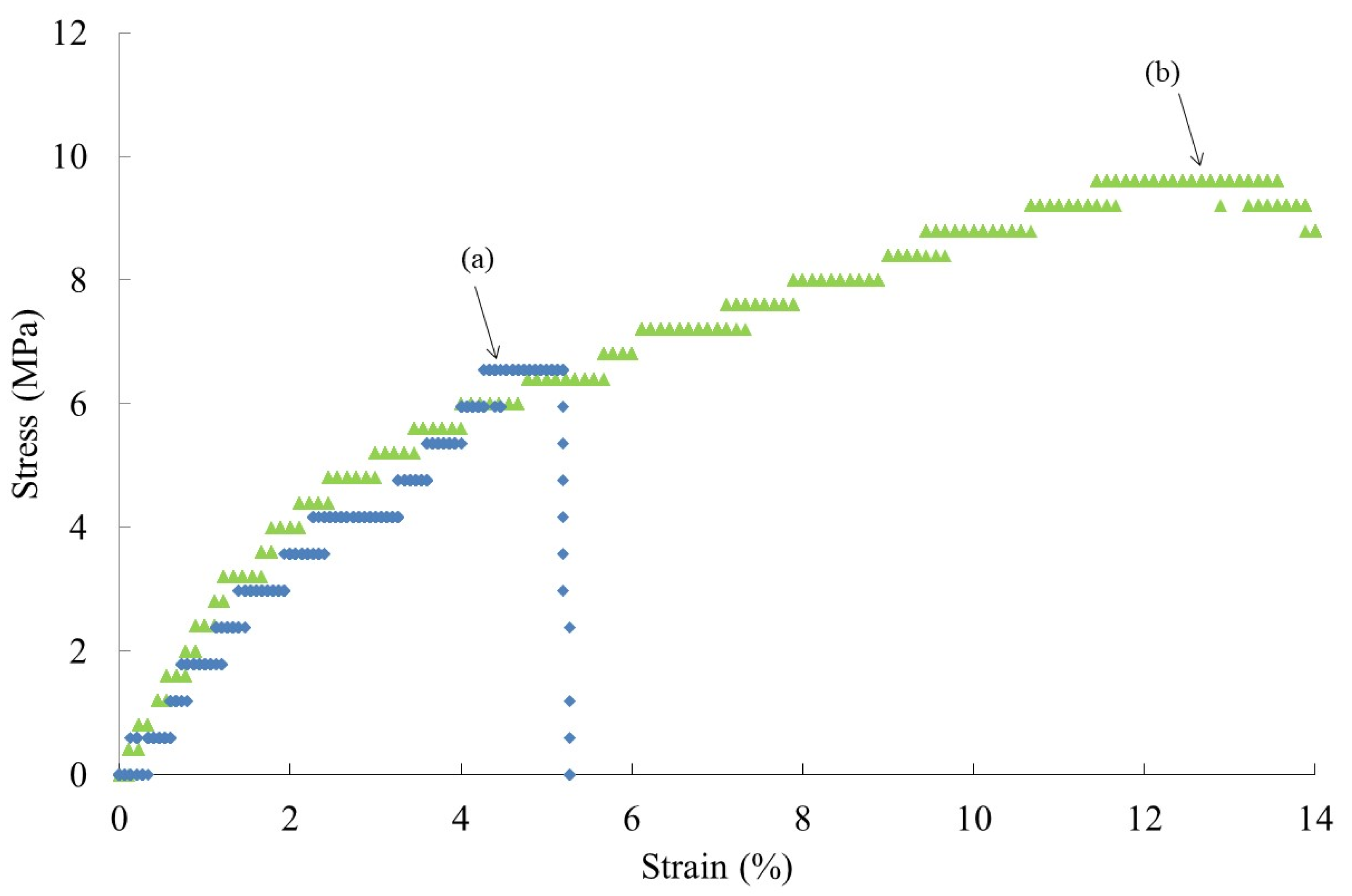
Figure 3b) did not show the morphology of the nanofiber. This is because the nanofiber structure, formed by the fibrous chitin chain assemblies, was not retained as a result of the disruption of chitin crystalline structure. The mechanical properties of the produced film were investigated by tensile testing. The resulting stress–strain curve (
Figure 6b) showed larger values in both tensile strength and elongation at break compared to the PDA-ChNF film (
Figure 6a). This result indicated that grafting of the oligochitin dihexanoate chains on ChNFs efficiently enhanced the mechanical property of the film, probably owing to a reduction of chitin crystallinity. Indeed, the ChNF film with the graft chains could facilely be bent, as shown in
Figure 1b.
The removal of hexanoyl groups (dehexanoylation) from the graft chains in the film was carried out by treatment with aqueous NaOH. The IR spectrum of the resulting film (
Figure 5c) did not detect the carbonyl absorption derived from the hexanoate groups, suggesting complete dehexanoylation to produce the ChNF film with free oligochitin graft chains. The reformation of the crystalline structure of chitin during dehexanoylation was suggested by the detection of diffraction peaks at 9° and 19° in the XRD profile of the resulting film (
Figure 2d), because it was composed of only chitin chains without any substituents, such as the hexanoyl groups.
4. Conclusions
We reported, herein, grafting of oligochitin dihexanoate on the PDA-ChNF film by reductive amination for enhancement of mechanical properties, because the parent ChNF film showed a quite brittle nature. The introduction of the oligochitin dihexanoate graft chains on ChNFs was confirmed by the 1H NMR and IR results. After the film was prepared from the aqueous acetic acid suspension of the product, the XRD analysis, SEM measurement, and tensile testing were conducted, which indicated a reduction of chitin crystallinity, disappearance of nanofiber morphology, and enhancement of flexibility. Alkaline treatment of the film was also performed for dehexanoylation in the graft chains. The XRD result of the dehexnoylated film suggested reformation of the crystalline structure of chitin. This fundamental study provided a method to fabricate new bio-based materials entirely composed of chitin moieties, leading to the efficient use of natural chitin resources as biomass in the future. As some limitations are present for practical application of the present approach in industrial field, of course, further studies are required, which will be conducted in our research group. The present approach, furthermore, has the potential to extend to grafting of other biopolymeric chains on ChNFs, as well as other bionanofibers, to fabricate new bio-based functional materials in the future.


