Article

Calcium Phosphate Cement Promotes Odontoblastic Differentiation of Dental Pulp Cells In Vitro and In Vivo

Shanghai Engineering Research Center of Tooth Restoration and Regeneration, Department of Pediatric Dentistry, School & Hospital of Stomatology, Tongji University, Shanghai 200072, China
*
Author to whom correspondence should be addressed.
These authors contributed equally to this work.
Coatings 2022, 12(4), 543; https://doi.org/10.3390/coatings12040543
Submission received: 26 March 2022 / Revised: 14 April 2022 / Accepted: 16 April 2022 / Published: 18 April 2022
(This article belongs to the Special Issue Antibacterial Coating in Biomedical Applications)

Abstract

:
In the case of pulp injury, odontoblastic differentiation of dental pulp cells (DPCs) at the site of the exposed pulp is necessary for a successful direct pulp capping treatment. Calcium phosphate cement (CPC), a kind of hydroxyapatite-like bone cement, exhibits therapeutic potential in osteogenesis by regulating cell cycle progression and promoting osteoblastic differentiation. Based on the similar biological process of osteo/odontoblastic differentiation, the present study evaluated the effects of CPC on odontoblastic differentiation of DPCs in vitro and in vivo, respectively. The morphology of CPC was observed by scanning electron microscopy. Colony-forming units were used to assess the antibacterial activity. The effects of CPC on cell proliferation and odontoblastic differentiation of human dental pulp cells (hDPCs) were also measured. Histological staining was performed to observe the reparative dentin formation in rat molars. In vitro, results of antibacterial studies showed that CPC significantly inhibited the growth of Streptococcus mutans. The appropriate concentration of CPC extract showed low cytotoxicity on hDPCs. Furthermore, CPC extract also promoted odontoblastic differentiation and mineralization compared with the control group, as shown by a dynamic increase in the expression of odontogenic marker genes and the increased number of mineralized nodules at 21 days. The pulpotomy models with CPC facilitated the formation of dentin bridge with the highly expressed dentin matrix protein 1 (DMP1) in odontoblast-like cells. In conclusion, the favorable biocompatibility, antibacterial property and bio-inductivity of CPC suggest that CPC can be used as a promising direct pulp capping material.

1. Introduction

With the widely accepted concept of minimum-intervention dentistry and the capacity of dentin-pulp complex (DPC) producing reparative dentin, exploiting suitable direct pulp capping materials to protect the vital pulp beneath the injured region is of great importance [1]. Based on this context, the materials with favorable surface characteristics, including biocompatibility, high alkaline, and antibacterial behavior, such as calcium hydroxide (CH), mineral trioxide aggregate (MTA), and Biodentine (BD), are suggested to be clinical choices [2,3]. Dressing the golden standard material MTA on the exposed pulp surface has been the first choice since 1988, but its high cost, long setting time without biodegradable ability and difficulty in handling are adverse effects associated with its clinical application [3,4]. Thus, some easily acquired, resorbable and odontoblast conductive materials applied in close contact with pulpal tissue are supposed to be novel approaches for conservative pulp therapy.
CPC, also named hydroxyapatite cement (HAC), is a type of non-ceramic and injectable bone cement that is used as a coating and scaffold material to enhance bone regeneration when placed on the implant or in a bony defect [5]. The fundamental roles of CPC are regulating osteoclast survival, facilitating osteoclast differentiation, and promoting osteoclast-mediated osseointegration [6]. Bone and dentin are both hard tissues with mineral (hydroxyapatite) and collagenous components that possess similar formation processes, that is, both osteoblasts and odontoblasts are responsible for the synthesis and secretion of unmineralized proteins [7]. Therefore, it is valuable to investigate if CPC participates in odontoblastic differentiation of dental pulp cells (DPCs) and promotes the formation of mineralized tissue in vivo. It was reported that CPC and chitosan-supplement CPC (CPC-Ch) could decrease the cell viability but enhance the odontoblastic differentiation of human dental pulp cells (hDPCs) in vitro under the effect of the ionic activities and pH changes that occurred during material setting [8]. Other studies focusing on the function of CPC extract with different calcium and phosphate ion concentrations and local pH values revealed that CPC extract did not negatively affect cell proliferation or apoptosis in human dental pulp stem cells (hDPSCs) [9]. Studies also demonstrated that the ability to release calcium and phosphate ions of CPC could facilitate osteoclast-mediated osseointegration [5,6]. Thus, based on the similar biological process of osteo/odontoblastic differentiation, the effect of CPC on DPCs behaviors in vitro should be further evaluated.
In recent years, CPC has been gradually applied in the oral field [10,11,12], such as maxillofacial bone defect repair, periodontal regeneration and dental implant stabilization. However, CPC has not been applied as a pulp capping agent in-clinic yet. The major challenge of a successful direct pulp capping treatment involves excellent remineralization with low inflammation. Therefore, in order to decrease the risk of chronic inflammation and necrosis caused by microbes, the antibacterial ability of the pulp capping agent is important to a large extent. At present, some novel materials, such as graphene oxide (GO) and quaternary ammonium salt (QAS) [13,14], have been developed as coatings on the surface of the basal material to enhance the antibacterial ability. A previous study has demonstrated the antibacterial activity of the clinical pulp capping agent, ProRoot MTA, using Streptococcus mutans (S.mutans), which is closely related to caries incidence [15], but the antibacterial effect of CPC still shows divergence [11,16,17]. Hence, colony-forming units (CFUs) and direct contact tests (DCT) were performed in this study to investigate the antibacterial properties of CPC against S.mutans.
The functions of CPC in odontoblastic differentiation and antibacterial properties, namely CPC itself or the released components in CPC extracts, are still elusive. The aim of this study was to elucidate the biological effects of CPC, including the proliferation and odontoblastic differentiation of hDPCs as well as the formation of reparative dentin after direct pulp capping of rat molars.

2. Materials and Methods

2.1. Morphological Analysis of CPC and MTA

The surface morphologies of CPC powder (kindly gifted by Professor Kaili Lin) and MTA powder (White ProRoot MTA; Dentsply Dental Specialties, Tulsa, OK, USA) were evaluated by using scanning electron microscopy (SEM, JEOL Ltd. JSM-820, Tokyo, Japan). After being fixed with 1% glutaraldehyde (Millipore) in PBS, both cements were dehydrated with 30–100% gradient ethanol and rinsed with hexamethyldisilazane (Millipore). The micrographs depicting magnifications of ×3.5 k and 35 k were chosen for the morphological characterization.

2.2. In Vitro Experiments

2.2.1. Evaluation of Antibacterial Activity

The antibacterial capability of CPC was tested by DCT and CFUs. Standard S. mutans was chosen as the experimental strain of the antibacterial activity test, with routine resuscitation and inoculation in a Brain Heart Infusion (BHI) agar culture dish for 24 h (37 °C, 80% N2, 10% CO2, 10% H2). Then the monoclonal clones were selected and cultured for 48 h. The optical density (OD) of the bacterial solution was found to be 105 CFUs/mL by using a microplate reader (Bio-Tek, Hercules, CA, USA) at the wavelength of 600 nm. The experiment contained 3 groups with 3 samples of each: Group 1 (50 μL bacteria suspension + 150 μL BHI), Group 2 (1 mg CPC + 50 μL bacteria suspension + 150 μL BHI), Group 3 (1 mg MTA + 50 μL bacteria suspension + 150 μL BHI). All groups were incubated at 37 °C for 24 h. Then, 5 μL aliquots of each group were added onto BHI agar plates and cultivated in an anaerobic incubator for 48 h. Bacterial growth was measured at OD 600 and the CFUs/mL was estimated by counting the number of colonies on nutrient agar (NA).

2.2.2. Cell Isolation and Culture

The study was approved by the Institutional Review Board of the Affiliated Stomatology Hospital of Tongji University, Shanghai, China, and Ethics Committee approval was obtained ([2019]-DW-059).
Healthy third molars without defects were collected from patients aged 14 to 25 years old (n = 5), and written informed consents were obtained in the Oral and Maxillofacial Surgery at the Affiliated Stomatology Hospital of Tongji University. Briefly, the healthy pulps were isolated from the extracted teeth and minced into pieces under sterile conditions and then treated with collagenase type I (Sigma-Aldrich, St. Louis, MO, USA). hDPCs were cultured in α-modified Eagle’s medium (α-MEM; Hyclone, Logan, UT, USA) supplemented with 10% fetal bovine serum (FBS, Gibco BRL, Gaithersburg, MD, USA), 100 U/mL penicillin and 100 mg/mL streptomycin (Sigma-Aldrich) at 37 °C in an atmosphere of 5% CO2 and 95% humidity. Fourth to sixth passages of hDPCs were used for in vitro experiments.

2.2.3. Extract Medium Preparation: CPC Extract (CPCe) and MTA Extract (MTAe)

The extraction condition and method were carried out according to previous studies [9,18] and the International Organization for Standardization (ISO) method 10993-12: sample preparation and reference materials. In brief, powders of CPC and MTA were previously sterilized through UV rays for at least 30 min and freshly configured into pastes with 0.9% physiology saline. There were 3 specimens per group. Both groups were placed at 37 °C with 95% relative humidity for 24 h, then ground into powder with the same particle size, and added to α-MEM, respectively. The solution at a concentration of 0.2 mg/mL (0.2 mg CPC or MTA:1 mL α-MEM) was vortexed until completely suspended, settled for 10 min, and incubated in the incubator (5% CO2, 37 °C) for 24 h. The supernatant was filtered before the use as a culture medium.

2.2.4. Cell Proliferation Assay

The hDPCs were seeded in 96-well plates (Corning Inc., Corning, NY, USA) at 1 × 10³ cells per well and were cultured in different concentrations of conditioned medium, including 0.2, 0.1, 0.05 mg/mL of CPCe or MTAe for 5 days. Medium without extracts was used as the control group. A cell counting kit-8 (Keygen) was used to analyze the cell numbers using a microplate reader at a wavelength of 450 nm. All reactions were performed in triplicate. After analyzing the results comprehensively, an optimal concentration was selected for mineralization experiments.

2.2.5. Alizarin Red Staining (ARS)

The control mineralization medium (MM) contained 0.05 mM ascorbic acid (Sigma-Aldrich, St Louis, MO, USA), 100 mM dexamethasone (Sigma-Aldrich), 10 mM β-glycerophosphate (Sigma-Aldrich), 10% FBS, 100 U/mL penicillin and 100 mg/mL streptomycin in α-MEM, as described previously [19]. The hDPCs were seeded in 24-well plates (Corning) at a density of 1 × 10⁵ cells per well and were cultured for 14 and 21 days. Samples were divided into 4 groups: negative control group, MM control group, the optical concentration of CPCe or MTAe with MM groups. Cells were treated every 2 days with freshly prepared CPCe and MTAe. Mineralization nodules were detected by ARS after 14 and 21 days. In brief, the fixed cells were stained using 2% ARS solution (Sigma-Aldrich) and were photographed under a microscope (Nikon Eclipse 80i, Tokyo, Japan) after several steps of washing.

2.2.6. Real-Time Quantitative Polymerase Chain Reaction (RT-qPCR)

The mRNA expression levels of dentin matrix protein 1 (DMP1), dentin sialophosphoprotein (DSPP), and alkaline phosphatase (ALP) were determined by RT-qPCR. Briefly, the samples were divided into 3 groups: control group, the optical concentration of CPCe or MTAe with MM groups. To extract total RNA, hDPCs treated with different medium were lysed by TRIzol reagent (Life Technologies, Carlsbad, CA, USA). Then, the extracted RNA was reverse transcribed into complementary DNAs using a PrimeScript RT reagent kit with gDNA Eraser (Takara Bio, Kusatsu, Japan). RT-qPCR was performed with HieffTM qPCR® SYBR Green Master Mix (Applied Biosystems, Foster City, CA, USA), and PCR product levels were normalized to that of the housekeeping gene glyceraldehyde-3-phosphate dehydrogenase (GAPDH). All reactions were performed in triplicate. The relative expression of each target gene was calculated by using the 2−ΔΔCt method. Primer sequences for DMP1, DSPP and ALP (Sangon Biotech, Shanghai, China) are listed in Table 1.

2.3. In Vivo Experiments

Sprague-Dawley rats (n = 12) weighing about 180–200 g at 6 weeks old were used for in vivo study. They were ordered from Slaccas (Shanghai, China) and maintained under standard conditions. The animal-use protocol conformed to the National Institutes of Health guidebook and was reviewed and approved by the Screening Committee for Animal Research of Tongji University.

2.3.1. Direct Pulp Capping Assay

Twenty-four maxillary first molars obtained from twelve rats were randomly divided into 3 groups: CPC pulp capping group (n = 8), MTA pulp capping group (positive control, n = 8) and negative control group without direct pulp capping material (n = 8). Rats were anesthetized with an intraperitoneal injection of 10% chloral hydras (Sangon Biotech, Shanghai, China). Cavities were prepared on the central fossa of the upper first molars using a ½ sterile stainless-steel round bur (0.6 mm in diameter; Dentsply Maillefer) with a high-speed handpiece, and sterile distilled water was irrigated to avoid pulp impairment. Then, pulps were exposed with the tip of a #15 sterile stainless file (Dentsply Tulsa Dental, Tulsa, OK, USA) through the bottom of each cavity. The procedure was conducted by an experienced operator to optimize the cavity size in all samples. After blow-drying, pulp perforation sites were directly capped with CPC paste (kindly gifted by Professor Kaili Lin) or MTA paste in both experimental groups, whereas the pulp was capped with nothing in the negative control groups. All cavities were subsequently sealed with glass ionomer cement (Fuji IX; GC International Corp, Tokyo, Japan). The cusp tips of the mandibular first teeth were ground to minimize occlusal forces.

2.3.2. Sample Preparation

Animals were anesthetized and internal fixed with 4% paraformaldehyde at 4 weeks after direct pulp capping. Bones were separated and fixed in 4% paraformaldehyde for 24–48 h at 4 °C. The samples were decalcified in 10% Ethylene Diamine Tetraacetic Acid (EDTA, pH = 7.4), embedded in paraffin, and cut into 4 μm thick sections in a mesiodistal direction.

2.3.3. Immunohistochemistry (IHC)

An UltraSensitiveTM IHC kit (Maxim Biotechnology, Fuzhou, China) was used for the IHC. The primary antibody was polyclonal rabbit anti-DMP1 (1:100, NBP 1-45525, Novus Biologicals Centennial, Centennial, CO, USA). Sections were deparaffinized in xylene and rehydrated by gradient ethanol. H2O2 3% was used to suppress endogenous peroxidase activity. For antigen retrieval, they were incubated in hyaluronidase (Sigma-Aldrich, St Louis, MO) at 37 °C for 1 h. Normal serum solution was dropped onto the slides for 30 min at room temperature prior to primary antibody incubation overnight at 4 °C. Sections were incubated with biotinylated secondary antibody (Maxim Biotechnology, Fuzhou, China) for 1 h and the DAB detection kit (DAB, Keygen, Nanjing, China) was used as a color developing agent; then, the sections were counterstained with hematoxylin. Finally, sections were observed on a microscope (Nikon Eclipse 80i, Tokyo, Japan). Negative controls were carried out by omitting the primary antibody. IHC was analyzed qualitatively with the mineralized areas.

2.4. Statistical Analysis

Statistical analysis was performed using SPSS Version 20.0 (SPSS Inc., Chicago, IL, USA), and the graphics software used was GraphPad Prism 9.0. Data were expressed as the mean value ± standard error of the mean (SEM) and subjected to a one-way analysis of variance (ANOVA). A probability value of p < 0.05 was considered statistically significant.

3. Results

3.1. Surface Morphology of the Cements

Representative SEM images (Figure 1) of CPC and MTA powders showed the variety in crystal shape and size within the particles. As shown in Figure 1b,d, the surface and internal morphology of cements with numerous ultrafine crystallites in needle shapes and globular angular shapes were different.

3.2. Antibacterial Activity

The antibacterial activity of CPC was tested by CFUs and DCT assay. Both cements of the same concentration were co-cultured with the bacteria in BHI medium for 24 h. The result of the CFUs assay was observed on the agar plates (Figure 2a), showing that the spread of bacterial colonies of CPC and MTA groups significantly decreased compared to that of the control group (Figure 2a,b), indicating the enhanced antibacterial activity. The result in Figure 2c also showed that both cements could inhibit the growth of S. mutans, and there was no significant difference between the CPC and MTA groups.

3.3. Effect of CPCe on Viability and Proliferation of hDPCs

To investigate the effects of CPCe and MTAe on hDPCs proliferation, the CCK-8 assay was conducted at 1, 3 and 5 days. The result showed that proliferation of hDPCs was not affected at 1 day, but at 3 and 5 days, the proliferation proportion of experimental groups was significantly higher than that of the control groups (p < 0.01, Figure 3b). In addition, there was no statistically significant difference between the groups of CPCe and MTAe. Meanwhile, we selected 0.1 mg/mL as the applicable concentration of CPCe or MTAe for the mineralization assay according to the results.

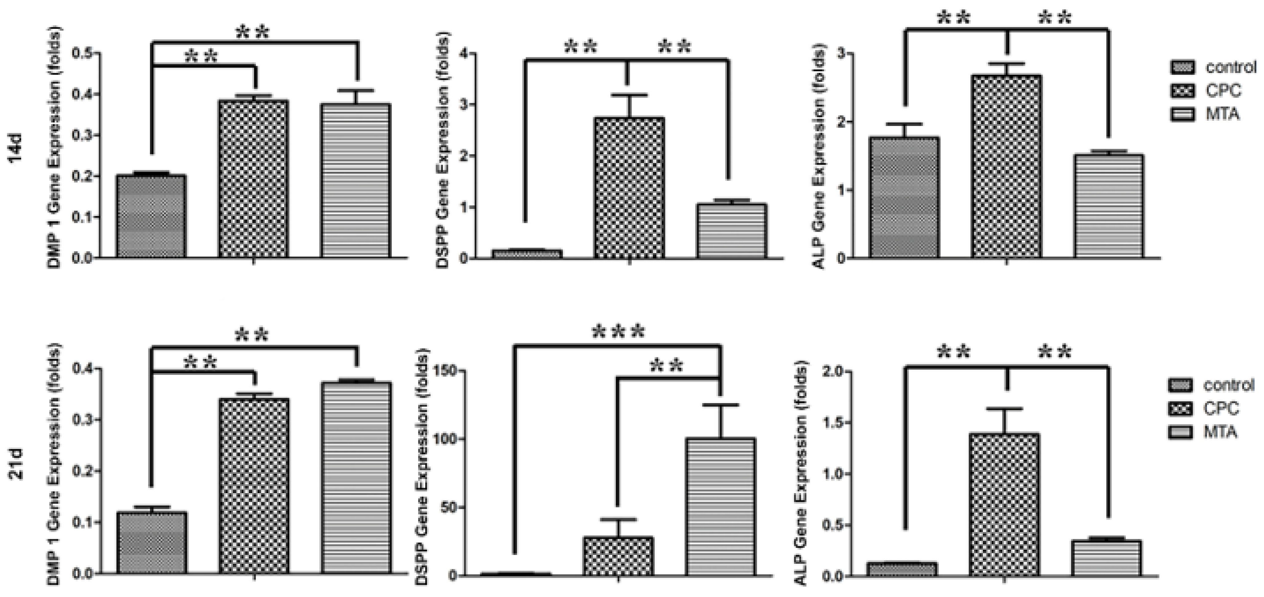
3.4. Effects of CPCe on the Odontoblastic Differentiation of hDPCs

The late stage of mineralization was assessed by ARS (Figure 4), which showed that the positive staining was denser in both groups of CPCe and MTAe with MM at 21 days compared with the MM group, whereas there was no noticeable staining difference in the experimental group at 14 days. Taken together, these results indicated that the extracts of CPC and MTA had the ability to induce odontoblastic differentiation of hDPCs.
To assess the odontogenic potential of CPC, the expression of related genes was conducted (Figure 5). The expression of DMP1, DSPP and ALP was up-regulated in the CPCe and MTAe groups after culturing for 14 days and 21 days compared with the control group. However, there was no significant difference between the groups of CPCe and MTAe in DMP1 expression. The expression of DSPP was higher in the CPCe group than in MTAe at 14 days, whereas the result was the opposite at 21 days. As for the expression of ALP, the CPCe group showed a higher level than that of the MTAe group both at 14 and 21 days.

3.5. CPC Stimulated Reparative Dentinogenesis in Rat Molars

To further elucidate the odontoblastic differentiation effect of CPC in vivo, we established experimental pulp exposure in rat maxillary first molars. The enamel and dentin of molars in this model were moved without damage to the pulp chamber. All groups showed various mineralized areas at 4 weeks, but reparative dentin in group CPC and MTA were better-distributed (dotted line). Although all groups could form mineralized nodules, inflammatory cell aggregation and necrosis were detected in the control groups (Figure 6b, indicated by asterisks) while this was not found in the experimental groups (Figure 6d,f). As for IHC, the expression of DMP1, a protein secreted by odontoblasts, was significantly higher in both CPC and MTA groups compared with the control groups (Figure 6g–l), and DMP1 was strongly localized to the odontoblast-like cells in the pulp chamber, especially around the newly formed dentin (Figure 6i–l).

4. Discussion

Hydroxyapatite (Ca10(PO4)6(OH)2) is the mineral component of teeth, making up 70% of dentin [20]. Previous studies have demonstrated that reparative dentin is secreted by odontoblast-like cells differentiated from resident DPSCs [21]. Based on the natural components of dentin, we evaluated the ability of CPC to promote odontoblastic differentiation in vitro and in vivo. MTA is the typical clinical pulp capping agent that was used as the positive control group in this study. The surface morphologies of CPC and MTA were different, as shown in the representative SEM images, which may represent different biological activities, but both cements are suitable for cell adhesion and growth [22,23]. CPC is a mixture of tetra-calcium phosphate (TTCP, Ca4(PO4)2O) and dicalcium phosphate anhydrous (DCPA, CaHPO4), whereas the compositions of MTA are tricalcium silicate ((CaO)3·SiO2), dicalcium silicate ((CaO)2·SiO2), tricalcium aluminate ((CaO)3·Al2O3), bismuth oxide (Bi2O3) and gypsum (CaSO4·2H2O) [24]. Therefore, the morphological and bioactive difference and similarity between CPC and MTA may be caused by the release in ion ratio, crystal structure, and solubility.
S. mutans has a pivotal role in the process of caries and also has the ability to maintain sugar metabolism in a low pH environment [1]. Furthermore, residual bacteria in exposed dentin tubules may lead to long-term treatment failure. Thus, we tested the antibacterial activity of CPC using S. mutans by CFUs and DCT assay, which can simulate the direct contact of bacteria with CPC and MTA in clinical treatment. Results in this experiment indicated that both the cements showed the antibacterial effect to some extent compared with the control group, which was consistent with previous studies [13,15,25]. CPC shared similar antibacterial capability with MTA, which may be due to the release of high concentration calcium and phosphate ions [17,26] and changes in osmotic pressure, subsequently affecting the structure of S. mutans. This result suggested that CPC is a pulp capping material since the antibacterial activity of CPC could also decrease the risk of inflammation during new dentin formation.
Direct pulp capping material should exhibit excellent biocompatibility because it is in direct contact with the pulp tissue. An in vitro cellular analysis was conducted to assess the biocompatibility of CPC. The result of CCK-8 showed that both cement extracts could significantly improve cell viability, and 0.1 and 0.2 mg/mL CPCe could promote proliferation, while the lower concentration of 0.05 mg/mL did not. This was in accordance with a previous report that CPC with a proper concentration showed no cytotoxicity to hDPCs [9].
Resident DPCs can be activated by pulp injuries and respond to the trauma by initiating the reparative function, containing a series of biological contexts, including cell proliferation, odontoblast-like cell differentiation, and mineralized tissue formation [27]. In terms of the effect on mineralization, accumulated evidence has focused on CPC-mediated osteogenesis by releasing calcium and phosphate ions [28,29,30]. The result of ARS confirmed that CPC could enhance the mineralization ability of hDPCs at 21 days in vitro. Furthermore, DMP1, DSPP, and ALP, specific markers of differentiated odontoblasts, are essential for the development of hard tissues [31,32]. In this context, the result of RT-qPCR showed dynamic changes of the mineralization-related factors, which illustrated that CPC facilitated the process of odontoblast-like cell differentiation. These results also demonstrated that CPC might be an effective pulp capping material. Although our results in the CPC groups were not totally superior to those in the MTA groups, several studies have found that the effect of odontoblastic differentiation would be better by modifying the component and chemical property of CPC [22,33]. Therefore, novel materials are required to construct bioactive coating surfaces that can release ions and promote the formation of reparative dentin in future investigations.
We further confirmed the potential application of CPC to induce odontoblastic differentiation in rat models. As for the histological result in rat models, CPC exhibited good biocompatibility with the pulp tissue without persistent necrosis, while the necrosis areas in the control group might result from the cytotoxicity of glass ionomer cement. Both CPC and MTA successfully induced tertiary dentin formation, and there was no significant difference between CPC and MTA groups. Thus, we speculate that before CPC setting, Ca2+, PO43− and OH ions from TTCP and DCPA might be dissolved out and recombined to form hydroxyapatite, which is the major inorganic constituent of dentin [34]. Meanwhile, the phosphate ions could promote macrophage polarization to alternatively activated (M2) macrophages, which are mainly involved in the anti-inflammatory response [9]. Wound healing of dental pulp may occur after the reduction in inflammation, differentiation of resident cells, and the formation of reparative dentin at the pulp-material interface [35,36]. DMP1 is a multifunctional phosphorylated non-collagenous matrix protein, which may regulate odontoblast-like cell differentiation, formation of the dentin tubular system and trigger the repair of exposed pulp [37,38,39]. A study has demonstrated that MTA could stimulate calcified granules following the induction of DMP1 in reparative dentin [40]. This is consistent with our present results, the deposition of DMP1 was significantly detected in the newly formed dentin, and we supposed that CPC could also stimulate reparative dentin formation. In the next investigation, we will try to establish a standard rat model by simulating the lesion size in human teeth and assess the effective volume of reparative dentin that can protect vital pulps.
At present, CPC is a widely used bone mineral-mimicking scaffold, which can enhance the bone repair process [41,42]. Meanwhile, the characteristic of pulp regeneration potential can lead to the development of therapeutic agents. The application of CPC will be more promising if it can be combined with bioactive coatings that can also promote odontoblastic differentiation and resist microbials. Thus, as a potential pulp capping agent, modified CPC materials with anti-inflammation and bioactive factors could provide necessary ions, work as a suitable scaffold for cell homing, and induce the generation of reparative dentin. However, the underlying mechanism remains to be further explored, especially the signaling pathways and the possible modification of the CPC-mediated dentin repair.

5. Conclusions

To conclude, this study provided insight into the capacity of CPC to modulate odontoblastic differentiation in vitro and promote the formation of reparative dentin in vivo. In addition, results in this study also indicated that CPC with suitable concentration showed biocompatibility and antibacterial activity. There was no significant difference between the CPC group and the golden standard clinical material MTA group. However, the assessment of CPC seems inadequate in limited animal studies, and the application evaluation of CPC needs more human clinical trials in the future. All the results suggest the potential application of CPC as a future direct pulp capping material and expand the range of raw materials in conservative pulp therapy.

Author Contributions

Conceptualization, B.J.; methodology, investigation, H.H., L.L. (Lefeng Li) and Y.G.; software, H.H. and L.L. (Linjuan Luo); writing—original draft preparation review and editing, H.H. and L.L. (Linjuan Luo); data curation, H.H. and L.L. (Lefeng Li); visualization, B.J., Y.Y. and Z.J.; supervision, B.J. and Z.J.; project administration, B.J. and Y.Y.; funding acquisition, B.J. and Y.Y. All authors have read and agreed to the published version of the manuscript.

Funding

This study was supported by the Shanghai Municipal Health Commission Program (Nos. 202040095, Nos. 202140357).

Institutional Review Board Statement

The animal-use protocol conformed to the National Institutes of Health guidebook and was reviewed and approved by the Screening Committee for Animal Research of Tongji University ((2019)-DW-059, (2021)-DW-70).

Informed Consent Statement

Informed consent was obtained from all subjects involved in the study. Written informed consent has been obtained from the patient(s) to publish this paper.

Data Availability Statement

Not applicable.

Acknowledgments

We would like to thank the Shanghai Engineering Research Center of Tooth Restoration and Regeneration. We also thank Kaili Lin for generously providing CPC powder.

Conflicts of Interest

The authors declare that there are no competing financial interests or personal relationships that appeared to influence the work reported in this paper.

References

  1. Bjorndal, L.; Simon, S.; Tomson, P.L.; Duncan, H.F. Management of deep caries and the exposed pulp. Int. Endod. J. 2019, 52, 949–973. [Google Scholar] [CrossRef] [PubMed]
  2. Hilton, T.J.; Ferracane, J.L.; Mancl, L.; Northwest Practice-based Research Collaborative in Evidence-based D. Comparison of CaOH with MTA for direct pulp capping: A PBRN randomized clinical trial. J. Dent. Res. 2013, 92 (Suppl. S7), 16S–22S. [Google Scholar] [CrossRef] [PubMed] [Green Version]
  3. Zhang, W.; Yelick, P.C. Tooth repair and regeneration: Potential of dental stem cells. Trends Mol. Med. 2021, 27, 501–511. [Google Scholar] [CrossRef] [PubMed]
  4. Lee, B.S.; Lin, H.P.; Chan, J.C.; Wang, W.C.; Hung, P.H.; Tsai, Y.H.; Lee, Y.L. A novel sol-gel-derived calcium silicate cement with short setting time for application in endodontic repair of perforations. Int. J. Nanomed. 2018, 13, 261–271. [Google Scholar] [CrossRef] [Green Version]
  5. Hoshikawa, A.; Fukui, N.; Fukuda, A.; Sawamura, T.; Hattori, M.; Nakamura, K.; Oda, H. Quantitative analysis of the resorption and osteoconduction process of a calcium phosphate cement and its mechanical effect for screw fixation. Biomaterials 2003, 24, 4967–4975. [Google Scholar] [CrossRef]
  6. Wang, X.; Yu, Y.; Ji, L.; Geng, Z.; Wang, J.; Liu, C. Calcium phosphate-based materials regulate osteoclast-mediated osseointegration. Bioact. Mater. 2021, 6, 4517–4530. [Google Scholar] [CrossRef]
  7. Moradian-Oldak, J.G.A. Biomineralization of enamel and dentin mediated by matrix proteins. J. Dent. Res. 2021, 100, 1020–1029. [Google Scholar] [CrossRef]
  8. Lee, S.K.; Lee, S.K.; Lee, S.I.; Park, J.H.; Jang, J.H.; Kim, H.W.; Kim, E.C. Effect of calcium phosphate cements on growth and odontoblastic differentiation in human dental pulp cells. J. Endod. 2010, 36, 1537–1542. [Google Scholar] [CrossRef]
  9. Gu, Y.X.X.; Zhuang, R.; Weir, M.D.; Oates, T.W.; Bai, Y.; Zhao, L.; Xu, H.H.K. A biphasic calcium phosphate cement enhances dentin regeneration by dental pulp stem cells and promotes macrophages M2 phenotype in vitro. Tissue Eng. Part A 2021, 27, 1113–1127. [Google Scholar] [CrossRef]
  10. Shirakata, Y.; Yoshimoto, T.; Goto, H.; Yonamine, Y.; Kadomatsu, H.; Miyamoto, M.; Nakamura, T.; Hayashi, C.; Izumi, Y. Favorable periodontal healing of 1-wall infrabony defects after application of calcium phosphate cement wall alone or in combination with enamel matrix derivative: A pilot study with canine mandibles. J. Periodontol. 2007, 78, 889–898. [Google Scholar] [CrossRef]
  11. Qiu, G.; Wu, H.; Huang, M.; Ma, T.; Schneider, A.; Oates, T.W.; Weir, M.D.; Xu, H.H.K.; Zhao, L. Novel calcium phosphate cement with biofilm-inhibition and platelet lysate delivery to enhance osteogenesis of encapsulated human periodontal ligament stem cells. Mater. Sci. Eng. C Mater. Biol. Appl. 2021, 128, 112306. [Google Scholar] [CrossRef] [PubMed]
  12. Schickert, S.L.; Jansen, J.A.; Bronkhorst, E.M.; van den Beucken, J.J.; Leeuwenburgh, S.C. Stabilizing dental implants with a fiber-reinforced calcium phosphate cement: An in vitro and in vivo study. Acta Biomater. 2020, 110, 280–288. [Google Scholar] [CrossRef] [PubMed]
  13. Esteki, P.J.M.; Tahmourespour, A. In vitro antimicrobial activity of mineral trioxide aggregate, Biodentine, and calcium-enriched mixture cement against Enterococcus faecalis, Streptococcus mutans, and Candida albicans using the agar diffusion technique. Dent. Res. J. 2021, 18, 3. [Google Scholar]
  14. Sun, N.; Yin, S.; Lu, Y.; Zhang, W.; Jiang, X. Graphene oxide-coated porous titanium for pulp sealing: An antibacterial and dentino-inductive restorative material. J. Mater. Chem. B 2020, 8, 5606–5619. [Google Scholar] [CrossRef] [PubMed]
  15. Bossu, M.; Mancini, P.; Bruni, E.; Uccelletti, D.; Preziosi, A.; Rulli, M.; Relucenti, M.; Donfrancesco, O.; Iaculli, F.; Di Giorgio, G.; et al. Biocompatibility and Antibiofilm Properties of Calcium Silicate-Based Cements: An In Vitro Evaluation and Report of Two Clinical Cases. Biology 2021, 10, 470. [Google Scholar] [CrossRef] [PubMed]
  16. Wu, S.; Lei, L.; Zhang, H.; Liu, J.; Weir, M.D.; Schneider, A.; Zhao, L.; Liu, J.; Xu, H.H.K. Nanographene oxide-calcium phosphate to inhibit Staphylococcus aureus infection and support stem cells for bone tissue engineering. J. Tissue Eng. Regen. Med. 2020, 14, 1779–1791. [Google Scholar] [CrossRef]
  17. Al-Qarni, F.; Weir, M.; Melo, M.A.; Al-Dulaijan, Y.; Almulhim, K.S.; Xu, H.H.K. Novel calcium phosphate ion-rechargeable and antibacterial adhesive to inhibit dental caries. Clin. Oral Investig. 2022, 26, 313–323. [Google Scholar] [CrossRef]
  18. Hakki, S.S.; Bozkurt, S.B.; Hakki, E.E.; Belli, S. Effects of mineral trioxide aggregate on cell survival, gene expression associated with mineralized tissues, and biomineralization of cementoblasts. J. Endod. 2009, 35, 513–519. [Google Scholar] [CrossRef]
  19. Yasuda, Y.; Ogawa, M.; Arakawa, T.; Kadowaki, T.; Saito, T. The effect of mineral trioxide aggregate on the mineralization ability of rat dental pulp cells: An in vitro study. J. Endod. 2008, 34, 1057–1060. [Google Scholar] [CrossRef]
  20. Carvalho, F.B.; Barbosa, A.F.; Zanin, F.A.; Brugnera Junior, A.; Silveira Junior, L.; Pinheiro, A.L. Use of laser fluorescence in dental caries diagnosis: A fluorescence x biomolecular vibrational spectroscopic comparative study. Braz. Dent. J. 2013, 24, 59–63. [Google Scholar] [CrossRef]
  21. Babb, R.; Chandrasekaran, D.; Neves, V.C.M.; Sharpe, P.T. Axin2-expressing cells differentiate into reparative odontoblasts via autocrine Wnt/beta-catenin signaling in response to tooth damage. Sci. Rep. 2017, 7, 3102. [Google Scholar] [CrossRef] [PubMed] [Green Version]
  22. Xia, Y.; Chen, H.; Zhang, F.; Bao, C.; Weir, M.D.; Reynolds, M.A.; Ma, J.; Gu, N.; Xu, H.H.K. Gold nanoparticles in injectable calcium phosphate cement enhance osteogenic differentiation of human dental pulp stem cells. Nanomedicine 2018, 14, 35–45. [Google Scholar] [CrossRef] [PubMed]
  23. Wang, M.C.; Tu, H.F.; Chang, K.W.; Lin, S.C.; Yeh, L.Y.; Hung, P.S. The molecular functions of Biodentine and mineral trioxide aggregate in lipopolysaccharide-induced inflamed dental pulp cells. Int. Endod. J. 2021, 54, 1317–1327. [Google Scholar] [CrossRef] [PubMed]
  24. Primus, C.M.; Tay, F.R.; Niu, L.N. Bioactive tri/dicalcium silicate cements for treatment of pulpal and periapical tissues. Acta Biomater. 2019, 96, 35–54. [Google Scholar] [CrossRef]
  25. Mao, B.; Xie, Y.; Yang, H.; Yu, C.; Ma, P.; You, Z.; Tsauo, C.; Chen, Y.; Cheng, L.; Han, Q. Casein phosphopeptide-amorphous calcium phosphate modified glass ionomer cement attenuates demineralization and modulates biofilm composition in dental caries. Dent. Mater. J. 2021, 40, 84–93. [Google Scholar] [CrossRef]
  26. Kaur, M.; Singh, H.; Dhillon, J.S.; Batra, M.; Saini, M. MTA versus Biodentine: Review of literature with a comparative analysis. J. Clin. Diagn. Res. 2017, 11, ZG01–ZG05. [Google Scholar] [CrossRef]
  27. Morotomi, T.; Washio, A.; Kitamura, C. Current and future options for dental pulp therapy. Jpn. Dent. Sci. Rev. 2019, 55, 5–11. [Google Scholar] [CrossRef]
  28. Lodoso-Torrecilla, I.; Van Den Beucken, J.; Jansen, J.A. Calcium phosphate cements: Optimization toward biodegradability. Acta Biomater. 2021, 119, 6985. [Google Scholar] [CrossRef]
  29. Wong, S.K.; Wong, Y.H.; Chin, K.Y.; Ima-Nirwana, S. A review on the enhancement of calcium phosphate cement with biological materials in bone defect healing. Polymers 2021, 13, 3075. [Google Scholar] [CrossRef]
  30. Wu, J.; Liu, F.; Wang, Z.; Liu, Y.; Zhao, X.; Fang, C.; Leung, F.; Yeung, K.W.K.; Wong, T.M. The development of a magnesium-releasing and long-term mechanically stable calcium phosphate bone cement possessing osteogenic and immunomodulation effects for promoting bone fracture regeneration. Front. Bioeng. Biotechnol. 2021, 9, 803723. [Google Scholar] [CrossRef]
  31. Arora, S.; Cooper, P.R.; Ratnayake, J.T.; Friedlander, L.T.; Rizwan, S.B.; Seo, B.; Hussaini, H.M. A critical review of in vitro research methodologies used to study mineralization in human dental pulp cell cultures. Int. Endod. J. 2022, 55, 3–13. [Google Scholar] [CrossRef] [PubMed]
  32. Suzuki, S.; Haruyama, N.; Nishimura, F.; Kulkarni, A.B. Dentin sialophosphoprotein and dentin matrix protein-1: Two highly phosphorylated proteins in mineralized tissues. Arch. Oral Biol. 2012, 57, 1165–1175. [Google Scholar] [CrossRef] [PubMed] [Green Version]
  33. Gallinetti, S.; Mestres, G.; Canal, C.; Persson, C.; Ginebra, M.P. A novel strategy to enhance interfacial adhesion in fiber-reinforced calcium phosphate cement. J. Mech. Behav. Biomed. Mater. 2017, 75, 495–503. [Google Scholar] [CrossRef] [PubMed] [Green Version]
  34. Meyer, F.; Amaechi, B.T.; Fabritius, H.O.; Enax, J. Overview of calcium phosphates used in biomimetic oral care. Open Dent. J. 2018, 12, 406–423. [Google Scholar] [CrossRef]
  35. Tran, X.V.; Gorin, C.; Willig, C.; Baroukh, B.; Pellat, B.; Decup, F.; Vital, S.O.; Chaussain, C.; Boukpessi, T. Effect of a calcium-silicate-based restorative cement on pulp repair. J. Dent. Res. 2012, 91, 1166–1171. [Google Scholar] [CrossRef]
  36. Aryal, Y.P.; Yeon, C.Y.; Kim, T.Y.; Lee, E.S.; Sung, S.; Pokharel, E.; Kim, J.Y.; Choi, S.Y.; Yamamoto, H.; Sohn, W.J.; et al. Facilitating reparative dentin formation using apigenin local delivery in the exposed pulp cavity. Front. Physiol. 2021, 12, 773878. [Google Scholar] [CrossRef]
  37. Saito, K.; Nakatomi, M.; Ohshima, H. Dentin matrix protein 1 compensates for lack of osteopontin in regulating odontoblastlike cell differentiation after tooth injury in mice. J. Endod. 2020, 46, 89–96. [Google Scholar] [CrossRef]
  38. Shigetani, Y.; Yoshiba, K.; Kuratate, M.; Takei, E.; Yoshiba, N.; Yamanaka, Y.; Ohshima, H.; Okiji, T. Temporospatial localization of dentine matrix protein 1 following direct pulp capping with calcium hydroxide in rat molars. Int. Endod. J. 2015, 48, 573–581. [Google Scholar] [CrossRef]
  39. Lu, Y.Y.L.; Yu, S.; Zhang, S.; Xie, Y.; Mckee, M.D.; Li, Y.C.; Kong, J.; Eick, J.D.; Dallas, S.L.; Feng, J.Q. Rescue of Odontogenesis in Dmp1-deficient mice by targeted reexpression of DMP1 reveals roles for DMP1 in early odontogenesis and dentin apposition in vivo. Dev. Biol. 2007, 303, 191–201. [Google Scholar] [CrossRef] [Green Version]
  40. Yamada, M.; Nagayama, M.; Miyamoto, Y.; Kawano, S.; Takitani, Y.; Tanaka, M.; Ehara, M.; Nakao, J.; Ochiai, T.; Shibukawa, Y.; et al. Mineral trioxide aggregate (MTA) upregulates the expression of DMP1 in direct pulp capping in the rat molar. Materials 2021, 14, 4640. [Google Scholar] [CrossRef]
  41. Ginebra, M.P.; Canal, C.; Espanol, M.; Pastorino, D.; Montufar, E.B. Calcium phosphate cements as drug delivery materials. Adv. Drug Deliv. Rev. 2012, 64, 1090–1110. [Google Scholar] [CrossRef] [PubMed]
  42. Thein-Han, W.; Xu, H.H. Prevascularization of a gas-foaming macroporous calcium phosphate cement scaffold via coculture of human umbilical vein endothelial cells and osteoblasts. Tissue Eng. Part A 2013, 19, 1675–1685. [Google Scholar] [CrossRef] [PubMed] [Green Version]
Figure 1. Typical SEM images of CPC and MTA powders. The magnifications ranging from ×3.5 k (a,c) to 35 k (b,d). (b,d) represented the high magnification of the white box in a and c. Scale bar = 10 µm (a,c) and 1 µm (b,d). SEM: scanning electron microscope.
Coatings 12 00543 g001
Figure 2. Antimicrobial activity of the CPC. Representative plates (a) and the statistical analysis (b) of the CFUs assay. Bacterial growth by DCT assay (c) was measured by a spectrophotometer at 600 nm. Asterix (*) indicates a statistically significant difference compared with the control group (* p < 0.05, **** p < 0.0001).
Coatings 12 00543 g002
Figure 3. Schematic illustration of CPCe and MTAe preparation (a). Proliferation assessment of the hDPCs after culturing with a series of concentrations of CPCe and MTAe (0.2, 0.1, 0.05 mg/mL) for 1, 3 and 5 days (b). (** p < 0.01).
Coatings 12 00543 g003
Figure 4. ARS of the hDPCs induced by the 0.1 mg/mL CPCe and MTAe with MM for 14 and 21 days. Mineralized nodules formed by induced hDPCs stained red. Pictures were taken by electron microscope and stereomicroscope (bottom right). MM, mineralization medium. Scale bar = 100 μm.
Coatings 12 00543 g004
Figure 5. Effects of 0.1 mg/mL CPCe and MTAe on DMP 1, DSPP, ALP mRNA levels in hDPCs. Cells were treated with each of the two extracts for 14 and 21 days before the analysis by RT-qPCR. Data were expressed as the mean ± SEM. (n = 3; ** p < 0.01, *** p < 0.01).
Coatings 12 00543 g005
Figure 6. The effect of CPC on the formation of reparative dentin (dotted line) at 4 weeks in vivo. HE staining is shown in pictures (af), and (b,d,f) represents the high magnification of the box in (a,c,e). Necrosis areas (asterisks) in the control group. As for IHC staining, DMP1 (gl) was highly expressed in the pulp tissue at the entrance of the root canal in CPC and MTA groups (il), and the strong expression in the nucleus of odontoblast-like cells (arrow). Scale bar = 25 μm.
Coatings 12 00543 g006
Table 1. PCR primer pairs for hDPC genes.
GeneForward PrimerReverse Primer
DMP1ACATCAACCTGATTTTTGAGACTTGGGTCTTCATTTGCCAAGGG
DSPPTGGCGATGCAGGTCACAATCCATTCCCACTAGGACTCCCA
ALPTGGCTTCAGGTCAAGAGGCTGGGTTCTCCTCCTCAACTGG
GAPDHGGAGCGAGATCCCTCCAAAATGGCTGTTGTCATACTTCTCATGG
Publisher’s Note: MDPI stays neutral with regard to jurisdictional claims in published maps and institutional affiliations.

