
Bimetallic Assembled Silver Nanoparticles Impregnated in Aspergillus fumigatus Extract Damage the Bacterial Membrane Surface and Release Cellular Contents

 , ,
, , 
 ,
,  ,
,  , , , and
Abstract
:1. Introduction
2. Materials and Methods
2.1. Chemical Reagents
2.2. Extracellular Mycogenic AgNPs Synthesis
2.3. UV-vis absorbance spectroscopy
2.4. Assessment of the XRD Pattern
2.5. SEM, EDX, and TEM Analysis
2.6. Antimicrobial Activity of Silver Nanoparticles
2.6.1. Agar Well Diffusion Method
2.6.2. Growth Curve Assay
2.7. Bacterial Cell Surface Potential
2.8. Cell Constituents Release
3. Results
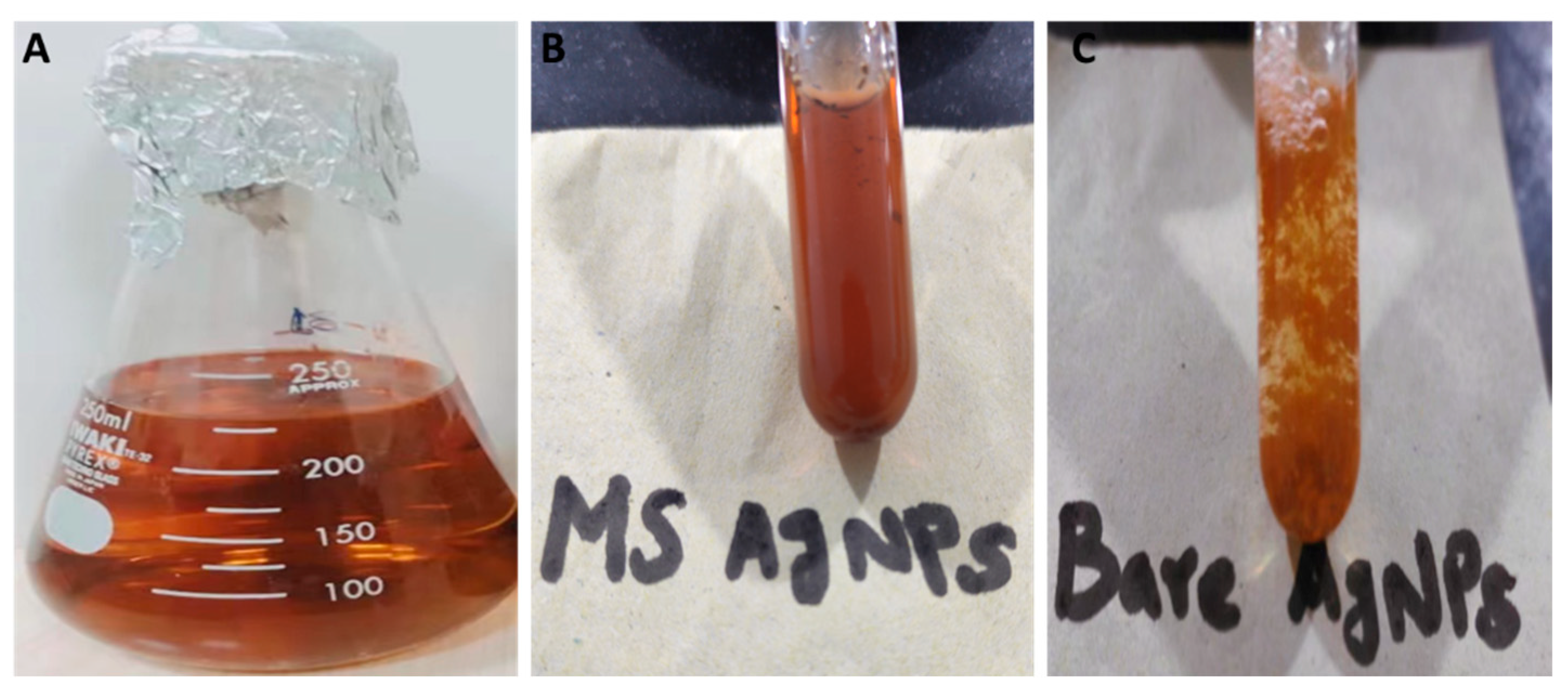
3.1. Myco-synthesis of AgNPs
3.2. Characterization of the Myco-synthesized Silver Nanoparticles (AgNPs)
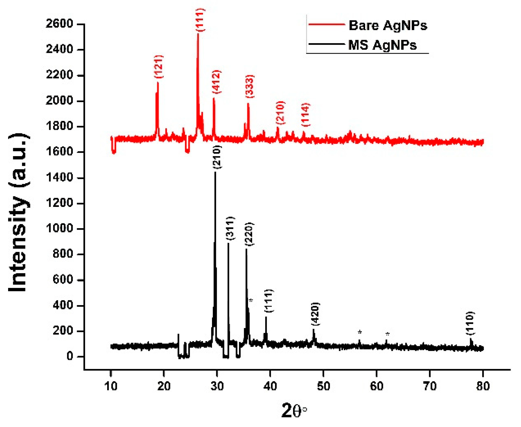
3.2.1. XRD
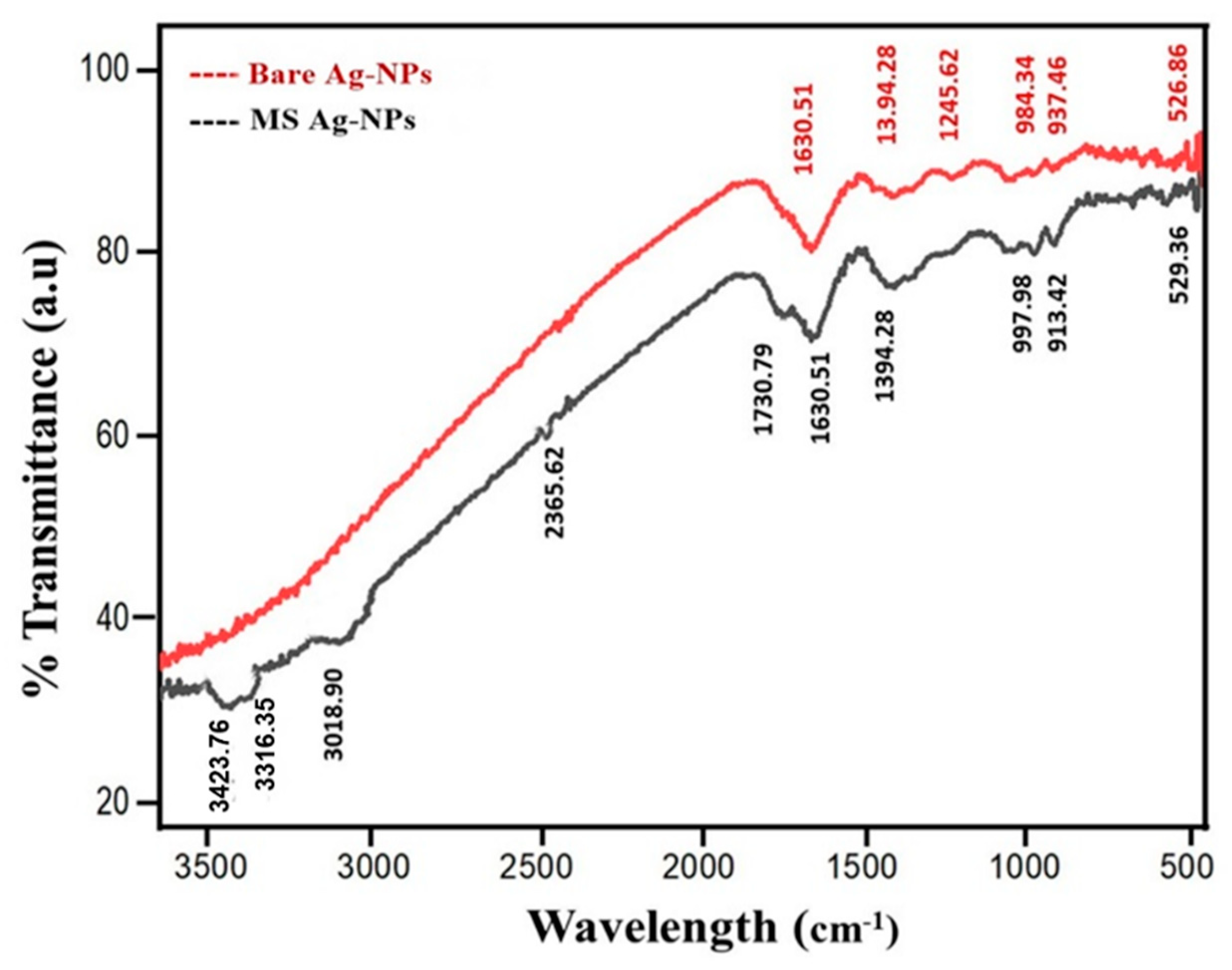
3.2.2. FTIR
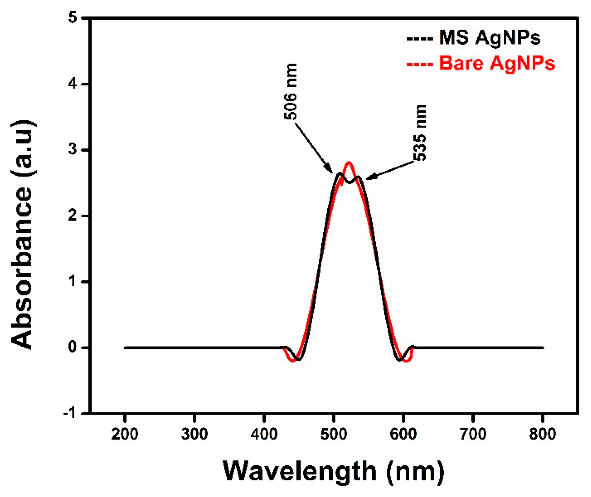
3.2.3. UV–Vis Spectroscopy
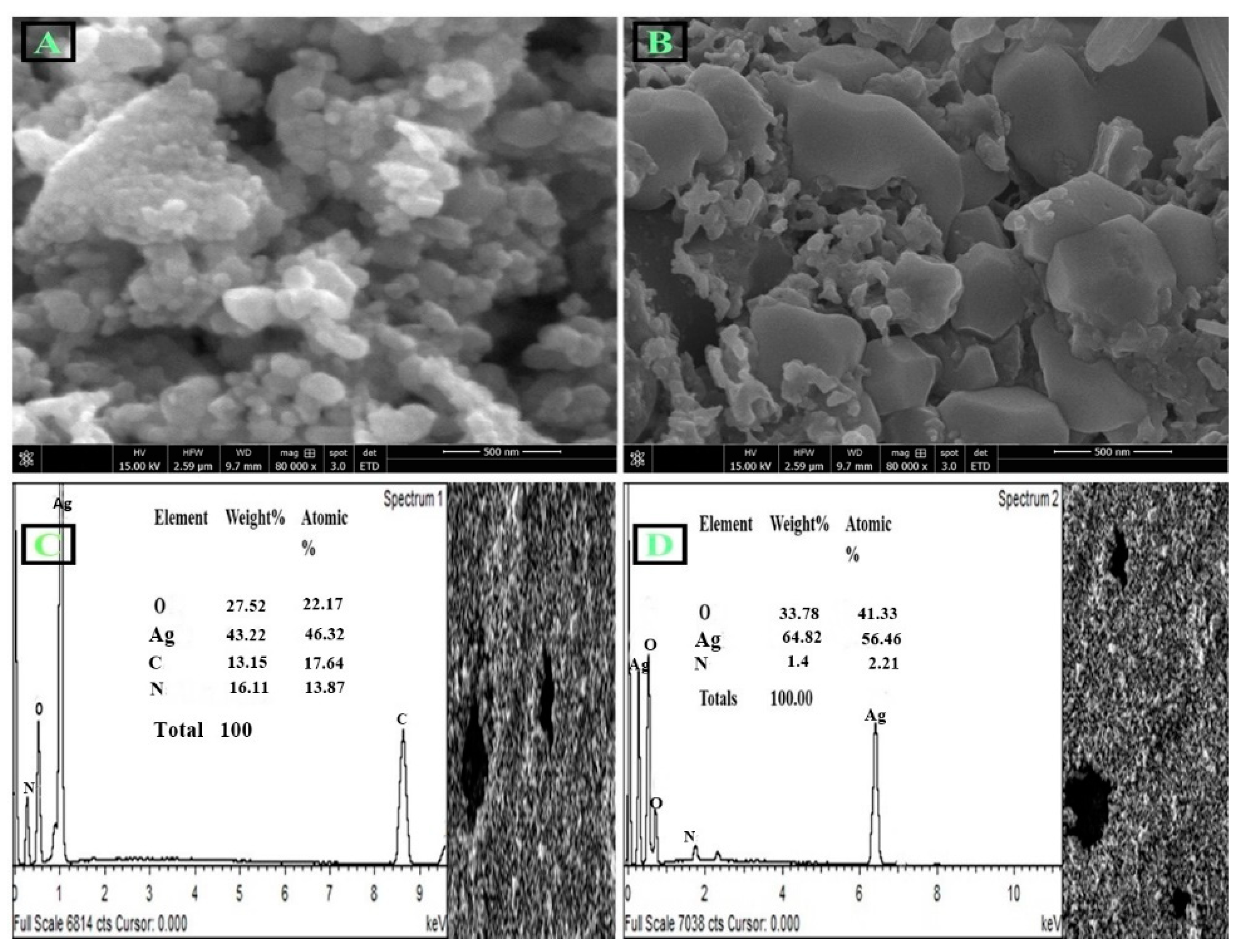
3.2.4. SEM Analysis
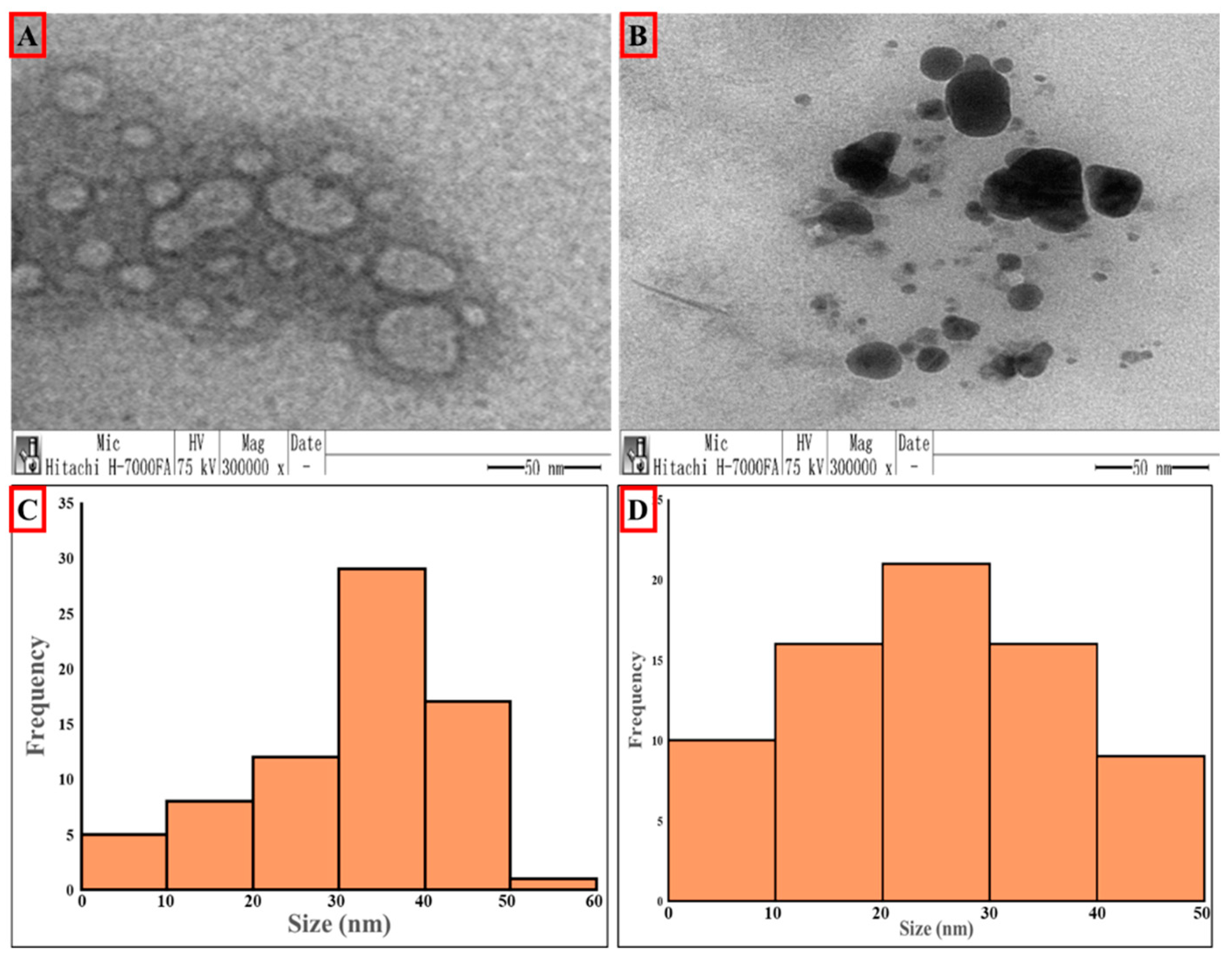
3.2.5. TEM Analysis
3.3. Antibacterial Activity
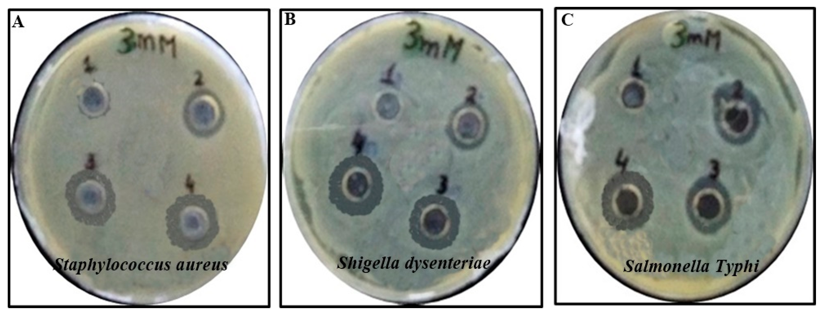
3.3.1. Agar Well Diffusion Method
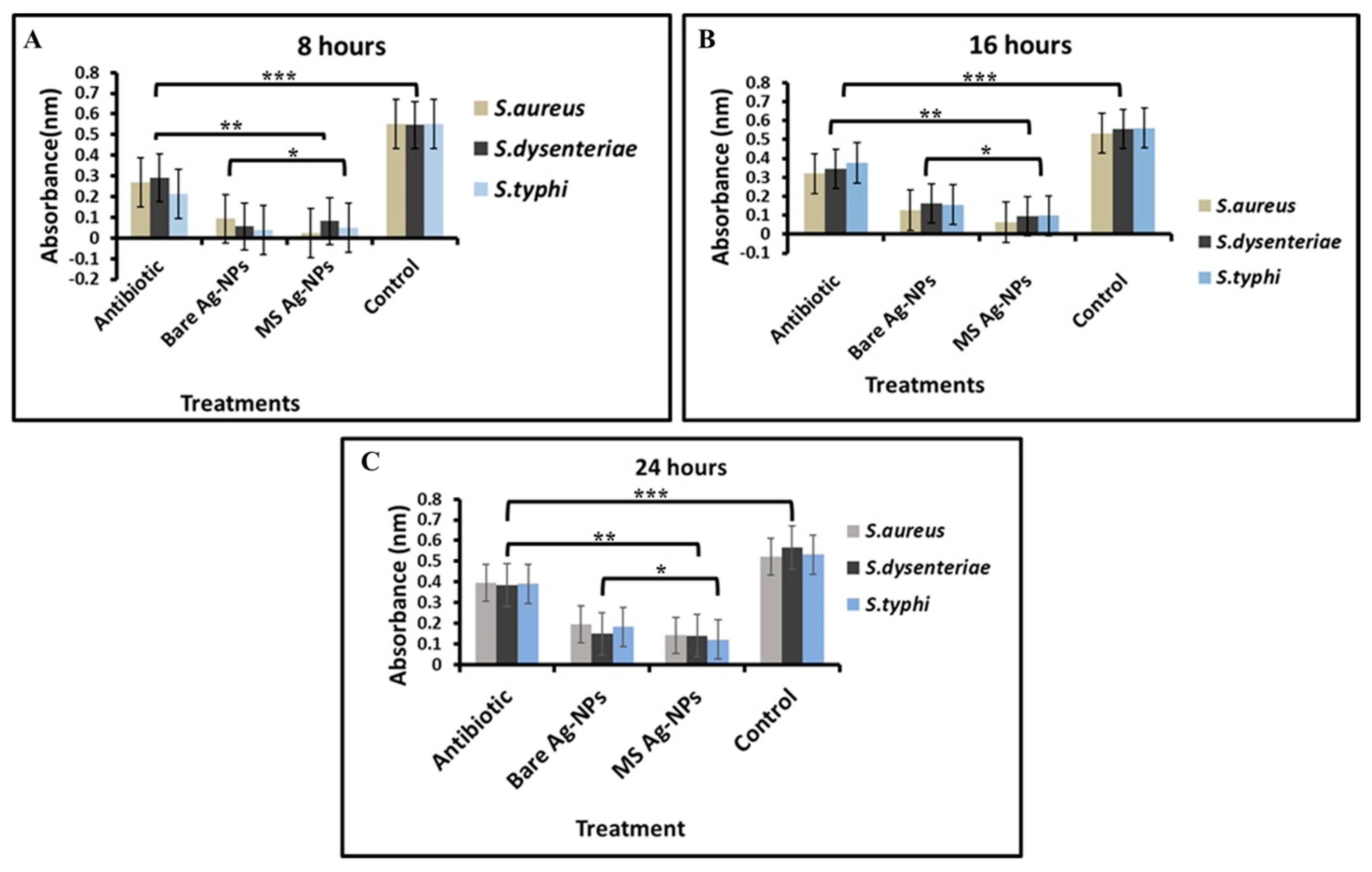
3.3.2. Optical Density Estimations
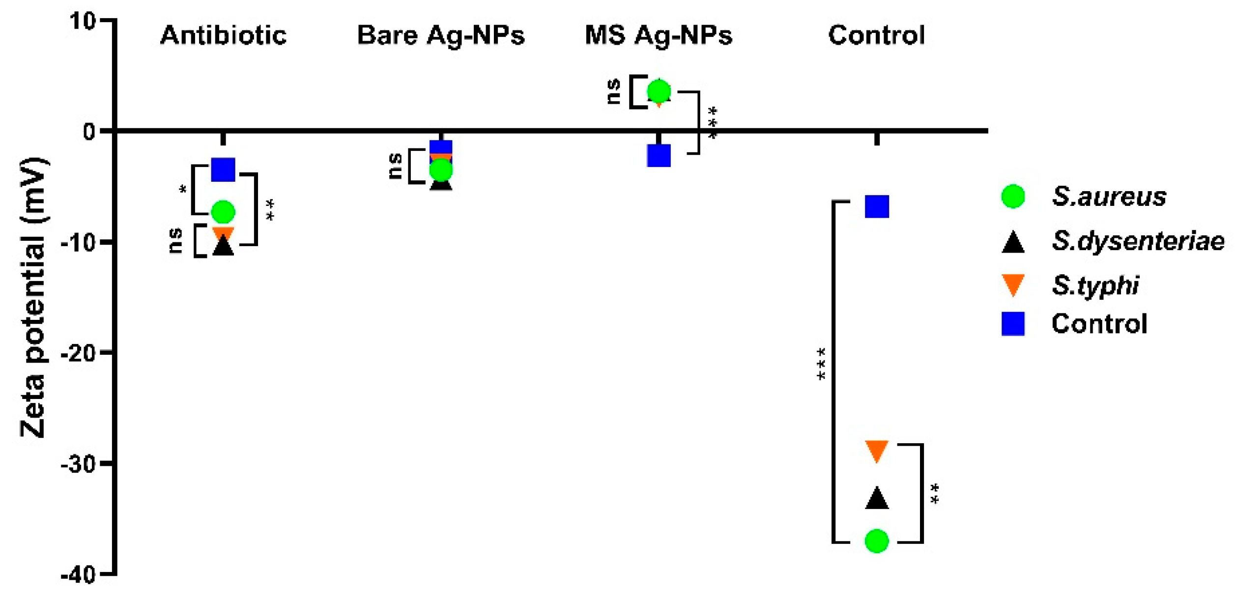
3.4. MS-AgNP’s Effect on Bacterial Membrane
3.5. Analysis of Cell Constituents Release
4. Discussion
5. Conclusions
Author Contributions
Funding
Institutional Review Board Statement
Informed Consent Statement
Data Availability Statement
Acknowledgments
Conflicts of Interest
References
- Regiel-Futyra, A.; Dąbrowski, J.M.; Mazuryk, O.; Śpiewak, K.; Kyzioł, A.; Pucelik, B.; Brindell, M.; Stochel, G. Bioinorganic antimicrobial strategies in the resistance era. Coord. Chem. Rev. 2017, 351, 76–117. [Google Scholar] [CrossRef]
- Afewerki, S.; Bassous, N.; Harb, S.; Palo-Nieto, C.; Ruiz-Esparza, G.U.; Marciano, F.R.; Webster, T.J.; Furtado, A.S.A.; Lobo, A.O. Advances in dual functional antimicrobial and osteoinductive biomaterials for orthopaedic applications. Nanomed. Nanotechnol. Biol. Med. 2020, 24, 102143. [Google Scholar] [CrossRef] [PubMed]
- Khan, I.; Saeed, K.; Khan, I. Nanoparticles: Properties, applications and toxicities. Arab. J. Chem. 2019, 12, 908–931. [Google Scholar] [CrossRef]
- Chand, K.; Cao, D.; Fouad, D.E.; Shah, A.H.; Dayo, A.Q.; Zhu, K.; Lakhan, M.N.; Mehdi, G.; Dong, S. Green synthesis, characterization and photocatalytic application of silver nanoparticles synthesized by various plant extracts. Arab. J. Chem. 2020, 13, 8248–8261. [Google Scholar] [CrossRef]
- Mohanta, Y.K.; Panda, S.K.; Bastia, A.K.; Mohanta, T.K. Biosynthesis of silver nanoparticles from Protium serratum and investigation of their potential impacts on food safety and control. Front. Microbiol. 2017, 8, 626. [Google Scholar] [CrossRef] [Green Version]
- Saqib, S.; Zaman, W.; Ullah, F.; Majeed, I.; Ayaz, A.; Hussain Munis, M.F. Organometallic assembling of chitosan-Iron oxide nanoparticles with their antifungal evaluation against Rhizopus oryzae. Appl. Organomet. Chem. 2019, 33, e5190. [Google Scholar] [CrossRef]
- Sunera, A.; Saqib, S.; Uddin, S.; Zaman, W.; Ullah, F.; Ayaz, A.; Asghar, M.; Rehman, S.; Munis, M.; Chaudhary, H. Characterization and phytostimulatory activity of bacteria isolated from tomato (Lycopersicon esculentum Mill.) rhizosphere. Microb. Pathog. 2020, 140, 103966. [Google Scholar] [CrossRef] [PubMed]
- Grün, A.Y.; App, C.B.; Breidenbach, A.; Meier, J.; Metreveli, G.; Schaumann, G.E.; Manz, W. Effects of low dose silver nanoparticle treatment on the structure and community composition of bacterial freshwater biofilms. PLoS ONE 2018, 13, e0199132. [Google Scholar] [CrossRef] [PubMed] [Green Version]
- Asghar, M.; Habib, S.; Zaman, W.; Hussain, S.; Ali, H.; Saqib, S. Synthesis and characterization of microbial mediated cadmium oxide nanoparticles. Microsc. Res. Tech. 2020, 83, 1574–1584. [Google Scholar] [CrossRef]
- Bocate, K.P.; Reis, G.F.; de Souza, P.C.; Junior, A.G.O.; Durán, N.; Nakazato, G.; Furlaneto, M.C.; de Almeida, R.S.; Panagio, L.A. Antifungal activity of silver nanoparticles and simvastatin against toxigenic species of Aspergillus. Int. J. Food Microbiol. 2019, 291, 79–86. [Google Scholar] [CrossRef]
- Azam, Z.; Ayaz, A.; Younas, M.; Qureshi, Z.; Arshad, B.; Zaman, W.; Ullah, F.; Nasar, M.Q.; Bahadur, S.; Irfan, M.M. Microbial synthesized cadmium oxide nanoparticles induce oxidative stress and protein leakage in bacterial cells. Microb. Pathog. 2020, 144, 104188. [Google Scholar] [CrossRef] [PubMed]
- Gopinath, V.; MubarakAli, D.; Vadivelu, J.; Kamath, S.M.; Syed, A.; Elgorban, A.M. Synthesis of biocompatible chitosan decorated silver nanoparticles biocomposites for enhanced antimicrobial and anticancer property. Process Biochem. 2020, 99, 348–356. [Google Scholar] [CrossRef]
- Saleh, H.E.-D.M.; Koller, M. Introductory chapter: Principles of green chemistry. In Green Chemistry; IntechOpen: London, UK, 2018. [Google Scholar]
- Hu, X.; Saravanakumar, K.; Jin, T.; Wang, M.-H. Mycosynthesis, characterization, anticancer and antibacterial activity of silver nanoparticles from endophytic fungus Talaromyces purpureogenus. Int. J. Nanomed. 2019, 14, 3427. [Google Scholar] [CrossRef] [Green Version]
- Prabakaran, K.; Ragavendran, C.; Natarajan, D. Mycosynthesis of silver nanoparticles from Beauveria bassiana and its larvicidal, antibacterial, and cytotoxic effect on human cervical cancer (HeLa) cells. RSC Adv. 2016, 6, 44972–44986. [Google Scholar] [CrossRef]
- Asghar, M.; Younas, M.; Arshad, B.; Zaman, W.; Ayaz, A.; Rasheed, S.; Shah, A.H.; Ullah, F.; Saqib, S. Bioactive potential of cultivated Mentha arvensis L. For preservation and production of health-oriented food. J. Anim. Plant Sci. 2022, 32, 835–844. [Google Scholar]
- Deplanche, K.; Merroun, M.L.; Casadesus, M.; Tran, D.T.; Mikheenko, I.P.; Bennett, J.A.; Zhu, J.; Jones, I.P.; Attard, G.A.; Wood, J. Microbial synthesis of core/shell gold/palladium nanoparticles for applications in green chemistry. J. R. Soc. Interface 2012, 9, 1705–1712. [Google Scholar] [CrossRef] [PubMed]
- Raftery, R.M.; Tierney, E.G.; Curtin, C.M.; Cryan, S.-A.; O’Brien, F.J. Development of a gene-activated scaffold platform for tissue engineering applications using chitosan-pDNA nanoparticles on collagen-based scaffolds. J. Control. Release 2015, 210, 84–94. [Google Scholar] [CrossRef] [PubMed]
- Dos Santos, C.A.; Seckler, M.M.; Ingle, A.P.; Gupta, I.; Galdiero, S.; Galdiero, M.; Gade, A.; Rai, M. Silver nanoparticles: Therapeutical uses, toxicity, and safety issues. J. Pharm. Sci. 2014, 103, 1931–1944. [Google Scholar] [CrossRef] [PubMed]
- Omran, B.A. Versatile Applications of Biosynthesized Nanoparticles, Global Safety Issues, Grand Challenges, and Future Perspectives Regarding Nanobiotechnology. In Nanobiotechnology: A Multidisciplinary Field of Science; Springer: Berlin/Heidelberg, Germany, 2020; pp. 185–221. [Google Scholar]
- Guilger-Casagrande, M.; de Lima, R. Synthesis of silver nanoparticles mediated by fungi: A Review. Front. Bioeng. Biotechnol. 2019, 7, 287. [Google Scholar] [CrossRef] [PubMed] [Green Version]
- Shahzad, A.; Iqtedar, M.; Saeed, H.; Hussain, S.Z.; Chaudhary, A.; Abdullah, R.; Kaleem, A. Mycosynthesis of size-controlled silver nanoparticles through optimization of process variables by response surface methodology. Pol. J. Microbiol. 2019, 68, 35–42. [Google Scholar] [CrossRef] [PubMed] [Green Version]
- Liaquat, F.; Munis, M.F.H.; Haroon, U.; Arif, S.; Saqib, S.; Zaman, W.; Khan, A.R.; Shi, J.; Che, S.; Liu, Q. Evaluation of metal tolerance of fungal strains isolated from contaminated mining soil of Nanjing, China. Biology 2020, 9, 469. [Google Scholar] [CrossRef] [PubMed]
- Palomo, J.M.; Filice, M. Biosynthesis of metal nanoparticles: Novel efficient heterogeneous nanocatalysts. Nanomaterials 2016, 6, 84. [Google Scholar] [CrossRef] [PubMed] [Green Version]
- Akhtar, M.S.; Swamy, M.K.; Sinniah, U.R. Natural Bio-Active Compounds: Volume 1: Production and Applications; Springer Nature: London, UK, 2019. [Google Scholar]
- Saqib, S.; Nazeer, A.; Ali, M.; Zaman, W.; Younas, M.; Shahzad, A.; Nisar, M. Catalytic potential of endophytes facilitates synthesis of biometallic zinc oxide nanoparticles for agricultural application. BioMetals 2022, 1–19. [Google Scholar] [CrossRef] [PubMed]
- Madakka, M.; Jayaraju, N.; Rajesh, N. Mycosynthesis of silver nanoparticles and their characterization. MethodsX 2018, 5, 20–29. [Google Scholar] [CrossRef]
- Liaquat, F.; Munis, M.F.H.; Arif, S.; Haroon, U.; Shi, J.; Saqib, S.; Zaman, W.; Che, S.; Liu, Q. PacBio single-molecule long-read sequencing reveals genes tolerating manganese stress in Schima superba saplings. Front. Genet. 2021, 12, 635043. [Google Scholar] [CrossRef] [PubMed]
- Khan, A.U.; Malik, N.; Khan, M.; Cho, M.H.; Khan, M.M. Fungi-assisted silver nanoparticle synthesis and their applications. Bioprocess Biosyst. Eng. 2018, 41, 1–20. [Google Scholar] [CrossRef] [PubMed]
- Jampilek, J.; Kralova, K. Advances in Biologically Applicable Graphene-Based 2D Nanomaterials. Int. J. Mol. Sci. 2022, 23, 6253. [Google Scholar] [CrossRef] [PubMed]
- El-Bendary, M.A.; Moharam, M.E.; Hamed, S.R.; Abo El-Ola, S.M.; Khalil, S.K.; Mounier, M.M.; Roshdy, A.M.; Allam, M.A. Mycosynthesis of silver nanoparticles using Aspergillus caespitosus: Characterization, antimicrobial activities, cytotoxicity, and their performance as an antimicrobial agent for textile materials. Appl. Organomet. Chem. 2021, 35, e6338. [Google Scholar] [CrossRef]
- Peters, R.J.; Bouwmeester, H.; Gottardo, S.; Amenta, V.; Arena, M.; Brandhoff, P.; Marvin, H.J.; Mech, A.; Moniz, F.B.; Pesudo, L.Q. Nanomaterials for products and application in agriculture, feed and food. Trends Food Sci. Technol. 2016, 54, 155–164. [Google Scholar] [CrossRef]
- Sharma, R.; Dewanjee, S.; Kole, C. Utilization of nanoparticles for plant protection. In Plant Nanotechnology; Springer: Berlin/Heidelberg, Germany, 2016; pp. 305–327. [Google Scholar]
- Bala, M.; Arya, V. Biological synthesis of silver nanoparticles from aqueous extract of endophytic fungus Aspergillus Fumigatus and its antibacterial action. Int. J. Nanomater. Biostructures 2013, 3, 37–41. [Google Scholar]
- Rafique, M.; Sadaf, I.; Rafique, M.S.; Tahir, M.B. A review on green synthesis of silver nanoparticles and their applications. Artif. Cells Nanomed. Biotechnol. 2017, 45, 1272–1291. [Google Scholar] [CrossRef]
- Gupta, M.; Shukla, K.K. Endophytic Fungi: A Treasure Trove of Novel Bioactive Compounds. In Bioactive Natural Products in Drug Discovery; Springer: Berlin/Heidelberg, Germany, 2020; pp. 427–449. [Google Scholar]
- Das, S.K.; Mahapatra, S. Isolation and Characterization of Bioactive Compound from Endophytic Fungus of Spoiled Fruits. Int. J. Res. Anal. Rev. 2019, 7, 65–72. [Google Scholar]
- Jyoti, K.; Baunthiyal, M.; Singh, A. Characterization of silver nanoparticles synthesized using Urtica dioica Linn. leaves and their synergistic effects with antibiotics. J. Radiat. Res. Appl. Sci. 2016, 9, 217–227. [Google Scholar] [CrossRef]
- Salleh, A.; Naomi, R.; Utami, N.D.; Mohammad, A.W.; Mahmoudi, E.; Mustafa, N.; Fauzi, M.B. The potential of silver nanoparticles for antiviral and antibacterial applications: A mechanism of action. Nanomaterials 2020, 10, 1566. [Google Scholar] [CrossRef] [PubMed]
- Fang, J.; Zhong, C.; Mu, R. The study of deposited silver particulate films by simple method for efficient SERS. Chem. Phys. Lett. 2005, 401, 271–275. [Google Scholar] [CrossRef]
- Feroze, N.; Arshad, B.; Younas, M.; Afridi, M.I.; Saqib, S.; Ayaz, A. Fungal mediated synthesis of silver nanoparticles and evaluation of antibacterial activity. Microsc. Res. Tech. 2020, 83, 72–80. [Google Scholar] [CrossRef]
- Ramesh, P.; Kokila, T.; Geetha, D. Plant mediated green synthesis and antibacterial activity of silver nanoparticles using Emblica officinalis fruit extract. Spectrochim. Acta Part A Mol. Biomol. Spectrosc. 2015, 142, 339–343. [Google Scholar] [CrossRef] [PubMed]
- Magdi, H.M.; Mourad, M.H.; El-Aziz, M. Biosynthesis of silver nanoparticles using fungi and biological evaluation of mycosynthesized silver nanoparticles. Egypt J. Exp. Biol. 2014, 10, 1–12. [Google Scholar]
- Alawfi, A.A.; Henari, F.Z.; Younis, A.; Manaa, H. Bio-inspired synthesis of silver nanoparticles using Hibiscus Tiliaceus L. flower extracts for improved optical characteristics. J. Mater. Sci. Mater. Electron. 2020, 31, 21073–21081. [Google Scholar] [CrossRef]
- Tyagi, S.; Tyagi, P.K.; Gola, D.; Chauhan, N.; Bharti, R.K. Extracellular synthesis of silver nanoparticles using entomopathogenic fungus: Characterization and antibacterial potential. SN Appl. Sci. 2019, 1, 1545. [Google Scholar] [CrossRef] [Green Version]
- Siddiqi, K.S.; Husen, A.; Rao, R.A. A review on biosynthesis of silver nanoparticles and their biocidal properties. J. Nanobiotechnology 2018, 16, 14. [Google Scholar] [CrossRef]
- Besinis, A.; Hadi, S.D.; Le, H.; Tredwin, C.; Handy, R. Antibacterial activity and biofilm inhibition by surface modified titanium alloy medical implants following application of silver, titanium dioxide and hydroxyapatite nanocoatings. Nanotoxicology 2017, 11, 327–338. [Google Scholar] [CrossRef] [PubMed] [Green Version]
- Nithya, R.; Ragunathan, R. Synthesis of silver nanoparticle using Pleurotus sajor caju and its antimicrobial study. Dig. J. Nanomater. Biostructures 2009, 4, 623–629. [Google Scholar]
- Kalia, S.; Kaith, B.; Kaur, I. Cellulose Fibers: Bio-and Nano-Polymer Composites: Green Chemistry and Technology; Springer Science & Business Media: Berlin, Germany, 2011. [Google Scholar]
- Das, R.K.; Brar, S.K. Plant mediated green synthesis: Modified approaches. Nanoscale 2013, 5, 10155–10162. [Google Scholar] [CrossRef] [PubMed]
- Saadat, S.; Pandey, G.; Tharmavaram, M.; Braganza, V.; Rawtani, D. Nano-interfacial decoration of Halloysite Nanotubes for the development of antimicrobial nanocomposites. Adv. Colloid Interface Sci. 2020, 275, 102063. [Google Scholar] [CrossRef]
- Sarsar, V.; Selwal, M.K.; Selwal, K.K. Biofabrication, characterization and antibacterial efficacy of extracellular silver nanoparticles using novel fungal strain of Penicillium atramentosum KM. J. Saudi Chem. Soc. 2015, 19, 682–688. [Google Scholar] [CrossRef] [Green Version]
- Khatami, M.; Varma, R.S.; Zafarnia, N.; Yaghoobi, H.; Sarani, M.; Kumar, V.G. Applications of green synthesized Ag, ZnO and Ag/ZnO nanoparticles for making clinical antimicrobial wound-healing bandages. Sustain. Chem. Pharm. 2018, 10, 9–15. [Google Scholar] [CrossRef]
- Taglietti, A.; Diaz Fernandez, Y.A.; Amato, E.; Cucca, L.; Dacarro, G.; Grisoli, P.; Necchi, V.; Pallavicini, P.; Pasotti, L.; Patrini, M. Antibacterial activity of glutathione-coated silver nanoparticles against gram positive and gram negative bacteria. Langmuir 2012, 28, 8140–8148. [Google Scholar] [CrossRef]
- Dey, A.; Pandey, G.; Rawtani, D. Functionalized nanomaterials driven antimicrobial food packaging: A technological advancement in food science. Food Control 2022, 131, 108469. [Google Scholar] [CrossRef]
- Abdel-Hafez, S.I.; Nafady, N.A.; Abdel-Rahim, I.R.; Shaltout, A.M.; Daròs, J.-A.; Mohamed, M.A. Assessment of protein silver nanoparticles toxicity against pathogenic Alternaria solani. 3 Biotech 2016, 6, 199. [Google Scholar] [CrossRef]


| Treatments (3 mM) Concentration | S. aureus | S. dysenteriae | S. typhi |
|---|---|---|---|
| Zone Inhibition Value (mm) | Zone Inhibition Value (mm) | Zone Inhibition Value (mm) | |
| MS Ag-NPs | 18.21 ± 2.1 a | 16.18 ± 1.3 b | 14.41 ± 1.7c |
| Bare Ag-NPs | 13.73 ± 1.4 b | 14.84 ± 1.8 c | 13.25 ± 1.9ab |
| Ampicillin | 7.31 ± 2.1 c | 7.80 ± 1.4 d | 5.51 ± 1.3ac |
| Control | 3.12 ± 1.4 d | 1.15 ± 1.3 e | 1.10 ± 1.4e |
| Bacterial Strains | MS-AgNPs (OD260 nm) | Bare AgNPs (OD260 nm) | Antibiotic (OD260 nm) | Control (OD260 nm) |
|---|---|---|---|---|
| S. aureus | 0.26 a | 0.22 b | 0.14 c | 0.006 d |
| S. dysenteriae | 0.21 a | 0.16 b | 0.15 c | 0.008 d |
| S. typhi | 0.18 a | 0.17 b | 0.17 c | 0.005 d |
| Bacterial strains | MS-AgNPs (OD260 nm) | Bare AgNPs (OD260 nm) | Antibiotic (OD260 nm) | Control (OD260 nm) |
|---|---|---|---|---|
| S. aureus | 0.32 a | 0.21 b | 0.11 c | 0.003 d |
| S. dysenteriae | 0.27 a | 0.19 b | 0.10 c | 0.006 d |
| S. typhi | 0.29 a | 0.18 b | 0.12 c | 0.004 d |
Publisher’s Note: MDPI stays neutral with regard to jurisdictional claims in published maps and institutional affiliations. |
© 2022 by the authors. Licensee MDPI, Basel, Switzerland. This article is an open access article distributed under the terms and conditions of the Creative Commons Attribution (CC BY) license (https://creativecommons.org/licenses/by/4.0/).
Share and Cite
Saqib, S.; Faryad, S.; Afridi, M.I.; Arshad, B.; Younas, M.; Naeem, M.; Zaman, W.; Ullah, F.; Nisar, M.; Ali, S.; et al. Bimetallic Assembled Silver Nanoparticles Impregnated in Aspergillus fumigatus Extract Damage the Bacterial Membrane Surface and Release Cellular Contents. Coatings 2022, 12, 1505. https://doi.org/10.3390/coatings12101505
Saqib S, Faryad S, Afridi MI, Arshad B, Younas M, Naeem M, Zaman W, Ullah F, Nisar M, Ali S, et al. Bimetallic Assembled Silver Nanoparticles Impregnated in Aspergillus fumigatus Extract Damage the Bacterial Membrane Surface and Release Cellular Contents. Coatings. 2022; 12(10):1505. https://doi.org/10.3390/coatings12101505
Chicago/Turabian StyleSaqib, Saddam, Saima Faryad, Muhammad Irfan Afridi, Bushra Arshad, Muhammad Younas, Muhammad Naeem, Wajid Zaman, Fazal Ullah, Momina Nisar, Sajid Ali, and et al. 2022. "Bimetallic Assembled Silver Nanoparticles Impregnated in Aspergillus fumigatus Extract Damage the Bacterial Membrane Surface and Release Cellular Contents" Coatings 12, no. 10: 1505. https://doi.org/10.3390/coatings12101505