Article

Chemical Vapor Deposition and Thermal Oxidation of Cuprous Phosphide Nanofilm

College of Materials and Environmental Engineering, Hangzhou Dianzi University, Hangzhou 310018, China
*
Author to whom correspondence should be addressed.
Coatings 2022, 12(1), 68; https://doi.org/10.3390/coatings12010068
Submission received: 30 November 2021 / Revised: 4 January 2022 / Accepted: 5 January 2022 / Published: 7 January 2022
(This article belongs to the Special Issue 10th Anniversary of Coatings: Invited Papers for Thin Films Section)

Abstract

:
Inorganic semiconductors usually show n-type characterization; the development of p-type inorganic semiconductor material will provide more opportunities for novel devices. In this paper, we investigated the chemical vapor deposition (CVD) of p-type cuprous phosphide (Cu3P) nanofilm and studied its thermal oxidation behavior. Cu3P film was characterized by optical microscopy, scanning electron microscopy (SEM), atomic force microscopy (AFM), laser Raman spectroscopy (Raman), and fluorescence spectroscopy (PL). We found that the thickness of film ranged from 4 to 10 nm, and the film is unstable at temperatures higher than room temperature in air. We provide a way to prepare inorganic phosphide nanofilms. In addition, the possible thermal oxidation should be taken into consideration for practical application.

1. Introduction

Due to the intrinsic band structure and crystal defects, inorganic semiconductors usually exhibit natural n-type transporting property [1]; therefore, the development of a p-type inorganic semiconductor attracts considerable interest for the development of novel devices based on p-n junctions [2,3,4]. Copper compounds generally show p-type properties, such as cuprous oxide, cuprous sulfide and cuprous halide [5,6,7], and cuprous phosphide (Cu3P) is one of them. Cuprous phosphide is a p-type, narrow band gap semiconductor with an energy gap of about 1.6 eV [8,9,10,11], which is of potential applications in light-emitting diodes, photodetectors, sensors and catalysis [12,13,14,15,16,17,18]. The morphologies of cuprous phosphide reported are usually thick film or nanosheets. For example, Pfeiffer et al. synthesized Cu3P particle film with a thickness of ca. 60 μm through a solid reaction between copper and red phosphorous and discussed the growth mechanism [19,20]. Lee et al. reported the growth of Cu3P nanosheets with thickness ranging from 28 nm to 440 μm by the reaction of copper foil and red phosphorous vapor [21]. Mu et al. used the solution method to obtain Cu3−xP hexagonal nanocrystals with a lateral size of 20 nm and thickness of 2 nm [22]. The solution method generally produces particles. Nanofilm of Cu3P with thickness of several nanometers has not been reported. In this paper, we prepared Cu3P nanofilm by a chemical vapor deposition (CVD) method. The successful preparation Cu3P nanofilm is meaningful to study novel device-based p-n junctions.
Cuprous phosphide is generally considered stable in air [20], so is it really stable when the temperature is slightly higher than the room temperature? Therefore, we studied the thermal oxidation properties of Cu3P. We found Cu3P is sensitive to the temperature. When temperature is higher than room temperature, Cu3P will be oxidized in the air, which should be taken into consideration during practical application.

2. Materials and Methods

CVD growth of nanofilm. Nonofilm with a thickness of 4–10 nm was prepared on the silicon wafer with a native oxide layer. Sodium hypohosphite and copper foil were used as precursors. Sodium hypohosphite (0.05 mol) was placed in a corundum crucible covered with copper foil (1 cm × 1 cm × 250 μm). They were placed in the centre of corundum tube, and heated to 800 °C with a rate of 10 °C/min and kept at 800 °C for 2 h. The silicon wafer was placed downstream of the tube. During the heating process of the precursor, the substrate was heated to ca. 700 °C. After that, the tube was cooled to room temperature and flushed with argon to remove gaseous phosphide residues before the sample was taken out of the tube. Note that gaseous phosphide requires purification treatment. The edge of Cu3P nanofilm grown on the silicon substrate will form a step on the substrate, and the height of the step is equal to the thickness of the nanofilm. The step height was measured by atomic force microscopy.
Growth and annealing of thick film. To facilitate the study of thermal stability, Cu3P film with thickness of ca. 2.7 μm was prepared by the reaction between copper foil and sodium hypophosphite according to the reference [9,23,24]. Sodium hypophosphite (Aladdin, Shanghai, China, 99.5%, 0.05 mol) was first placedin a corundum crucible covered with copper foil (Aladdin, Shanghai, China, 99.9%) with a size of 1 cm × 1 cm × 250 μm. Then, they were put into a corundum tube and heated to 300 °C in argon for 2 h before cooled to room temperature. After cooling, the tube was flushed with argon to remove gaseous phosphide residues. Films were annealed at 50, 100, 200, 300 and 400 °C, respectively, in air for 30 min to study thermal oxidation behaviour. The thickness of the Cu3P thick film was obtained by scanning the film cross section by scanning electron microscopy.
Optical microscope imaging was carried on a Jiangnan MV3000 digital microscope. Atomic force microscopy (AFM, tapping mode) was conducted on an Agilent 5500 (Agilent, Santa Clara, CA, USA). Scanning electron microscopy (SEM, Hitachi, Tokyo, Japan) was performed on Hitachi Su1510. Raman and photoluminescence spectra were carried on an HR Evo Nano (Horiba, Kyoto, Japan) with a 532 nm laser. The laser power was set at ca. 20 mW/mm2. All the above measurements were carried out at room temperature.

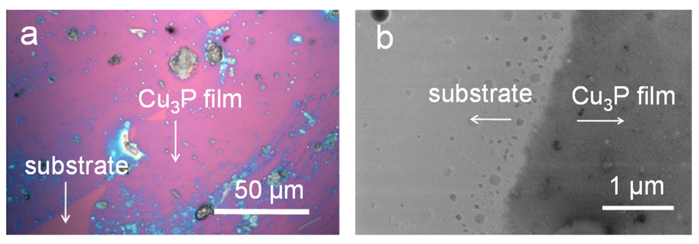
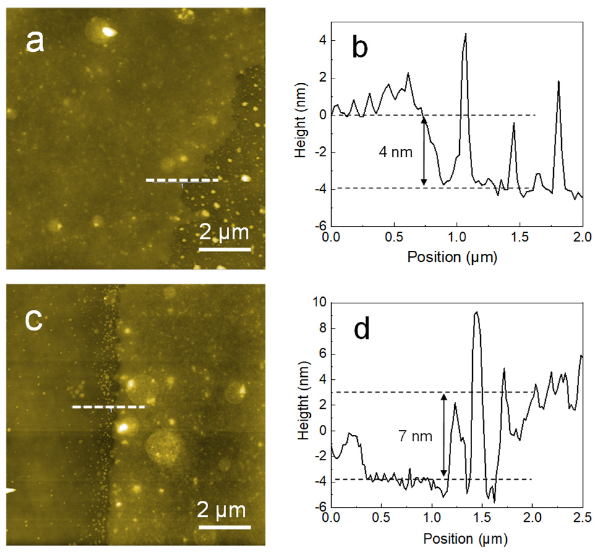
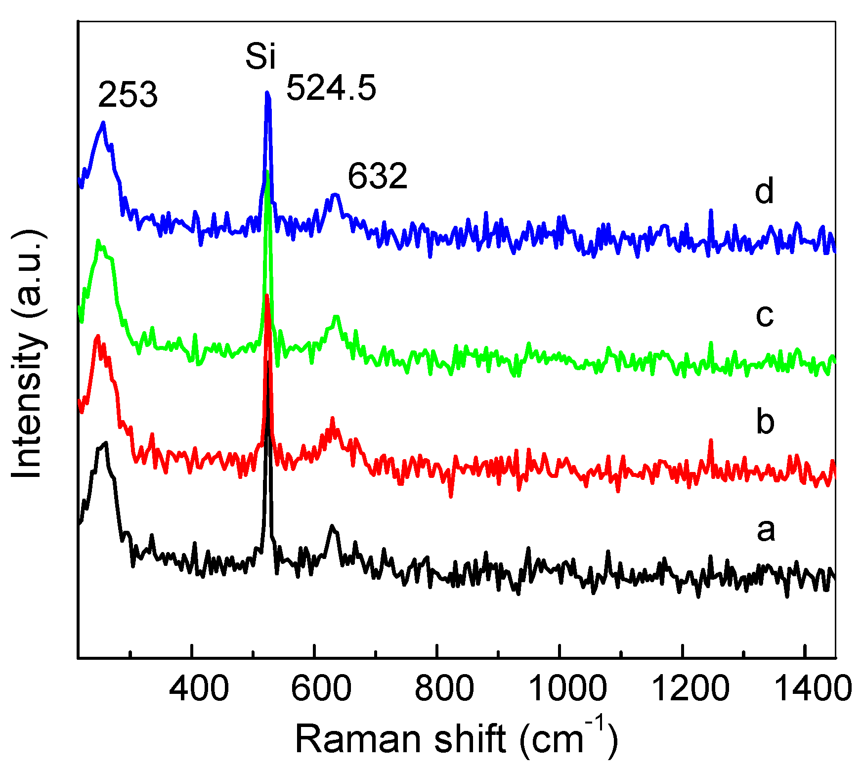
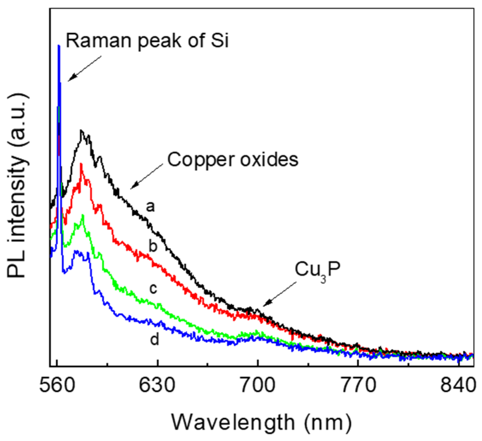
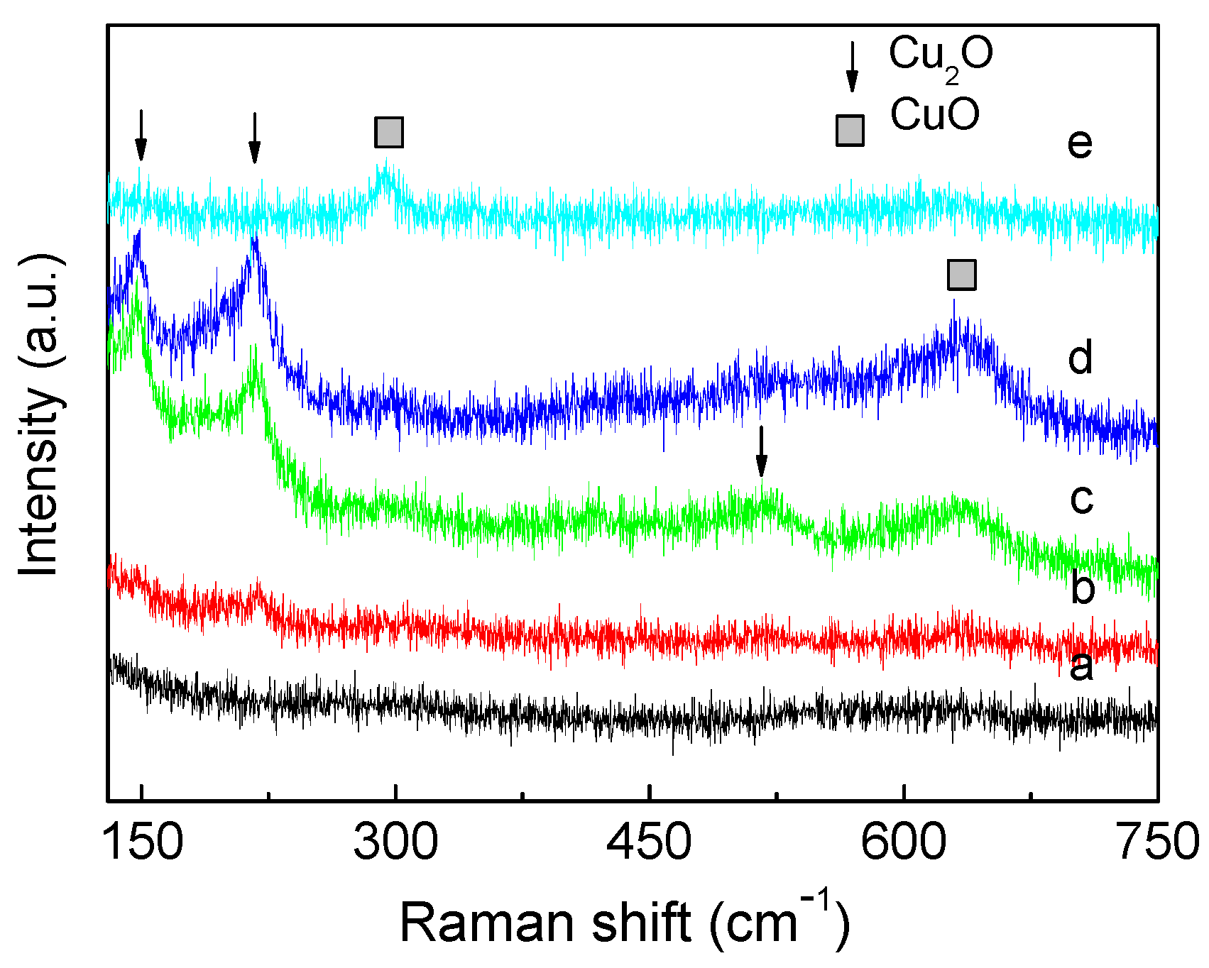
3. Results and Discussion

Sodium hypophosphite decomposed at a high temperature to produce phosphine gas, which reacted with copper to form solid Cu3P. Then, the solid Cu3P was vaporized and transported onto the surface of the cold substrate, where the deposition occurred. The substrate we used is a silicon single crystal wafer with a 300 nm SiO2 layer. Due to the interference of visible light, nanofilm grown on such substrate can be observed through an optical microscope. Figure 1a is the optical image of Cu3P, in which the area with purple color is corresponding to Cu3P film. From the optical image, we predict that the film is continuous. Some areas with cyan and grey colors are due to the thicker films or amorphous Cu3P deposited on the surface of the nanofilm. To further observe the details of the surface topography, we carried out the SEM. Figure 1b is the SEM image the Cu3P nanofilm taken at the edge of the film, in which the area with light-color area is corresponding to the substrate, and dark-colored area is Cu3P. We found that the Cu3P film is continuous. Many isolated Cu3P nanosheets were also observed at the edge of the continuous film. It is well known that the chemical potential of a small particle is larger than that of big one. Therefore, there is a trend that atoms will leave from the small particles to the continuous film, indicating that the growth is related to diffusion and growth processes.
The thickness and surface roughness were measured by AFM. Figure 2a,b show the typical AFM images of Cu3P nanofilm taken at two different locations on the same sample. The thickness of the film is ranging from ca. 4 to ca. 7 nm. The root-mean-square surface roughnesses of Figure 2a,b are 0.26 and 0.28 nm, respectively.
Figure 3 shows the Raman spectra of the Cu3P nanofilm taken at different locations with different thicknesses. The peak at 524.5 cm−1 is corresponding to silicon [25,26], which was used to calibrate the peak position. There are two peaks centred at 253 and 632 cm−1, which are due to Cu3P [9,27]. The frequency difference between the two peaks is constant, unlike Van Der Waals two-dimensional materials, such as graphene, hexagonal boron nitride and molybdenum sulfide, of which the frequency difference is a function of the film thickness [28]. Therefore, the frequency difference cannot be utilized to determine the thickness of Cu3P film. The Raman peaks of bulk Cu3P are centered at 273 and 607 cm−1, respectively [9]. Compared with the bulk material, peak positions of Cu3P nanofilm shift and the frequency difference increases, which indicates the existence of the lattice defects and disorder [29,30].
Figure 4 show the photoluminescence spectra of Cu3P film with different thickness corresponding to the data in Figure 3. We observed a weak peak centered at ca. 700 nm, which is possible due to the emission of Cu3P under the excitation of 532 nm laser. The weak photon emission indicates the Cu3P has a poor crystallinity. In addition, we found another broad-band emission ranging from ca. 555 to ca. 680 nm. We had discussed the photoluminescence behavior of Cu3P in the presence of copper oxide in our previous work [9]. Cuprous phosphide is a direct band gap semiconductor with a band gap of ca. 1.5 eV. The photon energy of the broad-band emission is higher than the band gap of Cu3P. Therefore, the broad-band emission is not due to Cu3P, but copper oxides [18,31,32]. So where do these copper oxides come from? In this paper, copper reacted with the decomposition of sodium hyphosphite to form Cu3P, and then Cu3P was evaporated and transported onto the surface of substrate, where the growth of Cu3P occurred. The growth took place in an inertatmosphere. The oxidation should not occur during this process. Szczuka et al. reported that diamond can be heated to 372 K by using a 532 nm laser with a power of 36 mW/mm2 in 2 min [33]. Therefore, the possible reason is due to the laser irradiation during the photoluminescence characterization. The laser irradiation increased the temperature of Cu3P. Then, Cu3P was oxidized by oxygen in the air. To test the hypothesis, we heated Cu3P using thick Cu3Pfilm in air. To choose thick film instead of nanofilm for the thermal oxidation experiments, is considering the Raman signal of thick film is stronger than that of nanofilm. Figure 5 shows the Raman spectra of Cu3P thick film annealed at higher temperatures. Peaks of Cu3P could not be found in the samples annealed at 50 and 100 °C. When the annealing temperature was increased above 200 °C, peaks of Cu2O (148, 220, and 512 cm−1) and CuO (291 and 636 cm−1) were observed [34,35]. These results indicate that Cu3P undergo thermal oxidation at temperatures higher than room temperature.

4. Conclusions

In this paper, we prepared cuprous phosphide nanofilm through the chemical vapor deposition method. We found the Raman peaks of the Cu3P nanofilm is independent on the film thicknessand the photoluminescence emission of the nanofilm is weak. In addition, we found the presence of copper oxides in PL and Raman spectra when the film was exposed to the laser irradiation or heated in air. Our study showed that Cu3Pnano film was unstable at temperatures higher than room temperature in air.

Author Contributions

S.Z. and Y.L. conceived and designed the experiments and wrote the paper; X.P. performed the experiments and wrote the paper. All authors have read and agreed to the published version of the manuscript.

Funding

This research received no external funding.

Institutional Review Board Statement

Not applicable.

Informed Consent Statement

Not applicable.

Data Availability Statement

Data sharing is not applicable to this article.

Conflicts of Interest

The authors declare no conflict of interest.

References

  1. Liu, A.; Zhu, H.H.; Noh, Y.Y. Solution-processed inorganic p-channel transistors: Recent advances and perspectives. Mat. Sci. Eng. R Rep. 2019, 135, 85–100. [Google Scholar] [CrossRef]
  2. Teng, F.; Hu, K.; Ouyang, W.X.; Fang, X.S. Photoelectric detectors based on inorganic p-type semiconductor materials. Adv. Mater. 2018, 30, 1706262. [Google Scholar] [CrossRef] [PubMed]
  3. Kim, T.; Jeong, J.K. Recent progress and perspectives of field-effect transistors based on p-type oxide semiconductors. Phys. Status Solidi R 2021, 2100394. [Google Scholar] [CrossRef]
  4. Zhang, P.; Yu, S.; Zhang, X.W.; Wei, S.H. Design of p-type transparent conductors from inverted band structure: The case of inorganic metal halide perovskites. Phys. Rev. Mater. 2019, 3, 055201. [Google Scholar] [CrossRef]
  5. Jayathilaka, K.M.D.C.; Kapaklis, V.; Siripala, W.; Jayanetti, J.K.D.S. Ammonium sulfide surface treatment of electrodeposited p-type cuprous oxide thin films. Electron. Mater. Lett. 2014, 10, 379–382. [Google Scholar] [CrossRef]
  6. Parreira, P.; Lavareda, G.; Valente, J.; Nunes, F.T.; Amaral, A.; de Carvalho, C.N. Optoelectronic properties of transparent p-type semiconductor CuxS thin films. Phys. Status Solidi A 2010, 207, 1652–1654. [Google Scholar] [CrossRef]
  7. Yamada, N.; Ino, R.; Tomura, H.; Kondo, Y.; Ninomiya, Y. High-mobility transparent p-type CuI semiconducting layers fabricated on flexible plastic sheets: Toward flexibletransparent electronics. Adv. Electron. Mater. 2017, 3, 1700298. [Google Scholar] [CrossRef]
  8. Sheets, E.J.; Yang, W.C.; Balow, R.B.; Wang, Y.; Walker, B.C.; Stach, E.A.; Agrawal, R. An in situ phosphorus source for the synthesis of Cu3P and the subsequent conversion to Cu3PS4 nanoparticle clusters. J. Mater. Res. 2015, 30, 3710–3716. [Google Scholar] [CrossRef]
  9. Peng, X.; Lv, Y.F.; Fu, L.; Chen, F.; Su, W.T.; Li, J.Z.; Zhang, Q.; Zhao, S.C. Photoluminescence properties of cuprous phosphide prepared through phosphating copper with a native oxide layer. RSC Adv. 2021, 11, 34095–34100. [Google Scholar] [CrossRef]
  10. Fu, Z.Y.; Ma, X.Y.; Xia, B.; Hu, X.Y.; Fan, J.; Liu, E.Z. Efficient photocatalytic H2 evolution over Cu and Cu3P co-modified TiO2 nanosheet. Int. J. Hydrogen Energy 2021, 46, 19373–19384. [Google Scholar] [CrossRef]
  11. Gaspari, R.; Labat, F.; Manna, L.; Adamo, C.; Cavalli, A. Semiconducting and optical properties of selected binary compounds by linear response DFT plus U and hybrid functional methods. Theor. Chem. Acc. 2016, 135, 73. [Google Scholar] [CrossRef]
  12. Li, H.; Jia, C.; Meng, X.W.; Li, H.B. Chemical synthesis and applications of colloidal metal phosphide nanocrystals. Front. Chem. 2019, 6, 652. [Google Scholar] [CrossRef] [Green Version]
  13. Kuwano, T.; Katsube, R.; Nose, Y. Improvement of ohmic behavior of back contact in ZnSnP2 solar cells by inserting Cu3P. In Proceedings of the 2019 IEEE 46th Photovoltaic Specialists Conference PVSC, Chicago, IL, USA, 16–21 June 2019; pp. 3007–3009. [Google Scholar]
  14. Zhu, S.; Wang, J.; He, Y.; Yu, Z.; Wang, X.; Su, W. In situ photodeposition of amorphous NixP on CdS nanorods for efficient visible-light photocatalytic H2 generation. Catal. Sci. Technol. 2019, 9, 5394–5400. [Google Scholar] [CrossRef]
  15. Tappan, B.A.; Chen, K.; Lu, H.; Sharada, S.M.; Brutchey, R.L. Synthesis and electrocatalytic HER studies of carbene-ligated Cu3−xP nanocrystals. ACS Appl. Mater. Interf. 2020, 12, 16394–16401. [Google Scholar] [CrossRef]
  16. Zhang, X.; Yan, J.; Lee, L.Y.S. Highly promoted hydrogen production enabled by interfacial P N chemical bonds in copper phosphosulfide Z-scheme composite. Appl. Catal. B 2021, 283, 119624. [Google Scholar] [CrossRef]
  17. Wolff, A.; Doert, T.; Hunger, J.; Kaiser, M.; Pallmann, J.; Reinhold, R.; Yogendra, S.; Giebeler, L.; Sichelschmidt, J.; Schnelle, W.; et al. Low-temperature tailoring of copper-deficient Cu3–xP-electric properties, phase transitions, and performance in lithium-ion batteries. Chem. Mater. 2018, 30, 7111–7123. [Google Scholar] [CrossRef] [Green Version]
  18. Hua, S.; Qu, D.; An, L.; Jiang, W.; Wen, Y.; Wang, X.; Sun, Z. Highly efficient p-type Cu3P/n-type g-C3N4 photocatalyst through Z-scheme charge transfer route. Appl. Catal. B 2019, 240, 253–261. [Google Scholar] [CrossRef]
  19. Pfeiffer, H.; Tancret, F.; Brousse, T. Synthesis, characterization and electrochemical properties of copper phosphide (Cu3P) thick films prepared by solid-state reaction at low temperature: A probable anode for lithium ion batteries. Electrochim. Acta 2005, 50, 4763–4770. [Google Scholar] [CrossRef]
  20. Pfeiffer, H.; Tancret, F.; Bichat, M.P.; Monconduit, L.; Favier, F.; Brousse, T. Air stable copper phosphide (Cu3P): A possible negative electrode material for lithium batteries. Electrochem. Commun. 2004, 6, 263–267. [Google Scholar] [CrossRef]
  21. Lee, S.W.; Kim, J.; Woo, S.G.; Park, Y.; Yoon, J.C.; Park, H.J.; Kim, N.Y.; Shin, H.S.; Lee, Z. Epitaxially grown copper phosphide (Cu3P) nanosheets nanoarchitecture compared with film morphology for energy applications. Surf. Interf. 2021, 26, 101369. [Google Scholar] [CrossRef]
  22. Mu, H.; Liu, Z.; Bao, X.; Wan, Z.; Liu, G.; Li, X.; Shao, H.; Xing, G.; Shabbir, B.; Li, L.; et al. Highly stable and repeatable femtosecond soliton pulse generation from saturable absorbers based on two-dimensional Cu3−xP nanocrystals. Front. Optoelectron. 2020, 13, 139–148. [Google Scholar] [CrossRef]
  23. Pawar, S.M.; Pawar, B.S.; Babar, P.T.; Ahmed, A.T.A.; Chavan, H.S.; Jo, Y.; Cho, S.; Kim, J.; Inamdar, A.I.; Kim, J.H.; et al. Electrosynthesis of copper phosphide thin films for efficient water oxidation. Mater. Lett. 2019, 241, 243–247. [Google Scholar] [CrossRef]
  24. Hao, J.H.; Yang, W.S.; Huang, Z.P.; Zhang, C. Superhydrophilic and superaerophobic copper phosphide microsheets for efficient electrocatalytic hydrogen and oxygen evolution. Adv. Mater. Interf. 2016, 3, 1600236. [Google Scholar] [CrossRef]
  25. Yoo, W.S.; Harima, H.; Yoshimoto, M. Polarized Raman signals from Si wafers: Dependence of in-plane incident orientation of probing light. ECS J. Solid State Sci. Technol. 2015, 4, 356–363. [Google Scholar] [CrossRef]
  26. Poborchii, V.; Tada, T.; Morita, Y.; Kanayama, T.; Geshev, P.I. High near-ultraviolet Raman efficiency of silicon nanowires with small cross sections. Phys. Rev. B 2011, 83, 153412. [Google Scholar] [CrossRef]
  27. Liu, S.L.; He, X.D.; Zhu, J.P.; Xu, L.Q.; Tong, J.B. Cu3P/RGO Nanocomposite as a new anode for lithium-ion batteries. Sci. Rep. 2016, 6, 35189. [Google Scholar] [CrossRef] [PubMed] [Green Version]
  28. Xu, Z.H.; Lv, Y.F.; Li, J.Z.; Huang, F.; Nie, P.B.; Zhang, S.W.; Zhao, S.C.; Zhao, S.X.; We, G.D. CVDcontrolled growth of large-scale WS2 monolayers. RSC Adv. 2019, 9, 29628. [Google Scholar] [CrossRef] [Green Version]
  29. Jovanovic, S.; Krasic, M.S. Asymmetric defects in one-dimensional photonic lattices. Laser Phys. 2021, 31, 023001. [Google Scholar] [CrossRef]
  30. Fedele, F.; Yang, J.K.; Chen, Z.G. Defect modes in one-dimensional photonic lattices. Opt. Lett. 2005, 30, 1506–1508. [Google Scholar] [CrossRef] [Green Version]
  31. Wu, T.; Zheng, H.; Kou, Y.C.; Su, X.Y.; Kadasala, N.R.; Gao, M.; Chen, L.; Han, D.L.; Liu, Y.; Yang, J.H. Self-sustainable and recyclable ternary Au@Cu2O-Ag nanocomposites: Application in ultrasensitive SERS detection and highly efficient photocatalysis of organic dyes under visible light. Microsyst. Nanoeng. 2021, 7, 1–10. [Google Scholar] [CrossRef]
  32. Singh, J.; Juneja, S.; Soni, R.K.; Bhattacharya, J. Sunlight mediated enhanced photocatalytic activity of TiO2 nanoparticles functionalized CuO-Cu2O nanorods for removal of methylene blue and oxytetracycline hydrochloride. J. Colloid Interf. Sci. 2021, 590, 60–71. [Google Scholar] [CrossRef] [PubMed]
  33. Szczuka, C.; Drake, M.; Reimer, J.A. Effects of laser-induced heating on nitrogen-vacancy centers and single-nitrogen defects in diamond. J. Phys. D Appl. Phys. 2017, 50, 395307. [Google Scholar] [CrossRef] [Green Version]
  34. Houng, B.; Wu, J.K.; Yeh, P.C.; Yeh, W.L.; Sun, C.K. Effect of Cu addition on the properties of the RF magnetron-sputtered Cu2O thin films. J. Electroceram. 2020, 45, 129–134. [Google Scholar] [CrossRef]
  35. Dubale, A.A.; Pan, C.J.; Tamirat, A.G.; Chen, H.M.; Su, W.N.; Chen, C.H.; Rick, J.; Ayele, D.W.; Aragaw, B.A.; Lee, J.F.; et al. Heterostructured Cu2O/CuO decorated with nickel as a highly efficient photocathode for photoelectrochemical water reduction. J. Mater. Chem. A 2015, 3, 12482–12499. [Google Scholar] [CrossRef]
Figure 1. Typical optical microscope (a) and scanning electron microscope (b) images of the cuprous phosphide (Cu3P) nanofilm. The scale bars in (a,b) represent 50 and 1 μm, respectively.
Coatings 12 00068 g001
Figure 2. Typical atomic force microscopy (AFM) images (a,c) and corresponding cross section curves (b,d) of Cu3P nanofilm. The dark area is due to the silicon substrate. Scale bars in AFM images represent 2 μm.
Coatings 12 00068 g002
Figure 3. Raman spectra of Cu3P nanofilm taken at different locations with different thickness: (a) 10 nm; (b) 7 nm; (c) 5 nm and (d) 4 nm, respectively.
Coatings 12 00068 g003
Figure 4. Photoluminescence spectra of Cu3P nanofilm with different thickness: (a) 10 nm; (b) 7 nm; (c) 5 nm and (d) 4 nm, respectively.
Coatings 12 00068 g004
Figure 5. Raman spectra of Cu3P films annealed at different temperatures: (a) 50 °C; (b) 100 °C; (c) 200 °C; (d) 300 °C and (e) 400 °C, respectively.
Coatings 12 00068 g005
Publisher’s Note: MDPI stays neutral with regard to jurisdictional claims in published maps and institutional affiliations.

