Article

Fabrication and Characterization of Antimicrobial Magnetron Cosputtered TiO2/Ag/Cu Composite Coatings

by
Dilyana Gospodonova
1,
Iliana Ivanova
2 and
Todorka Vladkova
3,*
1
Faculty of Electrical Engineering, Technical University, 8 “Kliment Ohridski” Blvd, 1756 Sofia, Bulgaria
2
Biological Faculty, Sofia University, “St. Kl. Ohridski”, 8 “Dragan Tsankov” Blvd, 1164 Sofia, Bulgaria
3
Laboratory for Advanced Materials Research, University of Chemical Technology and Metallurgy, 8 “Kliment Ohridski” Blvd, 1756 Sofia, Bulgaria
*
Author to whom correspondence should be addressed.
Coatings 2021, 11(4), 473; https://doi.org/10.3390/coatings11040473
Submission received: 26 March 2021 / Revised: 14 April 2021 / Accepted: 15 April 2021 / Published: 17 April 2021

Abstract

:
The aim of this study was to prepare TiO2/Ag/Cu magnetron co-sputtered coatings with controlled characteristics and to correlate them with the antimicrobial activity of the coated glass samples. The elemental composition and distribution, surface morphology, wettability, surface energy and its component were estimated as the surface characteristics influencing the bioadhesion. Well expressed, specific, Ag/Cu concentration-dependent antimicrobial activity in vitro was demonstrated toward Gram-negative and Gram-positive standard test bacterial strains both by diffusion 21 assay and by Most Probable Number of surviving cells. Direct contact and eluted silver/coper nanoparticles killing were experimentally demonstrated as a mode of the antimicrobial action of the studied TiO2/Ag/Cu thin composite coatings. It is expected that they would ensure a broad spectrum bactericidal activity during the indwelling of the coated medical devices and for at least 12 h after that, with the supposition that the benefits will be over a longer time.

1. Introduction

The increasing number of medical devices-associated infections and the microbial resistance to conventional drugs treatments raised the need for the efficient antimicrobial protection of medical devises, which is especially important for those in the urinary tract [1]. Surface coating technologies are a tool to mitigate the problem. They usually aim at the creation of an antiadhesive surface that does not allow the attachment of microbial cells and hence inhibits the initial stage of the biofilm formation; or at the creation of a contact killing or antibacterial agent delivering surface. Antibiotics, antimicrobial peptides and other organic compounds, such as enzymes, polymer gels, carbon materials, metal, metal oxide nanoparticles and ions and others, are in use in the development of antimicrobial coatings [2,3].
Due to their advantages, like low-toxicity, biocompatibility, high stability, etc. functional nanostructured TiO2 coatings are one of the most investigated among the metal oxide coatings [4,5,6,7]. Doping with other metal or/and metal oxide nanoparticles (NPs) to enhance their antimicrobial activity is widely investigated. Ag and Cu (which have been known for centuries for their good antibacterial properties) are one of the most often used. According to some current reports, Cu is also active against viruses [8,9,10,11]. The quantitative control over the incorporated Ag and Cu amount allows the realizing of both antibacterial activity and biocompatibility [12,13].
The development of Ag-doped TiO2 coatings and composites with improved antimicrobial activity continues [14,15,16,17,18,19,20,21,22,23,24]. Direct contact and released silver NPs and ions killing are proposed as a basis of the bactericidal action of such coatings [23]. The development of Cu-doped TiO2 coatings and composites also continues, and a large variety of such coatings with improved antimicrobial activity are reported in the literature [25,26,27,28,29,30,31,32,33,34,35,36,37,38,39,40].
Our short review demonstrated that different techniques are in use for preparation and deposition of antimicrobial coatings, each one with its own advantages and disadvantages. Among them are the Chemical Vapor Deposition (CVD) in different modes [18,19,20,21,22,23,24]; the sol–gel method [41,42,43]; the sputtering of multilayered and nanocomposite thin films [3,44,45,46,47,48,49]; and others. Magnetron sputtering is preferred for the fabrication of antimicrobial coatings because of its advantages. This is a dry method that ensures precise control over the plasma processing parameters and allows good control over the thickness of the deposited layers, their chemistry, charge, structure, morphology, hydrophilic/hydrophobic balance, etc. The long-term antimicrobial activity of the magnetron sputtered thin films, which could be deposited on any surface, is another advantage [48].
Triple TiO2/Ag/Cu magnetron cosputtered thin composite coatings are scarcely studied although they hold promise for a broad spectrum antibacterial activities due to the combination of three types biologically active nanoparticles: TiO2, Ag and Cu. In a former paper [50] we reported some preliminary results about the bioactivity of such coatings without correlation to their characteristics. Therefore, the fabrication of and search for a correlation between the biological activity and the characteristics of magnetron cosputtered TiO2/Ag/Cu composite coatings became the subject of this investigation, with our final aim being to demonstrate the ability of such coatings to be adjusted to the antimicrobial protection requirements of different medical devices.

2. Materials and Methods

2.1. Preparation of Coated Samples

Alcatel DION 450 magnetron installation, equipped with rotary and diffusion vacuum pumps and cylindrical glass camera was used to deposit thin coatings (50–60 nm, Tailor Hobzon prophilometer, Leicester, UK) on glass substrates (20 mm × 20 mm × 1 mm). RF magnetron cosputtering was performed under the following operation conditions: power of 50 W; vacuum in the sputtering chamber of 10−3 Pa before the film deposition; 0.8 Pa argon gas atmosphere; distance between the target and substrate of 7.5 cm; Ag- and Cu-plates, placed together with TiO2 target in its erosion zone; without heating. The glass substrates were 76 preliminary treated with Piranha etch, H2SO4:H2O2 (30%) = 3:1, to improve their adhesion with the coatings. The area of TiO2 was of 750 mm2, whereas that of both Ag and Cu sheds was varied between 40 and 190 mm2 to obtain thin coatings with different Ag and Cu content was confirmed by Energy Dispersive X-ray Detector (EDX) analysis. Five magnetron cosputtered TiO2/Ag/Cu coatings, deposited on glass substrates were selected for this investigation to evaluate the influence of the Ag and Cu content on their characteristics and antibacterial activity: three of them with varied Cu content at almost equal Ag amount (Samples 1, 2 and 3) as well as three of them with varied Ag content at almost equal Cu amount (Samples 3, 4 and 5). The exact content of Ti, Ag and Cu, at.%, was evaluated from the detailed Ti2p, Ag3d and Cu2p X-ray Photoelectron Spectroscopy (XPS) spectra of the corresponding coating.

2.2. Characterization the Coated Test Samples

2.2.1. X-ray Photoelectron Spectroscopy (XPS)

Kratos AXIS Supra spectrometer (Manchester, UK) with a monochromatic Al X-ray source under vacuum better than 10−8 Pa at 90° take-off angle was used for elemental analysis of the coatings. It started with a survey scan from 0 to 1200 eV, pass energy of 160 eV at steps of 0.5 eV with 1 sweep. For high resolution analyses the sweep number was increased, the pass energy was lowered to 20 eV at steps of 100 meV. The C1s photoelectron line at 285 eV was used for calibration of the spectra. Ag content, atomic %, was calculated from the area of the detailed Ag 3d3/2 and Ag 3d5/2 peaks; that of Cu—from the area of Cu2p3/2 and Cu2p1/2 peaks.

2.2.2. Scanning Electron Microscope (SEM) and SEM/EDX

SEM (JEOL, model JSM-35 CF, Tokyo, Japan) apparatus was used to observe the morphological features of the studied magnetron cosputtered TiO2/Ag/Cu composite coatings as well as of E. coli cells damaging and contact killing by them. The samples were gold sputtered coated and viewed in the second electron mode with field emission gun.
Lira/TEScan SEM (Brucker, Berlin, Germany) apparatus equipped with Quantax 200 EDX detector was used for preliminary evaluation of the elemental composition and to observe elements’ distribution in the studied magnetron cosputtered TiO2/Ag/Cu thin composite coating.

2.3. In Vitro Antibacterial Activity

2.3.1. Microbial Strains

Microbial strains used in this investigation were 3 standard Gram-negative bacteria: Esherihia coli 3548 NBIMCC (ATCC 10536); Pseudomonas putida 1090 NBIMCC (ATCC 12633); Pseudomonas aeruginosa ATCC 27853 NBIMCC (DSM4224) and 2 Gram-positive bacteria: Bacillus cereus ATTC 11778 and Staphylococcus epidermidis 3486, NBIMCC (ATCC 12228), all supplied by the National Bank of Industrial Microorganisms and Cells Cultures (NBIMCC). Every test culture was grown in a nutrient broth (Conda, Torrejon de Ardoz, Spain) on a rotary shaker (180 rpm) at 37 °C overnight followed by a subculturing of the bacteria in fresh medium until optical density reached 0.02–0.05, detected spectrophotometrically at 610 nm (0.5 McFarland). The bacterial suspension (100 μL) was used for inoculation of nutrient agar in Petri dishes.

2.3.2. Agar Diffusion Assay

Agar diffusion assay was used for a fast antibacterial activity screening of the studied TiO2/Ag/Cu coatings. The aliquots of 100 μL microbial suspension were randomly spread on a solid nutrient medium (Conda, Spain). The coated samples were sterilized by UV-irradiation for 30 min and placed on inoculated agar. The inoculated dishes were kept in refrigerator at 4 °C for 2 h to allow the diffusion of nanoparticles from the corresponding thin coating in the agar and after that cultivated for 24–48 h at 37 °C. The formed sterile zones around the samples were measured in mm.

2.3.3. Cell Growth Inhibition (Most Probable Number of Surviving Cells Test)

The Most Probable Number (MPN) of surviving cells [49] was used to evaluate the inhibition of bacterial growth above the coated samples, placed in Corning® Costar® (Northeim, Germany) TC-Treated, 6 well polystyrene plates. The cultivation of bacteria was performed in organic nutrient medium (NB, Conda, Spain) that is most suitable for bacterial growth and multiplication, to reliably prove the antibacterial effect. The concentration of the bacterial cells was 105–107 CFU·mL−1.
OD of bacterial suspension, in absence (control) and in presence of coated sample, was measured every hour (λ = 610 nm, Spekol-11, Carl Zeiss, Jena, Germany). For the MPN test, 10-fold dilutions were made in intervals of 2 h. Surviving cells were determined at different time points as described earlier [50]. All results are average of 3 measurements.

2.3.4. SEM Observation of E. coli on Coated Samples

E. coli on TiO2/Ag/Cu coated glass samples were observed after incubation in nutrient medium for 6 h, followed by distilled water rinsing and drying for 12 h under room conditions. Cathode sputtering of gold coating under vacuum in argon atmosphere was made before the SEM observation (Joel JSM 5510, Tokio, Japan).

3. Results

To evaluate the impact of both Ag and Cu content on the antibacterial activity of the TiO2/Ag/Cu composite coatings, five coated samples were selected for this investigation with varied Cu content at almost equal Ag content (Samples 1, 2, and 3) or with varied Ag content at almost equal Cu content (Samples 3, 4, and 5), based on EDX analyses. The exact content of Ti, Ag and Cu, in at.%, estimated from the corresponding detailed Ti2p, Ag3d and Cu2p XPS spectra, are presented in Table 1. Surface characterization of the studied coatings was carried out beforehand to evaluate their antimicrobial activity since the microbial adhesion depends on surface characteristics like surface chemistry and topography, hydrophilic/hydrophobic balance, surface energy, etc. [51,52,53,54,55,56,57].

3.1. Elemental Composition (XPS Analysis)

In the survey XPS spectra of all studied TiO2/Ag/Cu coated samples, the characteristic peaks of the elements C, O, Ti, Si and Ag were presented; the elements O, Ti, Ag and Cu originating from the cosputtered TiO2/Ag/Cu coatings and carbon, originating from the air. The results of the quantitative elemental analysis for Ti, Ag and Cu presenting in the magnetron cosputtered TiO2/Ag/Cu thin composite coatings, based on detailed Ti2p, Ag3d and Cu2p XPS spectra, are presented in Table 1.
It is evident that samples 1, 2 and 3 contain almost equal Ag amounts of about 14 at.%, whereas the Cu content varies between 13 at.% and 21 at.%; samples 3, 4, and 5 contain almost equal Cu amounts of 21 at.%–22 at.%, whereas the Ag content varies between 14 at.% and 23 at.%. These five samples were used in the further investigations.

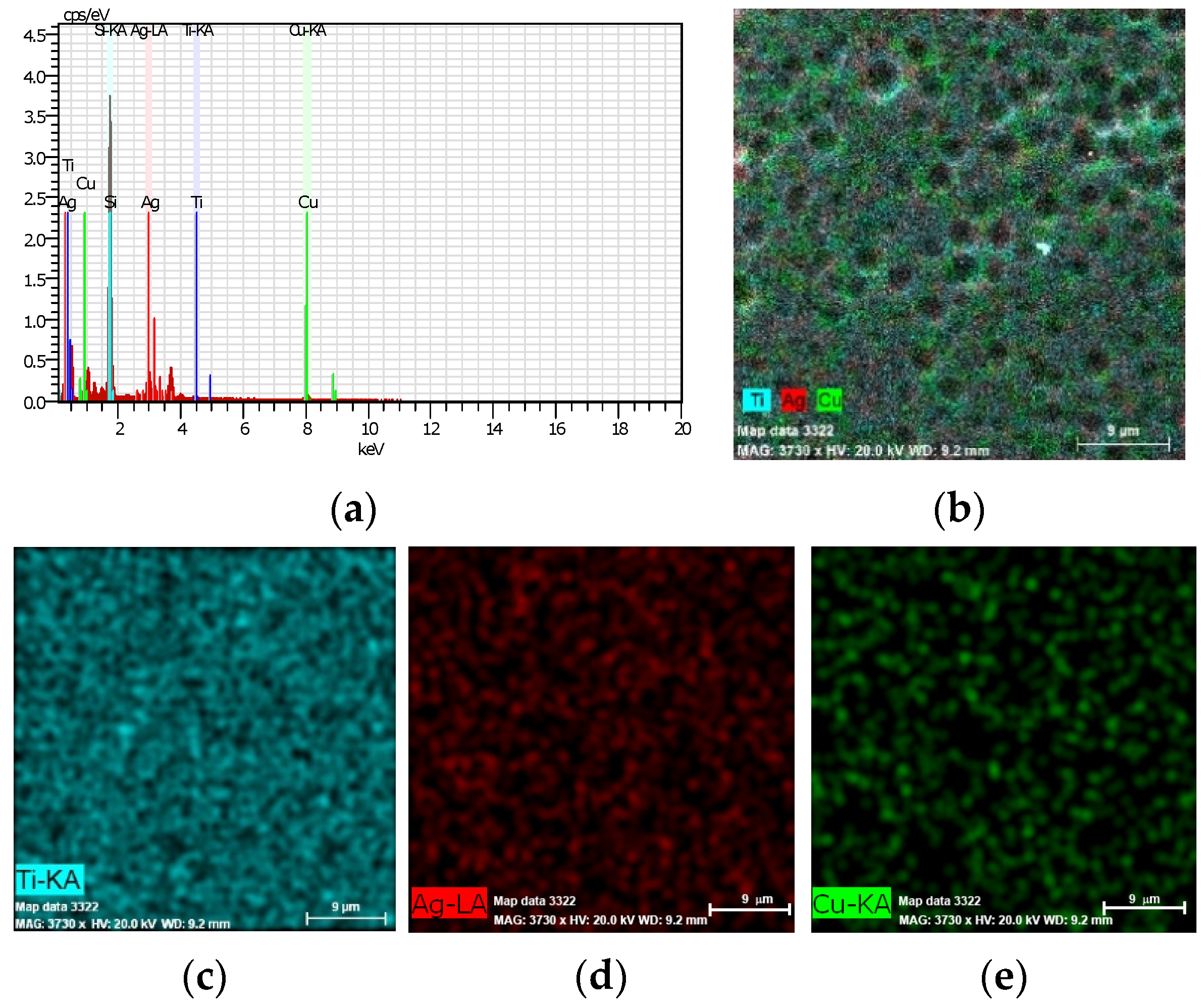
3.2. Elements Distribution (EDX)

The elements’ distribution is seen in Figure 1, which presents the EDX spectrum (a), an integrated map (b) and the individual elements’ distribution (Ti, Ag, and Cu (c, d, e, respectively)) of coated glass Sample 5—TiO2/Ag23.1 at.%/Cu21.6 at.%.
It is evident that all three elements (Ti, Ag and Cu) form chainlike aggregates (Figure 1c–e), specifically integrated in the TiO2/Ag23.1 at.%/Cu21.6 at.% coating to form dark depression wells, visible in the integrated map, Figure 1b. This picture is characteristic for all studied TiO2/Ag/Cu coatings, including Samples 1–4, and therefore their maps are not presented here.

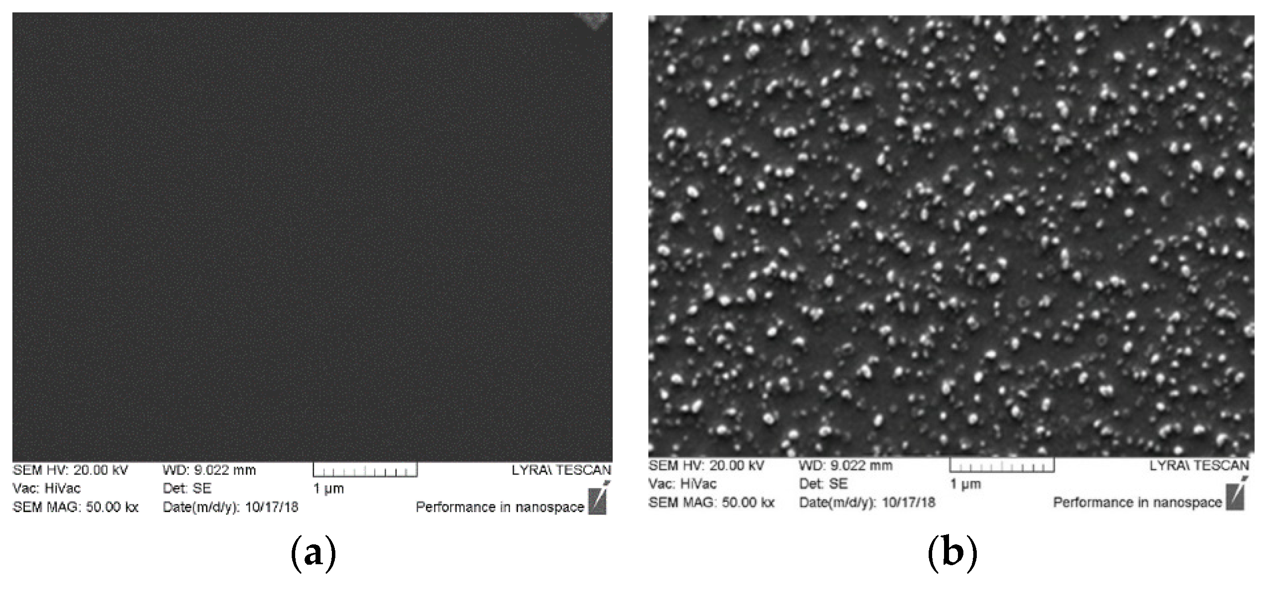
3.3. Surface Morphology (SEM)

The typical surface morphology of TiO2/Ag23.1 at.%/Cu21.6 at.% coated Sample 5 (Figure 2b) and that of TiO2 coated one (Figure 2a), for comparison, is presented in Figure 2. The TiO2 coated surface is flat (Figure 2a) whereas that of TiO2/Ag23.1 at.%/Cu21.6 at.% coated Sample 5 (b) characterizes by arrangements of chaotically distributed chainlike aggregates of Ag/Cu particles with different shapes and sizes, most of them submicron, which form dark walls, easily visible in Figure 1b. Such Ag and Cu particles’ distribution is characteristic for all TiO2/Ag/Cu magnetron cosputtered coatings. Therefore, their pictures are not presented here.

3.4. Water Contact Angle, WCA, Surface Energy, E and its Polar, Ep and Disperse, Ed Parts

As is evident from Table 2, the studied samples demonstrate slight deviations in the WCA (of 63.0–69.1°) and the surface energy, E (of 34.96 mN/m–39.21 mN/m), remaining in the region of moderate hydrophilic surfaces. This indicates that the insignificant impact of the surface hydrophilic/hydrophobic balance and surface energy could be expected on the initial microbial adhesion [58].

3.5. In Vitro Antimicrobial Activity

3.5.1. Diffusion Test

The diffusion test was employed for quick antibacterial screening of the studied TiO2/Ag/Cu magnetron cosputtered coatings. The results are presented in Table 3.
The data in Table 3 demonstrate a specific concentration of both the Ag- and Cu-dependent antimicrobial activity of the studied TiO2/Ag/Cu composite magnetron cosputtered coatings toward different microbial strains. The bactericidal activity of the test microbial species: (E. coli, P. aeruginosa, S. epidermidis and S. holeresius) is different on coatings with the same composition (Table 3, the sterile zones’ data in the rows). The sterile zones are different also for the test bacteria on the TiO2/Ag/Cu composite coatings containing different amounts of both Ag and Cu (Table 3, the sterile zones’ data in the columns). The inhibition zones of the studied bacteria are increasingly enlarged with the increasing Ag content (compare Samples 3, 4 and 5) and less enlarged with the increasing Cu content (compare Samples 1, 2 and 3), which indicates a stronger bactericidal effect of the Ag NPs as compared to that of the Cu NPs. The largest sterile zones appeared for all microbial strains in Sample 5—TiO2/Ag23.1 at.%/Cu21.6 at.%—the coating with the highest content of both Ag and Cu (Table 3, the last row).

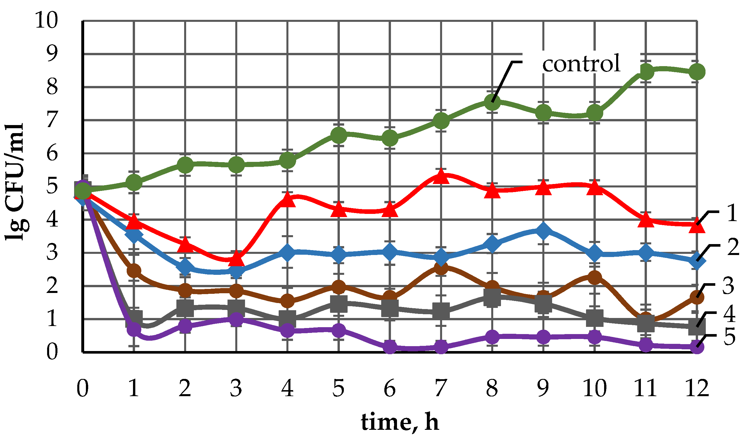
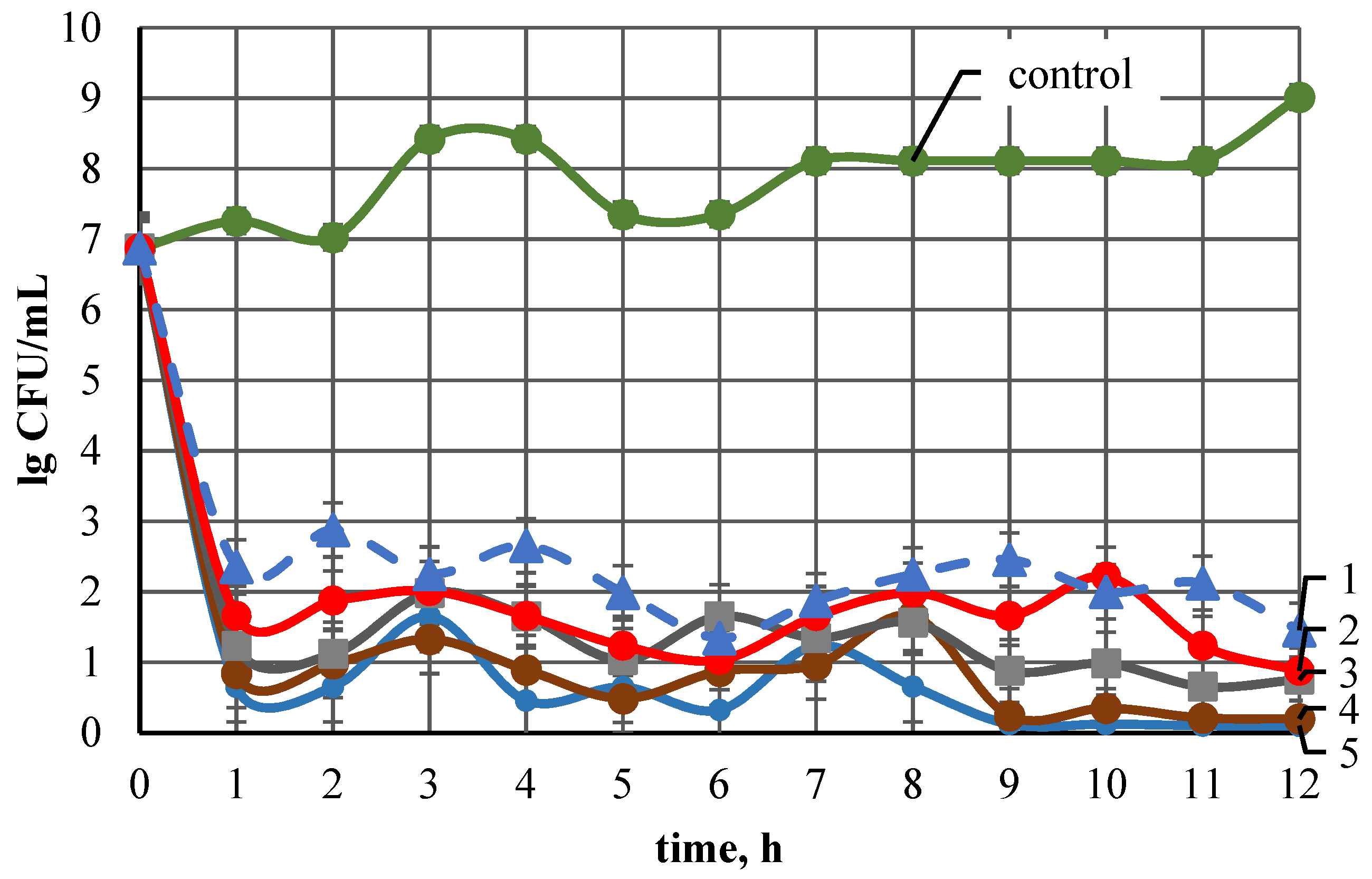
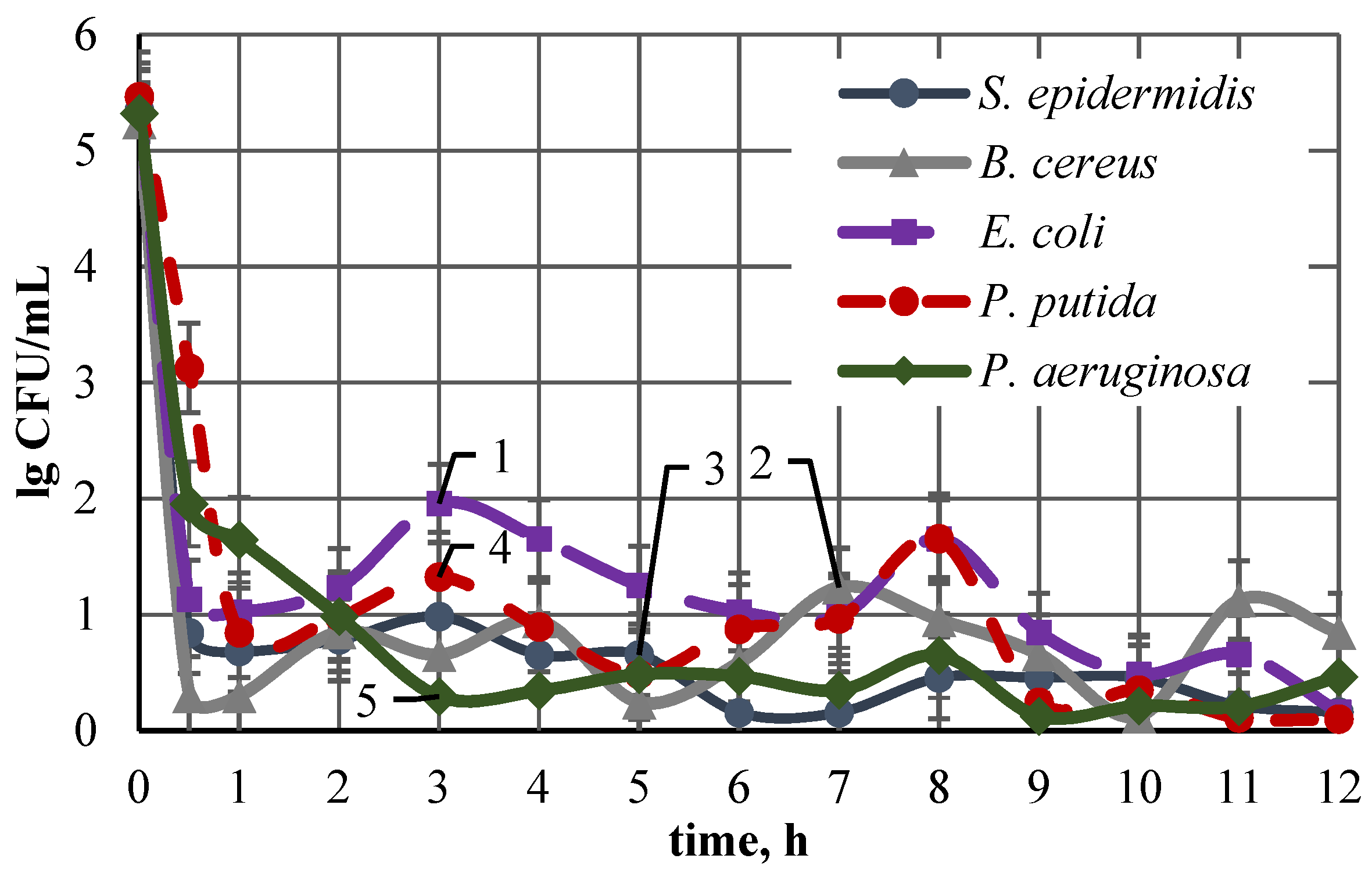
3.5.2. Cell Growth Inhibition

Two microbial strains, Gram-negative, E. coli and Gram-positive, S. epidermidis were used to study the cell growth inhibition on the studied TiO2/Ag/Cu cosputtered composition coatings with different Cu content and almost equal Ag content as well as with different Ag content at almost equal Cu content by MPN of the surviving cells. The dynamic of the E. coli growth inhibition is presented in Figure 3.
Figure 3 demonstrates a well expressed antimicrobial activity of TiO2/Ag/Cu coatings against E. coli, dependent on the concentration of both Ag and Cu. The significant inhibition of the E. coli growth happens in the first 1–2 h of exposure to the coated samples: the sharp slope of the curves 1–5 in the range of 1–3 h. The positions of curve 1, curve 2, and curve 3 in Figure 3 demonstrate the Cu concentration-dependent antibacterial activity of the coatings, containing almost equal amounts of Ag (about 14 at.%), and varied Cu content at: 13.0 at.% (curve 1), 17.9 at.% (curve 2), and 21.3 at.% (curve 3)—the increase in the Cu content leads to a stronger suppression of E. coli growth and the MPN of the living cells decreases. Similar dependence of the antimicrobial activity is observed for the TiO2/Ag/Cu coatings containing almost equal Cu amounts of about 21 at.%–22 at.% and varied Ag content at: 14.3 at.% (curve 3), 18.6 at.% (curve 4), and 23.1 at.% (curve 5). The MPN of the active cells presented by curve 4 is lower than that of the previous concentration of nanoparticles shown by curve 3 and curve 5 is below curve 4. The inhibitory effect toward E. coli is most strongly expressed in Sample 5 (curve 5), in which the amounts of both Ag and Cu in the TiO2/Ag/Cu coating are maximal: 23.1 at.% and 21.6 at.%, respectively. The MPN of the surviving E. coli cells tends to zero in this case. The data presented in Figure 3 (MPN of surviving cells) are in accordance with those obtained by the diffusion assay, presented in Table 3. The MPN of the surviving cells and the diffusion assay demonstrate good congruence and increased inhibitory effect against E. coli with the increase in the concentration.
Figure 4 represents the inhibitory effect of the same TiO2/Ag/Cu composite coatings toward S. epidermidis.
Figure 4 demonstrates a very well expressed antimicrobial activity of TiO2/Ag/Cu coatings against S. epidermidis, depending on the concentration of both Ag and Cu. A comparison of the group of curves 1–5 in Figure 3, to group of curves 1–5 in Figure 4 demonstrates the faster death and an MPN more closely approaching zero of the surviving cells in the second one (Figure 4), which indicates the higher sensitivity of S. epidermidis compared to E. coli, to both Ag NPs and Cu NPs. The strong inhibition of the S. epidermidis growth happens in the first 1–2 h of exposure to the coated samples, as indicated by the sharp slope of the curves 1–5 in Figure 4 in this time interval. The position of curves 1, 2 and 3 in Figure 4 (near each to other) demonstrates the slight increase in the antimicrobial activity with the increase in the Cu NPs concentration of the coatings, containing the same amount Ag of about 14 at.%, and varied Cu content: from 13.0 at.% (curve 1) to 17.9 at.% (curve 2) and 21.6 at.% (curve 3). The increase in the Ag NPs content from 14.3 at.% (curve 3); to 18.6 at.% (curve 4) and 23.1 at.% (curve 5) in the studied TiO2/Ag/Cu composite coatings at almost equal to Cu NPs content at 22 at.%–23 at.%, increases their activity against S. epidermidis, as indicated by the positions of the curves 3, 4 and 5 in Figure 4. The inhibitory effect toward S. epidermidis cell is most strongly expressed at Sample 5, i.e., at the maximal amounts of both Ag and Cu in the TiO2/Ag/Cu coating. The results of the MPN of surviving cells estimation confirm the findings obtained by the quick agar diffusion test (Table 3): higher sensitivity of S. epidermidis than E. coli to Ag NPs and Cu NPs dependent on the Ag and Cu concentration, and antimicrobial activity of the studied TiO2/Ag/Cu composite coatings.
Another demonstration of the specific antimicrobial activity of the studied TiO2/Ag/Cu composite coatings toward different bacterial species is evident in Figure 5, presenting the growth of different bacteria species in the presence of the same coating, Sample 5-TiO2/Ag23.1 at.%/Cu21.6 at.%.
The significant and specific antimicrobial activity of this coating toward large spectrum Gram-positive and Gram-negative microbial strains is clearly demonstrated by curves 1, 2, 3, 4, and 5. They show that the sharp decrease in the MPN of the surviving cells happens at different time intervals and it is down to different leaving cells number, both depending on the type of the microbial strain. The most sensitive to Ag/Cu as an antimicrobial agent is S. epidermidis, followed by B. cereus, P. putida, P. aeruginosa and E. coli.

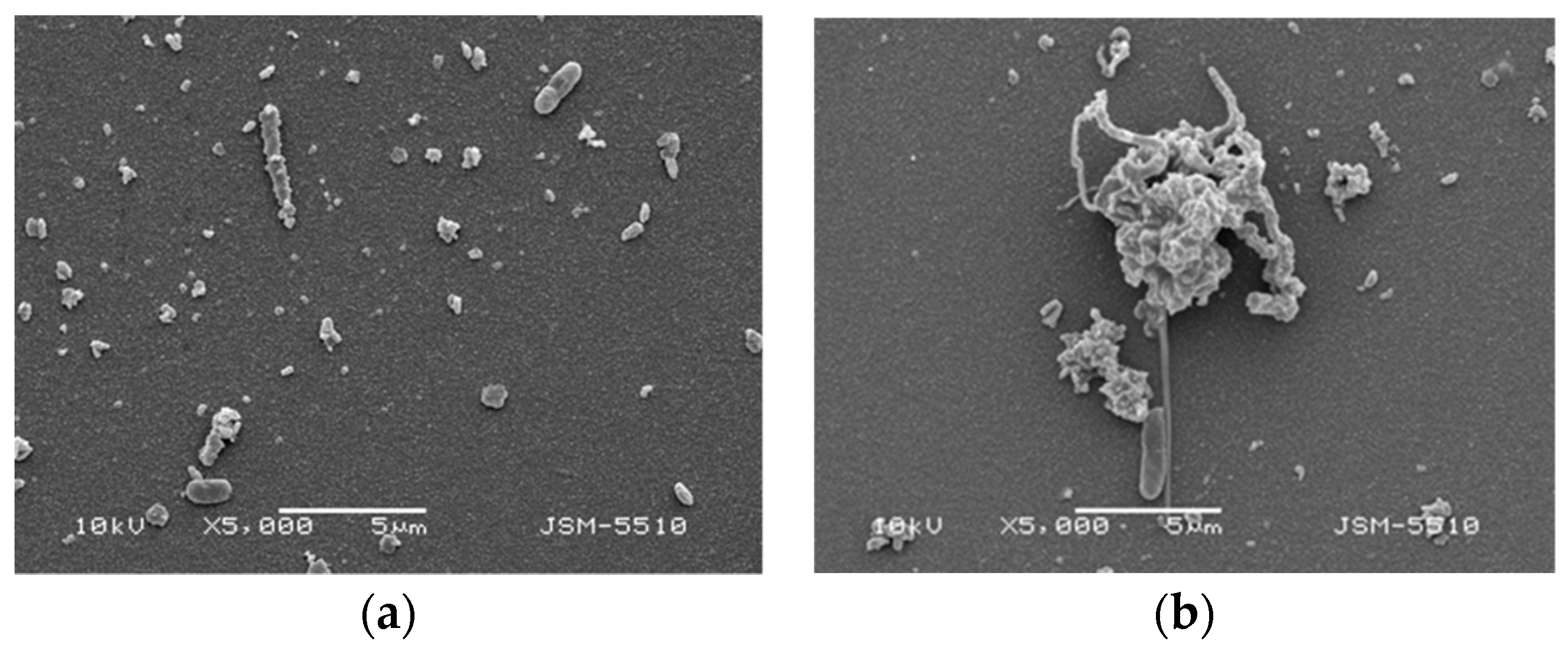
3.5.3. E. coli Cells on Coated Glass Samples

Significant damage to and killing of E. coli, after growth on TiO2/Ag/Cu coated glass samples, were observed by SEM. Typical images are presented in Figure 6.
Normally, the E. coli cells look like ovals with size of 0.8–1 μm. They seem to be damaged on the TiO2/Ag/Cu coated samples: deformed, with elongated original shape, dehydrated surface and multiple indentations (Figure 6a,b). Large colonies of damaged E. coli cells are also observed in some places (Figure 6b). This could be due to a poured cell content, the last one not seen in these pictures, because it was eluted after the samples had been washed in distilled water. The SEM observation of the E. coli cells on the coated samples demonstrates their contact killing.

4. Discussion

The surfaces of all TiO2/Ag/Cu coated samples (Samples 1–5) are characterized by similar morphology: Ag/Cu chainlike aggregates distributed chaotically in the coating (SEM, Figure 2b), and forming dark wells as visualized by the EDX maps (Figure 1b). All TiO2/Ag/Cu coated samples are moderately hydrophilic (WCA of 63.0°–69.1°, Table 2). Both the observed surface morphology and the moderate hydrophilicity [58] of the studied coatings suppose good interactions with living cells, i.e., the observed suppression of the test microbial cells growth could not be due to a lack of the conditions for initial attachment (initial adhesion). The suppression of the microbial cells’ growth by the studied TiO2/Ag/Cu composite coatings and its extent is determined by the chemical composition of the coating, i.e., by the presence of the antimicrobial agent Ag/Cu and its concentration
The well expressed cell growth inhibition could be due to cellular interactions with presenting on or/and eluted from the coated surface, Ag and Cu nanoparticles, ions and reactive oxygen species formed as a result of the oxidative processes. It is known that Ag NPs and Ag+, Cu NPs and Cu2+ are active antibacterial agents, although that the mechanism of their antibacterial action is not fully understood [59]. Our experimental results demonstrate direct contact (SEM observation, Figure 6) and released silver/coper killing (diffusion assay, Table 2; MPN of living cells, Figure 3, Figure 4 and Figure 5) of the bacterial cells by the studied TiO2/Ag/Cu coatings. A similar mode of bactericidal action was proposed earlier for magnetron sputtered TiO2 coatings, chemically incrusted with Ag NPs [12] and TiO2/SiO2/Ag magnetron cosputtered coatings [3,17].
There are a number of reports concerning the mechanism of action of Ag and Cu nanoparticles and ions. It was shown that silver ions (Ag+) interact with disulfide or sulfhydryl groups of enzymes, which leads to the inhibition of metabolic processes [60,61]; silver binds bacterial DNA (deoxyribonucleic acid), inhibiting replication and transcription [62,63] and copper leads to the collapse of some lipopolysaccharide (LPS) patches and alters the permeability and functionality of the outer cell membrane [64]. It was demonstrated that the activity of the NPs is size [27,65] and shape-dependent [66]. The free NPs penetrate inside the bacteria after their attachment to the plasma membrane and interact with sulfur containing proteins and the phosphorus containing DNA, which leads to blockage of the cell division [67]. It is difficult to distinguish the bactericidal activity of the NPs from that of the metallic ions released by the nanoparticles [68]. It was reported that the combined effect of the adhesion and penetration of the NPs and ions is crucial for the biocidal effect, with the plasma membrane being the target of the rapid antibacterial action of the NPs in E. coli [69]. The NPs also interact with the cell envelope of E. coli [70]. Morphological analysis reveals that the hallmarks of cell damage and NP toxicity include an increase in cell surface roughness and a change of the shape and size of the cells [71], such as were observed in our study. The mechanism of the antimicrobial action of Cu NPs and ions is less studied and reports on the mechanism of antimicrobial action of such coexisting with Ag NPs and ions are scarce.
The bactericidal activity of the magnetron cosputtered TiO2/Ag/Cu nanocomposite coatings could be due to a damage of the cell membrane and envelope (observed by SEM, Figure 6) usually resulting in a leakage of the cellular content, increased cell surface roughness, changed cell shape and size, and cell death [69,70,71]. The bonding of Ag NPs and Ag+, as well as of Cu NPs and Cu2+ from the studied coated samples to the microbial cells’ proteins, could also contribute, inactivating the electron transport chain and thereby inhibiting the respiration and growth of the cells, as was already reported [24]. The specific antimicrobial action of the studied coatings toward the used test bacterial strains could be explained with the specific features of their cells: rod or coil shape, presence or lack of flagella, cell envelope and membrane, density of the cell membrane, cell wall structure, etc. determining the engulf of the Ag/Cu NPs and ions [72,73].
The in-depth penetration of the mechanism of microbial cells’ interactions with Ag NPs and Ag+, Cu NPs and Cu2+, as well as with simultaneously presenting Ag NPs/Cu NPs and Ag+/Cu2+, needs additional, more in-depth investigations.

5. Conclusions

This investigation combines Ag and Cu as dopants in RF cosputtered TiO2/Ag/Cu functional coatings for medical devices and correlates their characteristics to the antimicrobial activity. The desired thickness, antibacterial activity and its durability can be obtained by the optimization of the operation parameters of the RF cosputtering and an accurately controlled composition of the TiO2/Ag/Cu coatings, especially of the Ag and Cu content as dopants. The last one is a convenient tool for the adjustment of the balance between antimicrobial activity and cytotoxicity to the requirements of different medical devices.
The magnetron cosputtered TiO2/SiO2/Ag composite coatings demonstrate a wide spectrum of antimicrobial activity, suppressing significantly the growth of Gram-negative (E. coli, P. aeruginosa) and Gram-positive (P. putida, B. cereus, S. epidermidis) bacteria.
The cells’ growth inhibition is specific and dependent on the Ag/Cu content for all used test bacterial strains. It is due to direct contact and eluted Ag/Cu-mediated killing, as experimentally demonstrated. The in-depth penetration in the mechanism of the microbial cells interaction with Ag and Cu nanoparticles and ions, as well as with simultaneously presented all of them needs in additional, more in-depth investigations.
It is expected that the studied magnetron cosputtered TiO2/Ag/Cu coatings would ensure a broad spectrum of bactericidal activity during the indwelling of the coated medical devices and for at least 12 h after that, with the supposition that the benefits would continue over a longer time.

Author Contributions

T.V. conceptualized this investigation; D.G. and T.V. performed the data curation, funding acquisition, coatings characterization, writing and illustrations of the original draft; T.V. and D.G. performed the project administration; I.I. carried out the antimicrobial testing. All coauthors participated in the discussion of the experimental results. All authors have read and agreed to the published version of the manuscript.

Funding

Nation Scientific Fund, Bulgaria (Grands DCOST 01/4/2018; KP-06-COST/20/2019).

Institutional Review Board Statement

Not applicable.

Informed Consent Statement

Not applicable.

Data Availability Statement

Not applicable.

Acknowledgments

Nation Scientific Fund, Bulgaria is gratefully acknowledged for its financial support (Grants DCOST 01/4/2018; KP-06-COST/20/2019) of this investigation. It is a part of the activities of COST Action CA 16217 ENIUS. O. Angelov is acknowledged for the performance of the magnetron cosputtering just before retiring.

Conflicts of Interest

The authors declare no conflict of interest.

References

  1. Ramstedt, M.; Ribeiro, I.A.C.; Bujdakova, H.; Mergulhão, F.J.M.; Jordao, L.; Thomsen, P.; Alm, M.; Burmølle, M.; Vladkova, T.; Can, F.; et al. Evaluating efficacy of antimicrobial and antifouling materials for urinary tract medical devices: Challenges and recommendations. Macromol. Biosci. 2019, 19, e1800384. [Google Scholar] [CrossRef] [PubMed] [Green Version]
  2. Adlhart, C.; Verran, J.; Azevedo, N.F.; Olmez, H.; Keinänen-Toivola, M.M.; Gouveia, I.; Melo, L.F.; Crijns, F. Surface modifications for antimicrobial effects in the healthcare setting: A critical overview. J. Hosp. Infect. 2018, 99, 239–249. [Google Scholar] [CrossRef] [Green Version]
  3. Murthy, S.P.; Raju, S.; Thiyagarajan, V. Biofilms Control: Biomedical and Industrial Environments; Narosa Publishing House: New Delhi, India, 2019. [Google Scholar]
  4. Khataee, A.; Mansoori, G.A. Nanostructured Titanium Dioxide Materials; World Scientific: Singapore, 2011. [Google Scholar] [CrossRef]
  5. Kartini, I.; Khairani, I.Y.; Triyana, K.; Wahyuni, S. Nanostructured titanium dioxide for functional coatings. In Titanium Dioxide: Material for a Sustainable Environment; Intech Open: Rijeka, Croatia, 2018. [Google Scholar]
  6. Watté, J.; Van Zele, M.; De Buysser, K.; Van Driessche, I. Recent advances in low-temperature deposition methods of transparent, photocatalytic TiO2 coatings on polymers. Coatings 2018, 8, 131. [Google Scholar] [CrossRef] [Green Version]
  7. Othman, S.H.; Salam, N.R.A.; Zainal, N.; Basha, R.K.; Talib, R.A. Antimicrobial activity of TiO2 nanoparticle-coated film for potential food packaging applications. Int. J. Photoenergy 2014, 2014, 945930. [Google Scholar] [CrossRef] [Green Version]
  8. Morrison, J. Copper’s Virus-Killing Powers were Known Even to the Ancients. Available online: https://www.smithsonianmag.com/science-nature/copper-virus-kill-180974655/ (accessed on 14 April 2020).
  9. Wilks, S.; Michels, H.; Keevil, C. The survival of escherichia coli o157 on a range of metal surfaces. Int. J. Food Microbiol. 2005, 105, 445–454. [Google Scholar] [CrossRef] [PubMed]
  10. Mathews, S.; Hans, M.; Mücklich, F.; Solioz, M. Contact killing of bacteria on copper is suppressed if bacterial-metal contact is prevented and is induced on iron by copper ions. Appl. Environ. Microbiol. 2013, 79, 2605–2611. [Google Scholar] [CrossRef] [Green Version]
  11. Mehtar, S.; Wiid, I.; Todorov, S. The antimicrobial activity of copper and copper alloys against nosocomial pathogens and mycobacterium tuberculosis isolated from healthcare facilities in the western cape: An in-vitro study. J. Hosp. Infect. 2008, 68, 45–51. [Google Scholar] [CrossRef]
  12. Shimabukuro, M. Antibacterial property and biocompatibility of silver, copper, and zinc in titanium dioxide layers incorporated by one-step micro-arc oxidation: A review. Antibiotics 2020, 9, 716. [Google Scholar] [CrossRef]
  13. Ning, C.; Wang, X.; Li, L.; Zhu, Y.; Li, M.; Yu, P.; Zhou, L.; Zhou, Z.; Chen, J.; Tan, G.; et al. concentration ranges of antibacterial cations for showing the highest antibacterial efficacy but the least cytotoxicity against mammalian cells: Implications for a new antibacterial mechanism. Chem. Res. Toxicol. 2015, 28, 1815–1822. [Google Scholar] [CrossRef] [Green Version]
  14. Reidy, B.; Haase, A.; Luch, A.; Dawson, K.A.; Lynch, I. Mechanisms of silver nanoparticle release, transformation and toxicity: A critical review of current knowledge and recommendations for future studies and applications. Materials 2013, 6, 2295–2350. [Google Scholar] [CrossRef] [PubMed] [Green Version]
  15. Marin, S.; Vlasceanu, G.M.; Tiplea, R.E.; Bucur, I.R.; Lemnaru, M.; Marin, M.M.; Grumezescu, A.M. Applications and toxicity of silver nanoparticles: A recent review. Curr. Top. Med. Chem. 2015, 15, 1596–1604. [Google Scholar] [CrossRef] [PubMed]
  16. Petica, A.; Florea, A.; Gaidau, C.; Balan, D.; Anicai, L. Synthesis and characterization of silver-titania nanocomposites prepared by electrochemical method with enhanced photocatalytic characteristics, antifungal and antimicrobial activity. J. Mater. Res. Technol. 2019, 8, 41–53. [Google Scholar] [CrossRef]
  17. Zawadzka, K.; Kisielewska, A.; Piwoński, I.; Kądzioła, K.; Felczak, A.; Różalska, S.; Wrońska, N.; Lisowska, K. Mechanisms of antibacterial activity and stability of silver nanoparticles grown on magnetron sputtered TiO2 coatings. Bull. Mater. Sci. 2016, 39, 57–68. [Google Scholar] [CrossRef] [Green Version]
  18. Brook, L.; Evans, P.; Foster, H.; Pemble, M.; Steele, A.; Sheel, D.; Yates, H. Highly bioactive silver and silver/titania composite films grown by chemical vapour deposition. J. Photochem. Photobiol. A Chem. 2007, 187, 53–63. [Google Scholar] [CrossRef] [Green Version]
  19. Sheel, D.W.; Brook, L.A.; Ditta, I.B.; Evans, P.; Foster, H.A.; Steele, A.; Yates, H.M. Biocidal silver and silver/titania composite films grown by chemical vapour deposition. Int. J. Photoenergy 2008, 2008, 1–11. [Google Scholar] [CrossRef]
  20. Page, K.; Palgrave, R.G.; Parkin, I.P.; Wilson, M.; Savin, S.L.P.; Chadwick, A.V. Titania and silver–titania composite films on glass—potent antimicrobial coatings. J. Mater. Chem. 2006, 17, 95–104. [Google Scholar] [CrossRef]
  21. Nigussie, G.Y.; Tesfamariam, G.M.; Tegegne, B.M.; Weldemichel, Y.A.; Gebreab, T.W.; Gebrehiwot, D.G.; Gebremichel, G.E. Antibacterial activity of ag-doped TiO2 and Ag-doped ZnO nanoparticles. Int. J. Photoenergy 2018, 2018, 5927485. [Google Scholar] [CrossRef] [Green Version]
  22. Mangalaraj, D.; Nithya Devi, D. Ag/TiO2 (Metal/Metal Oxide) Core Shell Nanoparticles for Biological Applications BT. In Recent Trends in Materials Science and Applications; Ebenezar, J., Ed.; Springer International Publishing: Cham, Switzerland, 2017; pp. 9–17. [Google Scholar]
  23. Wahyuni, E.T.; Roto, R. Silver Nanoparticle Incorporated Titanium Oxide for Bacterial Inactivation and Dye Degradation. In Titanium Dioxide: Material for a Sustainable Environment; Intech Open: Rijeka, Croatia, 2018. [Google Scholar]
  24. Vladkova, T.; Angelov, O.; Stoyanova, D.; Gospodinova, D.; Gomes, L.; Soares, A.; Mergulhao, F.; Ivanova, I. Magnetron co-sputtered TiO2/SiO2/Ag nanocomposite thin coatings inhibiting bacterial adhesion and biofilm formation. Surf. Coat. Technol. 2020, 384, 125322. [Google Scholar] [CrossRef]
  25. Desai, V.; Kaler, S.G. Role of copper in human neurological disorders. Am. J. Clin. Nutr. 2008, 88, 855S–858S. [Google Scholar] [CrossRef] [Green Version]
  26. Mungkalasiri, J.; Bedel, L.; Emieux, F.; Doré, J.; Renaud, F.; Maury, F. DLI-CVD of TiO2–Cu antibacterial thin films: Growth and characterization. Surf. Coat. Technol. 2009, 204, 887–892. [Google Scholar] [CrossRef] [Green Version]
  27. Mungkalasiri, J.; Bedel, L.; Emieux, F.; Doré, J.; Renaud, F.N.R.; Sarantopoulos, C.; Maury, F. CVD Elaboration of nanostructured TiO2-Ag thin films with efficient antibacterial properties. Chem. Vap. Depos. 2010, 16, 35–41. [Google Scholar] [CrossRef] [Green Version]
  28. Zielinska-Jurek, A.; Walicka, M.; Tadajewska, A.; Łacka, I.; Gazda, M.; Zaleska-Medynska, A. Preparation of Ag/Cu-doped titanium (IV) oxide nanoparticles in w/o microemulsion. Physicochem. Probl. Miner. Process. 2010, 45, 113–126. [Google Scholar]
  29. Sangchay, W.; Lek, S.; Kooptarnond, K. The photocatalytic and antibacterial activity of Cu-doped TiO2 thin films. Walailak J. Sci. Technol. 2013, 10, 19–27. [Google Scholar] [CrossRef]
  30. Varghese, S.; ElFakhri, S.O.; Sheel, D.W.; Sheel, P.; Bolton, F.J.E.; Foster, H.A. Antimicrobial activity of novel nanostructured cu- SiO2 coatings prepared by chemical vapour deposition against hospital related pathogens. AMB Express 2013, 3, 53. [Google Scholar] [CrossRef] [PubMed] [Green Version]
  31. Wei, X.; Gao, W. Polymer-Cu/TiO2 Antimicrobial Coatings BT. In Proceedings of the 8th Pacific Rim International Congress on Advanced Materials and Processing; Marquis, F., Ed.; Springer International Publishing: Cham, Switzerland, 2016; pp. 1663–1669. [Google Scholar]
  32. Mungkalasiri, J.; Bedel, L.; Emieux, F.; Cara, A.V.-D.; Freney, J.; Maury, F.; Renaud, F.N. Antibacterial properties of TiO2–Cu composite thin films grown by a one step DLICVD process. Surf. Coat. Technol. 2014, 242, 187–194. [Google Scholar] [CrossRef] [Green Version]
  33. Krishnakumar, V.; Boobas, S.; Jayaprakash, J.; Rajaboopathi, M.; Han, B.; Louhi-Kultanen, M. Effect of Cu doping on TiO2 nanoparticles and its photocatalytic activity under visible light. J. Mater. Sci. Mater. Electron. 2016, 27, 7438–7447. [Google Scholar] [CrossRef]
  34. Zhang, L.; Guo, J.; Huang, X.; Zhang, Y.; Han, Y. The dual function of Cu-doped TiO2 coatings on titanium for application in percutaneous implants. J. Mater. Chem. B 2016, 4, 3788–3800. [Google Scholar] [CrossRef] [PubMed]
  35. Mathew, S.; Ganguly, P.; Rhatigan, S.; Kumaravel, V.; Byrne, C.; Hinder, S.J.; Bartlett, J.; Nolan, M.; Pillai, S.C. Cu-doped TiO2: Visible light assisted photocatalytic antimicrobial activity. Appl. Sci. 2018, 8, 2067. [Google Scholar] [CrossRef] [Green Version]
  36. Alavi, M.; Karimi, N. Antiplanktonic, antibiofilm, antiswarming motility and antiquorum sensing activities of green synthesized Ag–TiO2, TiO2–Ag, Ag–Cu and Cu–Ag nanocomposites against multi-drug-resistant bacteria. Artif. Cells Nanomed. Biotechnol. 2018, 46, S399–S413. [Google Scholar] [CrossRef] [PubMed] [Green Version]
  37. Nißen, S.; Heeg, J.; Wienecke, M.; Behrend, D.; Warkentin, M.; Rokosz, K.; Gaiaschi, S.; Chapon, P. Surface characterization and copper release of a-C:H:Cu coatings for medical applications. Coatings 2019, 9, 119. [Google Scholar] [CrossRef] [Green Version]
  38. Alotaibi, A.M.; Williamson, B.A.D.; Sathasivam, S.S.; Kafizas, A.; Alqahtani, M.; Sotelo-Vazquez, C.; Buckeridge, J.; Wu, J.; Nair, S.P.; Scanlon, D.O.; et al. Enhanced photocatalytic and antibacterial ability of Cu-Doped anatase TiO2 thin films: Theory and experiment. ACS Appl. Mater. Interfaces 2020, 12, 15348–15361. [Google Scholar] [CrossRef] [PubMed] [Green Version]
  39. Tahmasebizad, N.; Hamedani, M.T.; Ghazani, M.S.; Pazhuhanfar, Y. Photocatalytic activity and antibacterial behavior of TiO2 coatings co-doped with copper and nitrogen via sol–gel method. J. Sol. Gel Sci. Technol. 2019, 93, 570–578. [Google Scholar] [CrossRef] [PubMed]
  40. Behnajady, M.A.; Eskandarloo, H. Characterization and photocatalytic activity of Ag-Cu/TiO2 nanoparticles prepared by sol-gel method. J. Nanosci. Nanotechnol. 2013, 13, 548–553. [Google Scholar] [CrossRef] [PubMed]
  41. Yuan, W.; Ji, J.; Fu, J.; Shen, J. A facile method to construct hybrid multilayered films as a strong and multifunctional antibacterial coating. J. Biomed. Mater. Res. Part B Appl. Biomater. 2008, 85, 556–563. [Google Scholar] [CrossRef]
  42. Akhavan, O. Lasting antibacterial activities of Ag–TiO2/Ag/a-TiO2 nanocomposite thin film photocatalysts under solar light irradiation. J. Colloid Interface Sci. 2009, 336, 117–124. [Google Scholar] [CrossRef]
  43. Akhavan, O.; Ghaderi, E. Bactericidal effects of Ag nanoparticles immobilized on surface of SiO2 thin film with high concentration. Curr. Appl. Phys. 2009, 9, 1381–1385. [Google Scholar] [CrossRef]
  44. Mazur, M.; Wojcieszak, D.; Domaradzki, J.; Kaczmarek, D.; Song, S.; Placido, F. TiO2/SiO2 multilayer as an antireflective and protective coating deposited by microwave assisted magnetron sputtering. Opt. Electron. Rev. 2013, 21, 233–238. [Google Scholar] [CrossRef]
  45. Khan, S.B.; Wu, H.; Pan, C.; Zhang, Z. A mini review: Antireflective coatings processing techniques, applications and future perspective. Res. Rev. J. Mater. Sci. 2017, 5, 1–19. [Google Scholar] [CrossRef] [Green Version]
  46. Yamaguchi, T.; Tamura, H.; Taga, S.; Tsuchiya, S. Interfacial optical absorption in TiO2–SiO2 multilayer coatings prepared by rf magnetron sputtering. Appl. Opt. 1986, 25, 2703–2706. [Google Scholar] [CrossRef]
  47. Vladkova, T.G.; Staneva, A.D.; Gospodinova, D.N. Surface engineered biomaterials and ureteral stents inhibiting biofilm formation and encrustation. Surf. Coat. Technol. 2020, 404, 126424. [Google Scholar] [CrossRef]
  48. Gao, A.; Hang, R.; Chu, P.K. Recent advances in anti-infection surfaces fabricated on biomedical implants by plasma-based technology. Surf. Coat. Technol. 2017, 312, 2–6. [Google Scholar] [CrossRef]
  49. Rijal, N. Most Probable Number (MPN) Test: Principle, Procedure and Results. Available online: https://microbeonline.com/probable-number-mpn-test-principle-procedure-results/ (accessed on 11 June 2017).
  50. Stoyanova, D.S.; Ivanova, I.A.; Angelov, O.I.; Vladkova, T.G. Antibacterial activity of thin films TiO2 doped with Ag and Cu on gracilicutes and firmicutes bacteria. Biodiscovery 2017, 20, e15076. [Google Scholar] [CrossRef] [Green Version]
  51. Wassmann, T.; Kreis, S.; Behr, M.; Buergers, R. The influence of surface texture and wettability on initial bacterial adhesion on titanium and zirconium oxide dental implants. Int. J. Implant. Dent. 2017, 3, 1–11. [Google Scholar] [CrossRef] [PubMed]
  52. Poncin-Epaillard, F.; Vrlinic, T.; Debarnot, D.; Mozetic, M.; Coudreuse, A.; Legeay, G.; El Moualij, B.; Zorzi, W. Surface treatment of polymeric materials controlling the adhesion of biomolecules. J. Funct. Biomater. 2012, 3, 528–543. [Google Scholar] [CrossRef] [Green Version]
  53. Speranza, G.; Gottardi, G.; Pederzolli, C.; Lunelli, L.; Canteri, R.; Pasquardini, L.; Carli, E.; Lui, A.; Maniglio, D.; Brugnara, M.; et al. Role of chemical interactions in bacterial adhesion to polymer surfaces. Biomaterials 2004, 25, 2029–2037. [Google Scholar] [CrossRef]
  54. Vladkova, T.G. Surface engineered polymeric biomaterials with improved biocontact properties. Int. J. Polym. Sci. 2010, 2010, 296094. [Google Scholar] [CrossRef] [Green Version]
  55. Vladkova, T.G. Surface Engineering of Polymeric Biomaterials; Smithers Rapra Technology: Shropshire, UK, 2013. [Google Scholar]
  56. Ploux, L.; Ponche, A.; Anselme, K. Bacteria/material interfaces: Role of the material and cell wall properties. J. Adhes. Sci. Technol. 2010, 24, 2165–2201. [Google Scholar] [CrossRef]
  57. Song, F.; Koo, H.; Ren, D. Effects of material properties on bacterial adhesion and biofilm formation. J. Dent. Res. 2015, 94, 1027–1034. [Google Scholar] [CrossRef]
  58. Altankov, G. Interaction of Cells with Biomaterial Surfaces; Bulgarian Academy of Science: Sofia, Bulgaria, 2003. [Google Scholar]
  59. Kędziora, A.; Speruda, M.; Krzyżewska, E.; Rybka, J.; Łukowiak, A.; Bugla-Płoskońska, G. Similarities and differences between silver ions and silver in nanoforms as antibacterial agents. Int. J. Mol. Sci. 2018, 19, 444. [Google Scholar] [CrossRef] [Green Version]
  60. Feng, Q.L. A Mechanistic study of the antibacterial effect of silver ions on escherichia coli and staphylococcus Aureus. J. Biomed. Mater. Res. 2000, 52, 662–668. [Google Scholar] [CrossRef]
  61. Butkus, M.A.; Edling, L.; Labare, M.P. The efficacy of silver as a bactericidal agent: Advantages, limitations and considerations for future use. J. Water Supply Res. Technol. 2003, 52, 407–416. [Google Scholar] [CrossRef]
  62. Lansdown, A. Silver I: Its antibacterial properties and mechanism of action. J. Wound Care 2002, 11, 125–130. [Google Scholar] [CrossRef]
  63. Castellano, J.J.; Shafii, S.M.; Ko, F.; Donate, G.; Wright, T.E.; Mannari, R.J.; Payne, W.G.; Smith, D.J.; Robson, M.C. Comparative evaluation of silver-containing antimicrobial dressings and drugs. Int. Wound J. 2007, 4, 114–122. [Google Scholar] [CrossRef] [PubMed]
  64. Nan, L.; Liu, Y.; Lü, M.; Yang, K. Study on antibacterial mechanism of copper-bearing austenitic antibacterial stainless steel by atomic force microscopy. J. Mater. Sci. Mater. Med. 2008, 19, 3057–3062. [Google Scholar] [CrossRef]
  65. Panáček, A.; Kvítek, L.; Prucek, R.; Kolář, M.; Večeřová, R.; Pizúrová, N.; Sharma, V.K.; Nevěčná, T.; Zbořil, R. Silver colloid nanoparticles: Synthesis, characterization, and their antibacterial activity. J. Phys. Chem. B 2006, 110, 16248–16253. [Google Scholar] [CrossRef]
  66. Pal, S.; Tak, Y.K.; Song, J.M. Does the antibacterial activity of silver nanoparticles depend on the shape of the nanoparticle? A study of the gram-negative bacterium escherichia coli. Appl. Environ. Microbiol. 2007, 73, 1712–1720. [Google Scholar] [CrossRef] [Green Version]
  67. Danilczuk, M.; Lund, A.; Sadlo, J.; Yamada, H.; Michalik, J. Conduction electron spin resonance of small silver particles. Spectrochim. Acta Part A Mol. Biomol. Spectrosc. 2006, 63, 189–191. [Google Scholar] [CrossRef]
  68. Kim, J.S.; Kuk, E.; Yu, K.N.; Kim, J.-H.; Park, S.J.; Lee, H.J.; Kim, S.H.; Park, Y.K.; Park, Y.H.; Hwang, C.-Y.; et al. Antimicrobial effects of silver nanoparticles. Nanomed. Nanotechnol. Biol. Med. 2007, 3, 95–101. [Google Scholar] [CrossRef]
  69. Bondarenko, O.M.; Sihtmäe, M.; Kuzmičiova, J.; Ragelienė, L.; Kahru, A.; Daugelavičius, R. Plasma membrane is the target of rapid antibacterial action of silver nanoparticles in escherichia coli and pseudomonas aeruginosa. Int. J. Nanomed. 2018, 13, 6779–6790. [Google Scholar] [CrossRef] [PubMed] [Green Version]
  70. Sondi, I.; Salopek-Sondi, B. Silver nanoparticles as antimicrobial agent: A case study on E. coli as a model for gram-negative bacteria. J. Colloid Interface Sci. 2004, 275, 177–182. [Google Scholar] [CrossRef] [PubMed]
  71. Prieto, E.; Kiat, A. The antimicrobial action of silver nanoparticles on E. coli as revealed by atomic force microscopy. Philipp. Sci. Lett. 2017, 10, 123–129. [Google Scholar]
  72. Albu, M.G.; Vladkova, T.G.; Ivanova, I.A.; Shalaby, A.S.A.; Moskova-Doumanova, V.S.; Staneva, A.D.; Dimitriev, Y.B.; Kostadinova, A.S.; Topouzova-Hristova, T.I. Preparation and biological activity of new collagen composites, part I: Collagen/zinc titanate nanocomposites. Appl. Biochem. Biotechnol. 2016, 180, 177–193. [Google Scholar] [CrossRef] [PubMed]
  73. Vladkova, T.G.; Ivanova, I.A.; Staneva, A.D.; Albu-Kaya, M.G.; Shalaby, A.S.A.; Moskova-Doumanova, V.; Kostadinova, A.S. Preparation and biological activity of new collagen composites, part III. collagen/(Ag/RGO) and collagen/(Ag/RGO/SiO2) composites. J. Arch. Mil. Med. 2017, in press. [Google Scholar] [CrossRef]
Figure 1. EDX spectrum (a), integrated map of Ti, Ag and Cu distribution (b) and maps of the individual elements Ti (c), Ag (d), Cu (e) distribution in Sample 5—TiO2/Ag23.1 at.%/Cu21.6 at.%.
Coatings 11 00473 g001
Figure 2. Morphology of: (a) TiO2 magnetron sputtered coating on glass substrate (×5000); (b) TiO2/Ag23.1 at.%/Cu21.6 at.% magnetron cosputtered coating on glass substrate, Sample 5.
Coatings 11 00473 g002
Figure 3. E. coli growth inhibition in presence of: (1) Sample 1-TiO2/Ag14.7 at.%/Cu13.0 at.%; (2) Sample 2-TiO2/Ag4.0 at.%/Cu17.9 at.%; (3) Sample 3-TiO2/Ag14.3 at.%/Cu21.3 at.%; (4) Sample 4-TiO2/Ag18.6 at.%/Cu22.4 at.%; (5) Sample 5-TiO2/Ag23.1 at.%/Cu21.6 at.%; or in absence of sample—control.
Coatings 11 00473 g003
Figure 4. S. epidermidis growth inhibition in presence of: (1) Sample 1-TiO2/Ag14.7 at.%/Cu13.0 at.%; (2) Sample 2-TiO2/Ag4.0 at.%/Cu17.9 at.%; (3) Sample 3-TiO2/Ag14.3 at.%/Cu21.3 at.%; (4) Sample 4-TiO2/Ag18.6 at.%/Cu22.4 at.%; (5) Sample 5-TiO2/Ag23.1 at.%/Cu21.6 at.%; or in absence of sample—control.
Coatings 11 00473 g004
Figure 5. E. coli (curve 1), B. cereus (curve 2), S. epidermidis (curve 3), P. putida (curve 4), P. aeruginosa (curve 5) growth inhibition in presence of Sample 5-TiO2/Ag23.1 at.%/Cu21.6 at.%.
Coatings 11 00473 g005
Figure 6. SEM images of significantly damaged E. coli cells (a) and E. coli cell aggregates (b) in contact with TiO2/Ag23.1 at.%/Cu21.6 at.% magnetron cosputtered composite coating.
Coatings 11 00473 g006
Table 1. Elemental composition of cosputtered TiO2/Ag/Cu nanocomposite coatings (from XPS) prepared under varied surface area of the Ag and Cu sheds.
Sample No.C1s, at.%Ti2p, at.%Ag3d, at.%Cu2p, at.%
1.4.1230.7814.713.0
2.3.1831.0014.217.9
3.5.2527.3114.321.3
4.8.2126.4818.622.4
5.6.3324.0423.121.6
Table 2. Water contact angle, WCA, surface energy, E and its polar Ep, and disperse part, Ed of TiO2/Ag/Cu composite coatings both with almost equal Ag content and varied Cu content (Samples 1, 2 and 3) or with almost equal coper content and varied silver content (samples 2, 4, and 5).
SamplesWCA, °E, mN/mEp, mN/mEd, mN/m
Sample 1-TiO2/Ag14.7 at.%/Cu13.0 at.%62.2 ± 0.6336.1211.1025.02
Sample 2-TiO2/Ag14.0 at.%/Cu17.9 at.%61.9 ± 0.3638.0312.3325.70
Sample 3-TiO2/Ag14.3 at.%/Cu21.3 at.%63.0 ± 0.7135.9210.1225.80
Sample 4-TiO2/Ag18.6 at.%/Cu22.4 at.%60.3 ± 0.5239.2111.2028.01
Sample 5-TiO2/Ag23.1 at.%/Cu21.6 at.%69.1 ± 0.8934.9617.9026.06
Table 3. Antimicrobial activity toward Gram-negative and Gram-positive microbial strains (as a sterile zone, mm; by agar diffusion test) of TiO2/Ag/Cu magnetron cosputtered thin coatings depending on the Cu-content at almost equal Ag content (Samples 1, 2, and 3) and depending on Ag content at almost equal Cu content (Samples 3, 4, and 5).
Sample No.Sterile Zone, mm
E.
coli
P.
aeruginosa
S.
epidermidis
S.
holeresius
Sample 1-TiO2/Ag14.7 at.%/Cu13.0 at.%1 ± 0.53 ± 0.52 ± 0.52 ± 0.5
Sample 2-TiO2/Ag14.0 at.%/Cu17.9 at.%3 ± 0.54 ± 0.55 ± 0.54 ± 0.5
Sample 3-TiO2/Ag14.3 at.%/Cu21.3 at.%2 ± 0.54 ± 0.55 ± 0.54 ± 0.5
Sample 4-TiO2/Ag18.6 at.%/Cu22.4 at.%5 ± 0.56 ± 0.57 ± 0.58 ± 0.5
Sample 5-TiO2/Ag23.1 at.%/Cu21.6 at.%6 ± 0.58 ± 0.59 ± 0.59 ± 0.5
Publisher’s Note: MDPI stays neutral with regard to jurisdictional claims in published maps and institutional affiliations.

