Article

Development of Adhesive, Bioactive and Antibacterial Titania Sol-Gel Coating on Titanium Substrate by Dip-Coating Technique

by
Diana Horkavcová
1,*,
Quentin Doubet
2,
Gisèle Laure Lecomte-Nana
2,
Eva Jablonská
3 and
Aleš Helebrant
1
1
Department of Glass and Ceramics, Faculty of Chemical Technology, University of Chemistry and Technology Prague, Technická 5, 16628 Prague, Czech Republic
2
Institut de Recherche sur les Céramiques, ENSIL-ENSCI, 12 rue Atlantis, Université de Limoges, 87068 Limoges, France
3
Department of Biochemistry and Microbiology, Faculty of Food and Biochemical Technology, University of Chemistry and Technology Prague, Technická 3, 16628 Prague, Czech Republic
*
Author to whom correspondence should be addressed.
Coatings 2021, 11(2), 243; https://doi.org/10.3390/coatings11020243
Submission received: 8 January 2021 / Revised: 8 February 2021 / Accepted: 9 February 2021 / Published: 18 February 2021
(This article belongs to the Special Issue Glass/Ceramic Coatings for Biomaterials and Biomedical Applications)

Abstract

:
The sol-gel method provides a wide variety of applications in the medical field. One of these applications is the formation of coatings on the metal implants. The coatings containing specific additive can enhance or improve the existing surface properties of the substrate. In this work, titania sol-gel coatings were doped with two forms of silver (AgNO3, Ag3PO4) and synthetic hydroxyapatite and applied on the titanium samples by dip-coating technique. After drying and slow firing, all coatings were characterized with scanning electron microscopy. Thin coatings were successfully prepared with excellent adhesion to the substrate (measured by ASTM D 3359-2), despite cracks. Coatings containing silver and hydroxyapatite demonstrated a 100% antibacterial effect against Escherichia coli after 24 h. The bioactivity of the coatings containing hydroxyapatite tested in modified simulated body fluid under static-dynamic conditions was confirmed by bone-like hydroxyapatite precipitation. To better understand the interaction of the coatings with simulated body fluid (SBF), changes of Ca2+ and (PO4)3− ions concentrations and pH values were studied.

1. Introduction

Metals are predominantly used for implants for decades because of their mechanical properties, i.e., great fatigue resistance and high strength. Titanium and its alloys are widely used to make hard tissues because of an excellent biocompatibility, good mechanical properties, a low Young’s modulus, a non-magnetic behavior, a good resistance to the corrosion, and a light weight. For medical applications, the titanium is often protected by coatings that can bring the benefits of bioactivity and antibacterial effect [1,2,3,4]. The sol-gel dip-coating technique is one of the simplest and enables to make homogenous layers, especially on regular samples like rectangular cuboids. It comprises three steps, the first one is the dipping, second is the dwelling, and the last is the withdrawing [5,6]. One of the most important properties is adhesion of the coating to the substrate [7]. Insufficient adhesion would not only lead to losing the properties linked to the coating, it could also be harmful for the living body. The layers prepared by sol-gel dip-coating technique are known to have excellent adhesive properties [8,9].
Bioactive sol-gel coatings can be achieved by addition of a hydroxyapatite reagent [10,11]. Bioactivity (ability of implants forming the apatite on its surface) can be monitored with in vivo test (on live organisms) or in vitro test (interaction with fluid that simulates the living body environment at specific conditions). Various solutions collectively called “simulated body fluid” (SBF) are used for in vitro test. SBF prepared according to Kokubo is one of the most frequently used [12]. Its chemical composition is very similar to the body fluid, however its concentration of Cl is higher than in the blood plasma while the concentration of HCO3 is lower. Thanks to SBF, it is possible to evaluate the bioactivity of the implant, i.e., the capacity of the implant to form apatite [13].
The antibacterial properties of the implant are beneficial, because despite sterile conditions, infection can occur, and it represents one of the most serious and devastating complications in implantology. Silver particles have a great antibacterial effect, however Ag also exerts toxicity towards eukaryote including human body and, therefore, the amount and form of Ag must be considered wisely [14,15]. It is not recommended to use more than 1.6 ppm Ag+ in bioactive materials [16]. The silver can be used in the form of Ag3PO4 [17,18] or AgNO3 [19,20] into biomedical coatings to be effective against bacteria and to reduce the risk of potential infection in the vicinity of the implant.
The aim of this work was to completely change titanium surface substrate properties by preparation of technically simple, cheap, adhesive, bioactive, and antibacterial sol-gel coatings. This improved coated titanium substrate can be used in biomedical applications in the field of orthopedics or dentistry.

2. Experimental

The titanium samples were rectangular cuboid of 30 mm × 10 mm × 1 mm. The first step consisted of grinding of all samples in order to homogenize the surface for better adhesion of the coating. Three sizes of abrasive SiC disc were used, namely: P400, 600, and 800. The second step was the removal of the free particles through the cleaning of samples in demi-water, acetone and denatured ethanol using ultrasound (each for 10 min). The third step was drying the samples at laboratory temperature for 10 min. The composition and preparation of the basic titania sol are described in article [20,21]. The sol was divided into six parts and two forms of silver at concentration of 0.09 mol/L and hydroxyapatite (HA) at the concentration of 0.2 mol/L were added (Table 1). Silver was added as AgNO3 (p.a. Lach-Ner) or as Ag3PO4 (Sigma-Aldrich, St. Louis, MO, USA). The particle size (median) of pure commercial hydroxyapatite (Riedel-de Haën, Seelze, Germany) was 12.337 µm. Identification of the individual sols and coatings prepared from them are shown in Table 1.
The speed of dipping was 20 cm/min, the time of dwelling was 10 s, and the withdraw speed was 6 cm/min. After the dip-coating, the samples were left to dry for 30 min at ambient temperature, then they were heated for 2 h at 400 °C with a ramp of 2 °C/min and cooled in the oven until the next day.
The antibacterial test was performed against gram-negative bacteria Escherichia coli with concentration of 104 colony forming unit (CFU, i.e., number of bacteria able to create colonies)/mL for 4 and 24 h at laboratory temperature in the dark as described in [20,21].
The in vitro test of bioactivity was performed under static-dynamic condition. In contrast to the static condition given in standard [22], in our case modified simulated body fluid (MSBF) was changed every day to simulate the circulation in the living body. MSBF solution was prepared from the reagents KCl, NaCl, NaHCO3, MgSO4. 7H2O, CaCl2, KH2PO4, TRIS, and NaN3. MSBF was buffered: with TRIS (Tris-hydroxymethyl aminomethane) and HCl to achieve pH = 7.40. Azide (NaN3) was added to prevent bacteria growth in the solution during the long-term test. The chemical composition of MSBF is shown in Table 2. Two coated samples of each type (THA, TANHA, TAPHA) were immersed into MSBF at 36.5 °C in a biological thermostat. The pH value of MSBF was measured with a pH meter (7110, Inolab, Weilheim, Germany), with the temperature being in the range of 33−34 °C. Concentration of calcium ions was measured by atomic absorption spectroscopy (AAS, VARIAN-Spectr AA 880, at wave 422.7 µm) and the concentration of phosphate ions was measured by UV-visible spectroscopy (UV-2450 spectrophotometer, at wave 830 nm).
The substrate after grinding and coatings after the heating and in vitro test were covered with Au-Pd layer and characterised by the scanning electron microscope (SEM, Hitachi S4700, Tokyo, Japan) at voltage of 15,000 V with an energy-dispersive spectroscopy analyser (EDS, D-6823, Thermo Fisher Scientific, Madison, WI, USA).
The adhesion of the coatings to the substrates was evaluated by the tape test (ASTM D 3359-2, Philadelphia, PA, USA). Lattice pattern of cuts 6 × 6 (space 1 mm) were made on the coated samples with scalpel. The tape (Permacell) was put on the sample for 60 sec and, thereafter, the tape was peeled off [7]. For the determination of the adhesion classification grade, the optical microscope (OM, Olympus Japan BX 50, Tokyo, Japan) was used.

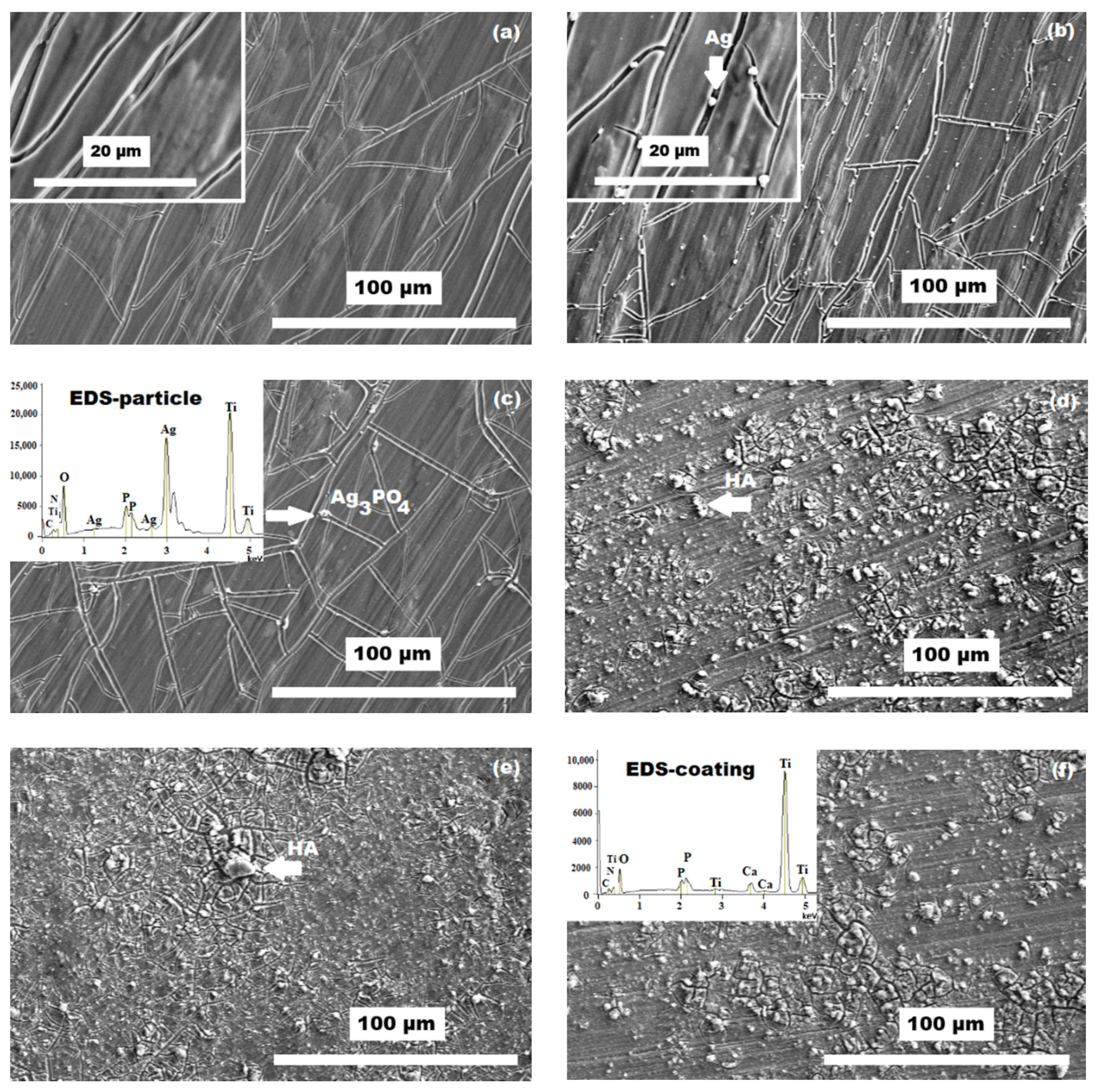
3. Results and Discussion

The surface of the titanium substrate after 3-step grinding (Figure 1) had the homogeneous roughness (profile) and provide better adhesion of the coatings to the substrate.
The titania sol-gel coatings developed by dip-coating technique on the titanium substrate after heating characterized with SEM-EDS are shown in Figure 2. The basic coating T (Figure 2a) contained cracks preferentially copied the grinding scratches. Cracks are interconnected. Particles of Ag from the dissolved silver nitrate (Figure 2b) and particles of silver phosphate (Figure 2c) were initiation points for cracks. Particles of silver were distributed mostly homogeneously in the basic titania coatings. The micro particles of hydroxyapatite were distributed around all coatings (Figure 2d–f). These three types of the coatings showed the lowest occurrence of cracks after heating.
The adhesion of all coatings (T, THA, TAN, TANHA, TAP, TAPHA) to the substrates measured by tape test was very good. The cracks in the coatings did not have any negative effect on the adhesion to the substrate. The particles of silver and hydroxyapatite were strongly fixed to the coatings after cross cut by a scalpel and peeling of the tape. In the case of TAN and TANHA coatings glue from the tape remained on the surface. The adhesion was evaluated by OM (always two samples of each type of the coating) and surfaces of the coatings were compared to a classification table. The coatings had the adhesion grade 5B (the best value in the classification table [7]).
All coatings were tested against gram-negative bacteria Escherichia coli. The Table 3 shows the antibacterial effect (%) after 4 and 24 h of interaction of all coatings with the bacteria. The values shown in the table are the average of four results from two independent measurements. The coatings with both forms of silver and hydroxyapatite TANHA and TAPHA had 100% antibacterial effect after 24 h of interaction. The coatings with silver but without hydroxyapatite had only 60% antibacterial effect after 24 h of interaction. The hydroxyapatite particles in titania-silver sol-gel coatings improved the antibacterial effect [17].
A positive control was not performed, however there is a clear decrease in number of surviving bacteria in case of TANHA and TAPHA samples compared to a negative control. A similar approach can be seen in a protocol [23]. Preliminary study of antibacterial properties was performed using a representative of gram-negative bacteria E. coli, as was also done by [24]. The study will be further extended using bacterial species similar to those which are most frequently found in peri-prosthetic infection.
The coatings containing hydroxyapatite particles (THA, TANHA, TAPHA) were tested for the apatite-forming ability by static-dynamic in vitro test for 25 days. The Figure 3a–c shows the surface of the coatings after exposition in MSBF. Hydroxyapatite particle aggregations and small cracks in the coatings (as showed after heating) did not have any negative effect on the bioactivity because bone-like hydroxyapatite with typical rosette-like crystals grouped into spherolites precipitated on the surface of all tested coatings.
The leachate analyses are described by the graphs in Figure 4a–c. The value of pH (Figure 4a) increased immediately after the exposition of the coatings in MSBF solution and kept this trend until the end of the in vitro test. This behavior of pH was very similar for all types of the coatings. The concentration of phosphate in MSBF slowly decreased after three days of immersion (Figure 4b). The concentration of calcium in MSBF for all types of the coatings decreased immediately after immersion (Figure 4c). The decreasing trend was maintained until the in vitro test ended for both ions. This nearly uniform ion removal means continuous precipitation of calcium phosphate (probably bone-like hydroxyapatite) on the surface of all types of the tested titania coatings (THA, TANHA, TAPHA).

4. Conclusions

We prepared and characterized an antibacterial bioactive titania coatings on titanium substrate with very good adhesive properties. The combination of hydroxyapatite with both forms of silver achieved a 100% antibacterial effect of the coatings after 24 h. Thanks to the static-dynamic conditions of the in vitro test, we found a daily decreasing of calcium and phosphate ions in MSBF. The results of the coating shows bioactive behavior almost from the beginning of the in vitro test. Visual characterization of the surfaces of the coated substrates by SEM-EDS confirmed the overall covering of the sol-gel coatings with a new layer of precipitated hydroxyapatite.
The concentration of silver and hydroxyapatite in the sols/coatings and the conditions of antibacterial (surface/volume) and bioactivity (static-dynamic mode) test developed by our laboratory is unique and therefore not easily comparable to the sols/coatings and test conditions developed by other laboratories.
Our results show that the sol-gel is a method with great potential, since it enables to make diverse coatings for many purposes, using a diversity of techniques. The dip-coating is one of them, it is simple, cheap, and results in homogeneous coatings. Moreover, it enables to modify properties of materials for various applications in the field of coated orthopedic and dental implants.

Author Contributions

Conceptualization, D.H., Q.D., and E.J.; methodology, D.H., Q.D., and E.J.; writing—original draft preparation, D.H.; writing—review and editing, D.H., E.J., G.L.L.-N., and A.H.; supervision G.L.L.-N. and A.H. All authors have read and agreed to the published version of the manuscript.

Funding

This research received no external funding.

Institutional Review Board Statement

Not applicable.

Informed Consent Statement

Not applicable.

Data Availability Statement

Data available by corresponding author on request.

Conflicts of Interest

The authors declare no conflict of interest.

References

  1. Keane, T.J.; Badylak, S.F. Biomaterials for tissue engineering applications. Sem. Ped. Sur. 2014, 23, 112–118. [Google Scholar] [CrossRef] [PubMed]
  2. Shan, D.; Gerhard, E.; Zhang, C.; Tierney, J.W.; Xie, D.; Liu, Z.; Yang, J. Polymeric biomaterials for biophotonic applications. Bioact. Mat. 2018, 3, 434–445. [Google Scholar] [CrossRef]
  3. Hussein, M.A.; Mohammed, A.S.; Al-Aqeeli, N. Wear characteristics of metallic biomaterials: A review. Materials 2015, 8, 2749–2768. [Google Scholar] [CrossRef]
  4. Milella, E.; Cosentino, F.; Licciulli, A.; Massaro, C. Preparation and characterisation of titania/hydroxyapatite composite coatings obtained by sol-gel proces. Biomaterials 2001, 22, 1425–1431. [Google Scholar] [CrossRef]
  5. Guo, L.; Feng, W.; Liu, X.; Lin, C.; Li, B.; Qiang, Y. Sol-gel synthesis of antibacterial hybrid coatings on titanium. Mat. Let. 2015, 160, 448–451. [Google Scholar] [CrossRef] [Green Version]
  6. Fu, T.; Shen, Y.; Alajmi, Z.; Wang, Y.; Yang, S.; Li, G. Sol-gel derived Ag-containing TiO2 films on surface roughened biomedical NiTi alloy. Ceram. Int. 2014, 40, 12423–12429. [Google Scholar] [CrossRef]
  7. ASTM International. Standard Test Methods for Measuring Adhesion by Tape Test; ASTM International: West Conshohocken, PA, USA, 2006. [Google Scholar]
  8. Cheng, K.; Ren, C.; Weng, W.; Du, P.; Shen, G.; Han, G.; Zhang, S. Bonding strength of fluoridated hydroxyapatite coatings: A comparative study on pull-out and scratch analysis. Thin Solid Films 2009, 517, 5361–5364. [Google Scholar] [CrossRef]
  9. Zhang, S.; Xianting, Z.; Yongsheng, W.; Kui, C.; Wenjian, W. Adhesion strength of sol-gel derived fluoridated hydroxyapatite coatings. Surf. Coat. Tech. 2006, 200, 6350–6354. [Google Scholar] [CrossRef]
  10. Wen, C.E.; Xu, W.; Hu, W.Y.; Hodgson, P.D. Hydroxyapatite/titania sol-gel coatings on titanium-zirconium alloy for biomedical applications. Acta Biomater. 2007, 3, 403–410. [Google Scholar] [CrossRef] [PubMed]
  11. Lim, P.N.; Teo, E.Y.; Ho, B.; Tay, B.Y.; Thian, E.S. Effect of silver content on the antibacterial and bioactive properties of silver-substituted hydroxyapatite. J. Biomed. Mater. Res. A. 2013, 101, 2456–2464. [Google Scholar] [CrossRef] [PubMed]
  12. Kokubo, T.; Takadama, H. How useful is SBF in predicting in vivo bone bioactivity? Biomaterials 2006, 27, 2907–2915. [Google Scholar] [CrossRef] [PubMed]
  13. Xu, W.; Hu, W.; Li, M. Sol-gel derived hydroxyapatite/titania biocoatings on titanium substrate. Mater. Lett. 2006, 60, 1575–1578. [Google Scholar] [CrossRef]
  14. Chaloupka, K.; Malam, Y.; Seifalian, A.M. Nanosilver as a new generation of nanoproduct in biomedical applications. Trends Biotechnol. 2010, 28, 580–588. [Google Scholar] [CrossRef] [PubMed]
  15. Bi, Y.; Ouyang, S.; Umezawa, N.; Cao, J.; Ye, J. Facet effect of single-crystalline Ag3PO4 sub-microcrystals on photocatalytic properties. J. Am. Chem. Soc. 2011, 133, 6490–6492. [Google Scholar] [CrossRef] [PubMed]
  16. Saravanapavan, P.; Gough, J.E.; Jones, J.R.; Hench, L.L. Antimicrobial macroporous gell-glasses: Disolution and cytotoxicity. key Eng. Mater. 2004, 254, 1087–1090. [Google Scholar] [CrossRef]
  17. Buckley, J.J.; Lee, A.F.; Olivi, L.; Wilson, K. Hydroxyapatite supported antibacterial Ag3PO4 nanoparticles. J. Mater. Chem. 2010, 20, 8056–8063. [Google Scholar] [CrossRef] [Green Version]
  18. Viana, M.M.; Mohallem, N.D.S.; Miquita, D.R.; Balzuweit, K.; Silva-Pinto, E. Preparation of amorphous and crystalline Ag/TiO2 nanocomposite thin films. App. Surf. Sci. 2013, 265, 130–136. [Google Scholar] [CrossRef]
  19. Babapour, A.; Akhavan, O.; Moshfegh, A.Z.; Hosseini, A.A. Size variation and optical absorption of sol-gel Ag nanoparticles doped SiO2 thin film. Thin Solid Films 2006, 515, 771–774. [Google Scholar] [CrossRef]
  20. Horkavcová, D.; Zmeškal, Z.; Šanda, L.; Cílová, Z.Z.; Jablonská, E.; Helebrant, A. Preparation of titania sol-gel coatings containing silver in various forms and measurement of their bactericidal effects against E. coli. Ceramics-Silikáty 2015, 59, 238–243. [Google Scholar]
  21. Horkavcová, D.; Novák, P.; Fialová, I.; Černý, M.; Jablonská, E.; Lipov, J.; Ruml, T.; Helebrant, A. Titania sol-gel coatings contain-ing silver on newly developed TiSi alloys and their antibacterial effect. Mater. Sci. Eng. C 2017, 76, 25–30. [Google Scholar] [CrossRef]
  22. ISO 23317:2014. Implants for Surgery—In Vitro Evaluation for Apatite-Forming Ability of Implant Materials; International Organization for Standard: Geneva, Switzerland, 2014. [Google Scholar]
  23. Martí, M.; Frígols, B.; Serrano-Aroca, A. Antimicrobial characterization of advanced materials for bioengineering applications. J. Vis. Exp. 2018, 138, e57710. [Google Scholar] [CrossRef] [PubMed] [Green Version]
  24. Zhang, S.; Liang, X.; Gadd, G.M.; Zhao, Q. A sol-gel based silver nanoparticle/polytetrafluorethylene (AgNP/PTFE) coating with enhanced antibacterial and anti-corrosive properties. App. Surf. Sci. 2021, 535, 147675. [Google Scholar] [CrossRef]
Figure 1. Surface of titanium substrate after grinding by SiC P800.
Coatings 11 00243 g001
Figure 2. Surface of coatings after heating: (a) T, (b) TAN (c) TAP, (d) THA, (e) TANHA (f) TAPHA.
Coatings 11 00243 g002
Figure 3. Surface of coatings after in vitro test: (a) THA, (b) TANHA, (c) TAPHA.
Coatings 11 00243 g003
Figure 4. Diagrams of: (a) pH value, (b) phosphate concentration, and (c) calcium concentration in MSBF during the static-dynamic in vitro test.
Coatings 11 00243 g004aCoatings 11 00243 g004b
Table 1. Composition of the sols and coatings prepared from them.
Type of the Sol (Coating)Composition of the Sol (Coating)
TTitania basic sol
TANTitania basic sol + AgNO3
TAPTitania basic sol + Ag3PO4
THATitania basic sol + hydroxyapatite
TANHATitania basic sol + AgNO3 + hydroxyapatite
TAPHATitania basic sol + Ag3PO4 + hydroxyapatite
Table 2. Composition of the modified simulated body fluid (MSBF).
SolutionIonic Concentration (mmol/L)
Na+K+Ca2+Mg2+ClHPO42−HCO3SO42−
MSBF142.05.02.51.01311.05.01.0
Table 3. Antibacterial effect of the coatings after interaction with E. coli for 24 and 4 h.
Type of the CoatingAntibacterial Effect (%)
After 4 hAfter 24 h
T1952
TAN777
TAP760
THA061
TANHA31100
TAPHA58100
Publisher’s Note: MDPI stays neutral with regard to jurisdictional claims in published maps and institutional affiliations.

